STUDY OF SERUM ZINC LEVEL IN DIABETES MELLITUS
AND ITS ROLE IN DEVELOPMENT OF COMPLICATIONS
Dissertation submitted in partial fulfillment of requirements for
M.D. DEGREE IN GENERAL MEDICINE
BRANCH I
Of
THE TAMILNADU DR. M.G.R. MEDICAL UNIVERSITY,
CHENNAI, INDIA.
MADRAS MEDICAL COLLEGE,
CHENNAI 600003
CERTIFICATE
This is to certify that the dissertation entitled “
STUDY OF SERUM
ZINC LEVEL IN DIABETES MELLITUS AND ITS ROLE IN
DEVELOPMENT OF COMPLICATIONS”
is a bonafide work done by
Dr.MAGESH.A.
, at Madras Medical College, Chennai in partial fulfillment
of the university rules and regulations for award of M.D., Degree in General
Medicine (Branch-I) during the academic year 2009 -2012.
Prof.K.SIVASUBRAMANIAN M.D.,
Professor and Unit Chief,
Research Supervisor and Guide, Institute of Internal Medicine, Madras Medical College &
Rajiv Gandhi Govt. General Hospital, Chennai – 3.
Prof.C.RAJENDIRAN M.D.,
Director and Professor,
Institute of Internal Medicine, Madras Medical College &
Rajiv Gandhi Govt. General Hospital, Chennai – 3.
Prof.V.KANAGASABAI M.D.,
The Dean
Madras Medical College &
Rajiv Gandhi Govt. General Hospital,
DECLARATION
I solemnly declare that this dissertation entitled “
STUDY OF
SERUM ZINC LEVEL IN DIABETES MELLITUS AND ITS ROLE IN
DEVELOPMENT OF COMPLICATIONS”
was done by me at Madras
Medical College and Rajiv Gandhi Government General Hospital, during
2009-2012
under
the
guidance
and
supervision
of
,
Prof.K.SIVASUBRAMANIAN, M.D.
This dissertation is submitted to the
Tamil Nadu Dr.M.G.R. Medical University towards the partial fulfillment of
requirements for the award of M.D. Degree in General Medicine (Branch-I).
Signature of Candidate
Place: Chennai-3
ACKNOWLEDGEMENT
At the outset, I thank
Prof.V.KANAGASABAI M.D.,
Dean, Madras
Medical College and Rajiv Gandhi Government General Hospital,
Chennai-3 for having permitted me to use hospital data for the study.
I am very much thankful to
Prof.V.PALANI M.S.,
Medical
Superintendent, Madras Medical College and Rajiv Gandhi Government
General Hospital, Chennai-3 for permitting me to carry out my study.
I am grateful to
Prof.C.RAJENDIRAN, M.D.,
Director and
Professor, Institute of Internal Medicine, Madras Medical College and Rajiv
Gandhi Government General Hospital, Chennai-3.
I am indebted to
Prof.K.SIVASUBRAMANIAN, M.D.,
Professor of
Medicine, Institute of Internal Medicine, Madras Medical College and Rajiv
Gandhi Government General Hospital, Chennai-3 for his valuable guidance.
I
would
like
to
thank
Dr.A.MURUGESAN,M.D.,
Dr.G.RAJAN,M.D.,Dr.S.GOPALAKRISHNAN,M.D.,
Assistant
Professors,
Madras Medical College and Rajiv Gandhi Government General
Hospital, Chennai-3 for their scrutiny.
I would also like to thank all the professors and assistant professors of
the Department of Cardiology for their continuous support and expert
guidance.
ABBREVIATIONS
ADA
:
American Diabetes Association
AGE
:
Advanced Glycosylation End products
BMI
:
Body Mass Index
CAD
:
Coronary Artery Disease
Cu
:
Copper
CVA
:
Cerebro Vascular Accident
DCCT
:
Diabetes Control and Complication Trial
DNA
:
DeoxyriboNucleic Acid
DAG
:
Diacyl Glycerol
DM
:
Diabetes Mellitus
ECM
:
Extra Celllular Matrix
FBS
:
Fasting Blood Sugar
HBA1C
:
Glycosylated Haemoglobin
HNF
:
Hepatocyte nuclear transcription factor
IL
:
Interleukin
MDA
:
Malondialdehyde
MODY
:
Maturity onset diabetes of Young
NCS
:
Nerve conduction study
PAD
:
Peripheral Artery Disease
RDA
:
Recommended daily allowance
SOD
:
Superoxide dismutase
STZ
:
Streptozocin
TNF
:
Tumor necrosis factor
CONTENTS
S.NO
CONTENT
PAGE NO.
1.
INTRODUCTION
1
2.
AIMS AND OBJECTIVES
4
3.
REVIEW OF LITERATURE
5
4.
MATERIALS AND METHODS
32
5.
RESULTS
37
6.
DISCUSSION
56
7.
CONCLUSION
59
8.
BIBLIOGRAPHY
9.
ANNEXURES
1. PROFORMA
2. MASTER CHART
INTRODUCTION
Diabetes mellitus (DM) is characterised by hyperglycaemia due to disturbances in the metabolism of carbohydrate, fat and protein because of abnormalities in the availability of insulin or insulin action (65).
Currently number of diabetic patients in worldwide is estimated to be 150 millions, two third of which are residing in developing countries. The number is predicted to double by 2025,
with the greatest number of cases in India and China alone.
Even though diabetes mellitus is an endocrine disease in origin, its major manifestations are those of a metabolic disease. The characteristic symptoms are excessive thirst, polyuria, pruritus, and otherwise unexplained weight loss. The secondary complications of diabetes is due to the thickening of basement membrane.
blood glucose level is the result of combination of both of these processes.
The secondary complications seen in diabetic patients are found to be due to alterations in vascular basement membrane composition as well as accumulation of glucose derived reaction products owing to over utilisation of glucose in insulin independent tissues [2].
Various authors have shown that hyperglycaemia leads to an increase in serum glycated proteins [3, 4, 5] along with alterations in other atherogenic risk factors. Disturbances in mineral metabolism are also noticed [6] and it is not known whether differences in trace element status are a consequence to the expression of the disease.
AIMS & OBJECTIVES
To detect serum zinc level in patients with diabetes mellitus.
To compare the serum zinc level in newly diagnosed diabetic patients & in those with complications.
REVIEW OF LITERATURE
ZINC
Zinc is the 24th most abundant element in the Earth's crust and has five stable isotopes. The element was probably named by the alchemist Paracelsus after the German word zinke. German chemist Andreas Sigismund Marggraf is normally given credit for discovering pure metallic zinc in 1746. Work by Luigi Galvani and Alessandro Volta uncovered the electrochemical properties of zinc by 1800. A variety of zinc compounds are commonly used, such as zinc carbonate and zinc gluconate (as dietary supplements), zinc chloride (in deodorants), zinc pyrithione (anti-dandruff shampoos), zinc sulfide (in luminescent paints), and zinc methyl or zinc diethyl in the organic laboratory.
BIOCHEMICAL FUNCTIONS OF ZINC
after iron and it is the only metal which appears in all enzyme classes [29].
There are 2–4 grams of zinc distributed throughout the human body. Most zinc is in the brain, muscle, bones, kidney, and liver, with the highest concentrations in the prostate and parts of the eye. Semen is particularly rich in zinc, which is a key factor for prostate gland function and reproductive organ growth.
Zinc plays ubiquitous biological roles in the prostate [34]. It interacts with a wide range of organic ligands[34] and has roles in the metabolism of RNA and DNA, signal transduction, and gene expression. It also regulates apoptosis.
A 2006 study estimated that about 10% of human proteins (2800) potentially bind zinc, in addition to hundreds which transport and traffic zinc; similar to the Silico study in the plant
Arabidopsis thaliana which found 2367 zinc-related proteins[29].
zinc homeostasis plays a critical role in normal functioning of the brain and central nervous system.
Zinc is an efficient lewis acid, making it a useful catalytic agent in hydroxylation and other enzymatic reactions. The metal also has a flexible coordination geometry, which allows proteins using it to rapidly shift conformations to perform biological reactions. Two examples of zinc-containing enzymes are carbonic anhydrase and carboxy peptidase, which are vital to the processes of carbon dioxide (CO2) regulation and digestion of proteins, respectively.
the protein being digested [39].Zinc also serves a purely structural role in zinc fingers twists and clusters[40].
METABOLISM OF ZINC
In blood plasma, zinc is bound to and transported by albumin (60%, low-affinity) and transferrin (10%). Since transferrin also transports iron, excessive iron reduces zinc absorption, and vice-versa. A similar reaction occurs with copper. The concentration of zinc in blood plasma stays relatively constant regardless of zinc intake. Cells in the salivary gland, prostate, immune system and intestine use zinc signaling as a way to communicate with other cells [42].
Zinc may be held in metallothionein reserves within microorganisms or in the intestines or liver of animals. Metallothionein in intestinal cells is capable of adjusting absorption of zinc by 15–40%. However, inadequate or excessive zinc intake can be harmful; excess zinc particularly impairs copper absorption because metallothionein absorbs both metals.
ZINC DEFICIENCY
Prevalence
In fact, one-third of the world population is at risk of zinc deficiency, ranging from 4 to 73% depending on the country. Zinc deficiency is the fifth leading risk factor for disease in the developing world. Providing micronutrients, including zinc, to humans is one of the four quick-win solutions to major global problems identified in the Copenhagen consensus from an international panel of distinguished economists. Conservative estimates suggest that 25% of the world's population is at risk for zinc deficiency[44]
Causes
Hypozincemia is usually a nutritional deficiency, but can also be associated with malabsorption, diarrhoea, acrodermatitis enteropathica, chronic liver disease, chronic renal disease, sickle cell diasease, diabetes, malignancy, and other chronic illnesses [45]. It can also occur after bariatric surgery.
that involve intestinal absorption promote zinc losses. Fecal losses of zinc caused by diarrhea is one contributing factor [55], often common in developing countries. Changes in intestinal tract absorbability and permeability due, in part, to viral, protozoal, and bacteria pathogens may also encourage fecal losses of zinc[49].Physiological states that require increased zinc include periods of growth in infants and children as well as in mothers during pregnancy[50].
SIGNS AND SYMPTOMS
Signs of zinc deficiency include hair loss,skin lesions, diarrhea, and wasting of body tissues. A lack of zinc can contribute to acne [51]. Vision[52][53], taste ,smell and memory are also connected with zinc. A deficiency in zinc can cause malfunctions of these organs and functions.
Cognitive and motor function impairment
Cognitive and motor function may also be impaired in zinc deficient children. Zinc deficiency can interfere with many organ systems especially when it occurs during a time of rapid growth and development, when nutritional needs are high, such as during infancy [57][43]. Low maternal zinc status has been associated with less attention during the neonatal period and worse motor functioning[59]. In some studies, supplementation has been associated with motor development in very low birth weight infants and more vigorous and functional activity in infants and toddlers[59].
Diarrhoea and pneumonia
disturbances in an individual may have potential for diagnosing its deficiency [54].
Dysmenorrhoea
High dose of zinc, 30 mg 1-3 times a day, prevents dysmenorrhoea[62] .
Pregnancy
Zinc deficiency during pregnancy can negatively affect both the mother and fetus. A review of pregnancy outcomes in women with acrodermatitis enteropathica, reported that out of every seven pregnancies, there was one abortion and two malfunctions, suggesting the human fetus is susceptible to the teratogenic effects of severe zinc deficiency. However, a review on zinc supplementation trials during pregnancy did not report a significant effect of zinc supplementation on neonatal survival.
Effect of zinc on lymphoid and myeloid cells
Treatment of zinc defeciency
Zinc supplementation has been shown to reduce diarrhea prevalence and mortality in children younger than 5 years of age [64]. To combat zinc deficiency, five intervention strategies can be used:
1) Supplementation using medicines
2) Food fortification through the incorporation of zinc additives in food
3) Dietary modification/diversification
4) Genetic biofortification through plant breeding
5) Agronomic biofortification through zinc fertilization.
EFFECTS OF DIABETES ON ZINC METABOLISM
El Yazigi et al evaluated both Type I and II diabetics and found that the absolute and creatinine corrected urinary excretions were greater in diabetics than in matched controls and found a positive correlation between Zn excretion and hemoglobin Alc concentrations [8].
In Type II Diabetes,insulin reduces the hyperzincuria while
other agents had no effect on zinc excretion[9].
It has also been postulated that hyperglycemia interferes with the active transport of Zn back into the renal tubular cells. Administration of insulin reduces but does not appear to completely ameliorate the hyperzincuria [11][12].
McNair et al [13] confirmed that hyperzincuria occurs in relationship to the degree of hyperglycemia, but not glycosuria. Kinlaw et al [10] demonstrated abnormal Zn tolerance tests in diabetic patients suggestive of decreased absorption.
absorption, coupled with hyperzincuria, could account for significant loss of intracellular Zn [14][13].
While it is clear that urinary excretion of Zn is markedly increased in individuals with diabetes, if hyperglycemia is the primary etiology, replacement with oral Zn supplementation should provide sufficient treatment[38].
EFFECTS OF ZINC ON DIABETES MELLITUS (PRIMARY DISEASE EFFECTS)
In 1966, Quarterman et al[15] demonstrated that diet induced zinc deficiency in rats resulted in a decrease in the ability of the pancreas to secrete insulin in response to a glucose load—a hallmark of diabetes.
Because zinc is a necessary factor in a variety of “antioxidant” enzymes, particularly superoxide dismutase, catalase and peroxidase, alterations of zinc metabolism such that adequate zinc is unavailable for these enzymes might be expected to contribute to the tissue damage observed in diabetes [18].
Rabinovitch et al [19] examined the relationship between cytokine induced (interleukin 1b, tumor necrotic factor (TNF) and interferon gamma) pancreatic beta cell destruction, production of malondialdehyde (MDA), an end product of lipid peroxidation, and nitrite, the end product of nitric oxide. These studies suggested that cytokines are toxic to the human beta cell by producing oxygen free radicals, lipid peroxidation, and aldehyde production in the islets and that MDA was one of the cytotoxic mediators [18][19].
Zimny et al have demonstrated induction of metallothionein production in the islet cell in response to Streptozocin (STZ) induced OH radical production [20]. Yang and Cherian [21] demonstrated STZ-induced lipid peroxidation and decreased superoxide dismutase (SOD). Roza showed marked decreases in pancreatic SOD and catalase antioxidant activity which preceded the loss of beta cell function suggesting increased beta cell vulnerability to free radical attack and cell destruction in genetically diabetic rats [22]. All of these data suggest a role for Zn in the protection of the beta cell against the immune-mediated free radical attack.
islet cell becomes more vulnerable to all sorts of damage. This matches the clinical picture in which, after prolonged hyperglycemia and inability of the islet cell to make enough insulin to control the glucose, there is a loss of islet cell altogether. This provides a mechanism by which Zn deficiency may affect the progress of Type II diabetes [23].
EFFECTS OF ZINC ON DIABETES MELLITUS (SECONDARY COMPLICATIONS)
It has become clear in recent years that hyperglycemia is a major culprit in the development of the microvascular complications which include retinopathy, nephropathy, neuropathy and the small vessel occlusions as well as birth defects including fetal malformations and macrosomia. It also appears clear that, while hyperglycemia is a major contributor, it is not the only contributor to the development of these complications.
radicals [24]. In other animal studies Minami et al [25] demonstrated an increase in the progression of diabetic nephropathy in STZ diabetic rats when Zn deficiency was induced either by increased renal excretion or by dietary induced deficiency.
DIABETES MELLITUS
DEFINITION
The term diabetes mellitus describes a metabolic disorder of multiple aetiology characterized by chronic hyperglycaemia with disturbances of carbohydrate, fat and protein metabolism resulting from defects in insulin secretion, insulin action[65], or both. The effects of diabetes mellitus include long–term damage, dysfunction and failure of various organs.
SYMPTOMS AND SIGNS
amputation, Charcot joints, and features of autonomic dysfunction, including sexual dysfunction.
PATHOGENESIS OF DIABETES
Several pathogenetic processes are involved in the development of diabetes. These include processes which destroy the beta cells of the pancreas with consequent insulin deficiency, and others that result in resistance to insulin action. The abnormalities of carbohydrate, fat and protein metabolism are due to deficient action of insulin on target tissues resulting from insensitivity or lack of insulin. The aetiological types designate defects, disorders or processes which often result in diabetes mellitus.
TYPE 1
Type 1 indicates the processes of beta–cell destruction [65] that may ultimately lead to diabetes mellitus in which insulin is required for survival to prevent the development of ketoacidosis, coma and death. An individual with a Type 1 process may be metabolically normal before the disease is clinically manifest, but the process of beta–cell destruction can be detected.
this clinical form of diabetes, particularly non–Caucasians, no evidence of an autoimmune disorder is demonstrable and these are classified as “Type 1 idiopathic”.
TYPE 2
It is due to predominant insulin resistance with relative insulin deficiency or predominant insulin secretory defect with/without insulin resistance[68]. Diabetes mellitus of this type was previously encompassed as non–insulin–dependent diabetes, or adult–onset diabetes. It is a term used for individuals who have relative (rather than absolute) insulin deficiency.
People with this type of diabetes frequently are resistant to the action of insulin. At least initially, and often throughout their lifetime, these individuals do not need insulin treatment to survive. This form of diabetes is frequently undiagnosed for many years because the hyperglycaemia is often not severe enough to provoke noticeable symptoms of diabetes. Nevertheless, such patients are at increased risk of developing macro vascular and micro vascular complications [69].
Many of those who are not obese by traditional weight criteria may have an increased percentage of body fat distributed predominantly in the abdominal region. Ketoacidosis is infrequent in this type of diabetes; when seen it usually arises in association with the stress of other illness such as infection .
The risk of developing Type 2 diabetes increases with age, obesity, and lack of physical activity . It occurs more frequently in women with prior GDM and in individuals with hypertension or dyslipidaemia .Some patients who present with a clinical picture consistent with Type 2 diabetes have auto antibodies similar to those found in Type 1 diabetes, and may masquerade as Type 2 diabetes if antibody determinations are not made. Patients who are non–obese or who have relatives with Type 1 diabetes and who are of Northern European origin may be suspected of having late onset Type 1 diabetes.
COMPLICATIONS OF DIABETES MELLITUS Acute complications
Diabetic ketoacidosis
Diabetic nonketotic, hyperosmolar coma
CHRONIC COMPLICATIONS Microvascular
Retinopathy
Nephropathy
Neuropathy
Macrovascular
Cerebrovascular
Cardiovascular
Peripheral vascular disease
MICROVASCULAR COMPLICATIONS
Microvascular complications are specific to diabetes and do not occur without longstanding hyperglycaemia. Other metabolic, environmental and genetic factors are undoubtedly involved in their pathogenesis.
The duration of diabetes and the quality of diabetic control are important determinants of microvascular disease but, because of other individual factors, do not necessarily predict their developent in individual patients.Different microvascular complications are commonly associated in individual patients, but their prevalence as a function of the duration or severity of diabetes may differ markedly.
Background retinopathy is rare before 5 years of diabetes but its prevalence increases steadily thereafter to affect over 90% of patients after 20 years. After several years of diabetes, the risk of proliferative changes is about 3% of patients per year, with a cumulative total of over 60% after 40 years.
Diabetic nephropathy affects 20-40% of patients with T1DM, particularly those presenting before puberty and possibly those with an inherited tendency to hypertension. T2DM patients are also susceptible to nephropathy.
PATHOPHYSIOLOGY OF MICROVASCULAR DISEASE In diabetes, the microvasculature shows both functional and structural abnormalities. The structural hallmark of diabetic microangiopathy is thickening of the capillary basement membrane. The main functional abnormalities include increased capillary permeability, blood flow and viscosity, and disturbed platelet function. These changes occur early in the course of diabetes and precede organ failure by many years.
Many chemical changes in basement membrane composition have been identified in diabetes, including increased type IV collagen and its glycosylation products. In patients with poorly controlled diabetes, even of short duration, blood flow is increased in many tissues including skin, retina and kidney. In the latter this is reflected by an elevated glomerular filtration rate. Increased capillary permeability is manifested in the retina by leakage of fluorescein and in the kidney by increased urinary losses of albumin which predict eventual renal failure. Both defects probably reflect a generalized vascular abnormality which may also involve the intima of the large vessels.
metabolism[71] . Plasma and whole blood viscosity are increased whereas red blood cell deformability is decreased in diabetes.
These defects together with the platelet abnormalities may cause stasis in the microvasculature, leading to increased intravascular pressure and to tissue hypoxia. The production by endothelial cells of von Willebrand factor and endothelial derived relaxing factors (mainly nitric oxide) may also be abnormal in diabetes and could contribute to tissue damage.
BIOCHEMICAL BASIS OF MICROVASCULAR DISEASE
Prolonged exposure to elevated glucose concentrations damages tissues by causing either acute, reversible metabolic changes (mostly related to increased polyol pathway activity and glycosylation of proteins) or cumulative irreversible changes in long lived molecules (formation of advanced glycosylation end products /AGE/ on matrix proteins such as collagen and on nucleic acids and nucleoproteins). In insulin independent tissues (nerve, lens, retina) hyperglycaemia causes elevated tissue glucose levels.
accumulation may cause damage by osmotic effect (e.g. in the lens). In addition, increased sorbitol production is partly responsible for tissue depletion of myoinositol, a molecule structurally related to glucose. Hypergylcaemia itself inhibits myoinositol uptake into cells. Animal studies indicate that tissue myoinositol depletion may cause abnormalities on peripheral nerve function.
ADVANCED GLYCOSYLATION END PRODUCTS
PATHOGENESIS OF DIABETIC RETINOPATHY
Histologically the earliest lesion is thickening of the capillary basement membrane. On fluorescein angiography the first abnormality is the capillary dilatation. Localised capillary dilatation is called microaneurysm. Microaneurysm may give rise to haemorrhage or exudate. Vascular occlusion, initially of capillaries and later of arteries and veins, leads to nonperfusion areas of retina. Large ischaemic areas are the stimulus of new vessel formation[1].
STAGES OF DIABETIC RETINOPATHY Background retinopathy
Capillary dilatation (later with leakage)
Capillary occlusion
Microaneurysms
Blot haemorrhages and lipid rich hard exudates.
Preproliferative retinopathy
Cotton-wool spots (retinal ischemia)
Venous abnormalities (loops, beading and reduplication)
Intraretinal microvascular abnormalities (clusters of dilated abnormal capillaries lying within the retina).
Proliferative retinopathy
New vessels arise in the periphery and/or on the optic disc, eventually with a fibrous tissue covering. The visual complications are caused by vitreous retraction which leads to haemorrhage and to traction and detachment of the retina.
Maculopathy
Heralded by rings of hard exudates approaching the fovea. Maculopathy occurs mostly in T2DM and can cause severe visual loss.
Thrombotic glaucoma is due to new vessels and fibrous tissue proliferating in the angle of the anterior chamber, preventing drainage of the aqueous. It is associated with rubeosis iridis (neovascularisation of the iris) and causes severe pain and irreversible blindness
DIABETIC NEPHROPATHY
an increased incidence of retinopathy and a ten-fold increase in cardiovascular mortality which is the major cause of death in nephropathic T2DM patients.
Diabetic nephropathy is defined by persistent albuminuria (>300 mg/day), declining glomerular filtration rate and rising blood pressure. Established nephropathy follows several years of incipient nephropathy, characterised by microalbuminuria (30 – 300 mg/day).
In T1DM, nephropathy develops in about 35% of cases, especially in males and in those whose diabetes presents before the age of 15 years. The incidence of nephropathy peaks after 15-16 years of diabetes and declines thereafter. In T2DM prevalence of nephropathy about 10%.
METHODS & METHODOLOGY
Study centre : Rajiv Gandhi Government General Hospital, Chennai – 3
Study duration : 6 months
Study design : Case control study.
Sample size : 100 patients ; 50 controls.
INCLUSION CRITERIA
Patients with newly diagnosed Diabetes mellitus attending Diabetology OPD and those who are admitted to the medical wards with complications of Diabetes Mellitus.
EXCLUSION CRITERIA
Infections: Tuberculosis,HIV.
Malignancy
Cirrhosis
Inflammatory bowel disease
Patient on ATT, OCP, Valproate, Penicillamine, Iron supplements
Pregnancy
DATA COLLECTION METHODS :
Collection of data as per proforma with consent from patients with diabetes mellitus in the Institute of Internal Medicine wards & in diabetology OP
SELECTION CRITERIA Diabetes
Diagnostic criteria[ 32].
Symptoms of diabetes mellitus plus random blood glucose concentration 11.1mmol/l (200mg/dl) or
Fasting plasma glucose 7mmol/l (126mg/dl) or
HbA1c >6.5% or
2 –hour plasma glucose 11.1mmol/l (200mg/dl) during an oral glucose tolerance test.
and acute metabolic decompensation, these criteria should be confirmed by repeat testing on a different day.
SYSTEMIC HYPERTENSION
Patients with Systolic BP > 130 & Diastolic BP > 80 mmHg
OBESITY
Patients with BMI > 25 Kg/sq.m.
DYSLIPIDEMIA
Total Cholesterol > 240 mg/dl
LDL – Cholesterol > 130 mg/dl
HDL – Cholesterol < 40 mg/dl(males) ; < 50 mg/dl (females)
Triglycerides > 150 mg/dl
MICROVASCULAR COMPLICATIONS
Retinopathy – Retinal changes in fundus examination.
Nephropathy – Presence of Microalbuminuria in urine.
Neuropathy – Symptoms ,Signs & NCS Abnormalities.
MACROVASCULAR COMPLICATIONS
Peripheral vascular disease – Examination & Doppler Study.
Cerebro vascular accident – History, Examination & Imaging Study.
STATISTICAL ANALYSIS
Following statistical methods have been employed in the present study.
Independent samples ‘t’ test-Unpaired.
Independent samples ‘t’ test-Paired.
One-way Analysis of Variance (ANOVA).
SPSS Version 13
EXCEL 2010
The study was approved by ethics committee of the hospital.
ZINC ESTIMATION:[58,66]
Method – calorimetric method:
Zinc + nitro-paps purple colour complex
NORMAL REFERENCE VALUES serum-60-120micro gram/dl.
OBSERVATION AND RESULTS
Table 1: Age incidenceChart showing age incidence in years:
In the study group of 100 patients with Diabetes,35% were seen in the age group of 51-60 years,24% were seen in the age group of 41-50 years.Maximum incidence-35% was seen in the age
Age(years) No.of patients %
10 to 20 4 4
21 to 30 11 11
31 to 40 13 13
41 to 50 24 24
51 to 60 35 35
61 to 70 8 8
Table 2: Age and sex distribution
Total Male Female Age
No. % No. % No. %
10 to 20 4 4 3 75 1 25
21 to 30 11 11 8 72 3 28
31 to 40 13 13 9 69 4 31
41to 50 24 24 17 70 7 30
51 to 60 35 35 22 62 13 38
61to 70 8 8 5 62 3 38
>70 5 5 2 40 3 60
Chart showingage and sex distribution
Table: 3 Type of diabetes
Total Male Female
No. No. % No. %
Type 1 15 11 73 4 27
Type 2 85 55 65 30 35
Chart showing the distribution of type of diabetes:
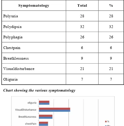
Table: 4 Symptomatology
Symptomatology Total %
Polyuria 28 28
Polydipsia 32 32
Polyphagia 26 26
Chestpain 6 6
Breathlessness 9 9
Visualdisturbance 21 21
Oliguria 7 7
Chart showing the various symptomatology
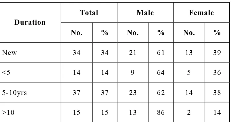
Table :5 Duration of diabetes
Total Male Female Duration
No. % No. % No. %
New 34 34 21 61 13 39
<5 14 14 9 64 5 36
5-10yrs 37 37 23 62 14 38
>10 15 15 13 86 2 14
Chart showing the duration of diabetes
Table: 6 Risk factors
Total Male Female
Risk factors
No. No. % No. %
Systemic hypertension 26 19 73 7 27
Dyslipidemia 11 7 64 4 36
Smoking 22 22 100 0 0
Alcoholism 13 13 100 0 0
Obesity 23 16 70 7 30
Total 95 77 18
Chart showing the various risk factors among patients
Table: 7 Microvascular complications
Total Male Female
No. No. % No. %
Retinopathy 22 20 91 2 9
Neuropathy 11 9 82 2 18
Nephropathy 28 22 79 6 21
Total 61 51 10
Chart showing the various microvascular complications
Table: 8 Macrovascular complications
Total Male Female
No. No. % No. %
CAD 10 7 70 3 30
CVA 11 9 82 2 18
PAD 2 2 100 0 0
Total 23 18 5
C hart showing the various macrovascular complications
Table: 9 Treatment regimen
Total Male Female
No. No. % No. %
Insulin 16 12 75 4 25
OHA 84 54 64 30 36
Total 100 66 34
Chart showing the treatment regimen
Table: 10 Comparison of serum zinc levels between patients and control
Parameter Subject (n=100)
Control
(n=50) P value
Mean Age 47 48 0.6379
Mean Zinc Levels 63.08 75.48 0.0001 In our study, 100 patients when compared to 50 controls had significant lower levels of zinc with a p value of <0.0001.
Table:11 Serum zinc levels in males and females
Male Female Zinc Levels[µg] Total
No. % No. %
10 - 20 2 2 100 0 0
21- 30 4 4 100 0 0
31 - 40 8 8 100 0 0
41- 50 14 11 78 3 22
51 - 60 19 12 63 7 37
61 - 70 27 15 55 12 45
71 - 80 11 7 63 4 37
81 - 90 6 2 33 4 67
91- 100 4 1 25 3 75
101 - 110 4 3 75 1 25
>110 1 1 100 0 0
TOTAL 100 66 34
[image:54.612.107.506.305.635.2]Table:11 Serum zinc levels in males and females
Male Female Zinc
Levels[µg] Total
No. % No. %
10 - 20 2 2 100 0 0
21- 30 4 4 100 0 0
31 - 40 8 8 100 0 0
41- 50 14 11 78 3 22
51 - 60 19 12 63 7
Chart showing the serum zinc levels in males and females
Table:12 Zinc level in nephropathy
Nephropathy
Males Females Zinc level
(µg)
Total
No. % No. %
0-20 1 1 100 0 0
20-40 7 7 100 0 0
40-60 15 10 75 5 25
60-80 5 4 80 1 20
Chart showing zinc level in nephropathy
Table:13 Zinc level in Retinopathy
Retinopathy
Males Females Zinc level
(µg)
Total
No. % No. %
0-20 1 1 100 0 0
20-40 7 7 100 0 0
40-60 10 8 80 2 20
60-80 4 4 100 0 0
Chart showing zinc level in retinopathy
Table: 14 Zinc level in neuropathy
Neuropathy
Males Females Zinc level
(µg)
Total
No. % No. %
20-40 3 3 100 0 0
40-60 7 5 71 2 29
60-80 1 1 100 0 0
Chart showing zinc level in neuropathy
Table: 15 Zinc level in CAD
Coronary artery disease
Males Females Zinc level
(µg)
Total
No. % No. %
0-20 1 1 100 0 0
20-40 4 4 100 0 0
40-60 4 2 50 2 50
60-80 1 0 0 1 100
Chart showing zinc level in CAD
Table: 16 Zinc level in CVA and PAD
Cerebro vascular disease
Peripheral Artery disease
Males Females Total
Zinc level (µg)
Total
No. % No. % M F
20-40 3 3 100 0 0 1 0
40-60 6 4 66 2 34 1 0
60-80 2 2 100 0 0 0 0
Chart showing zinc level in CVA and PAD
Table: 17 HbA1c level in males and females
Males Females HbA1c level
% Total
No. % No. %
5-5.5 2 1 50 1 50
5.6-6 36 22 61 14 39
6.1-6.5 28 17 60 11 40
6.6-7 19 15 78 4 22
7.1-7.5 9 7 77 2 23
>7.5 6 4 66 2 34
Chart showing HbA1C level in males and females
Table: 18 Correlation of zinc level in micro ¯o vascular complications
Parameter Microvascular Macrovascular
No.of patients 61 23
Mean Zn level (µg) 46.52 43.86
In our study patients with micro vascular complications, there was no significant variaton in serum zinc levels in each groups [retinopathy,nephropathy&neuropathy] with p value of 0.8431.In Patients with macro vascular complications also, there was no significant variation in serum zinc level in each groups[CAD,CVA&PAD] with p value of 0.4537.
Table: 19 Comparision of zinc level and HbA1c level
HbA1c level Total No.of patients Mean Zinc level
5-5.5 2 78
5.6-6 36 70
6.1-6.5 28 64
6.6-7 19 52
7.1-7.5 9 50
>7.5 6 46
[image:62.612.105.507.404.593.2]Table 20: Comparision of serum zinc level with duration of DM
Duration No.of patients
Serum mean zinc level
Newly diagnosed
34 69.74
1-4 years 14 69.78
5-10 years 37 56.16
>10 years 15 51.93
p
value<0.05
Patients with long standing diabetes had lower zinc levels than newly diagnosed patients, which was found to be statistically significant with p value < 0.0035.
DISCUSSION
The total number of patients analysed were 100,out of which 66% were males and 34% were females.The prevalence of diabetes was found to increase as the age advances.
This finding correlated with the study done by Ahuja mms[1996].Most of the patients in the study group were in the age group of 51-60 years[35%].85% of patients had type 2DM,of which 65% were males and 35% were females.
26% of patients in our study group had systemic hypertension, obesity was found in 23% of patients. Other risk factors like smoking [22%], alcohol [13%]& dyslipidemia [11%] were also present.
Of the 100 patients in the study, 84 were on Oral Hypoglycemic Agents and the remaining 16 were on Insulin therapy.
61% of the subjects had Micro vascular complications in the form of Nephropathy (28%), retinopathy (22%) and Neuropathy (11%)
The Mean Value for Serum Zinc levels in healthy individuals studied was within normal range as established by Study of Zinc Homeostasis in Humans [72]. Likewise our study showed decreased levels of Serum Zinc in Diabetic individuals compared to normal subjects. The minimum Zinc value found in the present study was similarly reported by a number of other studies [73], [74], [75]
It was also found in our study that compared to the newly diagnosed, patients with long standing Diabetes Mellitus had lower levels of Zinc.
Among the subjects with microvascular complications, 90% with neuropathy, 82% of those with nephropathy, 81% with retinopathy had low serum Zinc levels.
But there was no significant statistical variation in serum zinc levels between the various micro vascular complications.
But there was nosignificant statistical variation in serum zinc levels between the various macro vascular complications.
Similarly there was no significant statistical variation in serum zinc levels between the macro vascular and microvascular complications.
There was a significant negative correlation between HbA1C and serum Zinc levels.
Also, patients with poor glycemic control had lower Zinc levels compared to the subjects with a better glycemic control. This correlated with the conclusions of a study done by Al-Maroof and Al-Sabatti in 2006 for the Ministry of Health, Iraq.
LIMITATIONS OF THE STUDY
Zinc Supplementation was not done hence its effect on the subjects could not be ascertained.
CONCLUSION
Diabetes is more commonly seen in males (66%) compared to females (34%).
Diabetes is commonly diagnosed in the age group of 51-60 yrs.
Among those with Diabetes, the microvascular complications (61%) occur more often than the macrovascular complications (23%).
Nephropathy (28%) is the commonest microvascular complication closely followed by Retinopathy (22%).
Diabetic individuals have significantly lower levels of Zinc when compared to normal healthy individuals.
Patients with long standing DM have lower Zinc levels than those who are newly diagnosed.
Patients with poor glycemic control have lower Zinc levels compared to the subjects with a better glycemic control.
BIBLIOGRAPHY
1)
David M.Nathan Long term complications of diabetes mellitus. N Eng
J Med 1993:328:1676-1685.
2)
Spiro RG. Search for a biochemical basis of diabetic microangiopathy.
Diabetologia 1976; 12 : 1-14.
3)
Patel JJ, Mani UV. Measurement of serumglycosylated protein and
glycosylated low density lipoprotein fraction in the serum of
diabetics.Biochem 1987; 37 : 184-9.
4)
Mani I, Patel HR, Shah PB, Mani UV. Influence ofblood sugar and
duration of the disease on serum lipids in maturity onset diabetic
patients.
Ind J Clin Biochem
1986; 1 : 39-44.
5)
Mani I, Mani UV. Serum triglyceride, cholesterol,lipoprotein
cholesterol and urinary hydroxyl prolinelevels in insulin dependent
and Non-insulindependent diabetics.
J clin Biochem Nutr
1988; 4
:241-8.
6)
Walter RM, Uriu HJY, Olin KL, Oster MH, Anwalt BD, Critchfield
complications of diabetes mellitus. Diabetes Care 1991; 14(11)
:1050-56.
7)
Isbir T, Tamer A, Taylor A, Isbir M: Zinc, copper and magnesium
status in insulin dependent diabetes.
Diabetes Res
1994:26:41-45
8)
El-Yazigi A, Hannan N, Raines D: Effect of diabetic state and related
disorders on the urinary excretion of magnesium and zinc in
patients.
Diabetes Res
1993;22;67-75.
9)
HonNo.rat j,AccomiNo.tti M, Broussolle C, Fleuret A, Vallon J,
Orgiazzi J: Effects of diabetes type and treatment on zinc status in
diabetes mellitus.
Biol Trace Elem Res
1992;32;311-316
10)
Kinlaw WB, Levine S, Morley J, Silvis S, McClain C: Abnormal zn
metabolism in type II diabetes mellitus.
Am J Med
1983:75:273-277
11)
Lau A, Failla M: Urinary excretion of zinc, copper and iron in
streptozotocin diabetic rat.
J Nutr
1984;114;224
12)
Failla ML, Gardil C: Influence of spontaneous diabetes on tissue
status
of
zinc,
copper,
and
manganese
in
BB
Wistar
13)
McNair P, Kiilerich S, Christiansen C, Christiansen M, Madsbad S,
TransbolI: Hyperzincuria in insulin treated diabetes mellitus-its
relation to glucose homeostasis and insulin administration.
Clinica
Chimica Acta
1981:112:343-348.
14)
Escobar O, Sandoval M, Vargas A, Hempe J: Role of metallothioneien
and cysteine rich intestinal protein in the regulation of Zn absorption
by diabetic rats.
Ped Res
1995:37:321-327.
15)
Quarterman J, Mills C, Humphries W: The reduced secretion of and
sensitivity to insulin in Zn deficient rats.
BBRC
1966:25:354-358
16)
Falkmer S, Havu N, Pihl E: Insulin biosynthesis, storage and
secretion:
pancreatic
islet
cells
and
islet
cells.
Lakartidningen
1968:65:3603-3607.
17)
Engelbart K, Kief H: The functional behaviour of zinc and insulin
contain in the pancreatic islet cells of rats.
Virchows Archives, Cell
Pathol
1970;4:294-302.
18)
Sumovski W, Baquerizo H, RabiNo.vich A: Oxygen free radical
scavenger
protect
rat
islet
cells
from
damage
by
19)
Rabinovitch A, Suarez-Pinzon W, Strynadka K, Lakey J, Rajotte R:
Human pancreatic islet beta cell destruction by cytokines involves
oxygen free radicals and aldehyde production.
J Clin Endocrinol
Metab
1996:81;3197-3202
20)
Zimny S, Gogolin F, Abel J, Gleichmann H: Metallothionein in
isolated pancreatic islet cells of mice: induction by zinc and
streptozotocin,
a
naturally
occurring
diabetogen.
Arch
Toxicol
1993:67:61-65
21)
Yang J, Cherian G: Protective effects of metallothionein on
streptozotocin induced diabetes in rats.
Life Sciences
1994;55:43-51.
22)
Roza A, Pieper G, Johnson C, Adams M: Pancreatic antioxidant
enzyme
activity
in
normoglycemic
diabetic
prone
BB
rats.
Pancreas
1995:10;53-58.
23)
Spreitsma J, Schuitemaker G: Diabetes can be prevented by reducing
insulin production.
Medical Hypotheses
1994;42:15-23.
24)
Hagay Z, Weiss Y, Zusman I, Peled-Kamar M, Reese E, Erikson U,
Groner Y: Prevention of diabetes associated embryopathy by
dismutase
in
transgenic
mouse
embryos.
Am
J
Obstet
Gynecol
1995:173:1036-1041.
25)
Minami T, Ichii M, Okazaki Y, Kubo M, Kadota E, INo.ue T, Yamada
Y, Fushimi H: Renal changes of streptozotocin induced diabetic rats
fed a low Zn diet.
Renal Failure
1995:17:3489-363.
26)
Faure P, Ben Hamou P, Perard A, Halimi S, Roussel A: Lipid
peroxidation in insulin dependent diabetic patients with early retinal
degenerative lesions: effects of an oral Zn supplementation.
Eur J Clin
Nutr
1995:49:282-288.
27)
Powers MA. Handbook of Diabetes Nutritional Management. Aspen
Publishers Inc. Maryland 1987;195.
28)
Kinlaw WB, Levins AS, Morley JE. Abnormal zinc,metabolism in
Type 2 diabetes mellitus. Am J Med1983; 75 : 273-277.
29)
Broadley, M. R.; White, P. J.; Hammond, J. P.; Zelko I.; Lux -A.
"Zinc in plants".
New Phytologist
2007:173 (4): 677–702.
30)
Robert A.desilvestro,zinc in relation to diabetes and oxidative
31)
Kim, Jeung Gon; Walsh, Patrick J. "From Aryl Bromides to
Enantioenriched Benzylic Alcohols in a Single Flask: Catalytic
Asymmetric
Arylation
of
Aldehydes".
Angewandte
Chemie
International Edition
[2006],45 (25): 4175.
32)
Harrison’s principles of internal medicine Alvin c.powers, 18
thedition,chapter:344,page:2968-3001.
33)
Prasad A. S. Zinc in Human Health;Effect of zinc on immune
cells
Mol. Med.2008:
14 (5–6): 353–7.
34)
Hambidge, K. M. and Krebs, N. F. "Zinc deficiency: a special
challenge".
J. Nutr.
2007:137 (4): 1101–5. .
35)
Zinc's role in microorganisms is particularly reviewed in: Sugarman B
. "Zinc and infection".
Review of Infectious Diseases1983:
5 (1): 137–
147.
36)
Surender kumar,Kamala s,Jayarao.Blood and Urinary Zinc level in
DM.1974:17:231-235.
37)
Rink, L.; Gabriel P. "Zinc and the immune system".
Proc Nutr
38)
C.B.Niewoehner,John l allen,Levine,John E.Morley.Role of zn
supplementation
in
Type
2
Diabetes.American
journal
of
medicine1986:81(1):63-68.
39)
Bitanihirwe BK, Cunningham MG . "Zinc: The brain's dark
horse".
Synapse
2009:63 (11): 1029–49.
40)
Nakashima AS, Dyck RH . "Zinc and cortical plasticity".
Brain Res
Rev
2009:59 (2): 347–73.
41)
Stipanuk, Martha H. .
Biochemical, Physiological & Molecular
Aspects of Human Nutrition
. W. B. Saunders Company.2006:
pp. 1043–1067.
42)
Kohen, Amnon; Limbach, Hans-Heinrich (2006). Isotopes effect in
chemistry and biology. Boca Raton, Florida: CRC Press.2006: p. 850.
43)
Gadallah, M. A. A. "Effects of indole-3-acetic acid and zinc on the
growth, osmotic potential and soluble carbon and nitrogen components
of soybean plants growing under water deficit".
Journal of Arid
44)
Maret W, Sandstead HH . "Zinc requirements and the risks and
benefits
of
zinc
supplementation".
J
Trace
Elem
Med
Biol
2006:20 (1):3-18.
45)
Prasad AS 2003 BMJ 326[7386]:409
46)
Solomons, N.W. Dietary Sources of zinc and factors affecting its
bioavailability. Food Nutr. Bull.2001: 22: 138-154
47)
Sandstead HH, "Zinc deficiency. A public health problem?".
Am. J.
Dis. Child.
1991:145 (8): 853–9. .
48)
Castillo-Duran C, Vial P, Uauy R . "Trace mineral balance during
acute diarrhea in infants".
J. Pediatr.
1988;113 (3): 452–7. .
49)
Manary MJ, Hotz C, Krebs NF et al.
J. Nutr.2000:
130 (12): 2959–
64..
50)
Gibson RS ,
Proc Nutr Soc
2006:65 (1): 51–60.
51)
Gerd Michaelsson, "Diet and Acne".
Nutrition Reviews1981:
39 (2):
52)
Ikeda M, Ikui A, Komiyama A, Kobayashi D, Tanaka M . "Causative
factors of taste disorders in the elderly, and therapeutic effects of
zinc".
J Laryngol Otol
2008:122 (2): 155.
53)
Stewart-Knox BJ, Simpson EE, Parr H et al. "Taste acuity in response
to zinc supplementation in older Europeans".
Br. J. Nutr.
2008:99 (1):
129–36.
54)
Stewart-Knox BJ, Simpson EE, Parr H et al. "Zinc status and taste
acuity in older Europeans: the ZENITH study".
Eur J Clin
Nutr
2005:59 Suppl 2: S31–6.
55)
McDaid O, Stewart-Knox B, Parr H, Simpson E . "Dietary zinc intake
and sex differences in taste acuity in healthy young adults".
J Hum
Nutr Diet2007:
20 (2): 103–10.
56)
Nin T, Umemoto M, Miuchi S, Negoro A, Sakagami M . "[Treatment
outcome in patients with taste disturbance]" (in Japanese).
Nippon
Jibiinkoka Gakkai Kaiho2006:
109 (5): 440–6
57)
Sanstead, H. H. et al., Zinc nutriture as related to brain. J. Nutr.
2000:130: 140S-146S
59)
Black MM . "zinc deficiency and child development
Am. J. Clin.
Nutr.1998:
68 (2 Suppl): 464S–9S.
60)
Bhutta ZA, Bird SM, Black RE et al. Therapeutic effect of oral zinc in
acute and persistent diarrhea
Am. J. Clin. Nutr.2000:
72 (6): 1516–22.
61)
Garg HK, Singal KC, Arshad Z . "Zinc taste test in pregnant women
and its correlation with serum zinc level".
Indian J. Physiol.
Pharmacol.1993:
37 (4): 318–22.
62)
Eby
GA
.
"Zinc
treatment
prevents
dysmenorrhea".
Med.
Hypotheses
2007:69 (2): 297–301. .
63)
Shah D, Sachdev HP zinc deficiency in pregnancy and fetal
outcome.
Nutr. Rev.
2006:64 (1): 15–3.
64)
Fischer Walker CL, Black RE . "Micronutrients and diarrheal
disease".
Clin. Infect. Dis2007:.
45 Suppl 1: S73–7.
65)
Diabetic
care
–
volume
28
supplement.Jan
2005.s37,s5,s41(table2),s40(table1)
66)
Wild S, Roglic G, Green A, Sicree R, King H . "Global prevalence of
67)
Cooke DW, Plotnick L . "Type 1 diabetes mellitus in
pediatrics".
Pediatr Rev2008:
29 (11): 374–84; quiz 385.
68)
Boussageon
R,
Bejan-Angoulvant
T,
Saadatian-Elahi
M,
et
al.
(2011). 343: d4169.
69)
Adler AI, Stratton IM, Neil HA,
et al.
Association of blood pressure
with
micro
and
macro
vascular
complications
of
diabetes. 2000:
BMJ
321 (7258): 412–9.
70)
Narayan KM,Sorensen SW, Williamson DF . "Lifetime risk for
diabetes mellitus in the United States".
JAMA2003:
290 (14): 1884–
90.
71)
Pignone M, Alberts MJ, Colwell JA,
et al
.
Diabetes Care
2010:33 (6):
1395–402. .
72)
king JC, Sames DM, Woodhouse LR. Zinc homeostasis in humans J.
Nutr, 2000:130:1360S-1366S
73)
Gibson RS, Martinez OB, MacDonald AC Zinc Copper and Selenium
status of Selected sample of Canadian elderly women J Gerontol
74)
Pitch SM, Senti FR, Analysis of Zinc data from 2
ndNational Health
and Nutrition survey J, Nutr 1985:115(11):1393-7.
75)
Bales CW, Disilvestro RA, Currie KL, Plaisted CS, Joung H, Galanos
AN et al Marginal Zn deficiency in older adults J, Nutr
PROFORMA
NAME
AGE
SEX
I.P NO
OCCUPATION & ADDRESS
DURATION OF DIABETES MELLITUS:
SYMPTOMS
Polyuria
Oliguria Visual disturbances
Polydypsia Puffiness of Face Weight loss/gain
Polyphagia Swelling of legs High coloured urine
Giddiness Anorexia Headache/ LOC/
Seizures Altered sensorium
Chest Pain Vomiting/Hiccup Sensory disturbances
Palpitation Easy fatiguability Poor wound healing
Leg ulcers
Dyspnea Altered bowel habit Limb weakness
PAST HISTORY
•
* Hypertension * Renal disorders *CAD
•
* CVA *Seizure disorder * Pulmonary TB
•
*Surgical illness * Blood transfusion *Hypoglycemic attacks
•
*Thyroid Disorder * Liver disorders
•
* Drug intake- Ethambutol ,Penicillamine, Iron, Dietary fibres, Sodium
valproate
PERSONAL HISTORY
•
Smoking
•
Alcoholism
•
Drug abuse
•
STD
•
Prolonged Starvation
•
Exercise
ANTHROPOMETRY
•
Ht: cms
Wt: kgs
BMI:
•
Hydration Status
JVP
FUNDUS
•
Features of Thyroid Disorders:
•
Pulse:
•
Blood Pressure:
(Stage of Hypertension)
SYSTEMIC EXAMINATION:
CVS:
RS:
Abdomen:
CNS:
INVESTIGATIONS
•
Hb: TC: DC: P- L- E- M- Plt:
•
ESR :
•
Blood Glucose Fasting : Blood Glucose PP:
•
Blood Urea :
•
Serum Creatinine
:
•
Uric Acid
:
•
Serum Electrolytes :
Na+ K + HCO3-
•
Lipid Profile:
•
Total Cholesterol :
HDL
:
•
Triglycerides
:
LDL
:
•
Chol: HDL Ratio :
VLDL
:
•
Bilirubin Total
:
Direct
:
•
SGOT
:
•
SGPT :
•
Sr. Alkaline Phosphatase :
•
Total Protein
:
Albumin
:
Globulin
:
Urine examination:
•
Micro Albumin
:
Deposits:
•
Albumin
:
•
Sugar :
•
ECG:
X-ray chest pa view
:
•
Echo cardiogram :
•
USG abdomen
:
•
HIV serology
:
•
CT brain ( if indicated) :
•
Carotid & vertebral doppler (if indicated) :
POL Y UR IA PO LY PH A G IA PO LY D Y PS IA B R EA TH LE SS N ES S CHE ST P AIN O LIGU R IA VI SU A L DISTUR B A NCE S SE NSO R Y MO TO R DISTUR B A NCE S OB ES IT Y LE G UL C E R S R E TI N O P A THY CV A M IC R OA LB UM IN UR IA CAD PE RIPH E R A L AR TE R IAL DISE ASE NE U R O P AT HY DY SLIPIDE M IA AB NO R M AL RE N A L FU N C TI O N
1 GOVINDARAJ 57 M N P P P A A A A A A A 2 NIL OHA A A P A A A A A A A A 112 152 6 56
2 SUBBU 51 M N P P P A A A A A A A 2 NIL OHA A A P A A A A A A A A 142 181 6.5 62
3 KOILRAJ 32 M N P P P A A A A A A A 2 NIL OHA A A A A A A A A A A A 121 177 6.3 77
4 VENKATRAM 46 M O A A A A A A A A A A 2 10 INS A A A A A A A A A A A 156 233 7.5 52
5 ARUMUGAM 48 M O A A A A A A A A A A 2 5 OHA A A A A A A A A A A A 132 246 7.3 64
6 ARUNACHALAM 23 M N P P P A A A A A A A 1 NIL INS A A A A A A A A A A A 160 252 8.2 45
7 RAJA 42 M O A A P A A A A A A A 2 3 OHA A A A A A A A A A A A 144 198 7.6 48
8 KANNAN 56 M N P A P A A A A A A A 2 NIL OHA A A A A A A A A A A A 129 160 6.9 50
9 RASU 69 M O A A A A A A A P A A 2 16 OHA A A P A A A A A P A A 119 198 6.8 55
10 SUBRAMANI 51 M N P P P A A A A A A A 2 NIL OHA A A A A A A A A A A A 154 211 7.2 79
11 MAHENDRAN 32 M N P P P A A A A A A A 2 NIL OHA P A A A A A A A A A A 166 208 7 80
12 LAKSHMI 42 F N P P P A A A A A A A 2 NIL OHA A A P A A A A A A A A 134 186 6.7 68
13 BAGYAVADHI 26 F O A A A A A A A A A A 1 10 INS A A A A A A A A A A A 155 206 6.1 61
14 MURUGAN 72 M O A A P A A A A A A A 2 25 OHA P A A A A A A A A A A 134 178 6.8 59
15 KRISHNAN 39 M N P P P A A A A A A A 2 NIL OHA A A A A A A A A A A A 143 173 5.9 59
16 KADHIRESAN 61 M O A A A A A P A A P A 2 14 OHA A A A A A A A A A A P 119 165 6.7 61
17 MARY 48 F N P P P A A A A A P A 2 NIL OHA A A P A A A A A A A A 124 149 5.9 69
18 RAVI 14 M N P P A A A A A A A A 1 NIL INS A A A A A A A A A A A 133 167 7.2 29
19 VIJAYASANKARAN 49 M O A A A A A A A A A A 2 8 OHA A A A A A A A A A A A 124 154 6.1 79
20 GOWRIAMMAL 59 F N A A P A A A A A A A 2 NIL OHA A A P A A A A A A A A 169 244 8.9 51
21 DHANAPAL 34 M O A A A P P A A A A A 1 20 INS P A A A A A P A A A A 145 176 6.7 34
22 MARKANDEYAN 51 M N P A P A A A A A A A 2 NIL OHA A A A A A A A A A A A 140 189 6.2 77
23 KUPPUSAMY 68 M O A A A A A A A A A A 2 15 OHA A A P A A A A A A A A 117 134 5.8 67
24 THILAGAVATHY 49 F N P P A A A A A A A A 2 NIL OHA A A A A A A A A A A A 129 177 6.9 70
Symptoms Complications
MASTER CHART
NO NAME AGE SEX
POL Y UR IA PO LY PH A G IA PO LY D Y PS IA B R EA TH LE SS N ES S CHE ST P AIN O LIGU R IA VI SU A L DISTUR B A NCE S SE NSO R Y MO TO R DISTUR B A NCE S OB ES IT Y LE G UL C E R S R E TI N O P A THY CV A M IC R OA LB UM IN UR IA CAD PE RIPH E R A L AR TE R IAL DISE ASE NE U R O P AT HY DY SLIPIDE M IA AB NO R M AL RE N A L FU N C TI O N Symptoms Complications
NO NAME AGE SEX
NE W /O LD TYP E D U RA TI O N (Y E A RS ) TR E A TM E N T(OHA /I N SUL IN ) SM O K ING ALCO HO LISM SY STE M IC HT FBS PPBS HB A1 C SE R U M ZINC LE V E L( µ g/ d l)
25 RAMASAMY 53 M O A A A P P A A A A A 2 13 OHA P A P A A A P A A P P 144 189 6.3 38
26 SENTHILNATHAN 22 M O A A A A A A A A A A 1 5 INS A A A A A A A A A A A 132 176 6 69
27 RAJA SEKAR 43 M O A A A P A P A A P A 2 7 OHA A P A A P A P A A P P 126 188 6.9 44
28 ROSELIN MARY 37 F N P P A A A A A A P A 2 NIL OHA A A P A A A A A A A A 129 155 5.9 89
29 ANTONY 41 M N A A P A A A A A A A 2 NIL OHA A A A A A A A A A A A 119 177 6.4 51
30 FATHIMA BEE 59 F N P P P A A A A A A A 2 NIL OHA A A P A A A A A A A A 168 210 8 50
31 PARAMESWARAN 18 M N P P P A A A A A A A 1 NIL INS A A A A A A A A A A A 188 266 9 46
32 RAJAIYAH 59 M N A A A A A A A A P A 2 NIL OHA A P A A A A A A A A A 135 176 7 107
33 VARALAKSHMI 38 F N P P P A A A A A P A 2 NIL OHA A A P A A A A A A A A 121 155 6.3 78
34 PERUMAL 49 M N P P A A A A A A A A 2 NIL OHA A A P A A A A A A A A 119 145 6 80
35 GANDHIMATHI 55 F N A A A A A A A A A A 2 NIL OHA A A A A A A A A A A A 119 178 7.1 51
36 KUMARAN 25 M O A A A A A A A A A A 1 9 INS A A A A A A A A A A A 110 143 5.8 62
37 CHINNAMA 67 F O A A A P P A A A A A 2 7 OHA A A A A A A P A A P A 142 177 6.9 42
38 AYYAPAN 29 M N P P P A A A A A A A 1 NIL INS A A A A A A A A A A A 131 158 6.2 70
39 MALLIGA 46 F N P P P A A A A A P A 2 NIL OHA A A A A A A A A A A A 139 171 6 109
40 RAMALINGAM 56 M N P A A A A A A A A A 2 NIL OHA A A P A A A A A A A A 122 155 8.1 38
41 KAMARAJ 42 M N P P P A A A A A P A 2 NIL OHA A A A A A A A A A A A 119 149 6 70
42 DHANAM 39 F O A A A A A A A A A A 2 1 OHA A A A A A A A A A A A 110 158 6.1 95
43 VENKATACHALAM 51 M O A A A P A P A A P A 2 16 OHA P P P A A A P A A P P 155 190 7.3 18
44 GANESAN 52 M O A A A A A A A A A A 2 11 OHA A A P A A A A A A A A 118 175 6.2 94
45 MOORTHY 54 M O A A A A A A P A A A 2 7 OHA A P P P A P A A A A A 102 152 6.8 25
46 SANKARI 19 F O A A A A A A A A A A 1 13 INS A A A A A P A A A A A 114 137 5.3 55
47 SASIKALA 29 F O A A A A A A A A A A 1 11 INS A A A A A A A A A A A 102 130 5.8 79
48 SIVAKUMAR 38 M N P P P A A A A A P A 2 NIL OHA A A P A A A A A A A A 116 139 6 112
POL Y UR IA PO LY PH A G IA PO LY D Y PS IA B R EA TH LE SS N ES S CHE ST P AIN O LIGU R IA VI SU A L DIST