“A STUDY ON BACTERIAL, FUNGAL AND
PARASITIC AGENTS IN INFECTIOUS KERATITIS
PATIENTS DUE TO TRAUMA IN A TERTIARY
CARE OPHTHALMIC HOSPITAL”
Dissertation submitted to
THE TAMIL NADU DR. M.G.R. MEDICAL UNIVERSITY, CHENNAI, TAMILNADU
In partial fulfillment of the requirements for the degree of
BRANCH – IV – M.D. DEGREE (MICROBIOLOGY)
CERTIFICATE
This is to certify that the dissertation entitled “
A STUDY ON
BACTERIAL, FUNGAL AND PARASITIC AGENTS IN
INFECTIOUS KERATITIS PATIENTS DUE TO TRAUMA
IN A TERTIARY CARE OPHTHALMIC HOSPITAL
” is the bonafide work done by Dr.C.SENTHIL VADIVU, during her M.D. Degree Branch – IV (Microbiology) is a bonafide research work carried out by her under the direct supervision & guidance.Prof.V.KANAGASABAI The Dean
Madras Medical College &
Rajiv Gandhi Govt. General Hospital, Chennai – 3
DR.G.JAYALAKSHMI.,M.D Director,
DECLARATION
I, Dr.C.SENTHILVADIVU, declare that, I carried out this,
work on “
A STUDY ON BACTERIAL, FUNGAL AND
PARASITIC AGENTS IN INFECTIOUS KERATITIS
PATIENTS DUE TO TRAUMA IN A TERTIARY CARE
OPHTHALMIC HOSPITAL
” at the Institute of Microbiology, Madras Medical College, I also declare that this bonafide work or a part of this work was not submitted by me or any other for any award, degree or diploma to any other University, Board, either in India or abroad.This is submitted to the TamilNadu Dr.M.G.R.Medical University, Chennai in partial fulfillment of the rules and regulations for the M.D Degree examination in Microbiology.
Place : Chennai Dr. C.SENTHILVADIVU
ACKNOWLEDGEMENT
I humbly submit this work to the Almighty who has given the health and ability to pass through all the difficulties in the compilation and proclamation of this blue print.
I wish to express my sincere thanks to our Dean, Dr. V.KANAGASABAI M.D., for permitting me to use the resources of
this institutional for my study.
I feel indebted to Prof. Dr.G.JAYALAKSHMI M.D., Director
& Professor, institute of Microbiology for her constant encouragement, innovative ideas, erudite guidance in my study and for being a source of inspiration in my endeavours.
My sincere thanks to Dr. K.VASANTHA M.S., Former Director and Professor, Regional Institute of Opthalmology government Ophthalmic Hospital, Chennai for permitting to carry out my study.
I would like to thank My Professors Dr.NIRANJANADEVI
I extend my whole hearted gratitude to our Assistant professor Dr.LATA SRIRAM M.Sc.,Ph.D., for her valuable guidance
in my study.
I would like thank to our Assistant professor Dr.N.RATHNA PRIYA M.D., for her valuable guidance in my study.
I would like to thank to Dr.LILLY THERASAE M.D., Chief
Microbiologist, VRF REFERRAL LABORATORY (A unit of Medical Research Foundation (Sankara Nethralaya Hospital Chennai) for her valuable guidance in my study.
I would like to thanks to Dr.LALITHA M.D., Chief
Microbiologist Aravind Eye Hospital Madurai for an ideas and guidance to my study.
I also express my sincere thank to our Assistant Professors Dr.R.DEEPA M.D., Dr.USHA KRISHNAN M.D., Dr. LAKSHMI PRIYA M.D., DR.SRIPRIYA M.D., DR K.G.VENKATESH M.D., DR. AGATHADAVID M.D., DR. B.NATESAN M.D., for their
supporting in my study.
I express my thanks and gratitude to Dr.S.RAMKUMAR
I wish to thanks Mr.ALOCIUS SUKUMAR and Mrs. S. VIJAYALAKSHMI LAB TECHINNCIANS Microbiology laboratory RIOGOH Chennai for their Valuable help carrying out of this study.
I would like to thank all my colleagues and all staff of institute of Microbiology, Madras Medical College and Chennai – 3 for their help and encouragement.
I would like to thank to Institutional Ethical Committee for approving my study.
I also extend my thanks to all the patients who participated in my study.
CONTENTS
S.NO. TITLE PAGE NO.
1. INTRODUCTION 1
2. REVIEW OF LITERATURE 3
3. AIM OF THE STUDY 37
4. MATERIALS AND METHODS 38
5. RESULTS 60
6. DISCUSSION 80
7. SUMMARY 96
8. CONCLUSION 99
9. ANNEXURES
PROFORMA APPENDIX
INTRODUCTION
Inflammation of the cornea (keratitis) is characterized by corneal oedema, cellular infiltration and associated conjunctival reaction.
Corneal infection or infectious keratitis is one of the most important cause of preventable blindness in the developing world. Suppurative keratitis (corneal ulceration) occurs frequently subsequent to corneal injury. Delay in diagnosing the nature of infection is one of the paramount factors, which is responsible for inappropriate initial therapy and poor outcomes. Trauma maybe initiated by air-borne particles and is especially common in agricultural workers. Vegetable matter such as rice husks, soil, sand or dust, getting into the eye can cause damage to the ocular surface. Other risk factors include contact lens wearer, contamination of topical medications, lid abnormalities and ocular surface diseases such as dry eye.
2 reported to be 11.3 per 10,000. Infectious keratitis requires prompt diagnosis and treatment to prevent blindness or even enucleation.
Left untreated, or treated inappropriately, the patient can go blind in the affected eye. Corneal ulceration is an important cause of ocular morbidity. The scarring of the cornea when the ulcer heals can lead to significant visual impairment.
Infectious keratitis may be caused by bacteria, fungi, viruses or protozoa. A detailed work up is necessary to arrive at a proper diagnosis and to initiate appropriate treatment.
3
REVIEW OF LITERATURE
Corneal ulceration in the developing world is a silent epidemic. Corneal infection is a leading cause of ocular morbidity and blindness worldwide.56
Structure of the cornea:
The cornea consists of five layers namely: 1. The epithelium
2. Bowman’s membrane 3. Substantia propria or stroma 4. Descemet’s membrane 5. The endothelium
Nutrition of the Cornea
Cornea is an avascular structure. It derives nutrition from: 1. Perilimbal blood vessels: Anterior ciliary vessels invade the
periphery of the cornea (limbus) for about 1 mm.
2. Aqueous humor: It supplies glucose and other nutrients by process of simple diffusion or active transport.
Nerve Supply
The nerve supply is purely sensory. It is derived from the ophthalmic division of the 5th cranial nerve through the nasociliary branch.3
Functions
Two primary functions of the cornea are. 1. It acts as a major refracting medium.
2. It protects the intraocular contents.
This is possible by maintaining corneal transparency and replacement of its tissues. Transparency is maintained by:
i. Regular arrangement of corneal lamellae (lattice theory of cornea) ii. Avascularity
iii. Relative state of dehydration.3
DISEASES OF THE CORNEA
5 1. Inflammations (Kera`titis)
Bacterial keratitis Fungal keratitis Viral keratitis Parasitic keratitis
2. Degenerations
a) Arcus senilis
b) Arcus juvenilis
c) Band-shaped keratopathy
d) Hereditary corneal dystrophy
e) Reis-Bucklers’ dystrophy
f) Endothelial corneal dystrophy of Fuchs
3. Ectasias
a) Keratoconus
4. Pigmentations
a) Blood Staining b) Argyrosis
c) Kayser-Fleisher’s ring
Inflammations of the Cornea
Inflammations of the cornea (keratitis) is characterized by corneal oedema, cellular infiltration and associated conjunctival reaction.3
1. Exogenous infection e.g. Staphylococcus aureus, Streptococcus pneumoniae, Pseudomonas aeruginosa, E.coli, Proteus spp, Klebsiella spp, H.influenzae, etc. common. Usually organisms in the conjunctival sac, lacrimal sac (dacryocystitis), infected foreign body, etc. cause inflammation of the cornea.
2. From the ocular tissue
a. Conjunctival diseases spread to the epithelium. b. Scleral diseases spread to the stroma.
c. Uveal tract diseases spread to the endothelium.
7 Predisposing Factors
Keratitis due to microbial aetiology (Bacterial, Fungal, Viral and Parasitic) has the following predisposing factor. The following predisposing factors are the same for Bacterial, Fungal, Viral and Parasitic agents.3,67
1. Epithelial damage due to trauma, e.g. minute foreign body, misdirected eyelash, vegetative matter such as husks soil, sand, dust, air-borne particle.
2. Virulent organisms, e.g. Streptococcus pneumoniae, Pseudomonas spp, Neisseria gonorrohoae, etc.
3. Poor resistance
Xerosis and keratomalacia (vitamin A deficiency) Protein calorie malnutrition
Corneal oedema leads to desquamation of epithelium Neuroparalytic keratitis, e.g. Herpes zoster, leprosy Exposure of the cornea due to proptosis, facial nerve palsy.67
M.J. Bharathi et al,in 2003 from South India has reported that the epidemiology and etiology of bacterial keratitis is specific to the region. Screening patients for predisposing factors, treating the co-existing ocular diseases, and educating them about proper lens care and risk of infection may reduce the occurrence of bacterial keratitis9.
B.H.Jeng et al,in 2003 from UK has concluded that risk factors for infectious keratitis included contact lens use (55%), ocular surface disease (16.6%), trauma (11.9%), and bullous keratopathy (1.3%)47.
Reema nath et al from Upper Assam in 2011 has concluded that injury with vegetative matter as the most common risk factor81.
M.Srinivasan et al in 1997 from Madurai studied that corneal injury (65.4%) was the major predisposing factor in the aetiology of Infectious keratitis94.
9 Youhanna HW Ibrahuin et al in 2009 from UK as has concluded that wearing of the contact lens was the main predisposing factor (31%)in infectious keratitis patients 109.
Many studies have reported well recognized association of contact lens wear with Acanthamoeba keratitis and fungal keratitis
97
.
Ocular trauma particularly with vegetative matter is a well known predisposing factor in fungal keratitis54.
Dry, dusty and windy environment have a increased risk of microtrauma to the cornea, resulting in an increasing incidence of fungal keratitis during these seasons77.
Stages of Corneal Ulcer
Stages of Corneal Ulcer are categorised into 3 stages namely3
1. Progressive stage
There is grey zone of infiltration by polymorphs.
Localised necrosis and sloughing of sequestrum is present.
2. Regressive stage
The dead material is thrown off and the oedema subsides.
The floor and edges of the ulcer are smooth and transparent.
3. Healing stage
Minute superficial vessels grow in from the limbus near the ulcer.
There is formation of fibrous tissue which fills the gap. The irregular arrangement of fibrous tissue results in opacity, as the new fibres refract the light irregularly. As Bowman’s membrane never regenerates, permanent opacity remains, if it is damaged.
Symptoms
1. Pain - Cornea is richly supplied by ophthalmic division of the trigeminal nerve.
2. Photophobia – There is undue sensitivity to light.
11 Signs
1. Blepharospasm – There is tight closure of the eyelids specially in children.
2. Corneal opacification occurs due to infiltration and oedema. 3. Ciliary congestion with conjunctival hyperaemia is present. 4. Hypopyon or pus in the anterior chamber may be present.
COMPLICATIONS
1. Corneal opacity
2. Ectatic cicatrix (Keratectasia) 3. Descemetocele (Keratocele) 4. Perforation
EPIDEMIOLOGY
Dr. Rajan K. Anand in 2010 from Bihar studied that corneal ulcer is a common vision threatening condition among the rural population, next only to cataract. The annual incidence of corneal ulcer in India is reported to be 11.3 per 10,000080.
population per year. (Upadhyay et al, 2001) which is seven times higher than in South India (Gonzales et al , 1996) and seventy times greater than that reported in the USA (Erie JC et al, 1993)93.
M.Srinivasan et al in 1997 from Madurai has concluded increased incidence of infectious keratitis in males (65%)94.
Sadia Seth et al in 2010 from Peshawar (India) studied that the incidence of microbial keratitis was high in males (67%)84.
Youhanna HW Ibrahim et al in 2009 from UK has concluded predominance of corneal ulcer in female (54%)109.
B.H. Jeng et al in 2003 in US studied that the highest rate of infectious keratitis was found in females (63%)47,48.
13 PATHOGENESIS
Adherence of microbes to cornea
Invasion into corneal stroma
Inflammation and neovascularisation
Interruption of the host immune response
Stromal degradative process3
Reichert R et al in 1984 studied that the adherence of S.aureus, S.pneumoniae and Pseudomonas spp, to ulcerated corneal epithelium is significantly higher than other bacteria and may account in part for their frequent isolation82.
Acanthamoeba species enter through minor abrasions in the cornea produced by contact lens or external injury. In the cornea it elicits inflammation with hypopyon formation. Further progression of infection leads to perforation75.
BACTERIAL KERATITIS
Bacterial keratitis is a loss in the continuity of the corneal epithelium associated with tissue infiltration and necrosis.3
Etiology
Bacterial keratitis is always an exogenous infections are common due to pyogenic organisms which invade the cornea from outside such as Staphylococcus spp, Streptococcus pneumoniae, Pseudomonas spp, E.coli, etc.103
The common causative bacterial organisms of corneal ulcer are as follows:
i. Gram-positive cocci – Staphylococcus aureus, Staphylococcus epidermidis, Streptococcus hemolyticus, S.pneumoniae.
ii. Gram-negative cocci – Neisseria gonorrheae, N. meningitides.
15 iv. Gram-negative bacilli – Pseudomonas aeruginosa, Proteus spp, Klebsiella spp, Moraxella spp,Hemophilus spp, Escherichia coli, etc.
v. Mycobacteria –Mycobacterium tuberculosis, M.leprae.
Three pathogens can invade normal intact epithelium:
i. Neisseria Gonorrhoeae ii. Neisseria Meningitides
iii.Corynebacterium Diphtheriae
There has also been a change in the spectrum of bacteria causing keratitis with time88. Staphylococcus spp, Pseudomonas spp and Streptococcus spp appear to be the predominant causes of bacterial corneal ulcer in United States37.
Similarly common bacterial pathogens in most of the studies in India are Staphylococcus aureus, Staphylococcus epidermidis and Pseudomonas spp65.
FUNGAL KERATITIS
Etiology:
Fungal keratitis is commonly caused by Candida albicans, Aspergillus fumigatus, Fusarium spp, Pencillium spp, Acremonium spp, curvularia spp, Bipolaris, etc.3
Among the filamentous fungi, Aspergillus spp is the most common followed by Fusarium spp. Which are most prevalent in agricultural areas.
Candida albicans is the commonly affecting yeast like fungas in the immunocompromised host.3
Incidence:
Fungal keratitis is common in rural agricultural areas, and usually occurs due to ocular trauma involving vegetable matter, e.g. thorn, sharp wooden stick, wheat and paddy husk, branches of tree, etc.37
Predisposing Factors
17 Indiscriminate use of topical or systemic steroids alters host defence mechanism and hence fungal keratitis is found to be more common in immunocompromised host.51
Symptoms
Pain, Photophobia, Impairment of vision, Lacrimation are commonly seen. But they are less prominent than equal-sized bacterial ulcer.The presence of yellow patch in the cornea is mainly seen in the fungal keratitis.3
Signs
1. A typical lesion is a yellow-white coloured ulcer with indistinct margin, with minimum vascularization.
2. Fungal keratitis is dry in appearance with small satellite lesions around the ulcer due to the stromal infiltration with delicate feathery, finger-like hyphate edges protruding into adjacent stroma.
3. Ulcer margin is often elevated above the surface.
5. Slit-lamp examination – Endothelial plaque and immune ring may be seen around the ulcer. Some degree of iridocyclitis is usually present.
Jayahar bharathi et al in 2007 from south India studied that the incidence of fungal keratitis (66%) was more with agricultural workers where as the bacterial keratitis (57%) was more common in non - agricultural workers5.
Jagadish chander et al, from Chandigarh in 2008, reported that the prevalent organisms involved in microbial keratitis were Aspergillus spp. (41.18%), Fusarium spp (27%), Candida spp (8.82%), Curvularia spp(5.88%) and Bipolaris spp (5.88%)40.
Boucier T et al, in 2003 from US has concluded that the most common causative organisms are bacteria although fungi and protists are also pathogens12.
19 Usha Arora et al,in 2009 from Amristar has reported that Aspergillus spp was the most common isolate followed by Fusariuim spp, Penicillium spp and Curvularia spp102.
Species of Penicillium, Alternaria, Curvularia, Bipolaris, Acremonium, Aureobasidium were isolated frequently in studies conducted in various parts of India and Nepal15,52.
Feilmeir et al, in 2010 from Nepal has reported that fungal organisms are the most common cause of infectious keratitis inpatient population. Aspergillus spp (35%) among fungus and S.pneumoniae among bacteria were the most common organisms responsible for keratitis26.
Lai1a Aktar et al, in 2009 from Bangladesh studied that Pseudomonas spp (24%), S.pneumoniae (17%), Aspergillus spp (13%), Fusariuim spp (7 %) and Curvularia spp (6%) were found as pathogens causing suppurative corneal ulcer58.
Species of Aspergillus genus especially A.fumigatus, A.flavus and A.niger were the predominate fungal pathogens in studies conducted in most parts of India100.
Some studies in South India showed that Fusarium spp to be more common than Aspergillus spp. Fusarium spp have also been found to be the principal fungal pathogen in Florida, Paraguay, Singapore, Nigeria, Tanzania and Hong Kong. This phenomenon may be explained by difference in climate and the natural environment23.
Jagadish Chandar et al, in 1993 studied that 8% fungal corneal ulcer were caused by Acremonium spp in Chandigarh.41 Namrata Kumara et al, in 2002 reported mycotic keratitis in Patna documented 3.94% isolates as Acremonium spp.71
Verenkar M P et al, in 1998 study in Goa concluded 12.5% of corneal ulcer shows Penicillium spp103. Namrata kumara et al, in 2002 reported mycotic keratitis in Patna documented 7.89% isolates as of Penicillium spp.71
21 DIFFERENCES BETWEEN BACTERIAL AND FUNGAL CORNEAL ULCERS
S.
No. Bacterial Ulcers Fungal Ulcers
1. History of
Injury Non-specific With vegetable matter
2. Onset after
Injury 24-72 hours One to two weeks
3. Predisposing
factors Non-specific
Systematic
immunosuppressives, local or systemic steroids therapy
4. Course Rapid
Usually slow but can be rapid if activated by steroids
5. Clinical
features Proportionate
Signs out of proportion to symptoms.
6. Description of ulcer
i. Moist look ii. Soft slough iii. Marked infiltration with gross destruction of tissue
iv. Highly vascular v. Perforation common
Dry necrotic look, yellowish-white in colour
Thick solid slough Hyphate margins, satellite lesions, immune ring Minimum vascularization Perforation less common
Liesegang and Foster in South Florida in 1999 studied in six hundred and sixty three patients (663), the fungal isolates contribute 20.1%, among the isolates Fusarium spp were the most common and Aspergillus spp was the next pathogen being isolated.61
Upadhyay et al, in Nepal reported that Aspergillus spp was to be predominant fungal pathogen and Fusarium spp were less commonly isolated101.
Savithri Sharma et al, from Madurai studied that Fusarium spp showed high prevalence among the isolates88.
ACANTHAMOEBA KERATITIS
Acanthamoeba keratitis has gained importance recently because of its increasing incidence, difficulty in diagnosis and unsatisfactory treatment.3
Etiology
23 Predisposing Factors
1. Acanthamoeba keratitis may occur following a minor corneal abrasion.
2. Contact lens wearers who use distilled water and salt tablets instead of commercially prepared saline solutions for their lens care are at particular risk.
3. The fall of dust particles, trauma due to vegetable matter, contact with contaminated water etc have been found to be the predominant risk factors for Acanthamoeba keratitis.3
M.Jayahar Bharath et al,2009 reported that trauma due to vegetable matter was the major risk factor of Acanthamoeba keratitis.7 The study on Acanthamoeba keratitis by S.Sharma et al 2000 was similar to the above study.90
Symptoms
Signs
Acanthamoeba keratitis evolves over several months as a gradual worsening keratitis with periods of temporary remissions.3
1. Initial lesions of Acanthamoeba keratitis are in the form of coarse and opaque streaks. Fine epithelial and subepithelial opacities are also seen.
2. Advanced cases show a central or paracentral ring-shaped lesion with stromal infiltrates. There is an overlying epithelial defect.
3. Severe cases show associated radial keratoneuritis, in the form of perineural infiltrates along corneal nerves.
Diagnosis
1. Clinical Diagnosis: It is difficult and is usually made by exclusion and with strong clinical suspicion in non-responsive patients being treated for viral, bacterial and fungal keratitis.
25 a. Potassium hydroxide mount is reliable in experienced
hands for recognition of Acanthamoeba cysts.
b. Calcofluor white stain is a chemifluorescent dye which stains the cysts of Acanthamoeba bright apple green.
c. Lactophenol cotton blue stained film is also useful for demonstration of Acanthamoeba cysts in corneal scrapings.
d. Culture on non-nutrient agar (E.coli enriched) showed trophozoites within 48 hours which gradually become cysts. E.coli prevents other organisms to grow whereas Acanthamoeba thrives on it.
3. Confocal Microscopy: Acanthamoebae cysts can be demonstrated in optically cut parallel sections of cornea under confocal microscopy.3
DIAGNOSTIC TECHNIQUES
To determine the causative organism meticulous collection of microbiological specimens is of critical importance. The corneal ulcer is scraped for microscopy, culture and for further investigations if indicated74.
MICROSOPIC EVALUATION OF SMEARS Gram stain
The smear is prepared from corneal scrapings and Direct Gram staining done to observe the bacteria and yeast like cell51.
Bharathi et al, in 2006 studied 100% sensitivity of Gram stain procedure in the diagnosis of bacterial keratitis6.
Feilmeier, Michael R et al, from Nepal in 2010 studied that smear microscopy is reliable in determining the etiology of the corneal infection and can be used to help guide initial therapy in this setting26.
Noopur Gupta et al, in 2008 studied that smears prepared by corneal scraping and Gram staining done to observe the bacteria and yeast cells74.
27 with Giemsa stain79.
10% Potassium Hydroxide Mount (KOH)
Corneal scrapings were placed on a glass slide with 10% KOH to see the fungal elements.64
In 1985 Araffa et al, concluded that KOH staining was as effective, much easier and less expensive than calcofluor white staining for detection of fungi in corneal tissue2.
In 2007, Bharathi et al, reported that a potassium hydroxide smear is of greater diagnostic value in the diagnosis of fungal keratitis, Nocardia keratitis and Acanthamoeba keratitis5.
1988, Sharma et al, reported that KOH preparation demonstrated fungus in 100 percent of total culture positive cases.88
Lactophenol cotton Blue mount (LPCB)
Corneal scraping are placed over a clean glass slide and a drop of lactophenol cotton blue stain is added over the specimen and a coverslip is placed taking care to avoid trapping of air bubbles. It was found to be effective for demonstration of fungal structures and Acanthamoeba cysts in corneal scrapings.50
Calcofluor white stain
Calcofluor white is a water soluble colourless textile dye and fluorescent whitener. It selectively binds to chitin and cellulose of the fungal cell wall. It fluoresces light blue when exposed to ultra violet light (346-365nm)42.
The corneal scrapings are placed on a clean glass slide and 1 drop of 0.1% calcofluor white with 0.1% Evans blue and 1 drop 10% KOH are added. A coverslip is placed over the specimen and examined under fluorescent microscope. The morphology of smaller fungal elements was better appreciated in calcofluor white mount92.
Acridine orange stain
29 fluoresces with shades of orange red and DNA component fluoresces green under fluorescent microscope.42
Acanthamoeba cysts fluoresces bright yellow to orange. This stain has been used for direct examination of corneal scrapings in cases of Acanthamoeba Keratitis42.
Culture
Corneal scraping from patients with infectious keratitis due to trauma were inoculated on to Blood Agar Plate (BAP), Chocolate Agar Plate (CAP) for identify Bacterial etiology and to Sabouraud’s Dextrose Agar (SDA) for fungal etiology and to Non - Nutrient agar (NN) media with lawn culture of E.coli for parasitic etiology.
Wihelmus et al, in 1994 studied that the culture media recommended for evaluation of suspected microbial keratitis have the potential to support the growth of the principal bacteria and fungi responsible for keratitis.98,107
Slide culture technique
The slide culture is used to study morphology without disturbing, details particularly relationship between reproductive structure like conidia, conidiophores and hyphae. Fungal slide culture was performed in cases with doubtful morphology 55.
MOLECULAR DIAGNOSIS: Polymerase Chain Reaction (PCR)
Sujith venayil et al, in 2009 studied that although PCR has several advantages due to its rapid and wide spread applicability to bacteria, fungi and viruses, the technique has various reported complexities and drawbacks as evidenced from their study also some of the limitations are logistic and some are technical54,94.
TREATMENT OF BACTERIAL KERATITIS Principles
31 2. Cleanliness: Irrigation with warm saline or sodabicarb lotion is advised to wash away necrotic material, toxin, secretion and pathogenic organisms.
3. Heat: Heat prevents stasis and encourages repair of the ulcer. Hot fomentation may be given.
4. Rest: 1% atropine either as drops or ointment is applied 2-3 times a day. It paralyses the ciliary muscles and provides comfort to the eye by preventing ciliary spasm. There is associated iritis always in cases of corneal ulcer due to penetration of endotoxin across the endothelium in the anterior chamber. It also prevents most of the dangerous complications of iritis. Pad and bandage give rest to the eyeball by restricting its movements.
5. Protection: Pad and bandage protect the eye from dust, wind and harmful external agencies. A shield of dark glasses are used, if there is associated conjunctival discharge to avoid retention of secretion, which in turn favours bacterial growth due to warmth and stasis.
‘O’ Brien TP et al, in 1995 and Panda A et al, in 1991 concluded that initial regimens of fluroquinolone or aminoglycoside combined with a cephalosporin is effective in approximately 95% of cases of bacterial keratitis.76,77
Amikacin is a semi synthetic aminoglycoside that is useful in the treatment of infection due to gram negative infection resistant to gentamycin.79
In Pseudomonas keratitis ciprofloxacin is the drug of choice.79
TREATMENT OF FUNGAL KERATITIS Principles
1. Scraping and debridement of the ulcer is useful in drug penetration.
2. 1% atropine eyedrops or ointment controls associated iritis and prevents synechiae formation.
33 MEDICAL THERAPY
Antifungal drugs
The role of these drugs is limited due to few approved antifungal drugs and their poor penetration. Topical antifungals are to be instilled for a long-time, as the response is often delayed.3
ANTIFUNGAL THERAPEUTIC REGIMEN USED IN FUNGAL KERATITIS
1. Amphotericin-B
Topical – 0.3% every hour. Taper over several weeks
Subconjunctival 100-300mg on alternate day x1-2 doses
Intravitreal 5-10ug
Systemic – by infusion, 5-10mg total dose is given/day.
2. Ketoconazole Oral 200-400 mg/day for atleast 14 days
3. Miconazole Topical – 10mg/ml eyedrops every hour, taper gradually.
4. Clotrimazole Topical 1% eyedrop every hour. taper over several weeks
5. Natamycin Topical 5% drops every hour. taper over several weeks
6. Flucytosine Topical 10 mg/ml eye drop every hour, then taper gradually.
a. Topical
i. Natamycin (5%) eyedrops is instilled 1 hourly. It is effective against the most common fungi.
ii. Miconazole (1%) eye ointment is applied 5 times daily.
iii. Nystatin eye ointment is applied 5 times daily. It is only effective against Candida spp and is less potent.
iv. Topical Amphoterecin B (0.25%) is instilled 1 hourly and is effective against Aspergillus spp and Candida spp.
SYSTEMIC
Systemic antifungals are indicated, if the infection spreads to the sclera and there is impending perforation. e.g. oral ketoconazole or fluconazole 200 mg daily may be given for 2-3 weeks.
35 Corticosteroids are contraindicated as they enhance fungal growth.
Thomas PA et al, in 2003 from India reported that Natamycin (5%) (or) Amphotericin B (.15%) remain the drug of choice for superficial keratitis.99
Pankaj K Agarwal et al, in 2001 from Calcutta concluded that Itraconazole is effective in treating mycotic corneal ulcers.78
Mohan et al, in 1989, reported success rate of 64.7%, when 1% Miconazole was used to treat smear positive keratitis.68
Newer agents such as triazoles (Posaconazole and Ravuconazole), Echinocandins, Sodarin derivates and the Nikkomycins might improve the treatment of fungal keratitis in future.24,27
TREATMENT OF ACANTHAMOEBA KERATITIS
Treatment of acanthamoeba keratitis is usually unsatisfactory, but the treatment mentioned below is usually followed.3
1. Non-specific treatment is on the general lines for corneal ulcer.
a. Topical treatment
1. Propamidine isethionate (Brolene) 0.1% drops are used hourly.
2. Neomycin drops
3. Polyhexamethylene biguanide (0.01-0.02%) drops are used hourly
4. Chlorhexidine drops 5. Paromomycin drops 6. 1% Clotrimazole drops 7. Polymyxin B drop b. Systemic treatment
Oral ketoconazole 200mg may be given four times a day for 2-3 weeks.
c. Penetrating ketatoplasty is frequently required in non-responsive cases.
AIM OF THE STUDY
To isolate and identify the bacterial, fungal and parasitic aetiological agents in patient with infectious keratitis due to trauma.
To analysis the correlation between aetiological agents of infectious keratitis due to trauma with the occupation of the patients.
To evaluate the antibiotic susceptibility pattern of the bacterial isolates and study the beta lactamase production for their resistant isolates.
To study the antibiotic susceptibility pattern of the fungal isolates by agar dilution and broth micro dilution method.
MATERIALS
AND
MATERIALS AND METHODS
PERIOD OF STUDY
This is a cross sectional study undertaken over a period of one year from October 2011 to September 2012.
PLACE OF STUDY
This study was carried out at the Institute of Microbiology, Madras Medical College, Chennai and Regional Institute of Ophthalmology and Government General Hospital, Chennai.
STUDY GROUP
All patients presenting in the outpatient department with the history of trauma and signs and symptoms of infectious corneal ulcer such as pain, redness, watering of the eye, diminished vision and photophobia were included in the study.
ETHICAL CONSIDERATIONS
39 explanation of the study. This study was reviewed and approved by the Institutional Ethical Committee, Madras Medical College & Rajiv Gandhi Government General Hospital, Chennai-3. All data were handled confidentially and anonymously.
COLLECTION OF SPECIMENS:
Written consent from the participants (or) their guardians included in the study, was obtained after providing full explanation of the current study in their local language. All the data collected were kept confidential.
Specimens were collected from patients with infectious keratitis due to trauma, as per the proforma (Annexure) Informed consent from the patients and data were collected. Corneal scrapings were collected for the investigations by the Ophthalmologist.
contaminate the specimens. Eye speculum was used whenever necessary. No.15 and Bard Parker Blades were used to obtain scrapings from the ulcer.
The scraping were inoculated in a C-streak pattern on culture media (Blood Agar, Potato Dextrose Agar, Chocolate Agar and Non-nutrient Agar). The scraping is done by the Ophthalmologist under an operating microscope.
Direct Gram’s stain and and 10% KOH wet mount were made on the direct scraping.
Incubate the Blood Agar and Chocolate Agar Plates at 37oC in the presence of 5% CO2 for 2-7 days.
Sabouraud’s Dextrose Agar slant and Potato dextrose agar slant were incubated at 250C and 350C aerobically.
The culture plates and slants were observed for the growth of organisms every day / week. If bacterial growth was observed, staining (Gram’s, Acid Fast, Modified Acid Fast) was performed.
41 to identify the susceptibility pattern of the isolates to the antibiotics.
If the fungal growth was observed, Lactophenol Cotton Blue (LPCB) staining was performed and the fungus was identified based on the spore morphology.
After 48-72 hours observe non - nutrient agar plate under low power and high power for the presence of Acanthomoeba cyst. The cyst present were further confirmed by calcofluor stain.
SPECIMEN PROCESSING
The following test were performed on the scrapings that were collected.
1. Gram Staining
2. Modified Acid Fast Staining 3. KOH Wet Mount
4. Giemsa Staining
5. Lactophenol Cotton Blue Staining(LPCB) 6. Slide culture method
1. Gram Staining Procedure
Thin smear of the specimen was prepared on a clean and sterile glass slide. Then the smear was fixed by heating over a Bunsen burner flame. The smear was flooded with 1% crystal violet for 1 minute and washed with distilled water. The smear was flooded with Gram’s iodine for 1 minute and washed with distilled water and decolorized with acetone, washed with distilled water and counter stained with dilute carbol fuschin for 30 seconds.
2. Modified Acid Fast Staining
Thin smear of the specimen was prepared and dried in the air. The smear was fixed by heating over a Bunsen burner flame. The smear was flooded with strong carbol fuschin stain for 5 minutes. Washed with distilled water and flooded with 1% sulphuric acid for 3 minutes. Washed with distilled water and counter stained with 3% methylene blue for 3 minutes. Washed with distilled water dried and examined under oil immersion objective of the microscope.
43 the centre of the slide. A drop of 10% KOH was added and a coverslip was placed over that and observed under the low and high power of the microscope.
4. Giemsa Staining
Air dried and fixed smear with absolute methanol for 2-3 minutes, was stained with Giemsa stain for 30 minutes, then the smear was allowed to air dry and observed under the oil immersion objectives of the microscope.
EXAMINATION OF INOCULATED MEDIA:
Sabouraud’s Dextrose Agar slopes were observed periodically for growth at 25oC and 35oC and if it was inadequate, they were reincubated. The Sabouraud’s dextrose agar slopes were examined daily during first week and twice a week for the next three weeks. Failure of growth even after six weeks was considered as negative for fungal growth and were discarded.
LACTOPHENOL COTTON BLUE STAIN:
spread over the slide and coverslip was placed without trapping any air bubbles. The morphology of hyphae, conidia were observed under microscope and was correlated with macroscopic features.
RIDDLE’S SLIDE CULTURE METHOD:
This method was used to study the undisturbed morphological details of fungi particularly relationship between reproductive structures like conidia, conidiophores and hyphae. Fungal slide culture was performed in cases with doubtful morphology.
A round piece of filter paper was placed on the bottom of a sterile Petri dish. A pair of thin glass rods was placed on top of the filter paper to serve as support for the 3 inch x 1 inch glass microscope slide. 3 to 4 coverslips were placed within the Petridish and sterilized as a whole.
45 Four sides of the agar block were inoculated with the fungal colony which is to be studied by using heavy 24 gauge nichrome wire. The agar block was covered with sterile coverslip in the Petridish. Moisten the filter paper with sterile water and place the lid on the Petridish.
The Petridish was incubated at room temperature and examined periodically for growth. When the growth appeared to be mature, the coverslip was gently lifted from the surface of the agar with a pair of forceps taking care not to disturb the mycelium adhering to the bottom of the coverslip.
The coverslip was placed on a small drop of Lactophenol cotton blue on a second glass slide. Likewise, the mycelium adhering to the surface of the original glass slide after the block was removed, also was stained with Lactophenol cotton blue stain and a fresh coverslip was overlaid.
The characteristic shape and arrangement of hyphae, conidia were observed microscopically.
needles are used to tease, as it happens in the routine Lactophenol cotton blue mounts. The slide culture may also be seen directly by placing under the low power of the microscope.
The cellophane tape preparation has come into greater use to overcome the obstacles of time consumption and requirement of extra equipment to prepare the slide culture. A piece of tape is gently laid over a portion of the fungal colony and slowly lifted to remove an area of the colony and placed on a microscope slide with a drop of Lactophenol cotton blue stain and examined under the low power objective of the microscope. This preparation becomes an instant slide culture, revealing relationship of the various fungal structures.
INTERPRETATION OF BACTERIAL CULTURE :
47 Interpretation of Fungal Culture
Inoculated SDA slopes were incubated at 250C and 350C for a minimum of six weeks before discarding as negative. These slants were inspected daily during the first week and twice weekly during the next three weeks for growth.
Identification of filamentous fungi was done by preparing Lacto Phenol Cotton Blue mount and studying the morphology of hyphae and conidial arrangement. In difficult, ambiguous cases where sporulation was inadequate, Riddle’s slide culture technique was performed.
In case of yeasts, identification was done by Gram’s stain morphology, germ tube test, morphology on corn meal agar, and biochemical tests by standard microbiological techniques as recommended by CLSI.
INTERPRETATION OF PARASITIC CULTURE
objective of the microscope for the presence of Acanthaomoeba cyst. The Acanthaomoeba cyst present were further confirmed by calcofluor stain.
Trophozoites of Acanthamoeba are 14-15 micrometer in size and actively motile at 37oC and have numerous spiny acanthopodia. Cysts are smaller 10-25 mm, double walled, with wrinkled outer wall (ecto cyst) and a stellate polygonal inner wall (endo cyst)
ANTIMICROBIAL SUSCEPTIBLITY TESTING:
Antibiotic susceptibility testing was performed by the Kirby Bauer method on Mueller Hinton Agar (MHA) according to the CLSI protocols. The diameters of zones of inhibition were interpreted according to CLSI standards for each organism. Media and discs were tested for quality control using standard strains.
The following standard strains were used
1. Staphylococcus aureus - ATCC 25923
2. Escherichia coli - ATCC 25922
49 THE ANTIBIOTIC DISCS USED FOR BACTERIAL ISOLATES WERE:
Antimicrobial Agent
Inhibition zone in mm
Resistant Intermediate SensitiveOxacillin 1µg 10 11 – 12 13
Ciprofloxacin 5 µg 15 16 – 20 21
Amikacin 30 µg 14 15-16 17
Ceftazidime 30µg 14 15-17 18
Cefataxime 30µg 14 15-22 23
Gentamicin 10µg 12 13-14 15
Imipenem 10µg 13 14-15 16
Ofloxacin 5µg 12 13-15 16
Cotrimoxazole 1.25 µg /23.75µg
10 11-15 16
Chloramphenicol 30µg 12 13-17 18
Penicillin 10 units 28 - 29
Detection of
lactamase enzyme production in gram
negative bacilli:
A) DETECTION OF EXTENDED SPECTRUM LACTAMASES (ESBL) PRODUCTION:
1. Isolates of gram negative bacilli showing the following resistance pattern were considered to be possible ESBL producing strains.
Antibiotic Zone diameter for possible ESBL producing strain
Ceftazidime (30µg) 22mm
Cefataxime (30µg) 27mm
Ceftriaxone (30µg) 25mm
Aztreonam (30µg) 27mm
2. CLSI phenotypic confirmation method :
51 5ml of nutrient broth. The broth was incubated at 350C for 2-4 hrs and the turbidity matched with 0.5 McFarland’s standard. Lawn culture of the test organism was made on to MHA plate. Antibiotic discs like Ceftazidime (CAZ 30µg) and Ceftazidime / Clavulanic acid (CAZ / CA / 30µg / 10 µg) (Himedia, Mumbai) were placed onto the plate and incubated at 350C overnight. A 5mm increase in zone diameter for Ceftazidime tested in combination with Clavulanic acid than its zone when tested alone confirmed an ESBL producing organism.
DETECTION OF METHICILLIN RESISTANCE IN
STAPHYLOCOCCUS AUREUS :
1. Disc Diffusion Method
of 19 or 20mm corresponded to resistant or susceptible to Oxacillin.
2. Minimum Inhibitory Concentratin (MIC) for detecting Vancomycin resistance :
1. Culture media cation adjusted Mueller Hinton Broth (pH 7.2-7.4) 2. Preparation of stock antibiotic solution :
Antibiotic stock solution can be prepared using the formula
= 1000 x V x C = w
P
Where p= potency of the antibiotic in relation to the base. (For vancomycin, p=950/1000 mg : Himedia)
V= Volume of the stock solution to be prepared (10 ml)
C = final concentration of the antibiotic solution (1024 µg /ml).
W=weight of the antibiotic to be dissolved in the volume
3. Scheme for preparing dilution of antibiotics
53 (128µg/ml concentration) From this transfer 1 ml to the first tube in each row. Now we have 2ml of the diluted antibiotic in the uricol container. Using sterile syringe add 2ml of MH broth to the 2ml to the second tube in each row. Repeat this procedure till the 8th tube. Place 1 ml of the antibiotic free broth in the last tube in each row (growth control). The sterility controls for the antibiotic solution is kept.
4. Inoculum preparation for the test and ATCC control and incubation
ANTI FUNGAL SUSCEPTIBILITY TESTS
The antifungal susceptibility testing testing was done by two methods
1.Disc diffusion method
2.Agar dilution method
3.Broth microdilution method
The Clinical Laboratory Standards Institute (CLSI) subcommittee on Antifungal susceptibility Tests has developed a reproducible procedure for antifungal susceptibility testing of filamentous fungi by a broth microdilution. Recently an agar dilution method has been developed for testing filamentous fungi by diffusion methodology.
Inoculum preparation :
55 and hyphal elements was withdrawn and transferred to a sterile tube and allowed to settle. The uniform suspension was transferred to a screw capped tube and vortexed. The densities of the conidia or the sproangiospore suspensions were read and adjusted to a optical density of 0.09-0.11 for Aspergillus spp.
Disc Diffusion Method :
Disc Diffusion test was performed on Mueller-Hinton agar plates supplemented with 2% Glucose and 0.5µg/ml of Methylene Blue.
The entire dried agar surface was evenly streaked in three different directions with a sterile cotton swab dipped into the inoculums suspension. The plate was allowed to dry for 20 minutes. Using a pair of flame sterilized forceps the antifungal discs were applied onto the surface of the inoculated plate. The plates were incubated at 35oC for 48 hours. The plates were read at 24 hrs and 48 hrs.
Amphotericin B 100 units Itraconazole 10µg
Fluconazole 10 µg Nystatin 10 µg
The following standard strains were tested each time to ensure quality control.
Aspergillus flavus ATCC 204304
Aspergillus fumigates ATCC 204305
INTERPRETATION:
Zone diameters were measured at the point where there was prominent reduction of growth. The results were compared with broth microdilution method for respective fungal isolates.
AGAR DILUTION METHOD PROCEDURE
1.8 ml of nutrient agar poured in the test tube and allowed to cool at 50oc.
From the stock solution, 0.2ml of drug dilution added in the descending concentration to NA slope
57 All the test tubes incubated 300c for 2 days.
Examining macroscopically for growth
Lowest concentration of the drug which permitted no growth after 2-3 day is taken as MIC.
BROTH MICRODILUTION METHOD:
Amphotericin B and Itraconazole powders and voriconazole powders were obtained form HiMedia, Mumbai and Pharma Fabricon respectively. Their essay potency were 750 µg/mg each.
Weight (mg) = volume (ml)x desired concentration (µg/ml)
Assay potency (µg/ml)
Volume (ml) = weight (mg) x assay potency (µg/ml) Concentration (µg/ml)
STOCK SOLUTION
final concentration was prepared from the antifungal stock solution in the same solvent. Each intermediate solution was then further diluted to final strength in the test medium. This procedure was done to avoid dilution artifacts that result from precipitation of compounds with low solubility in aqueous media.
Media : RPMI 1640 (with glutamine, without bicarbonate and phenol red as pH indicator) Hi-Media, Mumbai.
INCUBATION :
All microtitre plates were incubated at 350C and examined for MIC determination at 48 hrs.
INTERPRETATION OF MIC :
By visual examination, MIC was defined as the lowest drug concentration that showed 100% growth inhibition compared to the growth control well.
59 controls showed the visible growth and then 24 hours later.
Scores were given as follows, (1) 0-optically clear (2) 1 + = slightly hazy (3) 2+ prominent reduction in turbidity compared with that of the drug-free growth control. 3+ = slight reduction in turbidity compared with that of the drug-free growth control. 4+= no reduction in turbidity compared with that of the drug-free growth control.
STATISTICAL ANALYSIS :
60
TABLE OF RESULTS
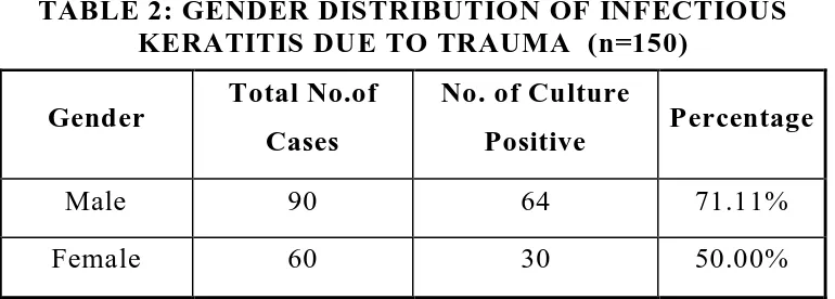
TABLE 1: CULTURE POSITIVITY IN INFECTIOUS KERATITIS DUE TO TRAUMA (n=150)
Total No.of Samples No.of Culture Positive Samples Percentage of culture Positivity Total No. of Bacterial agent Total No. of Fungal agent Total No. of Parasitic agent Total No. of Mixed growth
150 94 62.66% 31 54 6 3
[image:73.612.122.507.454.592.2]Out of total 150 samples, 94 aetiological agents were isolated, samples leading to 62.66% culture positivity. Among Bacterial fungal Parasitic agents isolated fungi was the predominant aetiological in patient with infectious keratitis due to trauma.
TABLE 2: GENDER DISTRIBUTION OF INFECTIOUS KERATITIS DUE TO TRAUMA (n=150)
Gender Total No.of Cases
No. of Culture
Positive Percentage
Male 90 64 71.11%
Female 60 30 50.00%
No of culture Negative, 37.34
[image:74.612.135.490.133.653.2]No of culture Positive, 62.66
CULTURE POSITIVITY IN INFECTIOUS
KERATITIS DUE TO TRAUMA
0 20 40 60 80 100
Total No.of Cases No. of Culture Positivity 90
64 60
30
[image:74.612.138.489.417.657.2]GENDER DISTRIBUTION OF
INFECTIOUS KERATITIS DUE TO
TRAUMA
Male Female
FIGURE - 1
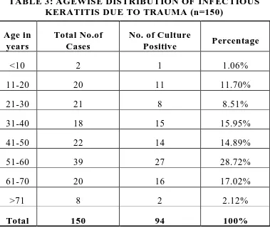
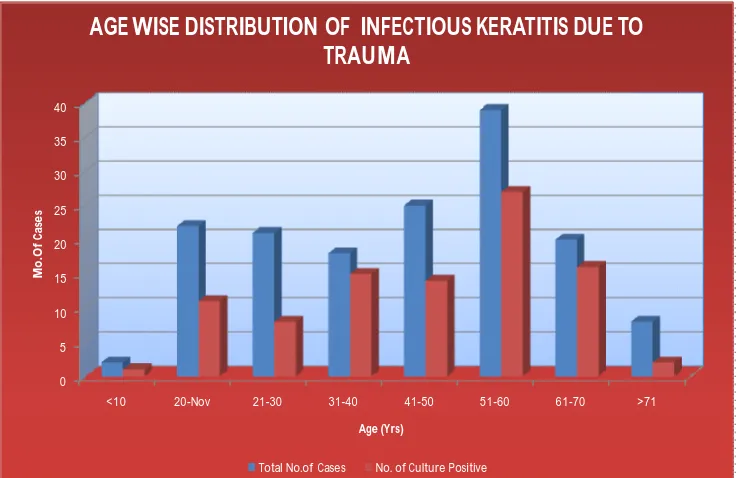
61 TABLE 3: AGEWISE DISTRIBUTION OF INFECTIOUS
KERATITIS DUE TO TRAUMA (n=150)
Age in years
Total No.of Cases
No. of Culture
Positive Percentage
<10 2 1 1.06%
11-20 20 11 11.70%
21-30 21 8 8.51%
31-40 18 15 15.95%
41-50 22 14 14.89%
51-60 39 27 28.72%
61-70 20 16 17.02%
>71 8 2 2.12%
Total 150 94 100%
Prevalence of infectious keratitis due to trauma was more common in 51-60 yrs age group (28.72%)
TABLE 4: DISTRIBUTION OF INFECTIOUS KERATITIS DUE TO TRAUMA IN RURAL AND URBAN AREAS (n=150)
Total Rural Urban
150 97 53
Percentage 64.66% 35.34%
[image:75.612.113.511.468.588.2]0 5 10 15 20 25 30 35 40
<10 20-Nov 21-30 31-40 41-50 51-60 61-70 >71
[image:76.612.129.498.134.373.2]M o. O f C as es Age (Yrs)
AGE WISE DISTRIBUTION OF INFECTIOUS KERATITIS DUE TO TRAUMA
Total No.of Cases No. of Culture Positive
Rural, 64.66% Urban,
[image:76.612.126.500.147.583.2]35.34%
DISTRIBUTION OF INFECTIOUS KERATITIS DUE TO TRAUMA IN RURAL AND URBAN
AREAS
FIGURE - 3
62 TABLE 5: GENDER DISTRIBUTION OF AETIOLOGICAL
AGENTS
Group Agent Male Female Grand
Total
Fungal Agents
Aspergillus flavus 8 6 14
Aspergillus niger 7 4 11
Aspergillus fumigatus 5 3 8
Fusarium spp 9 3 12
Curvularia spp 1 1 2
Penicillium spp 2 1 3
Graphium spp 2 - 2
Scopulariopsis spp 1 1 2
Samples with pure fungal growth 35 19 54 (57.44%)
Bacterial Agents
Staphylococcus. aureus 5 2 7
Staphylococcus. epidermidis
4 2 6
Pseudomonas aeruginosa 3 2 5
Klebsiella Pneumoniae 3 2 5
Acinetobacter boumannii 2 2 4
Escherichia coli 2 2 4
Sample with pure bacterial growth 19 12 31 (32.97%) Mixed
growth
Pseuodmonas aeruginosa + A.fumigatus
2 0 2
Staph. aureus + A.niger 0 1 1
Samples with mixed Microbial Growth
2 1 3
(3.19%) Parasitic
Agent
Acanthamoeba spp 4 2 6
0 5 10 15 20 25 30 35
NO
O
F CA
SES
[image:78.612.133.494.153.677.2] [image:78.612.140.488.442.671.2]TRAUMA FACTOR
DISTRIBUTION OF TRAUMA FACTOR IN
INFECTITIOUS KERATITIS PATIENTS
Male Female
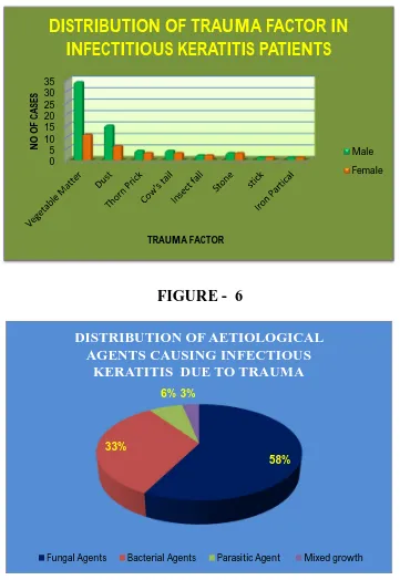
58% 33%
6% 3%
DISTRIBUTION OF AETIOLOGICAL AGENTS CAUSING INFECTIOUS
KERATITIS DUE TO TRAUMA
Fungal Agents Bacterial Agents Parasitic Agent Mixed growth
FIGURE - 5
63 TABLE 6: DISTRIBUTION OF TRAUMA FACTORS IN
INFECTIOUS KERATITIS DUE TO TRAUMA
Trauma Factor
Culture +ve Cases
Percentage Male Female Total
Vegetable Matter 34 11 45 47.87%
Dust 15 6 21 22.34%
Thorn Prick 4 3 7 7.44%
Cow’s tail 4 3 7 7.44%
Stone 3 3 6 6.38%
Insect fall 2 2 4 4.25%
stick 1 1 2 2.12%
Iron Partical 1 1 2 2.12%
Total 64 30 94 100%
0 0.5 1 1.5 2 2.5 3 3.5 4
Male Female
N
O
OF
CAS
[image:80.612.128.497.138.336.2]ES
DISTRIBUTION OF PARASITIC AGENTS CAUSING INFECTIOUS KERATITIS DUE TO TRAUMA
Acanthamoeba spp
FIGURE - 7
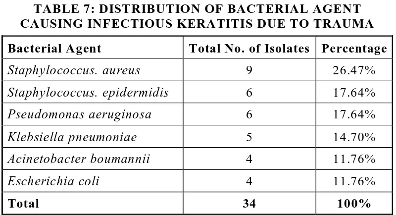
[image:80.612.139.488.411.642.2]64 TABLE 7: DISTRIBUTION OF BACTERIAL AGENT CAUSING INFECTIOUS KERATITIS DUE TO TRAUMA Bacterial Agent Total No. of Isolates Percentage
Staphylococcus. aureus 9 26.47%
Staphylococcus. epidermidis 6 17.64%
Pseudomonas aeruginosa 6 17.64%
Klebsiella pneumoniae 5 14.70%
Acinetobacter boumannii 4 11.76%
Escherichia coli 4 11.76%
Total 34 100%
Staphylococcus aureus was the most common bacterial agent (26.47%) causing infectious keratitis due to trauma.
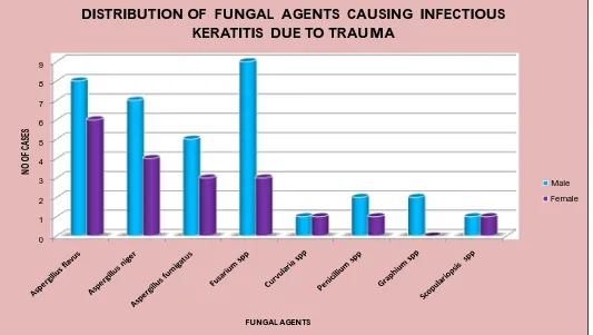
TABLE 8: DISTRIBUTION OF FUNGAL AGENT CAUSING INFECTIOUS KERATITIS DUE TO TRAUMA
Fungal Agent Total No. of Isolates Percentage
Aspergillus flavus 14 24.86%
Aspergillus niger 12 21.05%
Aspergillus fumigatus 10 17.52%
Fusarium spp 12 21.05%
Curvularia spp 2 3.50%
Penicillium spp 3 5.26%
Graphium spp 2 3.50%
Scopulariopsis spp 2 3.50%
Total 57 100%
[image:81.612.121.510.391.627.2]0 1 2 3 4 5 6 7 8 9
N
O OF
CASES
[image:82.792.132.665.154.455.2]FUNGAL AGENTS
DISTRIBUTION OF FUNGAL AGENTS CAUSING INFECTIOUS KERATITIS DUE TO TRAUMA
Male Female
65 TABLE 9: DISTRIBUTION OF PARASITIC AGENT CAUSING INFECTIOUS KERATITIS DUE TO TRAUMA Parasitic agent Total No. of Isolates Percentage
Acanthamoeba spp 6 6.38%
TABLE 10: ANTIBIOTIC SUSCEPTIBILITY PATTERN OF BACTERIAL ISOLATES IN INFECTIOUS KERATITIS DUE TO TRAUMA
Organism Total
No. Amikacin Gentamicin Ciprofloxacin Ofloxacin Cefataxime Cotrimoxazole Cephalexin Vancomycin Oxacillin
Staphylococcus.
aureus 9 9 100% - - 7 77% 7 77% 8 88% 5 55.5% 7 77% 9 100% 7 77% Staphylococcus.
epidermidis 6 6 100% - - 6 100% - - 6 100% 4 66% 4 66% 6 100% 4 66% Pseudomonas
aeruginosa 6 6 100% 5 83% 4 66% 5 83% 4 66% - - 5 83% - - - -Klebsiella
pneumoniae 5 5 100% 4 80% 5 100% 4 80% 3 60% - - 4 80% - - - -Acinetobacter
boumanii 4 3 75% 3 75% 2 50% 2 50% 3 75% - - 3 75% - - - -Escherichia
coli 4 3 75% 3 75% 4 100% 3 75% 3 75% - - 3 75% - - - -Percentage of
Sensitivity 34 94.11% 44.11% 82.35% 61.76% 79.41% 26.47% 76.47% 100% 32.35%
0.00% 10.00% 20.00% 30.00% 40.00% 50.00% 60.00% 70.00% 80.00% 90.00% 100.00%
Amikacin Gentamicin Ciprofloxacin Ofloxacin Cefataxime Cotrimoxazole Cephalexin Vancomycin Oxacillin
94.11% 44.11% 82.35% 61.76% 79.41% 26.47% 76.47% 100% 32.35% PERC E N TA GE
ANTIBIOTICS SUSCEPTIBILITY PATTERN OF BACTERIAL ISOLATES IN INFECTIOUS KERATITIS DUE TO TRAUMA
[image:85.792.107.689.169.478.2]ANTIBIOTICS
TABLE 11: MIC OF VANCOMYCIN FOR STAPHYLOCOCCUS AUREUS
Organism
Minimum inhibitory concentration break- point 0.25 g/ml 0.5 g/ml 1 g/ml >2 g/ml
Staph.auerus (2) 1 1 -
-MIC for the MRSA isolates were studied and found to be 100% sensitive to vancomycin. The break point concentration were 0.25 g/ml and 0.5 g/ml for the isolates.
TABLE 12: ANTIFUNGAL SUSCEPTIBILITY PATTERN BY DISC DIFFUSION TEST METHOD
Agent No.of
isolates
Amphotericin-B (20 g) S>15
mm Itraconazole (10 g) S>23 mm Fluconazole (10 g) S>19 mm Aspergillus flavus
14 9 (64%) 11 (78%) R
Aspergillus niger
12 7 (58%) 12(100%) R
Aspergillus fumigatus
10 7 (70%) 10 (100%) R
Fusarium spp 12 8 (66%) 10 (83%) R
Curvularia spp 2 1 (50%) 1 (50%) R
Penicillium spp 3 1 (33%) 2 (66%) R
Graphium spp 2 1 (50%) 2 (100%) R
Scopulariopsis spp
0 2 4 6 8 10 12
SE
NS
IT
IV
IT
Y
O
F FU
NGAL ISO
LA
TE
S
ANTI FUNGAL SUSCEPTIBILITY TESTING BY DISK DIFFUSION
METHOD
[image:87.792.109.705.168.476.2]Amphotericin-B Itraconazole Fluconazole
Itraconzole was found to be more effective against Aspergillus spp. All fungal isolates were resistant to fluconazole (<15 mm)
TABLE 13: MINIMUM INHIBITORY CONCENTRATION FOR AMPHOTERICIN B - AGAR DILUTION METHOD
Organism 0.25 g 0.5 g 1 g 2 g 4 g 8 g 16 g 32 g 64 g
Aspergillus Flavus 3 2 5 2 1 1
-Aspergillus niger 2 3 3 1 1 1 1
-Aspergillus Fumigatus
1 3 1 2 1 1 1
-Fusarium spp 3 1 3 3 1 1
-Curvularia spp - 1 1
-Penicillium spp - 1 1 1
-Graphium spp - 1 1
-Scopulariopsis spp - 1 1
69 TABLE 14: MINIMUM INHIBITORY CONCENTRATION
FOR ITRACONAZOLE - AGAR DILUTION METHOD
Organism 0.25 g 0.5 g 1 g 2 g 4 g 8 g 16 g 32 g 64 g
Aspergillus Flavus 3 4 2 4 1
-Aspergillus niger 2 3 2 3 1 1
-Aspergillus
Fumigatus - 2 3 2 1 - - 1 1
Fusarium spp 2 3 2 3 1 1
-Curvularia spp 1 1
-Penicillium spp 1 1 1
-Graphium spp - 1 1
-Scopulariopsis spp - 1 1
TABLE 15: MINIMUM INHIBITORY CONCENTRATION FOR
VORICONAZOLE - AGAR DILUTION METHOD
Organism 0.25 g 0.5 g 1 g 2 g 4 g 8 g 16 g 32 g 64 g
Aspergillus Flavus 4 3 4 2 1
-Aspergillus niger 3 2 4 3
-Aspergillus
Fumigatus 1 3 4 2
-Fusarium spp 4 3 3 1 1
-Curvularia spp - 1 1
-Penicillium spp 1 1 1
-Graphium spp - 1 1
-Scopulariopsis spp - 1 1
71 TABLE 16: MINIMUM INHIBITORY CONCENTRATION FOR
AMPHOTERICIN B - BROTH MICRODILUTION METHOD
Organism 0.25 g 0.5 g 1 g 2 g 4 g 8 g 16 g 32 g 64 g
Aspergillus Flavus 2 3 4 2 1 1 1
-Aspergillus niger 1 2 4 2 1 1 1
-Aspergillus
Fumigatus 1 2 3 1 1 - - 1 1
Fusarium spp 3 2 3 3 1
-Curvularia spp - 1 1
-Penicillium spp - 1 1 1
-Graphium spp - 1 1
-Scopulariopsis spp - 1 1
TABLE 17: MINIMUM INHIBITORY CONCENTRATION FOR ITRACONAZOLE - BROTH MICRODILUTION
METHOD Organism 0.25 g 0.5 g 1 g 2 g 4 g 8 g 16 g 32 g 64 g
Aspergillus Flavus 2 3 4 4 1 - - -
-Aspergillus niger 2 3 2 3 1 1
-Aspergillus
Fumigatus 1 2 3 2 - - - 1 1
Fusarium spp 2 1 3 5 1 - - -
-Curvularia spp - 1 1
-Penicillium spp - 1 1 1 - - - -
-Graphium spp - 1 1
-Scopulariopsis spp - 1 1
73 TABLE 18: MINIMUM INHIBITORY CONCENTRATION
FOR VORICONAZOLE - BROTH MICRODILUTION METHOD Organism 0.25 g 0.5 g 1 g 2 g 4 g 8 g 16 g 32 g 64 g
Aspergillus Flavus 2 3 4 5 - - - -
-Aspergillus niger 2 3 4 3 - - - -
-Aspergillus
Fumigatus 2 3 3 1 1 - - -
-Fusarium spp 3 2 3 4 - - - -
-Curvularia spp - 1 1
-Penicillium spp - 1 1 1 - - - -
-Graphium spp - 1 1
-Scopulariopsis spp - 1 1
-One isolate of
Aspergillus Fumigatus
which had MIC of
0 2 4 6 8 10 12 14
MINIMUM INHIBITORY CONCENTRATION BY AGAR DILUTION METHOD
Amphatericin B Itraconazole Voriconazole 0 2 4 6 8 10 12 14
MINIMUM INHIBITORY
CONCENTRATION BY MICRO
DILUTION METHOD
[image:94.612.137.488.116.636.2]Amphatericin B Itraconazole Voriconazole
FIGURE - 12
[image:94.612.140.488.407.632.2]74 TABLE 19: COMPARISION OF MIC IN AGAR DILUTION AND
BROTH MICRODILUTION
Drug Concentration
Amphatericin B MIC < 2 g
Itraconazole MIC < 2 g
Voriconazole MIC<2 g
Organism
Agar Dilution Method Broth Microdilution Method Agar Dilution Method Broth Microdilution Method Agar Dilution Method Broth Microdilution Method Aspergillus
Flavus 12 11 13 13 13 14
Aspergillus
niger 9 9 10 10 12 12
Aspergillus
Fumigatus 7 7 7 8 10 9
Fusarium spp 10 11 10 11 11 12
Curvularia
spp 2 2 2 2 2 2
Penicillium spp 2 3 3 3 3 3
Graphium spp 2 2 2 2 2 2
Scopulariopsis
spp 2 2 2 2 2 2
Total 46 47 49 51 55 56
Percentage 80.70% 82.45% 85.96% 89.47% 96.49% 98.24%
TABLE 20: EVALUATION OF 10% POTASSIUM HYDROXIDE
MOUNT SCREENING TEST
10% KOH Mount
Culture
Total
Positive Negative
Positive 50 2 52
Negative 4 94 99
Total 54 96 150
Sensitivity = TP/ (TP+FN) = 92.59% Specificity = TN/(TN+FP) = 97.91%
76
RESULTS
In this study, 150 cases of traumatic infectious keratitis patients, 94 cases were culture positive (Table 1).
The cases were analyzed under the following parameters.
The age and sex distribution of infectious keratitis was analysed 28.72% of infectious keratitis case were in 51-60 years of age group. Extremes of Age group showed low prevalence (<10 and >70%) (Table 3).
In this study male predominated in all forms of keratitis 71.11% (90/150) were males, and 50% (30/150) were females. (Table 2)
The urban and rural distribution of cases showed higher prevalence of infectious keratitis in rural population accounting for 64.66% (Table 4)
Majority of the aetiological agents were fungi (57.44%) followed by bacteria (32.97%), mixed bacteria and fungal growth was observed in (3.19%) of the cases. The parasitic growth was observed in 6.38% of the cases (Table 5).
Among the fungal isolates, 33 out of 54 (63.15%) cases were due to Aspergillus spp. The next common agent was Fusarium spp 12 out of 54 (21.05%), followed by Pencillium spp 3 out of 54 (5.26%) Graphium spp 2 out of 54 (3.50%), and Scopulariopsis spp 2 out of 54 (3.50%).(Table 8)
Staphylococcus aureus was the most common bacterial isolate, accounting for 26.47%, followed by Pseudomonas aeruginosa (17.64%) Acinetobacter boumannii was isolated in 4 cases (11.76%), Staphylococcus epidermidis (17.64%), Klebsiella pneumoniae (16.70%), Escherichia coli (11.76%) (Table 7).
78 Two Staphylococus aureus isolates exhibited to oxacillin resistant and both isolates were sensitive to vancomycin (Table 11)
Antifungal sensitivity pattern of fungal isolates by disc diffusion test showed that 64% (9) Aspergillus flavus, 58.3% (7) Aspergillus niger isolates, 70% of (7) Aspergillus fumigatus isolates and 66% (8) of Fusarium spp, 50% (1) of Curvularia spp, 33% (1) of pencillium spp, 50% (1) of Graphium spp, and 50% (1) Scopulariopsis spp were sensitive to Amphotericin B (Table 12)
Itraconazole was sensitive in 78% (9) of Aspergillus flavus, 100% (12) of Aspergillus niger isolates, 100% of (10) Aspergillus fumigatus isolates and 83% (10) of Fusarium spp, 50% (1) of Curvularia spp, 66% (2) of pencillum spp, 100% (2) of of graphium spp and 100% (2) of Scopulariopsis spp were sensitive to Amphotercin B (Table 12)
10% Potassium hydroxide mount preparation used as screening test for rap