ROLE OF MICROVESSEL DENSITY, HER-2/ NEU
EXPRESSION AND CD-34 IN PROGNOSTICATION
AND GRADING OF BLADDER CARCINOMAS
Dissertation submitted in partial
fulfilment of the requirements for the degree of
M.D. (PATHOLOGY) BRANCH - III
GOSCHEN INSTITUTE OF PATHOLOGY AND
ELECTRON MICROSCOPY
MADRAS MEDICAL COLLEGE
CHENNAI – 600 003
THE TAMIL NADU
DR. M.G.R. MEDICAL UNIVERSITY
CHENNAI
CERTIFICATE
This is to certify that this Dissertation entitled “ROLE OF MICROVESSEL
DENSITY, HER-2/ NEU EXPRESSION AND CD-34 IN
PROGNOSTICATION AND GRADING OF BLADDER
CARCINOMAS” is the bonafide original work of Dr.SURYALAKSHMI. S,
in partial fulfillment of the requirement for M.D., (Branch - III) in Pathology
examination of the Tamilnadu Dr.M.G.R Medical University to be held in
April 2013.
Prof. Dr. SUDHA VENKATESH, M.D., Prof. Dr. P. KARKUZHALI, M.D.,
Professor of Pathology, Director,
Institute of Pathology and EM, Institute of Pathology and EM,
Madras Medical College, Madras Medical College,
Chennai – 600003. Chennai – 600003.
Prof. Dr. KANAGASABAI, M.D., DEAN,
DECLARATION
I, Dr. Suryalakshmi. S, solemnly declare that the dissertation titled “ROLE OF MICROVESSEL DENSITY, HER-2/ NEU
EXPRESSION AND CD-34 IN PROGNOSTICATION AND GRADING OF BLADDER CARCINOMAS” is the bonafide work done by me at Institute of Pathology, Madras Medical College under the
expert guidance and supervision of Prof. Dr. Sudha Venkatesh, M.D., Professor of Pathology, Institute of Pathology and Electron Microscopy,
Madras Medical College. The dissertation is submitted to the Tamilnadu
Dr. M.G.R Medical University towards partial fulfillment of requirement
for the award of M.D., Degree (Branch III) in Pathology.
Place: Chennai
ACKNOWLEDGEMENT
I take the opportunity to thank Prof. Dr. KANAGASABAI, M.D.,
Dean, Madras Medical College and Government General Hospital, for the
permission given to utilize the vast resources of the Institution.
I also express my sincere thanks to Prof.Dr.P.KARKUZHALI, M.D.,
Director of Institute of Pathology and Electron Microscopy, Madras Medical
College, Chennai for her valuable opinions and encouragement throughout the
study.
I express my heartfelt thanks to Prof.Dr.SUDHA VENKATESH, M.D.,
Professor of Pathology, Institute of Pathology and Electron Microscopy,
Madras Medical College for her valuable advice, encouragement and
suggestions during the study.
I thank all the Professors, Prof.Dr.SHANTHA RAVISHANKAR, M.D.,
Neuropathology, Prof.Dr.GEETHA DEVADAS, M.D., D.C.P.,
Prof.Dr.M.P.KANCHANA, M.D., Institute of Obstetrics & Gynaecology,
Prof.Dr.K.RAMA, M.D., Government Kasturba Gandhi Hospital,
Prof.Dr.INDIRA, M.D., Regional Institute of Ophthalmology,
Prof.Dr.T.CHITRA, M.D., Institute of Child Health and
Prof.Dr.S.PAPPATHI, M.D., D.C.H. who provided constant support and
I express my thanks to all my Assistant Professors for their help and
encouragement during the past 3 years.
I also extend my thankfulness to all my friends, colleagues, technicians
of the Institute of Pathology for their help and support for this dissertation.
I express my heartfelt sincere thanks to all my Assistant Professors for
their help and suggestions during the study.
I also express my thanks to my family for their constant support during
ABBREVIATIONS
Her-2 neu - Human Epidermal Growth Factor Receptor 2
TCC - Transitional cell carcinoma
SCC - Squamous cell carcinoma
MVD - Microvessel density
PUNLMP - Papillary urothelial neoplasm of low malignant potential
SD - Standard deviation
N - Number of cases
IHC - Immunohistochemistry
WHO - World Health Organisation
Rt lateral wall - Right lateral wall
CONTENTS
S. NO. TITLE PAGE
NUMBER
1 INTRODUCTION 1
2 AIMS AND OBJECTIVES 4
3 REVIEW OF LITERATURE 5
4 MATERIALS AND METHODS 35
5 OBSERVATION AND RESULTS 41
6 DISCUSSION 68
7 SUMMARY 79
8 CONCLUSION 82
BIBLIOGRAPHY
ANNEXURES
INTRODUCTION
Bladder cancer is the sixth common cancer in developed
countries(1) and comprises 3.2% of all cancers. Though the mortality due
to bladder cancer shows a steady decrease, there is an increase in its
incidence, especially in females. It is more common in westernised
countries like North America, United kingdom and Australia. The
incidence of bladder cancer in India is 3.2% in males and 0.7% in females
among all malignancies. The two main causes of bladder cancer have
been smoking and industrial exposure. The industrialised countries show
a heavy burden of bladder cancer due to increase in smoking, especially
in females.(3)
Transitional cell carcinoma (TCC) is the commonest carcinoma in
urinary bladder. However, in countries like Egypt and in parts of African
continent, squamous cell carcinoma (SCC) predominates.(2) This is due to
the endemic presence of schistosomiasis which causes chronic irritation
of the bladder.
Though there are many prognostic factors in bladder carcinoma,
histological stage and grade of the tumour have been considered as
important factors. In some cases, tumours with higher grade and stage
show high incidence of recurrence. Hence additional prognostic
information is required to guide clinicians in the management of these
patients.
Microvessel density (MVD) has been considered as an important
prognostic factor in many solid tumours and also in carcinomas of
bladder.(4). Immunohistological methods can be used to identify the
density of newly formed blood vessels. The hot-spot method is
commonly employed where the areas of high MVD are identified in low
power and then they are counted in high power. Though there are many
other methodologies in assessing microvessel density, no standard
technique has yet been adopted either in urology or in general medicine.(4)
Increased MVD has been found to be associated with increased stage,
grade and increased chances of recurrence.
The HER-2/neu gene is located on chromosome 17 which encodes
a 185 kilodalton transmembrane protein. The role of HER-2/neu
over-expression has been reported in many tumours like breast, lung, gastric,
ovarian and oral cancers. HER-2/neu expression in bladder carcinoma has
been studied by many authors and its expression has been found to range
from 9% to 81%.(5) This over-expression has been found to be associated
with increased grade, stage and poorer outcome. Also, targeted therapy is
In this study of 50 cases, an attempt is made to study the role of
microvessel density and overexpression of HER-2/neu
immuno-histochemically in correlation with stage, grade, recurrence, positive
AIMS AND OBJECTIVES
• To identify the incidence and distribution of bladder carcinoma in
patients admitted in Government General Hospital, Chennai during
the year 2011-12.
• To study the histo-morphological features of bladder carcinoma
including tumour size, tumour location, number, histological type,
grade, depth of infiltration, stage, squamous metaplasia and
necrosis.
• To study the immunological expression of HER-2/neu in bladder
carcinoma.
• To study the immunological expression of CD34 in bladder
carcinoma and to find the microvessel density (MVD) by CD 34
expression.
• To evaluate the association of HER-2/neu over expression and
MVD in relation to clinical stage, histological grade, urine
cytology positivity and with other prognostic factors.
• To assess their prognostic significance by following up the patients
REVIEW OF LITERATURE
Bladder carcinomas are neoplasms of the bladder arising from the
lining transitional epithelium. More than 90% of bladder cancer cases are
urothelial (transitional cell) carcinomas which originate in the epithelial
cells that line the bladder wall internally.(7) About 5% of bladder
carcinomas are squamous cell carcinomas, and less than 2% are
adenocarcinomas; small cell carcinoma is less common.(7,9) Bladder
carcinomas can be classified into two types-superficial (80%) and
invasive disease(20%), on the basis of their histological appearance. The
non-muscle-invasive disease has a high recurrence of about 50–70% but
have a low progression rate of about 15–25% of the patients.(1,3)
EPIDIMEOLOGY:
Bladder cancer is the sixth most common malignancy in developed
countries.(1,3) It ranks as the fourth and ninth most frequently diagnosed
cancer in men and women, respectively, in the United States.(1) The most
common type of bladder cancer is transitional cell carcinoma, now
commonly called the urothelial carcinoma, which is derived from the
urothelium, constituting more than 90% of bladder cancer cases.
Squamous cell carcinoma accounts for 1.1% and 2.8% of all bladder
increased in countries where schistosomiasis is endemic.(2)
Adenocarcinoma of the bladder constitutes 1.5% and 1.9% in men and
women respectively, of all bladder tumours worldwide.(15)
ETIOLOGY:
The etiology of bladder cancer is not yet fully understood; it has
been classically associated with both exogenous and environmental risk
factors. The two well known risk factors for bladder carcinoma are
smoking and occupational exposure.(3)
Smokers have 2-4 times the risk of the general population, with
heavy smokers being at five times the risk. The exact mechanism by
which tobacco causes bladder cancer is not yet known, but urothelial
carcinogens such as acrolein, 4-amino-biphenyl, arylamine, and oxygen
free radicals have been implicated. The experimental work of McDonald
and Lund(10) confirmed the carcinogenic properties of β-naphthylamines
in the production of bladder cancer. Importantly, they also demonstrated
that the route of exposure to the carcinogen was not the blood stream but
the urine.
Occupational exposure to aniline dyes, aromatic amines such as
2-naphthylamine and benzidine has been implicated as the second most
common risk factor for bladder cancer. Benzidine, which is the most
and as a hardener in the rubber industry. Aromatic amines are now
prohibited, but people exposed to chemical substances from the
combustion of coal are also known to have an increased risk of bladder
carcinoma. Occupational bladder cancer has also been observed in gas
workers, painters, and hairdressers.
Nutrition also plays a role in bladder cancer. Vitamin A
supplement showed a reduced risk of bladder cancer, while fried food and
fat caused an increased risk.(3) A high intake of fluid was shown to reduce
the risk of bladder cancer in one study but this remains
controversial.(3)Research from Taiwan and Chile has shown an increased
risk of bladder cancer in populations using drinking water with a high
content of arsenic. Hence, these and other water contaminants are being
actively investigated.(3) Consumption of coffee and artificial sweeteners
were implicated in bladder Carcinoma.(11,12)
Other factors implicated in developing bladder cancer and its
progression include analgesic use, chemotherapeutic agents such as
cyclophosphamide,(13,14) pelvic radiation, urinary tract infections
including bacterial, parasitic, fungal, and viral infections and stones in
bladder. There is a causative relationship between the parasite bilharzias
and squamous cell carcinoma of the bladder, frequently seen in the
HISTOLOGICAL TYPES:
The WHO classification is used for bladder carcinomas (Annexure
I). Approximately 90% of bladder neoplasms are urothelial carcinomas.
The remaining 10% comprise all other types of carcinoma, a small
number of sarcomas, and miscellaneous tumours. The classification of
urothelial neoplasms is
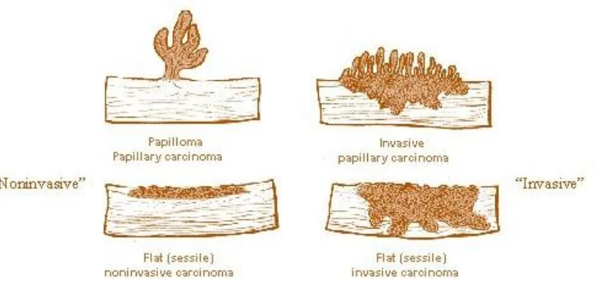
Non-invasive urothelial neoplasms(76):
Urothelial papilloma is a benign neoplasm composed of a delicate fibrovascular core covered by normal looking urothelium. The incidence
is <1% of all bladder tumours.(78) It was first introduced in WHO
classification 1973 and is defined as the same in 2004 WHO/ISUP
classification.
Inverted urothelial papilloma is a benign neoplasm resembling papilloma but having an inverted growth pattern with normal to minimal
cytologic atypia of the cells.(79)
Papillary urothelial neoplasm of low malignant potential (PUNLMP) is a non-invasive papillary urothelial tumour resembling the exophytic urothelial papilloma but shows increased layers of epithelium
PUNLMP was first introduced to replace the previously designated WHO
grade 1 urothelial carcinoma that was defined by the 1998 WHO/ISUP
classification system. Murphy(81) in 1999, interpreted some of the 1973
WHO grade 1 tumours as PUNLMP. Later, Cheng et al(84) reported a
series of 112 patients in 2007 whose bladder tumours showed findings
consistent with PUNLMP, with up to 35 yr of follow-up (median, >12
yr), with tumour recurrence in 29% of patients. Bostwick and Mikuz(82)
translated the 1973 WHO grade 1, 2, and 3 tumours as PUNLMP, low
grade papillary urothelial carcinoma, and high grade papillary urothelial
carcinoma respectively in the year 2002. Reuter and Melamed(83)
interpreted the 1973 WHO grade 1 tumours as PUNLMP, grade 2
tumours as low grade papillary urothelial carcinoma, and grade 3 tumours
as high grade papillary urothelial carcinoma.
Non-invasive low grade papillary urothelial carcinoma is a neoplasm composed of transitional cells lining papillary fronds showing
an orderly appearance, but the variations in architecture and cytologic
features are easily recognisable.(75)
Non-invasive high grade papillary urothelial carcinoma is a neoplasm composed of urothelium lining the papillary fronds with
Carcinoma-in-situ is a non-papillary, flat, lesion in which the surface epithelium contains cells that are cytologically malignant.(73,74)
Primary carcinoma-in-situ which arises de novo and accounts for 1–3%
of urothelial neoplasms is most commonly seen in the bladder. The distal
ureters can be involved in 6–60%, the prostatic urethra in 20–67%, and
the prostate ducts and acini in up to 40%.(74)
Infiltrating urothelial carcinoma:
These neoplasms are similar to non infiltrating neoplasms except
for the key feature of invasion which is characterized by nests, clusters, or
the presence of single cells within the papillary cores, lamina propria or
into the muscle.
This type has numerous variants(50) which include
Infiltrating urothelial carcinoma with squamous differentiation(51) which shows the presence of nests of malignant squamous epithelium,
characterized by polygonal cells that frequently display dyskeratosis,
keratin pearl formation, and occasional intercellular bridges. Squamous
differentiation within urothelial carcinoma occurs in about 21% of
urothelial carcinomas of the bladder and is associated with poorer
prognosis due to the resistance of squamous elements to chemotherapy
Glandular differentiation(51) Glandular differentiation within urothelial carcinoma was first described in the literature in 1968. It occurs
in 6% of urothelial carcinomas and is defined by the presence of true
glandular spaces within urothelial carcinoma. These glandular structures
consist of tubular glands or glands resembling enteric epithelium, often
associated with variable mucin production.
Trophoblastic differentiation(52) was described in 1904. Since then, more than 30 cases of tumours with trophoblastic differentiation,
including cases of pure choriocarcinoma, have been reported. It
encompasses a wide range of patterns, including formation of
syncytiotrophoblast, formation of areas resembling pure choriocarcinoma,
and urothelial carcinomas without giant cells that express human
chorionic gonadotropin. The presence of trophoblastic differentiation is
associated with poorer prognosis.
Carcinomas with a nested pattern(53) are very rare with an incidence of 0.3%. In 1979, Stern made the initial observation of an
unusual benign-looking bladder carcinoma of “Brunn's nest origin,” and
since then more than 50 cases have been reported. It is characterized by
tumour cells with a nested growth pattern, which can be confused with
von Brunn nests, cystitis cystica, and nephrogenic adenoma. These
that nested variant tumours are associated with progressive or recurrent
disease.
The microcystic variant, has micro and macrocysts and tubular structures containing granular eosinophilic material which is PAS and
Alcian blue positive and necrotic cellular debris. Atleast 25% of the
tumour should be comprised of microcystic component to designate a
tumour as microcystic urothelial carcinoma. Microcystic urothelial
carcinoma was first described in 1991 by Robert H. Young and Lawrence
R. Zukerberg.(55) Till now, 10 cases(56,57,58) have been reported. The
presentation and prognosis are similar to conventional invasive urothelial
carcinoma.(56,57,58)
The micropapillary variant is characterized by a conventional infiltrating urothelial carcinoma with an admixed micropapillary pattern
involving the surface urothelium which consist of slender, delicate
filiform papillary processes that do not contain distinct fibrovascular
cores which on cross section, have a glomeruloid appearance.
Micropapillary variant of urothelial carcinoma was first described by
Amin and colleagues(59) in the urinary bladder in 1994. At least 115
cases(59,60,561) have been described so far. The micropapillary variant of
in the absence of muscularis propria in the biopsy, the muscle invasion is
considered as present.
Lymphoepithelioma-like variant is defined as a urothelial carcinoma that histologically resembles lymphoepithelioma of the
nasopharynx. It is characterized by proliferation of primitive anaplastic
cells infiltrating with a prominent, lymphocytic background. Unlike
lymphoepithelioma of the nasopharynx, Epstein Barr virus has not been
found either using immunohistochemistry or in situ hybridization
technology.(62) Pure and predominant forms of lymphoepithelioma-like
carcinoma have a more favourable prognosis. When the
lymphoepithelioma - like morphology is only focally present, the
expected behavior is the same as for conventional urothelial carcinoma of
the same grade and stage.
The lymphoma-like and plasmacytoid variants of urothelial carcinoma exhibit the morphologic features of lymphoma or
plasmacytoma. This variant of urothelial carcinoma was first described in
1991 by Zukerberg et al(63) and Sahin et al.(64) Till now, at least 30 cases
of this variant of urothelial carcinoma have been reported.(64,65,66) Of the
cases reported, the clinical course following diagnosis has been variable
with some patients progressing rapidly to death, whereas others have
The giant cell variant of urothelial carcinoma is characterized by the presence of tumour giant cells exhibiting marked nuclear atypia,
along with a component of conventional urothelial carcinoma. These
tumours have some similarity to the giant cell tumours of the lung. The
significance of diagnosing the giant cell variant of urothelial carcinoma is
that it is associated with a poor prognosis.(67,68)
Sarcomatoid variants are a group of biphasic malignant neoplasms exhibiting morphologic evidence of both epithelial and
mesenchymal differentiation. More than 100 cases have been reported
since the mid 1800s, many originally classified as carcinosarcoma, but in
general this is a rare tumour.(69,70) The significance of diagnosing this
lesion lies in its association with a poor prognosis. These tumours are
typically diagnosed at advanced local stage, and they often exhibit nodal
or distant metastases. 70% of patients will die within 2 years of diagnosis.
The undifferentiated carcinoma(71) category includes rare tumours that cannot be otherwise classified and usually exhibit
high-grade malignant morphology.
Clear cell differentiation has been discussed as a distinct variant of urothelial carcinoma in 1995 since the presentation of 2 cases was
incidence is unclear because of the recent recognition of this type as a
[image:22.595.113.531.155.353.2]separate entity.
Figure 1: Gross types of bladder cancers
HISTOLOGICAL GRADING:
Histologic grading is one of the most important prognostic factors
in bladder cancer. Several attempts at grading were done by many
pathologists.
The main such systems were those of Ash in 1940, Mostofi’s
modification of Ash in 1960 (adopted by the American Bladder Tumour
Registry), Bergkvist et al. in 1965, and Malmstrom et al. in 1987. The
first widely accepted grading system for papillary urothelial neoplasms
was the WHO (1973) classification system, which divided urothelial
grade 3.(16)Histologic grading is based on the degree of cellular anaplasia,
with grade 1 tumours having the least degree of anaplasia, but compatible
with a diagnosis of malignancy; grade 3 tumours have the most severe
degree of anaplasia, and grade 2 have an intermediate degree of cellular
anaplasia. Anaplasia is further defined by the authors of the WHO (1973)
classification as increased cellularity, nuclear crowding, alteration in
polarity of cells, failure of differentiation from the base to the surface,
nuclear pleomorphism, variations in nuclear chromatin pattern, displaced,
abnormal mitotic figures, and giant cells. The WHO grading system
divides bladder cancer into 4 types based on grade such as papilloma,
grade1TCC, grade 2 TCC and grade 3 TCC.
PAPILLOMA:
This is a benign neoplasm having papillae lined by normal
appearing transitional epithelium.
GRADE 1 UROTHELIAL CARCINOMA:
Grade 1 papillary carcinoma consists of an orderly arrangement of
transitional cells lining delicate papillae with minimal architectural
abnormality and minimal nuclear atypia. The urothelium is often
thickened to more than seven cell layers but there is minimal complexity
and cohesiveness, with intact superficial layer of cells. They have fine
granular chromatin and mitotic figures are usually rare. They are
commonly seen around the ureter (69%). Since recurrence can still occur
in these patients, long term follow-up is recommended for these cases.
GRADE 2 UROTHELIAL CARCINOMA:
Grade 2 carcinomas retain some of the orderly architectural
appearance and maturation of grade 1 carcinoma, but they display focal
moderate variation in orderliness. Cytologic abnormalities are present,
with moderate degree of nuclear crowding, moderate loss of cell polarity,
moderate degree of nuclear hyperchromasia, moderate anisonucleosis,
and occasional prominent nucleoli. Mitotic figures can be seen but are
usually limited to the lower half of the urothelium. Superficial cells
(umbrella cells) are usually present.
Some authors consider both nuclear pleomorphism and mitotic
count as criteria for subdividing grade 2 urothelial cancer (grade 2A and
2B), and they have been successful in identifying groups of patients with
urothelial cancers with different outcomes.(17,18) However, sub
classification of grade 2 urothelial carcinoma is not recommended by
recurrence in patients of grade 2 urothelial carinoma is 45–67% and
progression to invasion can occur in about 20% of patients.
GRADE 3 UROTHELIAL CARCINOMA:
There is obvious loss of normal architecture and cell polarity, and
frequent atypical mitotic figures. The superficial cell layer is partially or
completely absent, accompanied by prominent cellular dyscohesion.
Cellular anaplasia characterised by hyperchromasia, nuclear crowding,
failure of differentiation and giant cells is frankly evident. Mitosis can be
seen in all levels of epithelium and multiple nucleoli may be evident. The
papillae may appear fused and branching. The recurrence risk for patients
with non-invasive grade 3 cancer is 65–85%, with in cases of invasive
cancer recurrence occurs in 20–52% of patients.
In 1998, a revised system for grading non-invasive papillary
urothelial neoplasms of the urinary bladder was proposed and was
subsequently formally adopted by the World Health Organization.
In 2004, a grading system for non-invasive papillary urothelial
neoplasms was given by International Society of Urologic Pathology
(ISUP), which was later accepted by WHO. According to this new
system, noninvasive papillary urothelial neoplasms are divided into four
malignant potential (PUNLMP), low-grade papillary urothelial
carcinoma, and high-grade papillary urothelial carcinoma.
INTERNATIONAL SOCIETY OF UROLOGICAL PATHOLOGY/
WHO(2004) CLASSIFICATION:
PAPILLOMA:
This entity is similar to the one described in 1973 WHO
classification consisting of papillae lined by benign looking transitional
epithelium.
PAPILLARY UROTHELIAL NEOPLASM OF LOW MALIGNANT
POTENTIAL (PUNLMP):
PUNLMP is a low-grade urothelial tumour with a papillary
architecture. This lesion is histologically defined by the WHO (2004)
classification system as a papillary urothelial tumour resembling the
exophytic urothelial papilloma, but with increased cellular proliferation
exceeding the thickness of normal urothelium and minimal atypia;
polarity is generally preserved in these tumours. All such tumours would
have been considered grade 1 urothelial carcinomas by the WHO 1973
LOW GRADE UROTHELIAL CARCINOMA:
A low-grade papillary urothelial carcinoma shows slender papillae
with frequent branching and variation in nuclear polarity, nuclei show
enlargement and irregularity; chromatin is vesicular, and nucleoli are
often present. Mitotic figures may occur at any level. The majority of
these cases would have been considered as grade 2 in the WHO (1973)
classification. Most patients have a single tumour in the posterior or
lateral bladder wall. However, 22% of patients with low-grade papillary
urothelial carcinoma have two or more tumours. Tumour recurrence,
stage progression and tumour-related mortality are 50%, 10% and 5%,
respectively.
HIGH GRADE UROTHELIAL CARCINOMA:
The cells lining the papillary fronds show an obviously disordered
arrangement with cytologic atypia. All tumours classified as grade 3 in
the 1973 WHO scheme, as well as some assigned grade 2 in that
classification, would be considered high grade carcinoma in the 2004
WHO classification. The papillae are frequently fused. The nuclei are
pleomorphic with prominent nucleoli. Polarity is altered. Mitotic figures
are frequent. Carcinoma in situ is frequently evident in the adjacent
progression and death due to disease can be seen in as many as 65% of
patients.
Other recent proposals for bladder cancer grading:
The Ancona 2001 refinement of the 1973 WHO classification(19)
divides urothelial tumours into two main groups based on growth pattern:
flat and papillary. Flat tumours include reactive changes, dysplasia, and
carcinoma in situ. Papillary tumours include papilloma, grade 1 papillary
carcinoma, grade 2 papillary carcinoma, and grade 3 papillary carcinoma.
The publication of the 1999 WHO blue book introduced a new
grading scheme.(20) This new classification retained the three-tiered
numbering system (grade 1, grade 2, and grade 3 carcinoma), but tumours
formerly classified as 1973 WHO grade 1 were subdivided into PUNLMP
and grade 1 tumours. Papillary tumours were subclassified as papilloma,
papillary urothelial neoplasm of low malignant potential, grade 1, grade
2, and grade 3 papillary urothelial carcinoma
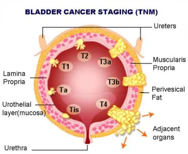
STAGING:
The TNM staging system (Annexure II) is widely used in western
countries. It is the best available predictor of prognosis and is
Early/Superficial bladder cancer is otherwise called as non-muscle
invasive cancer carcinoma in situ, Ta tumours and T1 tumours are
included under superficial bladder carcinoma.
Invasive bladder cancer includes T2 and T3 tumours. In T2 lesion
the tumour has spread to the muscle layer whereas in case of T3 tumours,
it has it has grown through the muscle layer.
Advanced bladder cancer is the one which is widespread outside
[image:29.595.124.496.364.667.2]the bladder. It includes T4 bladder tumours.
PROGNOSTIC FACTORS IN BLADDER CARCINOMA:
Prognostic factor is defined as any variable that provides
information useful in assessing the outcome at the time of diagnosis of
the disease. The prognostic factors are classified as clinical factors,
pathological factors and genetic/molecular factors.
The clinical factors(80) include patient age and a previous history of
bladder cancer.
AGE: Younger patients with bladder cancer appear to have a
favourable prognosis, because they usually present as superficial tumours
and low-grade tumours.
RECURRENCE: Mullerad et al and Kang et al reported that a
previous history of bladder cancer was an independent predictor of
cancer-specific survival.
The pathological factors play more useful role in assessing prognosis. It
includes the following
1. SIZE: Tumour size > 5 cm has been associated with 35% of
invasion compared to 9% invasion in small bladder tumours and is
2. LOCATION: Tumours arising from the bladder neck are
associated with a poorer prognosis. Tumours of the bladder dome tend to
present as higher-grade lesions, whereas tumours of the lateral walls and
the ureteric orifices tend to be of lower grade.(26)
3. NUMBER: Multiple tumours present more frequently with
recurrence (40-90%) when compared to single tumours (18-60%). (22)
3. HISTOLOGICAL TYPE: Non-papillary TCCs tend to present as
higher grade, stage and more aggressive tumours.(107)
4. LYMPH NODE INVOLVEMENT: Lymph node involvement is
associated with increased chances of recurrence and disease progression
and has an overall poor outcome.(108)
5. STAGE: Since Ta lesions are confined to basement membrane
they are associated with good prognosis. As the tumour invades the
muscle layer the prognosis become poorer.(23)
6. GRADE: Grade is an important prognostic indicator for
progression, mortality and recurrence.(24) High grade tumours are
associated with increased chances of recurrence and have a high risk of
7. CARCINOMA-IN-SITU CHANGES: Recurrence rates are
higher in transitional cell carcinomas which have associated
carcinoma-in-situ changes involving the adjacent mucosa.(25)
8. LYMPHO-VASCULAR INVASION: This feature, as
determined microscopically with H&E stain or by vascular stains in either
lymph vessels or blood vessels, is associated with an increased rate of
recurrence.(27)
9. SQUAMOUS METAPLASIA: Squamous metaplasia in
transitional cell carcinomas are fairly common and this squamous
epithelium resist radiotherapy. Hence they have a poor prognosis when
they present in inoperable stage.(90)
The molecular factors which are associated with prognosis are
1. MICROVESSEL DENSITY: This feature is alleged to be an
independent prognostic indicator which is associated with increased
stage, and more chances of recurrence.(28)
2. P53 OVEREXPRESSION: Nuclear over expression of P53 is
related to both grade and stage of bladder carcinoma.(29).
3. ALTERED EXPRESSION OF RB GENE: Tumours exhibiting
reduced expression of the RB protein have an aggressive behaviour than
4. LOSS OF E-CADHERIN: Tumours showing loss of E-cadherin
has worse prognosis than those in which this surface antigen was
present.(31)
5. HER 2/NEU EXPRESSION: Increased HER-2/neu expression
of this marker is associated with higher grade, stage and metastatic
growth.(32)
IMMUNOHISTOCHEMISTRY (IHC):
Albert Coons et al in 1941 first labelled antibodies directly with
fluorescent isocyanate. Nakane and Pierce et al in 1966, introduced the
indirect labelling technique in which the unlabelled antibody is followed
by second antibody or substrate. Various stages of development of
immunohistochemistry include peroxidase – antiperoxidase method
(1970), alkaline phosphatase labelling (1971), avidin biotin method
(1977) and two layer dextrin polymer technique (1993).(33)
Antigen retrieval:
Antigen retrieval can be done by the following different techniques to
unmask the antigenic determinants of fixed tissue sections.
1. Proteolytic enzyme digestion
3. Pressure cooker antigen retrieval
4. Microwave and trypsin antigen retrieval
PROTEOLYTIC ENZYME DIGESTION:
Huank et al in 1976 introduced this technique to breakdown
formalin cross linkages and to unmask the antigen determinants. The
most commonly used enzymes include trypsin and proteinase. The
disadvantages include over digestion, under digestion and antigen
destruction.
MICROWAVE ANTIGEN RETRIEVAL:
This is a new technique most commonly used in current practice.
Microwave oven heating involves boiling formalin fixed paraffin sections
in various buffers for rapid and uniform heating.
PRESSURE COOKER ANTIGEN RETRIEVAL:
Miller et al in 1995 compared and proved that pressure cooking
method has fewer inconsistencies, less time consuming and can be used
to retrieve large number of slides than in microwave method.
PITFALLS OF HEAT PRETREATMENT:
Drying of sections at any stage after heat pretreatment destroys
and fatty tissues tend to detach from slides while heating. Not all antigens
are retrieved by heat pretreatment and also some antigens like PGP 9.5
show altered staining pattern.
DETECTION SYSTEMS:
After addition of specific antibodies to the antigens, next step is to
visualize the antigen antibody reaction complex. The methods employed
are direct and indirect methods.
In the direct method, primary antibody is directly conjugated with
the label. Most commonly used labels are flouro-chrome, horse radish
peroxidase and alkaline phosphatase. Indirect method is a two-step
method in which labeled secondary antibody reacts with primary
antibody bound to specific antigen. The use of peroxidase enzyme
complex or avidin biotin complex further increases the sensitivity of
immunohistochemical stains(33).
In 1993, Pluzek et al introduced enhanced polymer one step
staining, in which large numbers of primary antibody and peroxidase
enzymes are attached to dextran polymer back bone. This is a rapid and
Dextran polymer conjugate two step visualization system is based
on dextran technology in Epos system. This method has greater
sensitivity and is less time consuming.
Uses of IHC in bladder pathology (34)
1. Assessment of prognosis by using markers for microvessel density
(CD 34, CD 31, VEGF), HER-2/neu, P53, EGFR and other markers.
2. Assessment of metastatic lesions of possible bladder origin by
using antibodies to CK7 and CK20, uroplakin, thrombomodulin as well
as 34βE12 and 4A4.
3. Evaluation of spindle cell lesions to distinguish sarcomatoid
carcinoma from mesenchymal lesions.
MICROVESSEL DENSITY:
Tumour stage and histopathological grade are the most important
prognostic factors affecting the survival of bladder carcinoma, but
tumours with similar stage and grade may show different outcome. Thus
there is some evidence that some other prognostic factors also play a role
in prognosis of patients. A majority of studies have assessed the
prognostic value of measuring tumour angiogenesis (i.e., measurement of
tumour microvessel densities) and have found a positive association
between increasing microvessel densities and prognosis.(28) Angiogenesis
pre-existing blood vessels. The process of neovascularization in tumours
is regulated by the combined action of tumour cells, stromal cells and
inflammatory cells.(38) Neovascularisation is essential for both benign and
malignant tumours especially when it grows beyond 2 mm3. Microvessel
density has been shown to add to prognostic information in a number of
solid tumours including prostate (Weidner et al, 1993), colon (Takahashi
et al, 1995), lung (Mattern et al, 1996) and breast cancer (Linderholm et
al, 1999). Microvessel density, has also recently been proven to add
prognostic information in bladder carcinoma. However, contradictory
results have also been reported;(39) this may be due to significant
differences in the methods employed for sample selection, techniques of
immunostaining, counting of vessels and statistical analysis, although a
number of biological differences may account for the discrepancy.
MICROVESSEL DENSITY BY IMMUNOHISTOCHEMISTRY:
Immunohistochemically, the blood vessels are identified by
highlighting them using specific markers. Targeted therapy has been
useful in cases of marker positive tumours. The markers can be divided
into panendothelial markers and markers which bind selectively with the
activated endothelium. The examples of panendothelial markers are CD
31 and CD34 which bind with all small and large vessels. The problems
CD 31 and staining of inflammatory cells by CD 34. These can be
reduced to some extent by pre treatment with microwave oven. The
activated endothelial markers like CD 105 bind specifically to the
proliferating endothelium and hence do not stain for normal blood vessels
or lymphatics. Thus they are more specific as a marker of angiogenesis.
Vascular hot spots are defined as regions of high vascular density
within the tumour. This was first described by Weidner et al, 1991in
breast cancer.(85) These hot spots can be identified by inspecting the
sections under low power. Atleast 10 of these hot spots should be
analysed so that the chance of missing them is reduced. They are seen
predominantly at the peripheral margins of the tumour. Once the vascular
hot spot is identified, they are viewed under high power to count
individual microvessels. The counting can be done either manually or by
using Computerised Image Analysis systems. Magnifications of the order
of 200 – 400X and field sizes ranging from 0.12 to 1.00 mm2 have been
used by Vermeulen et al, (1996). A higher magnification improves the
detail of the image and allows the identification of even single endothelial
cell. According to Weidner et al (1991), any highlighted endothelial cell
or cell cluster clearly separate from adjacent microvessels, tumour cells
and other connective tissue elements should be regarded as a distinct
necessary to identify a microvessel. However the cut-off calibre size
necessary to designate a microvessel has not been specified by many
authors and hence even single endothelial sprout and a large calibre
vessel are included in counting. Atleast three different areas of vascular
hot spots are counted in 200X magnification and the highest of these is
taken as the microvessel density.
The mean microvessel density can be calculated after the tumour is
divided into characteristic groups. The values are compared and statistical
significance is calculated.
Computerised Image Analysis Systems is an automated counting
technique that improves reproducibility and reduces inter-observer
variability. According to Wakui et al (1992) and Visscher et al (1993), it
has been considered as a more objective method of assessing microvessel
density. Additional parameters like the number of vessels with a certain
dimension range, the vessel luminal area, vessel luminal perimeter and
the number of immunostained areas per microscopic field can also be
HER-2/NEU RECEPTORS:
The HER-2/neu gene was originally called ‘neu’ as it was first
derived from rat neuro/glioblastoma cell lines. Coussens and coworkers
named it HER2 because its primary sequence was very similar to Human
Epidermal Growth Factor receptor (EGFR or ERBB or ERBB1).(86) This
human proto-oncogene, also known as c-erbB2, ErbB-2 is a
185-kilodalton transmembrane receptor tyrosine kinase located at
chromosome 17q. These proteins belong to subclass I of the super-family
of receptor tyrosine kinases. They are expressed in many tissues of
epithelial, mesenchymal, and neuronal origin and are critical for cell
proliferation and tissue differentiation. The clinical significance of
HER-2/neu has already been evaluated in colorectal, breast, stomach, lung,
head and neck, pancreatic, urothelial carcinoma, and gliomas and prostate
cancers; patients with elevated HER-2/neu demonstrate poor survival
compared with patients with lower level of HER-2/neu.(46,47,48)
Several studies have reported an association between prognosis and
HER-2/neu gene over expression in bladder cancers. HER 2/neu protein
over expression and HER-2/neu gene amplification were correlated with
higher histologic tumour grade and invasion in many studies. The first
report of increased amplification and over expression of the HER-2/neu
Eschenbach AC, et al in 1990.(40) Till then there were many studies which
showed an association of HER-2/neu expression with increased grade and
stage in bladder carcinomas.(41,42,43)Some studies also showed that
HER-2/neu was an independent variable in determining patient survival.(44,45)T
he prevalence of HER-2/neu expression in bladder carcinomas has ranged
from 2% to 81% . In a recent study by Lae M, Couturier J, Oudard S,
Radvanyi F et al in 2010, HER-2/neu protein over-expression was found
in 9.2%, while HER-2/neu gene amplification was found in 5.1% of
tumour specimens.(49) HER-2/neu can be assayed by immuno
histochemistry for protein over expression and by fluorescent in situ
hybridization for gene amplification.
The scoring system which is used for HER-2/neu expression in
bladder cancers is
Staining pattern Score Her2neu expression
No staining 0 Negative
Weak staining in part of membrane of
less than 10% of the cells. 1 Negative Complete membranous staining of weak
or moderate intensity in >10% of tumour cells.
2+ Positive
Strong complete membranous staining in more than 10 % of tumour cells creating a fish net pattern.
3+ Positive
MATERIALS AND METHODS
This study is a combined retrospective and prospective descriptive
study of bladder carcinomas conducted in the Institute of Pathology,
Madras Medical College and Rajiv Gandhi Government General
Hospital, Chennai during the period between January 2010 and December
2011.
A total of 19,898 cases were submitted to the Department of
Pathology, Rajiv Gandhi Government General hospital during the period
of January 2010 to December 2011 for histopathological examination.
Out of that, 143 cases were bladder cases. Among them, 13 specimens
were radical cystectomies, 2 specimens were simple cystectomies, 90
specimens were TURBT and 38 specimens were small biopsies. The total
number of non- neoplastic and malignant cases of the urinary bladder was
27 and 116 respectively.
Out of the malignant cases, 114 cases were carcinomas, one was a
case of inflammatory myofibroblastic tumour and another one was a case
of leiomyosarcoma of bladder. Out of the 114 cases, 101 were TCCs, 7
cases were adenocarcinomas and 6 cases were SCCs. The TCC cases
Source of data:
The bladder carcinoma cases reported in the Institute of Pathology,
Madras Medical College and Rajiv Gandhi Government General hospital
from January 2010 to December 2011 which have been sent by the
Department of Urology.
Inclusion criteria:
All the cases of transitional cell carcinomas reported in bladder
specimens irrespective of the age and sex and the procedure done were
included for the study.
Exclusion criteria:
Non-neoplastic lesions and benign neoplasms of the bladder.
Carcinomas other than transitional cell carcinomas.
Cases with inadequate material.
Method of data collection:
Detailed history of the cases regarding age, sex, history, type of
procedure, site, size, stage, previous surgery details and urinary cytology
were obtained for all the 101 transitional cell carcinomas reported during
the period of study from surgical pathology records. Hematoxylin and
Eosin stained 4 µ thick sections of the paraffin tissue blocks of specimens
evaluated: Age, gender, tumour size and tumour location (base, lateral,
posterior walls and trigone).
Carcinomas were classified as transitional cell type which includes
papillary/non-papillary, invasive/non-invasive and any other special types
like lymphoepithelial carcinoma, sarcomatoid carcinoma. Other
parameters like squamous metaplasia, necrosis and sarcomatoid
component were noted. Regarding the depth of invasion, the carcinomas
were classified into 4 groups: T1 (invasion of mucosa and submucosa),
T2 (invasion of muscle layer in which T2a is invasion of superficial
muscle and T2b is invasion of deep muscle), T3 (invasion of perivesical
tissue in which T3a means microscopic involvement and T3b means
macroscopic involvement) and T4 (invasion of adjacent organs in which
T4a is invasion of prostate, uterus and vagina and T4b is involvement of
pelvic and abdominal wall), and according to grade the carcinomas were
divided into 3 groups: PUNLUMP, low grade urothelial carcinoma and
high grade urothelial carcinoma according to the recommendations of the
WHO (2004). Carcinoma staging was done according to TNM
classification of bladder carcinomas (Annexure-II). The tumours were
further evaluated for the presence of infiltration, necrosis, squamous
metaplasia and were graded as present or absent. 50 cases of bladder
and their representative formalin fixed paraffin embedded tissue samples
were subjected to immunohistochemistry for a panel of 2 markers- CD 34
and HER-2/neu.
Immunohistochemical evaluation:
Immuohistochemical analysis of markers CD 34 and HER-2/neu
were done in paraffin embedded tissue samples using Super-sensitive
polymer HRP system based on non-biotin polymeric technology. 4 µ
thick sections from formalin fixed paraffin embedded tissue samples were
transferred on to gelatin coated slides. Heat induced antigen retrieval was
done. The antigen was bound with mouse monoclonal antibody
(Biogenex) against CD 34 protein and HER-2/neu protein and then
detected by the addition of secondary antibody conjugated with horse
radish peroxidase-polymer and diaminobenzidine substrate. The step by
step procedure of Immunohistochemistry is given in Annexure III.
Antigen Vendor Species(clone) Dilution Positive control
CD 34 Biogenex Mouse Ready to
use Bladder
HER-2/neu Biogenex Mouse Ready to
Interpretation and scoring system:
The immunohistochemically stained slides were analyzed for the
presence of reaction, cellular localization, percentage of cells stained and
intensity of reaction. Membrane staining was assessed for HER-2/neu and
cytoplasmic staining for CD 34.
HER-2/neu immuno-reactivity was assessed as being positive when
tumours exhibited intense nuclear staining and absent cytoplasmic
staining and was categorized into 2 groups: 2+ (complete membranous
staining of weak or moderate intensity in >10% of tumour cells) and
3+(strong complete membranous staining in more than 10 % of tumour
cells creating a fish net pattern)
CD 34 staining in the endothelial cells was noted. Any highlighted
endothelial cell or cell cluster clearly separate from adjacent
microvessels, tumour cells and other connective tissue elements were
regarded as a distinct countable microvessel. A lumen or the presence of
red blood cells was not taken as an essential criteria to identify a
microvessel. Hot spots were identified and atleast 3 such hot spots were
identified under 200X power and the vessels were counted. The
maximum of these was taken as microvessel density. The mean
Statistical analysis :
The statistical analysis was performed using statistical package for
social science software version 11.5 which consisted computing the
frequency counts and percentages for qualitative variables and mean for
the quantitative variables. The expression of microvessel density was
correlated with clinico- pathological factors like gender, tumour site,
tumour configuration, size, histological types, histological grade, depth of
infiltration, stage, squamous metaplasia, necrosis, sarcomatoid
component, urine cytology and recurrence using the Student t-test and
Anova t-test. HER-2/neu was also correlated with these parameters using
OBSERVATION AND RESULTS
In the study period of 24 months from January 2010 to December
2011, a total of 19,898 specimens were received in the Institute of
Pathology, Madras Medical College for histological examination. Total
numbers of bladder specimens received were 143. The total number of
non-neoplastic and malignant cases was 27 and 116 respectively. Out of
116 malignant cases 114 cases were carcinomas, 1 was a case of
Inflammatory myofibroblastic tumour of bladder and another 1 was a
case of leiomyosarcoma. Thus the distribution of non-neoplastic lesions
was 20.9%, and of malignant tumours were 81.1% among the bladder
specimens.
Among the 143 bladder specimens, there were 13 radical
cystectomies, 2 simple cystectomies, 90 TURBTs and 38 small biopsies.
All 13 radical cystectomies were done to treat bladder carcinoma. Out of
2 simple cystectomies 1 was done for carcinoma and 1 for inflammatory
myofibroblastic tumour.
Of the 114 total carcinomas arising from the bladder, transitional
cell carcinomas were the most common constituting 101 cases accounting
for 88.6% of carcinomas. Adenocarcinoma of bladder constituted 7 cases
accounting for 5.3% of carcinomas arising from urinary bladder (Table 1
and chart 1)
TABLE : 1 - HISTOLOGICAL SUBTYPES OF BLADDER CARCINOMAS
Histological subtypes Number of cases(N) Percentage Transistional cell carcinoma 101 88.6%
Adenocarcinoma 7 6.1%
Squamous cell carcinoma 6 5.3%
Total number of cases 114 100%
Transitional cell carcinomas had a peak incidence in the age group
of 61-70 years. The youngest age of presentation of bladder cancer was at
22 years in this study. The mean age was 58.74. (Table 2 and chart 2)
TABLE : 2 - AGE WISE DISTRIBUTION OF TRANSITIONAL CELL CARCINOMAS
Age group Number of cases Percentage
21 - 30 years 1 1%
31 - 40 years 7 7%
41 - 50 years 17 16.8%
51 - 60 years 29 28.7%
61 - 70 years 32 31.7%
71 - 80 years 13 12.9%
81 - 90 years 2 1.9%
Among the 101 cases of transitional cell carcinomas, 79 (78.2%)
cases were reported in males and 22 (21.8%) cases were reported in
[image:50.595.106.511.547.756.2]females. (Table 3 & Chart 3)
TABLE : 3 - SEX DISTRIBUTION IN TRANSITIONAL CELL CARCINOMAS
Sex Total number of cases Percentage
Male 79 78.2%
Female 22 21.8%
Total 101 100%
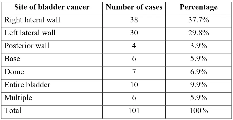
In this study, 38(37.7%) cases involved the right lateral wall,
30(29.8%) cases involved the left lateral wall, 4(3.9%) involved the
posterior wall, 6(5.9%) involved the base, 7(6.9%) involved the dome,
10(9.9%) involved the entire bladder and in 6(5.9%) cases the tumour
was multiple. (Table 4 and Chart 4)
TABLE : 4 - DISTRIBUTION OF SITE OF INVOLVEMENT IN TRANSITIONAL CELL CARCINOMAS
Site of bladder cancer Number of cases Percentage
Right lateral wall 38 37.7%
Left lateral wall 30 29.8%
Posterior wall 4 3.9%
Base 6 5.9%
Dome 7 6.9%
Entire bladder 10 9.9%
Multiple 6 5.9%
Among the 101 cases, 95 tumours (94%) were single and 6 tumours were multiple (6%) (Table 5 and chart 5).
TABLE : 5 - TUMOUR NUMBER IN TRANSITIONAL CELL CARCINOMAS
Tumour number Number of cases Percentage
Single 95 94%
Multiple 6 6%
Total 101 100%
The mean size of the tumours which ranged from 0.5 to 10 cm was
4.1 cm. 65(64.4%) were < 4cm and 36(35.6%) were > 4cm (Table 6 and
chart 6).
TABLE : 6 - DISTRIBUTION OF SIZE IN TRANSITIONAL CELL CARCINOMAS
Size Number of cases Percentage
<4cm 65 64.4%
>4cm 36 35.6%
Total 101 100%
Among the 101 transitional cell carcinomas, 83 cases were
TABLE : 7 - HISTOLOGICAL APPEARANCE OF TRANSITIONAL CELL CARCIINOMAS
Appearance Number of cases Percentage
Papillary 83 82.2%
Non-papillary 18 17.8%
Total 101 100%
Among the 101 transitional cell carcinomas, 74 cases were low
grade and 27 cases were high grade. (Table 8 and chart 8).
TABLE : 8 - DISTRIBUTION OF HISTOLOGICAL GRADE IN TRANSITIONAL CELL CARCINOMAS
Grade Number of cases Percentage
Low grade 74 73.3%
High Grade 27 26.7%
Total 101 100%
In this study, 71 cases were infiltrative and 30 cases were
non-infiltrative (Table 9).
TABLE : 9 - INFILTRATION IN TRANSITIONAL CELL CARCINOMAS
Infiltration Number of cases Percentage
Infiltrative 71 70.3%
Non-infiltrative 30 29.7%
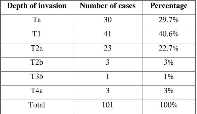
Out of the 71 infiltrative cases, 41 cases(57.8%) showed infiltration
upto subepithelial connective tissue, 23 cases(32.4%) showed infiltration
upto superficial muscle layer, 3 cases(4.2%) showed infiltration upto
deep muscle layer, 1 case(1.4%) showed microscopic involvement of
perivesical tissue and 3 cases(4.2%) showed involvement of prostrate.
[image:53.595.136.483.348.549.2]There were no cases with lymphnode involvement. (Table 10).
TABLE : 10 - DISTRIBUTION OF TRANSITIONAL CELL CARCINOMAS ACCORDING TO DEPTH OF INVASION
Depth of invasion Number of cases Percentage
Ta 30 29.7%
T1 41 40.6%
T2a 23 22.7%
T2b 3 3%
T3b 1 1%
T4a 3 3%
Total 101 100%
In the present study, 41 cases (57.7%) belonged to stage I, 26 cases
(36.6%) belonged to stage II and 4 cases (5.7%) belonged to stage III.
TABLE : 11 - DISTRIBUTION OF TRANSITIONAL CELL CARCINOMAS ACCORDING TO STAGE
STAGE NUMBER OF
CASES PERCENTAGE
0 30 29.7%
I 41 40.6%
II 26 25.7%
III 4 4%
Total 101 100%
In this study, squamous differentiation was seen in 15 cases,
necrosis was seen in 19 cases and sarcomatous component was seen in 3
cases. (Table 12 and chart 10)
TABLE : 12 - DISTRIBUTION OF OTHER PROGNOSTIC FACTORS IN TRANSITIONAL CELL CARCINOMA
Patient characteristics Present Absent Total Squamous metaplasia 15(14.9%) 86(85.1%) 101(100%) Necrosis 19(18.8%) 82(81.2%) 101(100%) Sarcomatous component 3(3%) 97(97%) 101(100%)
Among the 101 cases, urine cytology was done in 24 cases, out of
which 14 cases were positive for malignant cells and 10 cases were
TABLE : 13 - URINE CYTOLOGY IN TRANSITIONAL CELL CARCINOMAS
Urine cytology Number of cases Percentage
Positive 14 58.3%
Negative 10 41.7%
Total 24 100%
In our study, 30 cases were associated with recurrence. Among these cases, 6 cases showed recurrence in 1 month, 6 cases in 2 months, 3 case in 3 months, 1 case in 4 months, 1 case in 6 months, 1 case in 8 months, 7 cases showed recurrence in 1 year, and 1 case in 2 yrs. 3 of the cases were late recurrences with previous tumour occurring in 3 yrs and 1 case recurring after 7 years. (Table 14 and chart 12).
TABLE : 14 - RECURRENCE IN TRANSITIONAL CELL CARCINOMAS
Recurrence Number of cases Percentage
1 month 6 20%
2 month 6 20%
3 month 3 10%
4 month 1 3.3%
6 month 1 3.3%
8 month 1 3.3%
1 year 7 23.4%
2 year 1 3.3%
3 year 3 10%
7 year 1 3.4%
RESULTS OF IMMUNOHISTOCHEMICAL STUDIES
Of the total 101 transitional cell carcinomas, 50 cases of varying
grade and stage were selected in a random manner and subjected to
immunohistochemical analysis with a panel of 2 markers – CD 34 and
HER-2/neu. The microvessel density was found out by using CD 34.
Of the 50 cases, there were 42 males (84%) and 8 females (16%). The
ages ranged between 35 and 81 yrs with a mean age of 60.28. There were
3 cases (6%) of small biopsy, 44 cases of TURBT (88%), 2 cases (4%) of
simple cystectomy and 1 case (2%) of radical cystectomy. There were 37
cases (74%) below 66 years of age and 13 cases (26%) more than 66
years. The tumour was located in the right lateral wall in 16 cases, left
lateral wall in 13 cases, base in 4 cases, dome in 5 cases, posterior wall in
1 case, entire bladder in 6 cases and the tumour is multiple in 5 cases. The
tumours ranged in size from 0.5 to 8 cm with a mean size of 4.32.
Among the final study group, 30 cases (60%) were low grade and
20 cases (40%) were high grade. 45 cases (90%) showed papillary
morphology and 5 cases (10%) showed flat morphology. Squamous
metaplasia was seen in 9 cases (18%) and necrosis was seen in 8 cases
(16%). Sarcomatoid area was seen in 1 case (2%). 8 cases (16%)
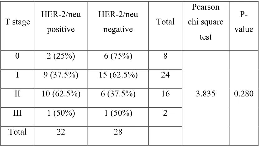
belonged to stage Ta (non-invasive), 23 cases (46%) belonged to T1, 16
belonged to T3b and 1 case (2%) belonged to T4a. 8 cases (16%)
belonged to stage 0, 23 cases (46%) belonged to stage 1, 17 cases (34%)
belonged to stage 2 and 2 cases (4%) belonged to stage 3.
Urine cytology was done in 12 cases and was found to be positive
in 8 cases (66.7%) and negative in 4 cases (33.3%).Among the 50 cases,
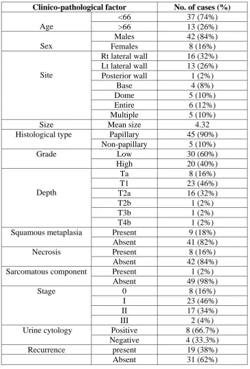
TABLE : 15 - DISTRIBUTION OF TRANSITIONAL CELL
CARCINOMA AMONG THE VARIOUS CLINICOPATHOLOGICAL GROUPS FOR THE IHC STUDY (50 CASES)
Clinico-pathological factor No. of cases (%)
Age
<66 37 (74%)
>66 13 (26%)
Sex
Males 42 (84%)
Females 8 (16%)
Site
Rt lateral wall 16 (32%) Lt lateral wall 13 (26%) Posterior wall 1 (2%)
Base 4 (8%)
Dome 5 (10%)
Entire 6 (12%)
Multiple 5 (10%)
Size Mean size 4.32
Histological type Papillary 45 (90%) Non-papillary 5 (10%)
Grade Low 30 (60%)
High 20 (40%)
Depth
Ta 8 (16%)
T1 23 (46%)
T2a 16 (32%)
T2b 1 (2%)
T3b 1 (2%)
T4b 1 (2%)
Squamous metaplasia Present 9 (18%)
Absent 41 (82%)
Necrosis Present 8 (16%)
Absent 42 (84%)
Sarcomatous component Present 1 (2%)
Absent 49 (98%)
Stage 0 8 (16%)
I 23 (46%)
II 17 (34%)
III 2 (4%)
Urine cytology Positive 8 (66.7%) Negative 4 (33.3%)
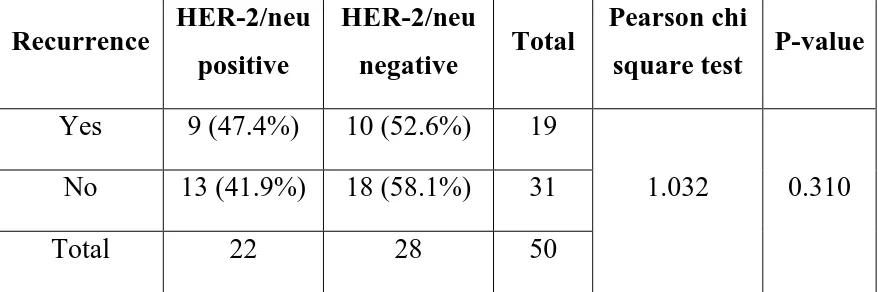
Recurrence present 19 (38%)
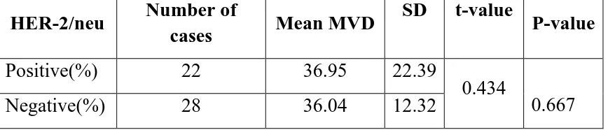
In this study, various prognostic factors were compared with mean
microvessel density and HER-2/neu expression. The microvessel density
ranged from 8 to 102 with a mean value of 36.44. 22 cases (44%) showed
strong expression for HER-2/neu (scoring 2+ and 3+) and were
considered positive and 28 cases (56%) showed weak expression (scoring
0+ and 1+) and were considered as negative (Table 16 & Chart 13).
TABLE : 16 - DISTRIBUTION OF HER-2/NEU EXPRESSION AND MICROVESSEL DENSITY IN TRANSITIONAL CELL
CARCINOMA
IHC parameters Result
MVD Mean- 36.44
HER-2/neu Positive-22(44%). Negative-28(56%)
CORRELATION OF MICROVESSEL DENSITY WITH VARIOUS CLINICO – PATHOLOGICAL PARAMETERS
In this study, the mean microvessel density for the male patients
was found to be 38.45 with the standard deviation of 18.0. The mean
MVD for the female patients was found to be 25.88 with the standard
deviation of 6.3. Since the standard deviation was found to be very high,
the logarithmic transformations was carried out before applying the
student’s t-test. The significant P-value infers that male patients have
TABLE : 17 - CORRELATION OF GENDER WITH MICROVESSEL DENSITY
Gender N Mean MVD
Standard deviation(SD)
t-value* P-value
Male 42 38.45 18.002
2.047 0.046
Female 8 25.88 6.379
For the logarithmic values of MVD the t-test has been applied.
This study showed that there is no statistically significant correlation
between MVD and site of tumour. Since only one patient had tumour at
the posterior wall, for statistical purpose its MVD value was combined
with that of the base. The mean values have been compared using
ANOVA test. The non-significant p-value infers that the site of the
occurrence of the tumour has no influence on the MVD level. (Table 18).
TABLE : 18 - CORRELATION OF TUMOUR SITE WITH MICROVESSEL DENSITY
Site N Mean
MVD SD
ANOVA F-value*
P-value Right lateral wall 16 32.69 9.884
0.472 0.795 Left lateral wall 13 37.54 20.630
Base & Posterior wall 5 40.40 18.756
Dome 5 30.60 13.722
Entire bladder 6 40.83 30.407
Multiple 5 42.20 11.256