CLINICAL STUDY, EVALUATION AND
MANAGEMENT OF UPPER
GASTROINTESTINAL BLEEDING
A DISSERTATION SUBMITTED TO
THE TAMILNADU DR.M.G.R MEDICAL UNIVERSITY In partial fulfilment of the regulations for the award of the
Degree of M.S., (GENERAL SURGERY)
BRANCH – I
DEPARTMENT OF GENERAL SURGERY
STANLEY MEDICAL COLLEGE AND HOSPITAL
THE TAMILNADU DR.M.G.R MEDICAL UNIVERSITY
CHENNAI
CERTIFICATE
This is to certify that the dissertation entitled “Clinical Study,
Evaluation and Management of Upper Gastrointestinal Bleeding”
is the bonafide work done by Dr. K.S.Saravana Krushna Raja.,
Post Graduate student (2010 – 2013) in the Department of General
Surgery, Government Stanley Medical College and Hospital, Chennai
under my direct guidance and supervision, in partial fulfilment of the
regulations of The Tamil Nadu Dr. M.G.R Medical University, Chennai
for the award of M.S., Degree (General Surgery) Branch - I, Examination
to be held in April 2013.
Prof. A. RAJENDRAN, M.S., Prof. P. DARWIN M.S.
Professor of Surgery, Professor and Head of surgery, Dept. of General Surgery, Dept. of General Surgery, Stanley Medical College, Stanley Medical College, Chennai-600001. Chennai-600001.
PROF. S. GEETHA LAKSHMI, M.D., PhD, The Dean,
DECLARATION
I, solemnly declare that this dissertation titled “Clinical Study,
Evaluation and Management of Upper Gastrointestinal Bleeding” is a
bonafide work done by me in the Department of General Surgery,
Government Stanley Medical College and Hospital, Chennai under the
guidance and supervision of my unit chief Prof. A. RAJENDRAN, M.S.,
and my Head of the Department Prof. P. DARWIN, M.S.
This dissertation is submitted to The Tamilnadu Dr. M.G.R.
Medical University, Chennai in partial fulfilment of the university
regulations for the award of M.S., Degree (General Surgery) Branch - I,
Examination to be held in April 2013.
Place : Chennai
ACKNOWLEDGEMENT
I am highly indebted to my guide Prof. A. Rajendran, Professor of
Surgery for his constant help, inspiration and valuable advice in preparing
this dissertation.
I express my deepest sense of thankfulness to my Assistant Professors
Dr. P. Balaji, Dr. G. V. Manoharan, Dr. G. Venkatesh and Dr. Vignesh for their valuable inputs and constant encouragement without
which this dissertation could not have been completed.
I consider it a privilege to have done this study under the supervision of
my beloved Professor and Head of the Department Prof. Darwin, who has
been a source of constant inspiration and encouragement to accomplish
this work.
I express my sincere gratitude to my mentor Prof. S Deivanayagam,
former Head of Department of General Surgery. I thank him for the
constant support, able guidance, inspiring words and valuable help he
rendered to me during my course.
I am grateful to the Dean Prof. S. Geethalakshmi for permitting to
I am very thankful to the Department of Surgical Gastroenterology and
Department of Medical Gastroenterology for their immense support and
guidance in completing this study.
I am particularly thankful to my fellow post graduate colleagues for their
valuable support and to all staff members who have made this study
possible.
It is my earnest duty to thank my wife and my parents, without whom
accomplishing this task would have been impossible.
I am extremely thankful to my patients who consented and participated to
CONTENTS
S. no. Chapter Page nos.
1. Introduction 1
2. Aims and Objectives 2
3. Review of Literature 3
4. Materials & Methods 53
5. Observation and Results 55
6. Discussion 68
7. Summary 78
8. Conclusion 80
9. References
10. Annexure
(i) Proforma
(ii) Institutional Ethical Committee approval Certificate
(iii) Plagiarism percentage (Screen Shot) (iv) Consent form
INTRODUCTION
Upper Gastrointestinal bleeding is a common potentially life threatening
condition associated with high morbidity, mortality and medical care
costs. Clinically manifests as haematemesis and, or melena and rarely
haematochezia with or without haemodynamic compromise.
Upper Gastrointestinal bleeding is defined as bleeding proximal to the
ligament of Treitz. The incidence of UGI bleeding is approximately 100
cases per 100,000 population per year. Mortality rates from UGI
bleeding are 6 – 10 % overall[1]. Accurate patient evaluation and
appropriate early management is critical to decrease the morbidity
and mortality. The foundation of diagnosis and management of
patients with Upper GI Bleeding is Oesophago-Gastro-Duodenoscopy
(OGD). Endoscopy has a sensitivity of 92% for identification of the site of
(AUGIB), with a specificity that approaches 100% , especially if it is done
within the first 24 hour of (AUGIB)[2] . Various scoring systems have
been used in the prediction of risk in patients with Upper GI
bleeding and early stratification in accordance with clinical symptoms.
Non variceal bleeding is due to arterial haemorrhage such as ulcers
hypertension cause variceal bleeding and should be managed
AIMS AND OBJECTIVES
1. This study reviews the clinical presentation , diagnostic
modalities and evaluation of Upper GI bleeding.
2. To analyse the incidence and severity of presentation.
3. To predict the prognosis and mortality risk of Non variceal
bleeding using Rockall scoring system.
4. To delineate the specific cause and bleeding site using OGD
Scopy.
5. To analyse various modalities in the early resuscitation and
REVIEW OF LITERATURE
HISTORICAL ASPECTS
For more than 5000 years, upper Gastro Intestinal bleeding is one of the
recognized causes of death. Various documents like Chinese manuscripts,
Egyptian papyri, medical works of Hippocrates and not to forget famous
Indian Surgeon,Sushruta, all have mentioned the upper gastrointestinal
bleeding(non variceal) as one of the conditions associated with very high
mortality. During ancient times, it was gradually brought to notice by
symptomatology of the patients[4]. There were lot of evidences regarding
the incidence of peptic ulcer were studied since from first century [5]
.Morgagni in 1700 was the first person to describe the gastrointestinal
bleeding because of portal hypertension[6]. Only at the turn of the 20th
century with advent of endoscopy, evidence of haematemesis due to
rupture of esophageal varices has been well established. Most of the earlier
pioneers in esophageal endoscopic procedures including Crafoord used
rigid endoscope (Negus Type). Now the flexible fibreoptic endoscope(
Introduced in 1980 ) has replaced the rigid scope in almost all centres,
which is supported by the result of controlled trial from Capetown.
Endoscopy is now 50 years old and has established strongly it’s place in
emergency and non emergency situations. Crafoord and Frenckner from
Sweden were first to use sclerotherapy in 1936 to treat esophagealvarices
in a 19 year old patient. In 1990, Stiegmann published the ligating device
using rubber or latex for varices. Spices and herbs were used in ancient
times to treat Peptic ulcer disease. The changes in our management of
gastrointestinal
bleedingoverthecenturieshavebeendrivenbynaturalalterationsinthespectrum
ofdiseases,expanding our understanding of these diseases and the never
ending advances in technology and pharmacology that have occurred
relative to Upper Gastrointestinal diseases.
ANATOMY
OESOPHAGUS
Oesophagus is a fibro muscular tube which extends from cricoid Cartilage
(C6, vertebra) to Oesophago gastric junction (T11, Vertebra)measuring
25cms. It is divided into cervical, thoracic and abdominal oesophagus. An
anatomical sphincter is at the upper end of the oesophagus and
physiological sphincter at the lower end of the oesophagus. In passing a
Oesophago Gastro Duodenoscope, Cricopharyngeus sphincter can be
BLOOD SUPPLY ARTERIAL SUPPLY Inferior thyroid arteries:
The paired inferior thyroid arteries supplies the cervical esophagus that
gives off branches called tracheoesophageal arteries
Tracheobronchial and Bronchoesophageal arteries:
The tracheobronchial arteries give off multiple small branches to the
esophagus which subdivide within the periesophageal tissue. Commonly,
one bronchoesophageal artery originates 1 cm to 3 cm caudal to the
vascular bundle from the anterolateral aspect of the descending aorta.
Aortic proper Oesophageal Artery:
It arises from the anterior aspect of the descending aorta.
Left Gastric and Splenic Arteries:
The left gastric artery mainly supplies the anterior and right lateral aspects
of the esophageal wall. The splenic artery primarily supports the posterior
and left lateral aspects (cardiac notch) by either one or two direct branches
or by vessels of the gastric fundus, including connections with the short
Venous Drainage:
The extrinsic veins drain into the locally corresponding large vessels. The
superior vessels drain to the jugular veins or the azygos and hemiazygos
veins. The inferior veins terminate in the left gastric and splenic veins.
STOMACH:
The stomach is divided into, Cardia, fundus, body and pylorus.
BLOOD SUPPLY: ARTERIAL SUPPLY: Left Gastric Artery:
In more than 90% of individuals, the left gastric artery is a branch of the
celiac axis. The left gastric artery commonly divides into an anterior and a
posterior branch before attaining the lesser curvature, it bifurcates into an
anterior branch which sends branches to the anterior gastric wall, and a
posterior branch which, similarly, supplies the posterior gastric wall. The
anterior branch of the left gastric artery angles rather obliquely across the
body of the stomach toward the greater curvature. It ends in numerous
small ramifications and forms a vascular "crow's foot" (of Payne) and the
posterior branch follows the lesser curvature a centimeter or two from its
posterior gastric branches may possess direct interconnections with one
another or with the continuing segment of the parent left gastric artery.
Right Gastric Artery:
The right gastric artery is a small branch which arises most
commonly from the proper hepatic artery( 62% ) but also arises from left
hepatic artery, rarely from common hepatic.A. It gives origin to one or
more suprapyloric branches.
Gastroduodenal Artery:
The gastroduodenal artery arises as one of the two terminal branches of
the common hepatic artery. It gives origin to the supraduodenal,
Right Gastorepiploic Artery:
The right gastroepiploic artery is a branch of the gastroduodenal
artery (or its continuation) in most cases. After giving origin to an
infrapyloric branch, the artery passes along the greater curvature of the
distal gastric surgical unit within the gastrocolic ligament. The gastric
branches of the right gastroepiploic artery pass mostly undivided in a
submucosal position about one-fifth of the distance from the greater
curvature. They anastomose extensively with branches from the left gastric
artery.
Left Gastroepiploic Artery:
It arises in most cases (75%) from the distal splenic, inferior splenic
terminal, middle part of the splenic trunk, or superior splenic terminal. It is
the largest branch of the splenic artery gives off the left epiploic and the
Short Gastric Arteries:
Approximately five to seven short gastric arteries arise from the terminal
branches of the splenic artery or from the left gastroepiploic artery.
Venous Drainage:
The venous supply of stomach usually accompany the corresponding
arteries. A great venous arch can develop between the left and right
gastroepiploic veins during portal hypertension, forming a congested
vascular bridge between the splenic and portal veins.
DUODENUM:
The duodenum has 4 parts:
The first (superior) part, or bulb (5 cm), The second (descending)
part or C loop (10 cm),The third (horizontal) part (7.5 cm), The fourth
(ascending) part (2.5 cm) continues into jejunum at duodenojejunal
Blood supply:
The first part of the duodenum is supplied by the supraduodenal artery and
the postero superior pancreaticoduodenal branch of the gastroduodenal
artery. The remaining three parts of the duodenum are supplied by an
anterior and a posterior arcade.
Venous Drainage:
Veins of the lower first part of the duodenum and the pylorus usually open
into the right gastroepiploic veins they are the subpyloric veins while the
upper first part of the duodenum is drained by suprapyloric veins. The
venous arcades draining the remaining duodenum follow the arterial
arcades and tend to lie superficial to them.
PORTAL VEIN:
The superior mesenteric and splenic veins join posterior to the neck of the
pancreas to form the main portal vein[9]. It receives pyloric and coronary
vein branches as it courses cephalad and obliquely to the right to form the
most posterior structure within the hepatoduodenal ligament (portal triad).
In the hilum of the liver, the main portal vein bifurcates into a short
oblique right portal vein and a longer, more transverse, and more
and become invested along with the other components of the portal triad
by extensions of Glisson's capsule.
Porto systemic collaterals:
Multiple anastamoses forming between the portal and systemic circulation
contributing the collateral circulation as follows,
• Lt gastric vein anastamose with oesophageal veins.
• Superior rectal vein with middle and inferior rectal veins.
• Para umbilical veins with anterior abdominal wall subcutaneous
veins.
• Splenic and pancreatic vein tributaries anastomose with left renal
vein retroperitoneal area[13].
• Veins over the bare area of liver anastomose with with the veins of
EPIDEMIOLOGY:
The age distribution varies depending on the studied population affecting
the elderly in the west [16,17]. The male:female ratio for (AUGIB) in
many European countries and the United States is approximately 2:1
.There is regional variation regarding the frequency of causes of (AUGIB)
depending on the demographic characteristics of the studied population,
risk factors of bleeding, timing of the study and pathological
classification[17].The incidence rates of UGIB demonstrates a large
geographic variation ranging from 50 to 160 cases per100 000 population,
with consistent reports of higher incidences among men and elderly
people. Possible explanations for this reported geographic variation in
incidence are differences in definition of UGIB in various studies,
population characteristics, prevalence of alcoholism, ulcerogenic
medication, in particular aspirin, nonsteroidal anti-inflammatory drugs
(NSAIDs), and Helicobacter pylori(H. pylori) prevalence[18,21]. Acute
variceal bleeding has a significant mortality which ranges from 5% to 50%
in patients with cirrhosis[23]. Some studies have reported a significant
decline in incidence of acute UGIB, especially peptic ulcer bleeding, in
recent years[22,23].This decline is likely due to a combination of factors,
including decreasing prevalence of gastric colonization with H. pylori, the
use of PPI therapy,both in general and in patients using aspirin and
NSAIDsin particular. Despite the introduction of therapeutic endoscopy
and acid-suppressive therapy, the overall mortality of UGIB has remained
stable over recent decades and is still 6%-14% in most studies[24] . As
such, mortality from UGIB is strongly associated with advanced age and
presence of severe comorbidity. The risk of mortality increases with
rebleeding, which is thus another major outcome parameter. It was noticed
that, mortality due to haemetemesis after admision was more than those
who admitted with symptoms[25] . The incidence of rebleeding in patients
with UGIB shows a wide range from 5% - 20%, depending on several
factors. One of the prognostic index of rebleeding was increased portal
pressure > 15 mmHg, and the same was also confirmed in a study of
Moitinho et al[26].
More than 25 percent of episodes of UGI bleeding were due to pepti
ulcers among 7800 individuals included in a database of USA between
1999 and 2001[27]. Nonspecific mucosal abnormalities appeared to be
commonest(40 percent), while Oesophagitis in 12 percent, and
Oesophageal and gastric varicesin about 10 percent. Other causes (AV
malformations, Mallory-Weiss tears, and GI tumors) were seen in less
gastric ulcers were seen in more than fifty percent than duodenal
ulcers[28].
In another study focused on OGD performedfor a period of 4 years from
the year 2000 in a setting, the endoscopic findings that was common in
patients with UGIB were Peptic ulcer (31 percent) followed by an Gastric
or duodenal erosion (20 percent). Gastric ulcers in OGD were dominant
than duodenal ulcers (54 vs 36 percent)[29].
Recent epidemiological studies had revealed a decrease in incidence of all
causes of UGIB except those of peptic ulcer bleed. Bleeding due to
varices seems be the leading cause of bleeding in cirrhotic patients in
50-60%[30]. Rebleeding in occurs in 10-16%, despite the therapeutic
modalities. Mortality ranges between 3 to 12 % and does not pose a change
in the last few years. NSAIDs was only used in 12% of the individuals
who were presented with bleeding. H. pylori infection is found in about
45% of individuals with bleeding peptic ulcer. H. pylori should be kept in
mind in all patients with peptic ulcer and eradication should be given.
Child, in his classic monograph emphasized the co-existence of hepatic
disease and manifestations of portal hypertension. It was only in the turn of
twentieth Century that Gilbert and associates, and Pichancourt coined the
ETIOLOGY
PEPTIC ULCER DISEASE
GASTRODUODENAL EROSIONS
OESOPHAGITIS
VARICES
MALLORY WEISS TEAR
UGI MALIGNANCIES the formation of esophago gastric varices to portal hypertension[30].The
incidence of varices from 5%to15% ofpatients with cirrhosis per annum.
ETIOLOGICAL FACTORS:
According to various studies, the incidence of etiological factors were as
shown below,
PEPTIC ULCER 40%
NO OBVIOUS CAUSE 15%
EROSIVE DISEASE 15%
OESOPHAGITIS 10%
OTHERS 6%
MALLORY WEISS TEAR 5%
VARICES 5%
Peptic ulcer disease:
Peptic ulcers are focal defects in the gastric or duodenal mucosa that
extend into the submucosa or deeper to it. They may be acute or chronic
caused by an imbalance between mucosal defenses and acid injury.
Common sites for peptic ulcers are the 1st part of the duodenumand the
lesser curve of the stomach, but they also occur on the stomal site
following gastric surgery[32]. It remains as the most common cause of life
threatening UGIB. Bleeding mainly occurs from the underlying arterial
erosion and it depends upon the size of the erosion and diameter of the
vessel. There are 5 types of gastric ulcer, classified depending upon the
anatomical location. Among these types, Type I seems to be the common
ulcer located near the incisura angularis of lesser curvature.
H.pylori is the most important factor in the development of peptic
ulceration. About 3 /4 of duodenal ulcers and 1 /4 of gastric ulcers were
caused by H.pylori infection. With the use of antibiotics, the prevalence of
the infection have been much decreased in USA. The other factors
associated with peptic ulcer formation are stress induced, NSAIDs intake,
smoking and alcoholism. even after treatment with proton pump inhibitor
Mallory weiss tear:
These are tears seen at the oesophago gastric junction, that cause UGIB.
Due to continuous vomiting or with retching, haemetemesis occurs and are
commonly associated with alcoholism and chemotherapeutic agents like
cisplatin etc[35].
Portal Hypertension:
The portal venous system contributes approximately 75% of the blood and
75% of the oxygen supplied to the liver[31]. In the average adult 1000 to
1500 mL/min of portal venous blood is supplied to the liver. However, this
amount can be significantly increased in the cirrhotic patient. The portal
venous system is without valves and drains blood from the spleen,
pancreas, gallbladder, and abdominal portion of the alimentary tract into
the liver[34]. Tributaries of the portal vein communicate with veins
draining directly into the systemic circulation. These communications
occur at the gastroesophageal junction, anal canal, falciform ligament,
splenic venous bed and left renal vein, and retroperitoneum . The normal
portal venous pressure is 5 to 10 mmHg, and at this pressure minimum
blood is shunted from the portal venous system into the systemic
circulation. As portal venous pressure increases, however, the
blood may be shunted around the liver and into the systemic
circulation[37].
A WHVP or direct portal venous pressure that is >5 mmHg greater than
the inferior vena cava (IVC) pressure, a splenic pressure of >15 mmHg, or
a portal venous pressure measured at surgery of >20 mmHg is abnormal
and indicates portal hypertension. A portal pressure of >12 mmHg is
necessary for varices to form and subsequently bleed[38].
Depending on this, the etiological factors implicated in portal
hypertension can be categorized into four major groups:
1. Increased Hepatic portal flow.
2. Extra Hepatic outflow obstruction.
3. Obstruction of the extrahepatic venous systems.
4. Intrahepatic obstruction.
90% of the cases with portal hypertension are due to intrahepatic
obstruction.
Causes for portal hypertension:
I. Cardiac diseases
2. Tricuspid stenosis
II. Vascular diseases:
1. Budd- chiari syndrome
2. Membranous obstruction of the Inferior Vena Cava
3. Thrombosis of the inferior vena cava.
III. Acute and chronic Liver diseases
1. Cirrhosis.
2. Idiopathic portal Hypertension.
3. Schistosomiasis.
4. Congenital hepatic fibrosis.
5. Exposure to environmental toxins.
6. Metastatic carcinoma.
IV. Venous Occlusion of Portal System.
1. Portal Vein
Oesophageal Varices
The shunting of esophageal collaterals or varices, are the most important
clinically, because of their predilection to bleeding. The development of
this portosystemic shunting depends upon a threshold portal pressure
below which varices do not occur unless the portal pressure, as measured
by the hepatic vein wedge pressure gradient is greater than 12mmHg,
varices and variceal haemorrhage do not develop[39]. As all the four layers
of veins in the wall of esophagus are intercommunicating, they all become
engorged and elongated, dilated and tortuous when the pressure is above
12 mm of Hg. However, the deep intrinsic veins seem to bear the brunt of
the insult and dilate massively becoming esophageal varices, which are
seen endoscopically. These vessels lie in the lamina propria, where they
are poorly supported by surrounding tissue bulge into the lumen.
Oesophageal varices are classified into,
• Grade 1: varices that looks small and straight.
• Grade 2: varices that are occupying less than 1 /3rd of the lumen
• Grade 3: varices that are large, coil shaped occupying > 1 /3rd of the
lumen.
Gastric Varices
Short gastric veins presenting at the fundus communicate with the deep
intrinsic venous plexus of the esophagus, which due to back pressure
changes result in gastric varices of fundal type. They are common in
extrahepatic obstruction like splenic vein thrombosis. There is diffuse
increase in arterio-venous communications between muscularis mucosa
and dilated pre-capillaries. This is termed as congestive gastropathy[37].
They have a particular risk of bleeding and of damage eg. By Aspirin or
NSAIDs. Therefore bleeding gastritis constitutes 30% of upper
gastrointestinal tract bleeding in portal hypertension patients.
Gastric varices are classified primarily by their location as,
A. Gastroesophageal varices
Type I (GEV 1)- along the lesson curve (2-5cm in length)
Type II (GEV 2) – along the greater curve extending towards the gastric
fundus
B. Isolated Gastric Varices
Type I (IGV 1) – Isolated cluster of varices in gastric fundus
Upper Gastrointestinal tract tumours:
Oesophagogastric tumours are uncommon cause of acute upper
gastrointestinal bleeding. The important benign type is gastrointestinal
stromal cell tumour (GIST) arising from the muscle layers of the gastric or
duodenal wall. Erosion through the mucosa gives a characteristic
umbilicated in endoscopy. These tumours may cause major bleeding by
eroding the underlying arteries. Acute GI bleeding due to malignant
lesions are unusual (6%) arising from the oesophageal malignancies
presents as massive bleeding due to aortic invasion. Significant
gastrointestinal bleeding is uncommon with gastric cancer; however,
hematemesis does occur in approximately 10% to 15% of patients[43].
Dieulafoy lesion:
It is rare cause of Upper GI bleeding. It comprise about 3 – 6 % of all
gastrointestinal bleeds in adults. They are thought to be of developmental
malformation and are often called as, cirsoid aneurysm, and submucosal
arterial malformation. They can occur anywhere in the GI tract, and most
Arterio venous malformations:
Arteriovenous malformations (AVMs) are abnormal blood vessels seen in
the gastrointestinal (GI) tract and are the source of bleeding. They are
identified nowadays by the use of angiography[41]. Presentation could be
of massive UGI bleeding or anaemia of chronic blood loss.
Other causes:
Other rare causes of UGIB should also be considered in patients with
UGIB. For example, In patients with chronic pancreatitis who presents
with UGIB, Hemosuccus pancreaticus must be excluded. Bleeding in these
patients can be secondary to a pseudoaneurysm in peripancreatic blood
vessels as a complication of pancreatic pseudocysts[42]. Hemobilia is
another rare cause of UGIB that should be considered in the setting of
recent hepatobiliary tree instrumentation, such as with ERCP ( Endoscopic
retrograde cholangio pancreatography) or laparoscopic cholecystectomy
possibly due to the injuries of bile duct and hepatic artery. Aortoenteric
fistula must be considered in patients with a history of intra abdominal
vascular surgery, such as Abdominal aortic aneurysm repair. Post
Iatrogenic injuries secondary to endoscopic procedures, such as
percutaneous endoscopic gastrostomy tube placement, are also rare causes
of UGIB. portal gastropathy, Ménétrier's disease, and watermelon
stomach(Gastric Antral Vascular Ectasia) should also be considered.
Clinical features:
All patients presenting with Haemetemis or Malena with associated
symptoms provides clue to the diagnosis as follows,
1. Symptoms due to Oesophagitis or Peptic ulcer disease.
2. Symptoms pertaining to Upper GI malignancies.
3. Symptoms consistent with the cause of portal hypertension
4. Symptoms of portal hypertension
5. Symptoms directed towards the esophageal Varices.
Clinical features vary depending upon the underlying causes like, Peptic
ulcer disease usually have relationship of symptoms pertaining to food
intake, Gastric malignancies have Ball rolling movements and symptoms
suggestive of Gastric outlet obstruction[43] and cirrhosis patients have
features of Liver failure like Spidernaevi, Foetor hepaticus,
Flapping Tremors and Hepatic encephalopathy [31]. Clinical features of
portal hypertension like Ascites, Splenomeagaly with Hyper splenism,
caput medusae should be noted. Patient may come with severe
Haematemesis and Melaena in the case of ongoing or massive bleeding
due to Erosive gastritis, Malignancies, Esophageal or Gastric varices.
Investigations:
Complete Blood Count:
Haemoglobin and hematocrit , Total Count, Differential count , E.S.R, Packed Cell volume (P.C.V), Platelet count.
Bleeding time, Clotting time, Pro thrombine Time
Renal Function Test:
Blood Sugar, Blood Urea, Sr. Craetinine.
Blood Grouping and Rh typing
Urine routine
Liver Function Test:
Serum Bilirubin
Albumin: Globulin ratio
Serum Asparate amino transferase (AST, SGOT )
Serum Alanine amino transferase (ALT, SGPT)
Serum Alkaline phosphatise
Chest X ray PA view.
X ray Abdomen AP erect view.
ECG.
Barium Swallow and Barium meal: Linear filling defects in the distal
esophagus and stomach can be made out depending upon the underlying
pathology.
Endoscopy( Oesophago Gastro Duodenoscopy) : Gold standard for the
diagnosis of varices . This is the single most important investigation when
the patient comes with upper G.I. bleeding and Malena. The bleeding site
and the underlying cause can be identified by the use of OGD. In
Emergency situations it can be used diagnostic tool to identify the spurting
artery, visible vessel / varices or stigmata of recent haemorrhage (SRH)
Ultra sonography of Abdomen with Doppler evaluation
Ascites
Splenomegaly
Hepato megaly
Portal vein Caliber, splenic, hepatic vein and infra and intrahepatic
IVC
Angiography
Measurement of portal pressure
Liver biopsy: To know the cause of intrahepatic portal hypertension.
MANAGEMENT:
Protocol for early management of acute upper gastrointestinal bleeding
Triage
Patients are prioritised depending upon the clinical presentation and
decisions to be taken accordingly whether surgical intervention is needed
Intensive care monitoring
Maintaining the circulation by accessing the central venous line, Nasal
oxygen , urinary catheterisation to monitor the output, ryles tube insertion
done to clear the blood filled stomach for endoscopy and also to asses the
ongoing bleeding. Vital signs should be monitored periodically by using
appropriate methods.
General supportive therapy
Endotracheal intubation should be attempted if needed[45]. Patients can
be resuscitated by using intravenous fluid administration, compatible blood
transfusion, cardio pulmonary resuscitation, and management of associated
comorbid diseases, such as sepsis, liver disease or coronary artery
disease[46]. OGD could be delayed until the patient is adequately
resuscitated and stabilized with the available measures. Nasal oxygen must
be given to counteract the blood loss which indirectly decreases the oxygen
carrying capacity. Patients with acute UGIB must be kept nil oral for the
purpose of the urgent need for OGD and for abdominal surgery if needed.
They are also assessed for hypovolemic shock to determine requirements
of intravenous fluid administration and blood transfusion, and attention
Intravenous access is secured at two or more sites using 16 or 18 -gauge
venflons.
Patients with active UGI bleeding should receive initial 500mL of
crystalloids, during the first half an hour to maintain the blood pressure,
while several units of packed redblood cells are cross matching and
typed[46]. Fluid administration is subsequently raised if the blood
pressure falls. Transfusion requirements are determined by many factors,
including the age of the patient, presence or absence of comorbid ilnesses,
cardiovascular status, hematocrit, and the quantity of blood loss, along
with the current hematocrit level. Packed redblood cells are transfused in
individuals who have significant blood loss, ongoing bleeding, and in
those patients who manifest cardiac, renal, or cerebral ischemia. Patients
who presents with variceal haemorrhage are conservatively transfused to a
hematocrit of only 26 to 28 to avoid exacerbating the bleeding by
increasing the portal pressure[47]. Fresh frozen plasma transfusion or
Platelets transfusion is individualized according to multiple factors like,
severity of bleeding, bleeding rate, presence of other bleeding diathesis,
ROLE OF ENDOSCOPY:
OGD stands as the prime diagnostic and therapeutic tool for UGIB[49] . It
accurately identifies the bleeding site and determines the specific cause and
in more than 90% of individuals with acute UGIB [50]. OGD is the
principal modality in diagnosing the type of bleeding and therapeutically it
reduces the surgical intervention. Befor OGD, it advisable to provide
proton pump inhibitor therapy[51]. Urgent OGD for UGIB is ideal, which
significantly improves the clinical outcome in certain conditions like
variceal bleeding and severe ongoing bleeding[52]. Stigmata of recent
haemorrhage could be identified by performing OGD as early as possible.
From this, the site and number of lesions are identified. Multiple scoring
systems like Rockall, forrest classification etc are used for prognostic
purposes and triage of patients with UGIB[53]. Local adrenaline injection,
electrocautery or argon plasma coagulation(APC), and mechanical
therapies like endoclips or banding are the available therapies with
endoscopy. Combined with the clinical presentation and OGD findings,
risk stratification can be made[54].
PEPTIC ULCER DISEASE:
These ulcers are seen as craters in endoscopy. For controlling the
at the site of bleeding or surrounding the ulcer. Sclerosants like ethanol,
sodium tetradecyl sulphate can also be useful [55]. The use of
electrocautery, argon plasma coagulation was also justified.
Reflux esophagitis:
The endoscopic findings are erythematous mucosa, edema with exudates,
ulceration and bleeding with increased vascularity. Ulcers due to
oesophagitis can be efficiently treated with adrenaline injection or by using
ablation therapies[56].
Mallory- weiss tear:
A linear and longitudinal tear that are seen over the osophago gastric
junction. Sometimes an erosion or scab are also identified during
endoscopy. The role of therapeutic endoscopy is under evaluation[57].
Cameron lesion:
The ulcers or erosions, that are seen over the gastric part within the hiatus
hernia are termed as Cameron lesions. They are asymptomatic and are
incidentally diagnosed during endoscopy. These lesions are uncommon
cause of acute bleeding[58] and the therapeutic modalities are injection
Portal gastropathy:
This condition looks like an intense red lesion in a mosaic
background, commonly noted in the fundus. Portal hypertension is
commonly associated with it. Due to the diffuse nature of the lesion,
therapeutic modalities in endoscopy are not useful[59].
Oesophageal and Gastric varices: Endoscopic sclerotherapy:
Obliteration of esophageal varices by injecting sclerosants directly into the
channel (Intravariceal), beside the channel (Paravariceal) or combination
Indications:
1) Emergency sclerotherapy can be performed immediately at the time
of the diagnostic endoscopy.
2) It can be delayed until after the variceal haemorrhage has been
controlled by conservative measures.
3) In patients for whom no other treatment is available, including those
in whom surgery carries high risk, those who have undergone
surgery but continue to bleed.
4) In patients prior to definitive surgery so that the patients condition
can be improved. Hence reduces the mortality.
Mechanism of action:
Intra variceal sclerosant injection acts by causing thrombosis (damage the
intima) thus stopping the bleeding and preventing rebleed from particular
vein. In paravariceal injection sclerotherapy bleeding stops by two
mechanisms:
1) By external compression of the bleeding varices (Peri vascular fibrosis)
Endoscopic variceal ligation:
Indications:
1. In acute variceal bleeding, the band ligation has to be done
immediately after the diagnosis, to control the bleeding.
2. In the long term eradication of the esophageal varices.
3. Prophylactic band ligation of the esophagal varices has also
been reported
4. In high-risk patients the band ligation is indicated, till the
patients recover from the risks and become fit for surgery.
5. Indicated in patients who are not fit for surgery (definitive
surgery).
6. Indicated in the esophageal variceal eradication along with the
Mechanism of action:
Band ligation of esophageal varices acts by mechanical obstruction,
leads to strangulation of variceal tissue. The strangulation of the varices
ligated is followed by ischaemic necrosis of the mucosa and sub mucosa of
the varix. At 3-7 days after treatment, sloughing of the ligated tissue, at the
site with shallow ulcerations occurs (1-2 mm deep) and they are 6-10 mm
in diameter. At 14-21 days minimal residual varices are present, the
vascular structures in the submucosa will be replaced by matured scar
tissue. After 50-60 days at variceal site mature scar tissue, without any
stricture is seen.
Benign and malignant Upper gastrointestinal tumors :
These are all rare causes of UGIB. Benign conditions like GIST and
leiomyomas are seen as extraneous compression in endoscopy.
MALTomas are seen as polypoidal mass with cerebroid like mucosal folds.
Malignancies, more commonly adenocarcinoma stomach are seen as an
ulcerative growth, ulcero proliferative lesion, bleeding irregular mass or
stricture. Linitis plastica, seen as a noncompliant stomach wall.
Secondaries in the stomach are visualised as polypoidal erosions or mass.
Dieulafoy lesion:
During endoscopy, these lesions are visible as an elevated lesion with
erosions around it. In majority of cases, dielaufoy lesions are seen in the
upper half of the stomach along the lesser curvature. It is about 3 – 6 mm
in diameter. Adrenaline injection, argon plasma coagulation, ligation with
bands or clips are useful to arrest bleeding[63].
Angiodysplasia:
These lesions appear as dark red, thick network of vessels, 2 – 6 mm in
diameter. Initially, adrenaline injection is used to control haemostasis
followed by APC[64].
Gastric antral vascular ectasia:
The lesions which are seen as multiple folds radiating from the pylorus
upto the antral region with red streaks at the proximal ends. They also
termed as watermelon stomach. Due to its superficial and diffuse nature of
the lesion, thermal ablation are useful[65].
Aortoenteric fistula:
It carries a very high mortlity as it necessitates the emergency endoscopy.
Sometimes, a prosthetic mesh can also be seen during endoscopy. OGD
must be deferred after identifying such lesion, as therapeutic procedures
may cause alarming haemorrhage on disturbing the lesion.
Repeat esophagogastroduodenoscopy
Repeat endoscopy is useful at times to identiy the missed out lesions.
Rebleeding from the lesions occurs with in three days of the first
endoscopy and the relook endoscopy is generally not advisable
regularly[67].
RISK STRATIFICATION:
Several scoring systems have been developed to help predict the outcome
of patients and to improve patient management and promote cost-effective
use of hospital resources .
The Rockall scoring system is used for risk stratification with the use of
clinical presentation, comorbid factors and endoscopy findings. It can be
done befor and after endoscopy for accurate calculation. Mortality can be
predicted with the use of it.
A total Rockall score of < 3 is predictive of low risk of adverse outcomes
a score of > 8 is predictive of high mortality[68]. With Rockall scoring
system, individuals with high and low risk strata are calculated.
ROCKALL RISK SCORING SYSTEM
Rockall Risk scoring system
Variable Scores
Age (Years)
< 60 Years 60 - 79 Years > 80 Years
0 1 2
Shock
Pulse < 100/min, SBP>100 mmHg Pulse > 100/min, SBP>100 mmHg Pulse < 100/min, SBP<100 mmHg
0 1 2
Co-Morbid Conditions
No Major Co-Morbidity
Cardiac Failure, Ischemic Heart Disease
Renal Failure, Liver Failure, Disseminated Malignancies 0 2 3
Diagnosis
Mallory Weiss tear, No Lesion Identified All other Diagnosis
Malignancy of Upper GI Tract
0 1 2
Major Stigmata of Recent Haemorrhage
None / Dark Spot Only
Blood in Upper GI Track, Adherent clot, Visible or Spurting Vessel
Evaluation of the bleeding lesion was determined in Forrest
classification by using OGD findings alone and does not include clinical
parameters[69].
Blatchford risk scoring system was used in patients with UGIB to predict
the clinical outcome , using only the clinical parameters without
endoscopic evaluation of bleeding lesion.
CONSERVATIVE MANAGEMENT: Management of Non-variceal bleeding:
Gastric acid inhibits platelet aggregation, impairs clot formation, and
promotes fibrinolysis; therefore, inhibiting gastric acid and raising the intra
gastric pH to 6 or more may promote clot formation and decrease the risk
of rebleeding.
High-dose PPI therapy is defined as an initial bolus (Pantoprazole 80 mg)
followed by continuous infusion (Pantoprazole 8 mg/h) for up to 72 h.
Omeprazole can also be used with best results.
Since H.Pylori is the leading causative factor in Peptic ulcer disease, the
Anti-H.Pylori regimen:
PROTON PUMP INHIBITORS THERAPY bd +
CLARITHROMYCIN 500mg bd + AMOXYCILLIN 1g bd
10 – 14 days
PROTON PUMP INHIBITORS bd +
CLARITHROMYCIN 500mg bd + METRONIDAZOLE 500 mg bd
10 – 14 days
PROTON PUMP INHIBITORS bd + BISMUTH SUBSALICYLTE 525mg qid + METRONIDAZOLE 250mg qid + TETRACYCLINE 500mg qid .
10 – 14 days
Radiological approach
Transcatheter coil embolisation is commonly used as a therapeutic
approach in patients with unidentifiable and uncontrollable bleeding with
the routine measures Bowel ischaemia is less as there is good collateral
supply of the stomach and duodenum[71].
Safety and effectiveness of the procedure was explained in recent
BEFORE AND AFTER EMBOLISATION OF GASTRODUODENAL. A.
Partial splenic artery embolization (PSE) has been performed to treat
Oesophageal and Gastric varices including portal hypertensive
gastropathy.
Management of Variceal bleeding:
Numerous agents have been studied for the prevention and treatment of
variceal hemorrhage. In practice, the list consists of vasopressin and its
analogues, somatostatin and octreotide, nonselective β- blockers and
nitrovasodilators.
I. Vasopressin and its analogues:
Vasopressin causes splanchnic arteriolar vasoconstriction and decreases
portal tributary inflow with a resultant decline in portal pressures Standard
infusion 0.2-0.4 unit/min with NTG 40 µg/min. The morbidity of
vasopressin has led to the development of analogues with fewer side
effects such as terlipressin. This drug does not increase plasminogen
activator activity but has the same effects as vasopressin on the coronary
vasculature.
II. Somatostatin and its analogues
It is a naturally occurring tetradecapeptide found in the GIT. Octreotide is
a structural analogue of somatostatin. Somatostatin is administered as a
250 µg /hour IV bolus followed by continuous infusion of 250µg/hour for
2-4 days.
Octreotide is given as 50µg IV bolus followed by infusion of 25-50 µg
/hour. Because somatostatin and octreotide have to be administered
parenterally, their role has primarily been restricted to the management of
the acutely bleeding cirrhotic patient[72].
III. β- adrenergic Antagonists
They cause splanchnic arteriolar vasoconstriction and decrease portal
venous inflow. Propranolol is the prototype nonselective β-blocker.
In the long term, propranolol maintains a portal hypotensive effect in most
is associated with numerous side effects such as bronchoconstriction, heart
failure and impotence in cirrhotics. Their administration does not impair
the hemodynamic response to acute blood loss.
IV. Nitrovasodilators:
Nitric oxide is one of the most potent vasodilators and plays an important
role in pathophysiology of portal hypertension. Patients treated with
isosorbide mononitrate together with either propranolol or nadolol had a
greater sustained portal hypotensive effect.
Balloon Tamponade:
They are highly effective in controlling esophageal variceal
bleeding, but temporarily. There are three types of tubes available
They are:
a) Linton-Nachlas tube: It has only gastric balloon and three
lumen.
b) Sengstaken Blakemore tube: Both gastric and esophageal
balloons are present and have three lumen.
It has four lumen, one excess to prevent aspiration pneumonia. After
testing the balloons for leak, it should be passed under sedation or
anaesthesia (Local or General) in operation theatre, through nostril. Once
the tube in the stomach is confirmed by aspiration and easy distensibility
of the gastric balloon, (300-400 ml of air is used for inflation). So that the
pressure is maintained between 20-30 mm of Hg. Tube is secured to the
forehead with slight traction. Inflate the esophageal balloon to a pressure
of 40 mm Hg.
Removal of the tube:
24 hrs after inflating the esophageal balloon if there is no bleeding, deflate
the bulbs and remove the tube. If bleeding is present, we can keep the tube
for further 24 hrs.
Results:
Several studies have shown 60-70% effectiveness. However, the rebleed
rate is as high as 40-60%. Hence, this is as effective as pharmacological
control.
Transjugular intrahepatic portosystemic shunts
In this procedure, a communication is made inbetween the portal vein (
cases. Model of End Stage Liver Disease is the good mortality predictor
after the procedure. It is not useful in patients with multi organ failure.
CCF, pulmonary hypertension and portal vein thrombosis are
contraindications of TIPSS[73].
SURGICAL MANAGEMENT:
Non varicel bleeding:
Despite major advances in endoscopic treatment, the incidence of
emergency surgery has not significantly changed. Today, most patients
undergoing operation for bleeding peptic ulcer have simple oversewing of
a bleeding ulcer, or simple patch of a perforated ulcer, Truncal vagotomy
or distal gastrectomy. Surgical interventions, such as ligation of the
bleeding vessel or excision of the aneurysm, should be considered if
embolization fails or is contraindicated in case of Hemosuccus
pancreaticus.
Vagotomy:
These surgical procedures are associated with ulcer recurrence as carried
out in emergency situations. Vagotomy with drainage procedures carry
Gastrectomy:
Surgical resection appears to be the only curative treatment for
gastric cancer and most patients with clinically resectable loco regional
disease should have gastric resection. The standard operation for gastric
cancer is radical subtotal gastrectomy. Reconstruction is usually by
Billroth II gastro jejunostomy, but if a small gastric remnant is left (<20%),
a Roux-en-Y reconstruction is considered. The operative mortality is
around 2 to 5%.
Variceal bleeding:
1. Portosystemic shunting: They are aimed at lowering the
portal pressure and diverting the portal flow from around the
A) Total shunts:
1) End to side portacaval shunt
2) Side-to-side portacaval shunt
3) Mesocaval shunt
4) Proximal splenorenal shunt
B) Selective shunts:
1) Distal splenorenal shunt
2) Coronary caval shunt
C) Partial shunts
Small diameter H-graft (sarfeh shunt)
This is the standard for both the emergency and elective treatment.
End to side Porta caval shunt was introduced in 1940’s, itself, the operative
mortality is much higher in emergency situation when used as a last resort
was 50% mortality rate, in elective patients it may be as low as 19% in best
2. Devascularization procedures:
a. Splenectomy
b. Esophageal transection
c. Esophageal transection and devascularization (Suguira
procedure)20
Many shunt techniques evolved of these distal splenorenal shunt is
said to be most advantageous.
The devascularization procedures carry mortality rate of 33% as
compared to endoscopic procedures which is 24%.
Contra-indications for emergency surgery are,
1. Presence of acute alcoholic hepatitis.
2. Marked coagulopathy that is uncorrectable
3. Presence of major systemic complications related indirectly to liver
disease such as acute renal failure, frank sepsis etc.
Child class ‘c’ is not a contraindication, infact they are resistant to
Liver Transplantation: Theoretically, it is the ideal therapy for all
patients with chronic liver disease complicated by portal hypertension and
variceal haemorrhage. This re-establishes low resistance portal outflow
through the liver. Considering the availability of the donor, cost of both the
procedures and immunosuppressive medication it is indicated for the
treatment of selected patients with end stage liver disease that is medically
MATERIALS AND METHODS
STUDY DESIGN : Prospective study.
SAMPLE SIZE : 100 patients admitted with upper GI
bleeding .
PLACE OF STUDY : Trauma ward, Department of General Surgery, Government Stanley Medical
College Hospital, Chennai.
PERIOD OF STUDY : 1 Year.
INCLUSION CRITERIA:
All patients with age groups 20 to 85 years admitted with Upper GI
bleeding in Trauma ward.
EXCLUSION CRITERIA:
Children and patients with age groups below 20 years with Upper GI
METHODS:
• A Proforma will be made that includes detailed history, physical
examination , basic investigations and other relevant investigations
required.
• Clinical diagnosis will be made accordingly.
• Risk stratification will be done using Rockall risk scoring system.
• Triage, intensive monitoring and general supportive therapy done
for the patients will be recorded.
• Endoscopic findings and various modalities of treatment are
RESULTS
Total number of cases
Mean Age of presentation
Male : Female ratio
CHART - 1
Male 89% Female
11%
Gender distribution
Total number of cases – 100.Mean Age of presentation – 45 years.
0 5 10 15 20 25 30 35
20 - 30 31 - 40 CHART – 2
Age distribution among males and females
40 41 - 50 51 - 60 61 - 70 71 - 80 ibution among males and females
MALES
Clinical severity of Upper Gastrointestinal bleeding: TABLE - 1
Mild ( < 500 ml ) 58
Moderate ( 500 – 1500 ml ) 36
Severe ( > 1500 ml ) 6
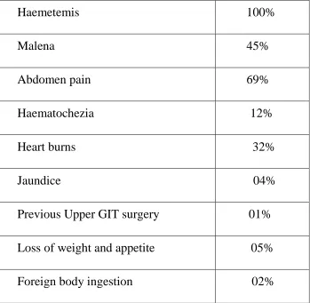
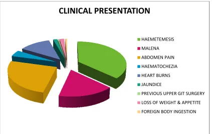
[image:70.595.131.480.373.714.2]Clinical presentation : TABLE.2
Haemetemis 100%
Malena 45%
Abdomen pain 69%
Haematochezia 12%
Heart burns 32%
Jaundice 04%
Previous Upper GIT surgery 01%
Loss of weight and appetite 05%
CHART - 3
[image:71.595.94.517.123.391.2]Co-morbidities: TABLE.3
Hypertension
Diabetes
Coronary Artery Disease
Chronic liver disease
CLINICAL PRESENTATION
Hypertension 12%
Diabetes 11%
Coronary Artery Disease 19%
Chronic liver disease 05%
CLINICAL PRESENTATION
HAEMETEMESIS MALENA ABDOMEN PAIN HAEMATOCHEZIA HEART BURNS JAUNDICEPREVIOUS UPPER GIT SURGERY
LOSS OF WEIGHT & APPETITE
FOREIGN BODY INGESTION HAEMETEMESIS
ABDOMEN PAIN
HAEMATOCHEZIA
HEART BURNS
PREVIOUS UPPER GIT SURGERY
LOSS OF WEIGHT & APPETITE
0.0 10.0 20.0 30.0 40.0 50.0 60.0 70.0 80.0 SMOKING 57.0 P e rc e n ta g e
Role of Risk factors
Smoking , Alcoholism and NSAIDS drug intake appears to be the major
risk factors in patients presenting with Upper GIT bleeding.
CHART - 4
Alcoholism is one of the
Gastrointestinal bleeding in our study.
SMOKING ALCOHOLISM NSAIDS DRUG
57.0
72.0
21.0 Incidence of risk factors
:
Smoking , Alcoholism and NSAIDS drug intake appears to be the major
risk factors in patients presenting with Upper GIT bleeding.
is one of the major risk factor associated with
Gastrointestinal bleeding in our study.
NSAIDS DRUG 21.0
Smoking , Alcoholism and NSAIDS drug intake appears to be the major
0 10 20 30 40 50 60 70 80 SMOKING 50 P e rc e n ta g e
Distribution of Death among Risk factors
CHART - 5
Alcoholism tends to be the major risk factor causing mortality in Upper
Gastrointestinal bleeding in our study.
SMOKING ALCOHOLISM NSAIDS DRUG 25
50 50
75
Risk factors
Distribution of Death among Risk factors
Alcoholism tends to be the major risk factor causing mortality in Upper
Gastrointestinal bleeding in our study.
NSAIDS DRUG 50
Distribution of Death among Risk factors No
Yes
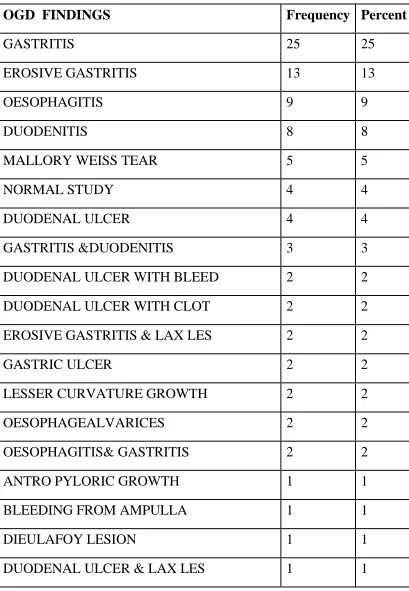
OGD findings in our study :
TABLE – 4
OGD FINDINGS Frequency Percent
GASTRITIS 25 25
EROSIVE GASTRITIS 13 13
OESOPHAGITIS 9 9
DUODENITIS 8 8
MALLORY WEISS TEAR 5 5
NORMAL STUDY 4 4
DUODENAL ULCER 4 4
GASTRITIS &DUODENITIS 3 3
DUODENAL ULCER WITH BLEED 2 2
DUODENAL ULCER WITH CLOT 2 2
EROSIVE GASTRITIS & LAX LES 2 2
GASTRIC ULCER 2 2
LESSER CURVATURE GROWTH 2 2
OESOPHAGEALVARICES 2 2
OESOPHAGITIS& GASTRITIS 2 2
ANTRO PYLORIC GROWTH 1 1
BLEEDING FROM AMPULLA 1 1
DIEULAFOY LESION 1 1
FOREIGN BODY - NEEDLE 1 1
FUNDAL VARICES&DUODENITIS 1 1
GASTRIC POLYP 1 1
GASTRIC ULCER WITH CLOT 1 1
GASTRIC ULCER WITH SLOUGH 1 1
GASTRIC VARICES 1 1
LOWER OESOPHAGEALVARICES 1 1
OESOPHAGEAL EROSION 1 1
OESOPHAGITIS&GERD 1 1
OESOPHAGITIS& LAX LES 1 1
STOMAL ULCER 1 1
CHART - 6
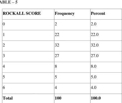
TABLE – 5
ROCKALL SCORE 0 1 2 3 4 5 6 Total
ROCKALL SCORE Frequency Percent
2 2.0
22 22.0
32 32.0
27 27.0
8 8.0
5 5.0
4 4.0
100 100.0
Impact of Rockalls score with mortality:
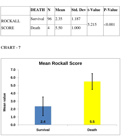
[image:77.595.91.514.228.704.2]Independent samples t-Test to compare mean rockall score between Survival and Death
TABLE – 6
DEATH N Mean Std. Dev t-Value P-Value
ROCKALL SCORE
Survival 96 2.35 1.187
5.215 <0.001 Death 4 5.50 1.000
CHART - 7
2.4 5.5 0.0 1.0 2.0 3.0 4.0 5.0 6.0 7.0 Survival Death M e a n v a lu e
Conservative management :
Initial Fluid Resuscitation ( crystalloids & colloids ) - 87%
Blood transfusion - 82%
Proton pump inhibitor therapy - 88%
CHART.8
Initial fluid resuscitation with crystalloids and colloids were given in 87%
of patients, 82% of patients were given blood transfusion and 88% of
Intervention procedures:
CHART - 9
* Therapeutic endoscopy
removal and variceal band ligation.
* Radiological intervention
* Laparotomy :
• Under running suture over the bleeding vessel with Truncal vagotomy and pyloroplasty.
• Subtotal Gastrectom
resection.
• Palliative Gastrojejunostomy.
• Feeding Jejunostomy.
Variceal band ligation 1% Therapeutic endoscopy 2%
Intervention Procedures
Intervention procedures:peutic endoscopy : - Gastric polypectomy , Foreign body
removal and variceal band ligation.
Radiological intervention procedures : Coil embolisation.
Under running suture over the bleeding vessel with Truncal
vagotomy and pyloroplasty.
Subtotal Gastrectomy with Roux-en-Y anastomosis and D 2
Palliative Gastrojejunostomy. Feeding Jejunostomy. None 91% Laparotomy 4% Radiological intervention 2% Variceal band ligation
Intervention Procedures
Gastric polypectomy , Foreign body
procedures : Coil embolisation.
Under running suture over the bleeding vessel with Truncal
TABLE - 7
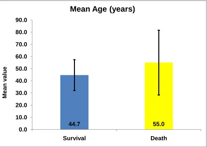
Independent samples t-Test to compare mean age between Survival and Death in Upper GI bleeding.
DEATH N Mean Std. Dev t-Value P-Value
AGE
No 96 44.72 12.677
0.769 0.497
Yes 4 55.00 26.608
CHART – 10 : MORTALITY IN UPPER GI BLEEDING
44.7 55.0 0.0 10.0 20.0 30.0 40.0 50.0 60.0 70.0 80.0 90.0 Survival Death M e a n v a lu e
DISCUSSION
Upper Gastrointestinal bleeding is one of the most common emergencies in
the surgical department that carries 6 to 10 % mortality worldwide[1]. Our
study was a prospective clinical study on evaluation and management of
Upper gastrointestinal bleding in patients admitted in our institute
(Government Stanley Medical College Hospital ) in one year. We excluded
children and patients under the age of 20 years from this study. We
assessed the clinical presentation, co-morbidities, associated risk factors,
OGD findings ( within 24 hours of presentation ), need of initial
resuscitation, blood transfusion and intervention procedures like
therapeutic endoscopy, radiological intervention or emergency laparotomy.
A total of 100 cases were take up for the study. In these patients, the mean
age of presentation was 45 years with a standard deviation of 13. Minimum
and maximum age of presentation in the final study was 20 and 79 years
respectively with sex distribution predominantly seen among males ( 89%
) than females ( 11% ). The maximum number of age group is between 31
– 40 years as shown in the chart.2.
The severity of bleeding was classified into mild, moderate and severe
depending upon the quantity of blood loss. Majority of patients
patients presented with moderate haemetemesis ( 500 – 1500 ml ) and only
6% presented with severe haemetemesis ( > 1500 ml ) as shown in Table.1.
We had 58 patients in mild, 36 patients in moderate and 6 patients in
severe form of which 4 patients were died inspite of resuscitative
measures.
All of our patients admitted with haemetemis had Abdomen pain in
69 of them. About 45 patients had malena, 12 patients were presented with
haematochezia and 32 patients had history of heart burns. History of
anorexia and weight loss were observed in 5 patients while jaundice were
noted in 4 patients as shown in Table.2 and Chart.3.
The mortality rate was 4% in our study with mean age of survival and
mortality being 44 and 55 year respectively. Thus elderly people withstand
less well to UGIB than younger patients. The major contributing factor that
leads to death was blood loss, which inturn leading to hypovolemic shock.
The incidence of risk factors like Smoking, Alcoholism and NSAIDs drug
intake were considered in our study and the impact with mortality have
also been been compared. Smoking is seen in 57 % of patients while the
incidence of alcohol was seen with 72 %. Around 21 % of patients were
having NSAIDs intake. This was surprisingly a small number compared to
patients took NSAIDS for arthritis and myalgia. Thus, it can be clearly
delineated from the Chart.4 that alcoholism is one of the major risk factors
causing Upper GI bleeding and almost, always associated with Peptic ulcer
disease. Mortality rate also stands high in patients with alcoholism ( 75% )
than the other two risk factors ( 50% ) as shown in Chart.5.
The associated co-morbid illness in our study were Hypertension
(12% ), Diabetes ( 11% ), Coronary Artery Disease ( 19% ) and Chronic
liver disease ( 5% ) as noted in Table.3. One of our patients died had
associated history of old coronary artery disease and another patient had
chronic liver disease leading to portal hypertension. Thus, it can be stated
that, associated co-morbid factors had a strong link with mortality.
OGD is very useful to delineate the site of bleeding and to facilitate
targeted therapy. It defines the low risk and high risk strata and helps to
identify the appropriate candidates for a period of hospital stay or
intervention procedures if neded. Risk – stratification scores that
incorporate endoscopic data, such as the complete Rockall score, propose
that such scores are superior to clinically based scores because they define
a greater proportion of all patients at low risk for adverse outcomes related
to acute upper gastrointestinal bleeding. The pattern of Upper GI bleeding