THE TAMILNADU
Dr. M.G.R. MEDICAL UNIVERSITY
CHENNAI
THE STUDY AND MANAGEMENT OF
NON TRAUMATIC ABDOMINAL
SURGICAL EMERGENCIES
Dissertation submitted for
MASTER OF SURGERY (BRANCH I)
ACKNOWLEDGEMENT
“Learn to see, learn to hear, learn to feel, and know that by practice alone you can become expert. Medicine is learned by the bedside and not in the class room”.
- Sir William Osler I am greatly indebted to my Head of the Department Prof. Dr.M.Gobinath, M.S., Department of General Surgery, Government Rajaji Hospital, Madurai for his excellent guidance in conducting this study.
I am grateful to my unit chief , Dr.R. Vijayan. M.S., Professor of Surgery, Department of Surgery, Govt. Rajaji hospital, Madurai for his
help and guidance throughout this study.
I am grateful to my unit Assistant Professors, Dr.P. Sundareswari. M.S. D.G.O., Dr. M. Muthukumar, M.S., Dr. P. Prabhakaran, M.S., for their help and guidance throughout this study.
I express my gratitude to Dr.S.M. Sivakumar, M.S., Dean, Government Rajaji Hospital, Madurai Medical College, Madurai for permitting me to use the clinical material for the study.
BONAFIDE CERTIFICATE
This is to certify that the dissertation entitled “THE STUDY AND MANAGEMENT OF NON TRAUMATIC ABDOMINAL SURGICAL EMERGENCIES ” submitted by Dr. D. SURESH to the Tamil Nadu Dr. M.G.R. Medical University, Chennai in partial
fulfillment of the requirement for the award of M.S Degree Branch – I
(General Surgery) is a bonafide research work were carried out under
his direct supervision & guidance.
Prof. Dr. R. Vijayan, M.S., Prof.Dr. Gopinath, M.S., Department of Surgery, Head of the Department,
Madurai Medical College, Department of Surgery,
DECLARATION
I Dr. D. SURESH declare that, I carried out this work on, “THE STUDY AND MANAGEMENT OF NON TRAUMATIC ABDOMINAL SURGICAL EMERGENCIES” at the Department of Surgery, Govt. Rajaji Hospital during the period of October 2008 to September 2009. I also declare that this bonafide work or a part of this work was not submitted by me or any others for any award, degree, diploma to any other University, Board either in India or abroad.
This is submitted to The Tamilnadu Dr. M. G. R. Medical University, Chennai in partial fulfillment of the rules and regulations for the M.S degree examination in General Surgery.
Place : Madurai Dr. D. SURESH
CONTENTS
S.No.
TOPIC
Page No.
1.
Introduction
1
2.
General
Background
2
3.
Historical
Background
3
4.
Aim
of
the
Study
5
5.
Review
of
Literature
6
6.
Materials
and
Methods
34
7.
Analysis
and
Discussions
37
8.
Results
57
9.
Conclusion
61
Bibliography
Proforma
INTRODUCTION
An imprecise term ‘ACUTE ABDOMEN’ leaves much to be desired from a semantic point of view but is useful in practice; included under the heading are all of the painful abdominal syndromes, sometimes benign; frequently potentially lethal and of several origins; characterized by sudden appearance of pain with accompanying local and general signs and symptoms.
“ The majority of abdominal pains which ensue patients who have been previously fairly well and which lasts as long as 6 hours are caused by conditions of surgical importance”.
GENERAL BACKGROUND
It is usually relatively easy to interpret the pain and associated signs and symptoms of acute abdomen. Some times an acute abdomen appears suddenly in a previously healthy individual; at other times, it can occur as a complication in a chronically debilitated patient. Because of the possible disastrous outcome, the examination of an acute abdomen should never be put off.
HISTORICAL BACKGROUND
Even in ancient times, surgeons were aware of the potential seriousness of a ‘Painful abdomen’. Surgical advances were scarce until the last part of the nineteenth century because of the absence of autopsies and clinico-pathological correlation. Reluctance to perform abdominal exploration existed until a little less than 100 years ago. The long debates, disagreements of the leading side have been described in a report “ the cliniques de lannelongue”. Less than 100 years ago, Lannelongue a surgeon, Cornil a pathologist demonstrated at autopsy that the starting point of a purulent peritonitis was a perityphilitis, convincing the contemporaries of its appendicular origin, Monder 1974.
AIM OF THE STUDY
1. To analyse the incidence of Non traumatic abdominal surgical emergencies treated in Govt. Rajaji Hospital, Madurai, in relation to Sex, age etc.
2. To compare the reliability of clinical signs versus radiological signs and preoperative findings versus peroperative findings and post operative outcome.
REVIEW OF LITERATURE
‘Acute abdomen’ means the patient complains of acute abdominal symptoms that suggest a disease which definitely or possibly threatens life and may or may not demand immediate interference.
Causes :
1. Inflammation : (eg) acute appendicitis, acute salpingitis acute pneumococcal peritonitis.
If the patient gets pain around the umbilicus or in the epigastrium in the beginning and later on this pain shifts to the right iliac fossa, he is undoubtedly suffering from an acute appendicitis.
2. Perforation of Peptic ulcer, Typhoid ulcer, Diverticular disease etc.
Stages of Perforation etc. i) moment of perforation ii) stage of peritonism iii) stage of reaction
3. Acute Intestinal obstruction:
Intestinal obstruction is a common surgical emergency. Because of the serious nature it demands early diagnosis and speedy relief.
a) Dynamic:
Here there is peristalsis working against obstructing agent which may be in the lumen of the bowel such as inspissated faeces or a gall stone; In the wall such as an inflammatory or malignant stricture; or outside the wall as in hernia, adhesions, volvulus or intussusception.
b) Adynamic :
In this condition peristalsis ceases and no true propulsive waves occur as in paralytic ileus or mesenteric vascular occlusion.
4. Obstructed Hernia :
that not the obstruction culminates in strangulation. A hernia becomes strangulated when the blood supply of its contents is seriously impaired rendering gangrene imminent. Strangulated hernia is dangerous and threatening to life.
PRESENTATION :
Acute abdominal pain may be presented in so many ways. 1. The pain may be one sided or both sided
2. Upper abdominal – related to stomach and duodenum
3. Lower abdominal – may be due to intestinal obstruction or acute appendicitis
4. Colicky – Intestinal or Renal 5. Pain and rigidity – perforation 6. Pain and restlessness – renal colic
1. Acute Appendicitis :
It is one of the commonest causes of acute abdomen.
Cause : The lesion is due to the lumen of the appendix being obstructed by a faecalith, worms rarely foreign body.
Symptoms :
Murphy’s Traid :
1. Pain 2. Vomiting 3. Rise of temperature
1. Pain vague, central abdominal pain to start with finally settling in the right iliac fossa when it becomes more intense 2. Vomiting is due to pylorospasm
3. The patient looks toxic, tongue white and furred, foetor oris and tachycardia present.
4. The right iliac fossa is tender with maximum tenderness in and around the Mcburney’s point.
5. Localised muscle guarding and rigidity is present with minimum movement with respiration.
6. Rebound tenderness may be present
7. Pressure on the left iliac fossa may cause pain in the right iliac fossa due to distension of caecum due to displaced gas – Rovsing’s sign.
9. Bowel sounds are usually heard if the infection is localized in the right iliac fossa.
10. Rectal examination may cause pain deep in the pelvis when the finger is pushed high upto the right.
Various Positions of appendix :
1. Retrocaecal 2. Paracaecal 3. Preileal 4. Postileal 5. Pelvic
Alvarado Score
Migratory RIF pain 1
Anorexia 1 Nausea vomiting 1
RIF tenderness 2 Rebound tenderness 1 Elevated temperature 1
Leucocytosis 2 Shift to Left 1
Score 7 to 10 - Strongly suggestive of appendicitis Score 5 to 7 - +/- appendicitis
Score < 5 - No appendicitis
Stages of acute appendicitis :
I Stage : Infection confined to the appendix II Stage : Stage of appendicular mass
III Stage : Appendicular mass with perforation requiring laparotomy.
Emergency Appendicectomy Indication : Stage I and Stage III
Conservative treatment for Stage II. (Ochsner – Sherren regimen) 1. Rest 2. IV fluids 3. Antibiotics
4. Half hourly pulse, temperature and respiratory rate chart. 5. Nothing by mouth 6. Mark the mass for regression 7. Watch for signs of general peritonitis
2. PERFORATED PEPTIC ULCER :
condition. Treated usually by surgical intervention. In delayed diagnosis after 24 hours the outcome is poor.
Incidence :
“It is better to look and see than to wait and see” – Lord Moynihan.
Illingworth and Dick give the sex ratio of men and women as 19:1. According to our data men and women ratio is 16:1. According to Brunner 1903 and Jamieson 1947, the incidence being less in the summer moths, greater in November and December.
Pathology:
Perforation is a terminal stage of an ulcer which continues to penetrate the deeper tissues. Duodenal contents escape through the perforation into the general peritoneal cavity resulting in peritoneal irritation. Peritoneum reacts to chemical irritation by secreting peritoneal fluid copiously. This reaction lasts for 3 to 6 hours and is followed by infection with Pyogenic organisms.
Clinical Features:
1. moment of perforation 2. Stage of peritonism
3. stage of reaction 4. stage of chemical peritonitis 5. stage of bacterial peritonitis
Subacute perforation or early sealed perforation is clinically indistinguishable from local periotonitis in the vicinity of the ulcer. Diagnosis of perforation is correctly made in 95% of cases clinically. Plain x ray abdomen erect reveals a. crescent shaped translucent area beneath the Right cupola of the diaphragm in about 70% of cases. PR : movements of finger in the pelvis causes pain.
Investigations :
1. Plain x ray abdomen erect posture – shows air under the diaphragm.
2. Polymorphonuclear Leucocytosis
3. Hb – PCV will show the amount of dehydration 4. Serum Electrolytes
5. Other Investigations - Urine albumin Sugar Deposits
Blood Grouping and Typing X ray Chest
ECG
Treatment :
This is an emergency
1. Continuous Ryles tube aspiration 2. IV alimentation
3. Antibiotics
4. Emergency laparotomy and sealing of perforation with omental plug
5. Large perforation can be managed with a) Serosal patch closure
b) Pyloric exclusion and gastro jejunostomy c) Lateral tube duodenostomy
6. Definite treatment like Gastro jejunostomy with vagotomy or Gastrectomy is done at a later date
3. INTESTINAL OBSTRUCTION:
Simple Occlusion :
is obstruction of the intestine not associated with vascular change. Strangulation does not occur and the sequence of event is less dramatic. It is usually due to bands or adhesions as a result of operations done previously.
Level of Obstruction :
Obstruction is more serious in the small intestine than in the large intestine and higher the obstruction more serious are the results.
Clinical Features :
The cardinal clinical features are colicky abdominal pain, distension, vomiting and constipation. But it is true that only one of these may be sufficient for diagnosis of intestinal obstruction.
a. Pain :
Abdominal pain is the first symptom. It commences suddenly and often without warning. If it becomes, increasingly severe, then passes off gradually only to return at intervals of few minutes. These attacks of intestinal colic, which lasts from 3 to 5 minutes, spread all over the abdomen, but are localized mainly at the umblicus. In between attacks the patient is often quite comfortable. Recurrent attacks of severe colicky or spasmodic abdominal pain is a leading feature of all varieties of acute intestinal obstruction with the sole exception of paralytic ileus.
b. Vomiting :
c. Distension :
In early cases of small gut obstruction, abdominal distension is often slight or even absent. In subacute or partial obstruction of the small gut, step ladder pattern type of distension may be visible over the abdomen.
Inside the lumen of the bowel there will be collection of fluid which is made up of various digestive juices which is about 8 litres per day.
d. Constipation :
This is classically absolute, with presence of Intestinal obstruction, neither flatus for faeces being passed.
e) Dehydration :
Management :
There are three measures for combating the effects of intestinal obstruction.
1. Gastroduodenal or gastrointestinal suction drainage. 2. Maintenance of water & electrolyte balance.
3. Relief of obstruction by surgical intervention.
4. Antibiotics to prevent complications from associated sepsis, either locally (peritonitis) or peripherally (chest complications) or generally (septicaemia).
The main indication for early operation as soon as the fluid and electrolyte depletion has been corrected are :
1. Obstructed or strangulated hernia 2. Internal intestinal strangulation
3. Acute or acute-on-chronic obstruction Relief of Obstruction by Operation :
small intestine. A search is made to find out a loop of collapsed small intestine somewhere in the pelvis or right iliac fossa.
This is followed proximally. This is done by holding the loop in the long axis of the body and a finger is passed deeply along the left side of its mesentery. The contracted bowel is followed proximally till the level of obstruction is reached. When the bowel is too much distended, it is deflated by means of an aspirating needle.
If the caecum is distended, the obstruction must be in the colon, and by far the commonest cause is colonic carcinoma.
Bands and Adhesions :
They are as a result of previous operation. These bands and adhesions are divided under direct vision.
Internal Strangulation :
Evaluation of the history and physical examination :
The diagnosis of most instances of acute abdomen requires a careful history and a complete physical examination. After making a rapid evaluation of the patient, his history should be taken in great detail despite the great pain, discomfort, vomiting and mental disturbances which some patients experience following a state of shock. Physical examination is carried out in two stages. First a preliminary survey of the patient, secondly after resuscitation of the patient. If the patient is in a state of shock, the patient is resuscitated; then sent for investigations like, urinary and Blood investigations and skiagrams. A preoperative diagnosis is made, patient is prepared and taken to the operation theatre for surgical intervention.
Immediate Resuscitative measures done are : 1. Assurance of patients air way
2. Control of visible bleeding
3. Assisted ventilation by Mask and bag for respiratory failure
4. Rapid infusion of sodium containing solutions
Rectal and Pelvic Examination :
Is a must in all cases of acute abdominal emergencies. Laboratory Investigations :
Leucocytosis with an associated neutrophilia is evidence for the presence of purulent inflammation. Laboratory investigations frequently confirm a clinically apparent diagnosis. Rapidly evolving appendicitis is sometimes accompanied by only a very modest neutrophilia. Leucocytosis that is disproportionate with the physical signs is a useful finding when ischaemic damage to the walls of the digestive tract is suspected, particularly the distal part.
The systematic measurement of serum and urinary amylase levels in patients suspected to have perforated duodenal ulcer, strangulated bowel, acute cholecystitis, acute pancreatitis revealed increased levels of this enzyme. Haematocrit gives precious information about plasma volume. Concentrated urine is an important sign of dehydration which increases blood viscosity, a significant factor in thrombus formation.
abdomen suspected of ischamic origin; monitoring and laboratory examination must be repeated often. The finding of an incipient metabolic acidosis in these patients indicates the possible development of a segmentary gangrenous necrosis of a part of the digestive tract.
RADIOLOGY:
Radiological examination is quite valuable. A plain film of the abdomen with the patient in erect posture showing chest and abdomen gives a great deal of information. In incapacitated patients a left lateral decubitus view should be used.
The presence of air in the peritoneal cavity confirms a diagnosis of perforation. Air is not usually found in the initial period of perforation in the small intestine. When the clinical signs and symptoms of the patient point strongly towards a diagnosis of perforation of a hollow organ, the absence of air in the peritoneal cavity is not an adequate argument to delay an emergency surgical intervention.
ABDOMINAL EXPLORATION :
recently had a meal it will be necessary to pass a stomach tube through which much of the gastric content, can be removed with a suction apparatus.
Liver Abscess :
Pyogenic Liver abscess :
Hippocrates first described liver abscess in the year 400 BC. Oshner described the same in the year 1938.
Untill 1953 recommended treatment open surgical drainage in 1953 Mc Fadzean and associates in Hongkong advocated closed respiration and antibiotics for solitary pyogenic liver abscess.
Etiology : Kuffer cells act as filter for clearance of micro organism in the liver. These organism in the liver via blood stream. The biliary Potential sources :
1. Bile ducts (Ascending cholangitis) 2. Postal pyaemia
3. Direct extension of contagious diseases 4. Hematoma (trauma)
Pre disposing factors : 1. Diabetes mellitus 2. Cirrhosis
3. Pancreatitis 4. IBD
5. Pyleonephritis
6. Peptic ulcer diseases 7. Lymphoma
Portal, traumatic, cryptogenic are large and solitary. Bacteriology :
Escherichia coli Klebsiella Pseudomonoas Bacteriodes
Clinical presentations :
Signs symptoms
Chills Pleural effusion Diarrhoea
Abcess ruptures :
Signs of peritonisim develops Laboratory Data :
Increased ALP WBC > 10,000 / mm3 Albumin < 3 g / dl Hematocrit < 36% Bilirubin 72 mg / dl
Radiology :
Plain CXR :
1. Elevated Right hemidiapharm 2. Right pleural effusion
3. Right lower lobe Atelectasis
CT Scan :
More sensitive (95 – 100%)
Lower attenuation than surrounding liver and wall of abscess may enhance with Intravenous contrast administration.
TREATMENT :
1. US or CT guided abscess drainage 2. Blood cultures drawn
3. Broad spectrum IV antibiotics
Classical Antibiotics regimen include 1. Aminoglycoside
2. Clindaycin 3. Metronidazole Treatment given for 4-6 weeks Surgical drainage include
Amebic liver abscess :
Caused by parasitic protozoas entomeba histolytica
Diagnosis established by detection of antibody to the ameba Difference between Amebic and pyogenic liver abscess
Amebic Pyogenic liver abscess 1. Age < 50 Age > 50 yrs
2. Hispanic No ethnic predispositions 3. Recent travel to determine area Malignancy
4. Pulmonary dysfunction Increased Temperature, Pruritis , Jaundice
5. Abdominal pain septic shock 6. Diarrhoea Palpale mass 7. Abdominal tenderness
8. Hepatomegaly
Amebic liver abscess odourless grams stain and cultures are negative.
Treatment :
Metronidazole
Positive responses should be seen by third day of treatment Response increases to 90% by 10 days
Therapeutic Aspiration :
¾ No clinical response to drug therapy with in 5-7 days
¾ Risk of abscess rupture
¾ Cavity 7.5 cm
¾ Lesion in single lobe
¾ Percutaneous aspiration of catheter drainage under us guided can be done.
Surgical :
1. Failed to respond to conservative therapy 2. Percutaneous treatment fails
Acute cholecystitis : 2 types
Pathology :
Attack develops due to gall stone, Impacting in the Harman’s pouch following obstruction. Gall bladder becomes hyperemic, odematous, Tense and distended. Initial inflammation is chemically induced. Trauma to the gall bladder wall due to gall stone impaction leads to mucosal damage through release of phospholipases that converts lecithin to lysolecithin a known mucosal toxins.
The release of prostaglandin precursor archidonic acid by the action of phospholipase A on lecithin may mediate inflammatory response by producing prostoglandins.
With conservative management 80% of patient as the rising tension is gall bladder lumen from the out pouring of the inflammatory exudates lifts the walls of Hartmann’s pouch of the impacting stone which then disengages and drops into gall bladder lumen cystic duct drainage leads to resolution.
20% patients may go for patchy gangrene and perforation. Signs and symptoms :
1. RUQ pain and tenderness 2. Pyrexia
4. Nausea 5. Vomiting
6. Ileus and abdominal distention 7. Jaundice
Laboratory tests :
Leucocytosis > 10 x 109 / l Abnormal LFT
Differential Diagnosis
1. Perforated peptic ulcer diseases 2. Acute pancreatitis
3. Retrocaecal appendicitis 4. Hepatitis
5. Right pyelonephritis 6. Lower lobe pneumonia 7. Myocardial infarction Imaging Tests :
1. Plain X ray
b) Calculi or sludge c) Thickened gall bladder d) Peri cholecsystic oedema 3. Intravenous cholongiography 4. CT scanning
5. MRI
Acute acalculus cholecystitis
Acute inflammation of the gall bladder arises in the abscess of gall stones although biliary sludege is often present encountered in critically ill patients.
Pre disposing factors :
1. Blood volume depletion 2. Prolonged ileus
3. Morphine administration 4. Intravenous hyperalimentation 5. Multiple blood transfusion 6. Sepsis
Pathology :
Inflammation develops as a consequences of prolonged distention of gallbladder bile stasis, biliary sludge results in mucosal injury and thrombosis of vessels thrombosis thought to be initiated by the activation of factor XII.
Diagnosis : 1. Fever
2. Leucocytosis
3. Tenderness in right hypochondrium CT Scan :
USG :
Isotope scintiscanning : US and CT findings :
Indication of acalculus cholecystitis include wall thickness greater than 4 mm, intramural gas, peri cholecystitis fluid sub serosal oedema sloughed mucosal membranes.
Complication :
Treatment :
1. Nil by mouth 2. IVF
3. Electrolyte replacement 4. Nasogastric aspirations 5. Systemic antibiotics 6. Parenteral analgesia
Indication for emergency surgical interventions :
¾ Progression of disease despite of conservative treatment
¾ Failure to improve within 24 hrs
¾ Age > 60 years
¾ Presence of inflammatory mass
¾ Detectable gas in gall bladder
¾ Established generalized peritonitis
¾ Development of intestinal obstruction.
METHODS AND MATERIALS
This study material consists of 365 cases of Acute abdomen admitted in seven surgical units in our Department of Surgery, Madurai Medical College, Govt. Rajaji Hospital during the period from Oct 2008 to Sep 2009.
All these cases of pain abdomen admitted in surgical ward were carefully and thoroughly examined to arrive at a clinical diagnosis. A preformed proforma was carefully filled up giving particulars. Importance to the duration of illness and general and special investigation wherever possible.
The following procedure were adopted according to the conditions of the patient ie. conservative management and emergency surgeries.
Based on clinical diagnosis, Investigation patients opted for emergency surgery.
The study included 365 patient with abdominal emergency and treated in all the seven surgical units in Govt. Rajaji Hospital, Madurai Medical College, Madurai.
The patient details were entered in the typed proforma with necessary details for the study preoperatively and were followed up post operatively till the time of discharge of the patients and for 3 months hence forth. The rest of the data were provided by the medical record office.
The age and sex incidence, the common clinical presentations, the different types of management was analysed and discussed in relevance to each of the patients. The various investigations that were available in our Institution has been used, which includes Bio chemical and radiological investigations. Radiological Investigations include plain radiographs and USG of abdomen when necessary.
The various treatment options were considered for each of the cases and each cases was provided best optimal treatment available in our Institution and the post operative complication was also discussed.
1. Acute Appendicitis - 130 Cases 2. Perforation peritonitis - 94 Cases 3. Intestinal Obstruction - 112 Cases 4. Acute cholecystitis - 12 cases 5. Liver Abscess - 17 cases
DISCUSSIONS
TABLE - I
ABDOMINAL SURGICAL EMERGENCIES SEX INCIDENCE
S.No. Types of Cases Total No.of cases
Male Female
1. Acute Appendicitis 130 91 39
2. Perforative peritonitis 94 81 13 3. Intestinal Obstruction 112 76 36
4. Cholecystitis 12 4 8
5. Liver abscess 17 14 3
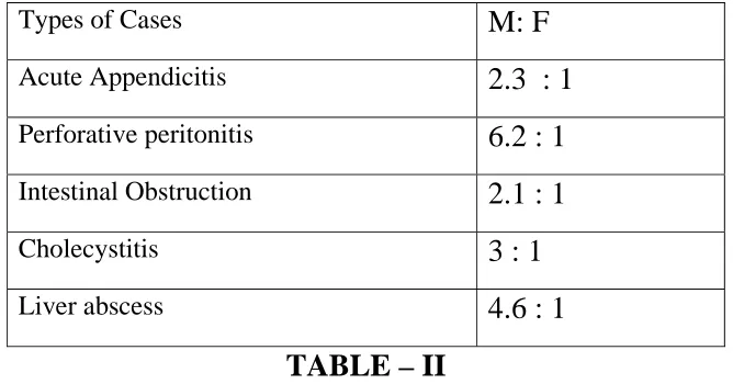
Male : Female ratio
Types of Cases M: F
Acute Appendicitis 2.3 : 1
Perforative peritonitis 6.2 : 1
Intestinal Obstruction 2.1 : 1
Cholecystitis 3 : 1
Liver abscess 4.6 : 1
TABLE – II
AGE INCIDENCE AGE DISTRIBUTION 44 7 2 46 28 25 28 40 31 2 5 12 19 22 6 8 32 4 4 0 5 10 15 20 25 30 35 40 45 50 APPENDICITIS PERFORATIVE PERITONITIS INTESTINAL OBSTRUCTION ACUTE CHOLECYSTITIS LIVER ABCESS
13-20 YRS 21-30 YRS 31-40 YRS 41-50 YRS >50 YRS
13-20 YRS 21-30 YRS 31-40 YRS 41-50 YRS >50 YRS APPENDICITIS
44
46 28 12
PERFORATIVE PERITONITIS
7 28 40 19
INTESTINAL OBSTRUCTION
2 25 31 22 32
ACUTE
CHOLECYSTITIS
2 6 4
ACUTE APPENDICITIS
Clinical Presentations No.of patients RIF Pain 112
Vomiting 86 Fever 56 Gaurding 100 Rebound tenderness 114
Per rectal tenderness 70
112
86
56
100
114
70
0 20 40 60 80 100 120
RIF Pain Fever Rebound
tenderness CLINICAL PRESENTATIONS
Among the 130 patients diagnosed as Acute appenditicits routine bio chemical investigation urea, sugar, creatinine and USG of abdomen done in all cases.
Acute Appendicitis 102 Appendicular abscess 17 Appendicular mass 11
Emergency appendicetomy done in 119 cases. In 17 cases of appendicular abscess. Appendicectomy done right drain was kept postoperatively patients were followed up.
2 patients found to have enterocutaneous fistula and 4 patient had wound infection.
Enterocutaneous fistula was treated conservatively.
PERFORATIVE PERITONITIS
CAUSES NO OF PT
DU PERFORATION
63
ILEAL PERFORATION
31
63 31 deodunal perforation ileal perforation
PERFORATIVE PERITONITIS
MALES FEMALES RATIO
DU PERFORATION 54 9 6 : 1
ILEAL PERFORATION
27 4 6.9 : 1
Total 81 13
6.2 : 1
s ex
inc idenc e
54
9 27
4
D u males D u female Ilel males Ileal fema
CLINICAL PRESENTATION
74
56
39
34
0 10 20 30 40 50 60 70 80
GAURDING AND RIGIDITY
VOMITING CONSTIPATION SHOCK
CLINICAL PRESENTATION NO.OF PATIENTS ABDOMINAL GAURDING AND
RIGIDITY
74
VOMITING
56
CONSTIPATION
39
CAUSES No. of patients
NSAIDS
32
ALCOHOL
18
PRE EXISTING ULCER
12
CAUSES OF PERFORATIVE PERITONITIS
32
18 12
Nsaids
Alcohol
preexisting ulcer
C AUS E S
F O R
IL E AL
P E R F O R AT IO N
20 7
4
enteric fever non s pecific inflamation T B
CAUSES NO OF PATIENTS
ENTERIC FEVER
20
NONSPECIFIC INFLAMATION
7
TB
OPERATED VS NON OPERATED
58 5
Operated
Non Operated
POST OPERATIVE MORBIDITY NO OF PATIENTS
ARDS
7
WOUND INFECTION
6
SEPSIS
5
ELECTROLYTE IMBALANCE
5
ABDOMINAL WALL DISRUPTION
2
POSTOPERATIVE
MORBIDITY
7
6 5
5
2 ARDS
WOUND INFECTION SEPSIS
ELECTROLYTE IMBALANCE ABDOMINAL WALL DISTRUPTION
Among the 94 cases of perforative peritonitis. 63 patients had Du perforation and 31 cases found to have ileal perforation. All the patient were subjected to routine blood investigation Urea, sugar, creatinine x ray abdomen erect, X ray chest were done.
Air under diapharagm seen in 78% of cases (69 patient)
Among the 63 patient, 4 patient were managed with flank drain since the general conditions was poor emergency laparotomy performed in 58 cases. Grahams live omental patch closure done in 53 patients in 5 cases jejunal serosal patch closure done. 5 patients could not operated because of poor general condition does not fit for anaesthesia. 8 patient (12%) had found to be expired.
Among the 31 patients diagnosed ileal perforation. Emergency laparotomy performed in all cases.
27 cases underwent primary closure. 4 cases underwent resection and anastomosis. Patient followed up in immediate post operative period and 3 months after.
Clinical presentation
Clinical features No.of patients
Abdominal distention 112
Nausea vomiting 72
Abdominal pain 90
Obstipation 86
112
72
90 86
0 20 40 60 80 100 120
Abdominal distention
Nausea Vomiting
Abdominal tenderness
CAUSES NO OF PATIENTS ADHESION 44 VOLVOLUS 5 HERNIA 22
PSEUDO OBSTRUCTION 13
MALIGNANCY 17 FECAL IMPACTION 8 SMA occulusion 3
CAUSES
FOR
DYNAMIC
INTESTINAL
OBSTRUCTION
44 5 22 13 17 8 3 Adhesion Volvulus hernia Inflamatory malignancy faecal impaction miscellaneousDiagnosis Number of cases
Operation Conservative management
Procedure done Mortality
Adhesion (44) 24 20 Resection and anastomosis
5
VOLVOLUS(5)
5 Hartman’s
procedure
HERNIA (22)
22 Reduction (18)
Herniaraphy – Resection & Anastomosis (4)
1
PSEUDO
OBSTRUCTION(13) Conservative MALIGNANCY (17) 17 Diversion colostomy (17)
FAECAL IMPACTION (8)
1 7 Enterostomy (1)
SMA OCCLUSION (3)
3 Resection &
Anastomosis (3) 3
Among 44 patient, who were diagnosed adhesive obstruction all were found to be due to previous abdominal surgery. Patient were managed with nil oral iv fluids, NGA, antibiotics followed which Operated - 24 patients
(Resection and Anastomosis) Conservative management - 20 patients
Voluvulus
operated on all 5 cases underwent Hartman procedure. Obstructed groin hernia :
Among the 22 cases reduction and herniaraphy done in 18 cases. Resection anastomosis done in 4 cases. These 4 cases found to be strangulated hernia. Mortality in one case.
Malignancy
Among the 17 cases, 16 found to be rectal carcinoma, and 1 case was analcanal melanoma.
Emergency sigmoid loop colostomy done in all cases.
Fecal impaction :
Among 8 cases, 7 cases managed conservatively and 1 case operated enterostomy done and obstruction relieved.
SMA occlusion :
LIVER ABSCESS :
Clinical presentation No.of patients
Fever 8 Abdominal pain 13
Jaundice 4 Mass 5 Diabetes 12
8
13
4
5
12
0 2 4 6 8 10 12 14
Fever Abdominal pain Jaundice Mass Diabetes Clinical features
All patients were managed with nil oral iv fluids, antibiotics USG done in all cases demonstrated multiple abscess in 14 cases and 3 cases with solitary abscess confined to single lobe. Routine investigation performed including LFT, BT, CT
Operated - 14 cases Therapeutic aspirations - 3 cases
Post operatively patient was managed with iv fluids, antibiotics, analgesis, FFP, Human albumin.
Post operative complication
Acute cholecystitis : Clinical Features
Clinical presentation No.of patients
Fever 12 Jaundice 3 Murphy’s sign 12
Ileus 3
12
3
12
3
0 2 4 6 8 10 12
Fever Jaundice Murphy’s sign Ileus No.of patients
All cases were managed with nil oral, iv fluids, antibiotics, NGA, analgesis.
Emergency cholecystectomy done in all 12 cases. Post operative complication :
Wound infection – 2 cases.
RESULTS
Acute appendicitis :
Among 130 cases, Male : Female incidence is 2.3 : 1. (79 %) 102 cases were acute appendicitis, (13% ) 17 cases were appendicular abscess, (8%) 11 cases were appendicular mass presented with RIF pain (86%) 112 cases, vomiting (66%) 86 cases, fever (43%) 56 cases. All cases underwent surgery. 2 cases 1.5% had enterocutaneous managed conservatively 4 cases (3%) had wound infection in immediate post operative period, managed by secondary suturing. Perforative peritonitis:
Among 94 cases, Male : Female incidence is 6.2 : 1. 63 found to be DU perforation with sex incidence of 6 : 1.
Clinical features 72 cases (78%) had abdominal pain, 34 cases (36%) presented with shock. In 63 cases of DU perforation NSIAD induced were (50%) (32 cases). Alcoholism in 16 cases (25%) and 16 cases (25%) found to have preexisting ulcer diseases among the DU perforation.
performed in 58 cases. Grahams live omental patch closure done in 53 patients in 5 cases jejunal serosal patch closure done. 5 patients could not operated because of poor general condition does not fit for anaesthesia. 8 patient (12%) had found to be expired.
31 cases of ileal perforation with sex incidence of 6.9: 1,
inflammatory 7 cases (33%), TB 4 cases (13%) enteric fever 20cases (74%) primary closure done in 27 cases(87%) resection
anostomosis in 4 cases (13%)
X ray abdomen revealed air under diaphragm in 86 cases (78%).
Intestinal Obstruction : Adhesion :
Among 44(40%) patients, who were diagnosed adhesive obstruction all were found due to previous abdominal surgery. Patient were managed with nil oral iv fluids, NGA, antibiotics followed which
Anastomatic Leak- 5 cases Voluvulus
operated on all 5 cases (4%) underwent Hartman procedure. Post operative wound dehiscence found in 5 patients.
Obstructed groin hernia :
Among the 22 cases (20%) reduction and hernioraphy done in 18 cases. Resection and anastomosis done in 4 cases. These 4 cases found to be strangulated hernia and mortality in one case.
Malignancy
Among the 17 cases (15%), 16 found to be rectal carcinoma, and 1 case was anal canal melanoma.
Emergency sigmoid loop colostomy done in all cases. Fecal impaction :
Among 8 cases (7%) 7 cases managed conservatively and 1 case operated enterostomy done and obstruction relieved.
Pseudo obstruction :
SMA occlusion :
All the 3 cases (3%) were operated extensive small bowel resection an anastomoses done. All three cases were expired due to sepsis, extensive bowel resection and electrolyte imbalance.
Liver Abscess :
Among the 14 cases, Male : Female ratio 4.6 : 1. USG done in all cases demonstrated multiple abscess in 14 cases and 3 cases with solitary abscess confined to single lobe.
Operated - 14 cases Therapeutic aspirations - 3 cases
Post operative complication
Wound infection was found in 2 cases Acute Cholecystitis :
Emergency cholecystectomy done in all 12 cases. Post operative complication :
Wound infection – 2 cases.
CONCLUSION
Judging from the results of this present study it comes to a firm conclusion that the
1. Acute abdominal emergency incidence is more common in males when compared to that of females.
2. Among the Duodenal perforation NSAIDs induced were more common.
3. Smaller perforation < 1 cm can be closed with Graham’s live omental patch closure.
4. In larger perforation, serosal patch closure can be done for better results.
5. Smaller the perforation and shorter the duration better the outcome with low morbidity and mortality.
6. Among the ileal perforation most common etiology found to be enteric fever. Prognosis is good if resection anastomosis done in larger perforation and in unhealthy bowel.
tachycardia, and fever in the immediate post operative period indicates anastomotic dehiscence.
8. Incase of liver abscess laparotomy and drainage in multiple and large abscess should be done for better results.
9. Early cholecystectomy is treatment of choice in patient with acute cholecystitis with acceptable risks.
ACUTE CALCULUS CHOLECYSTITIS
ILEAL PERFORATION
LIVER ABCESS – USG GUIDED ASPIRATION
BIBLIOGRAPHY
1. Bailey & Love’s Short practice of Surgery – Twenty fifth Edition.
2. Park MS, Yu JS, Kim YH et al. cholecytitis: comparison of MR, cholangiography and US. Radiology 1998; 209: 781 – 785.
3. Lau H, CY, Patil NG et al. Early versus delayed – interval cholecystectomy for acute cholecystitis: a meta – analysis. Surg Endosc 2006; 20: 82 – 87.
4. Andersson RE, Hugander AP, Ghazi SH et al. Why does the clinical diagnosis fail in suspected appendicitis? Eur J Surg 2000; 166: 796 -802.
5. Wagner JM, McKinney WP, Carpenter JL. Dose this patient have appendicitis? JAMA 1996; 276: 1589 - 1594.
7. De Dombal FT, Dallos V, McAdam WA. Can computer aided teaching packages improve clinical care in patients with acute abdominal pain? BMJ 1991; 302: 1495 – 1497. 8. Ohmann C, Yang Q, Franke K. Diagnostic scores for acute
appendicitis. Abdominal pain study Group. Eur J Surg 1995; 161: 273 – 281.
9. Puylaert JB, Rutgers PH, Lalisang RI et al. A prospective study of ultrasonography in the diagnosis of appendicitis. N Engl J Med 1987; 317: 666 – 669.
10. Orr RK, Porter D, Hartman D. Ultrasonography to evaluate adults for appendicitis: decision making based on meta – analysis and probabilistic reasoning. Acad Emaerg Med 1995; 2: 644 – 650.
11. Lau WY, Leung KL, Kwong KH et al. A randomized study comparing open repair of perforated peptic ulcer using suture or sutureless technique. Ann Surg 1996; 224: 131 – 138. 12. Siu WT, Leong HT, Law BK et al. repair for perforated
13. Lau H. repair of perforated peptic ulcer: a meta – analysis. Surg Endosc 2004; 18: 1013 – 1021.
14. Ghosheh B, Salameh JR. approach to acute small bowel obstruction: review of 1061 cases. Surg Endosc 2007; 21: 1945 – 1949.
15. Levard H, Boudet MJ, Msika S et al. treatment of acute small bowel obstruction: a multicentre retrospective study. Aust NZ J Surg 2001; 71: 641 – 646.
16. Bhansali K. – B.Y.S. Surgical Complications and Sequelae of Typhoid Fever
17. Boyee – F.F. 1959 – Acute Appendicitis
18. Clarke R. 1954 – “Closed Abdominal Injuries”. 19. Claudee Welch – Intestinal Obstruction
20. De Dombal, F.R. Horrocks – J.C. Staniland J.R. (1971) Construction and uses of Data Base of Clinical information concerning 600 patients of Acute Abdominal pain.
21. Mondor H (1974) Diagnosis in Acute Abdomen.
22. Ronald C. Merrieli – Gastroenterological Emergencies (June 1988).
24. Saegesser F. & Besson (1977) Acute Abdomen
25. Saegesser F & Robinson J.W.L. (1979) – Ischaemic Diseases of the Large Intestine
26. Staniland J.R. Ditchbun.J. & De Dombal P.T. (1972) Clinical Presentation of Acute Abdomen
27. Wangen Steno K. (1955) Intestinal Obstruction.
PORFORMA Name Age Sex Ip no Clinical presentation graded as follows
ACUTE APPENDICITIS
CLINICAL PRESENTATION GRADES RIF PAIN 1 VOMITING 2 FEVER 3 GAURDING 4 REBOUND TENDERNESS 5 PER RECTAL TENDERNESS 6 CLINICAL DIAGNOSIS ACUTE APPENDICITIS APPENDICULAR MASS APPENDICULAR ABCESS COMPLICATION WOUND INFECTION ‐ WI ENTEROCUTANEOUS FISTULA ‐ ECF
PERFORATIVE PERITONITIS
SUFFIX
NSAIDS N
ALCHOLISM A
PEPTIC ULCER DISEASES P
ILEAL PERFORATION
ENTERIC FEVER E
INFLAMATORY I
TB T
COMPLICATION COMPLICATION GRADES ARDS 1 WOUND INFECTION 2 SEPSIS 3 ELECTROLYTE IMBALANCE 4 ABDOMINAL WALL DISRUPTION 5 OPERATED
OMENTAL PATCH CLOSURE OMC
SEROSAL PATCH CLOSURE SPC
FLANK DRAIN FD
NOT OPERATED NP
RESECTION&ANOSTOMOSIS R&A
PRIMARY CLOSURE PC
INTESTINAL OBSTRUCTION
CLINICAL PRESENTATION
CLINICAL PRESENTATION GRADES
ABDOMINAL DISTENTION 1
NAUSEA&VOMITING 2
ABDOMINAL TENDERNESS 3
ADHESION HERNIA VOLVOLUS PSEUDOOBSTRUCTION MALIGNANCY FECAL IMPACTION SMA OCCULSIONS SURGERY
RESECTION & ANOSTAMOSIS R&A
HERNIORAPHY HRY
HARTMANS PROCEDURE HARTMAN
COLOSTOMY
ENTEROSTOMY
CONSERVATIVE MANAGEMENT CM
COMPLICATION
WOUND INFECTION WI
ANASTOMATIC LEAK AL
LIVER ABCESS
COMPLICATION
WOUND INFECTION WI
ACUTE CHOLECYSTITIS
CLINICAL PRESENTATION
CLINICAL PRESENTATION GRADES
FEVER 1
JAUNDICE 2
MURPHYS SIGN 3
ILEUS 4
ACUTE CHOLECYSTITIS
SURGERY CHOLECYSTECTOMY
COMPLICATION
WOUND INFECTION WI
PORFORMA
Name
Age
Sex
Ipno
Clinicalpresentationgradedasfollows
ACUTEAPPENDICITIS
CLINICALPRESENTATION GRADES
RIFPAIN 1
VOMITING 2
FEVER 3
GAURDING 4
REBOUNDTENDERNESS 5
PERRECTALTENDERNESS 6
CLINICALDIAGNOSIS
ACUTEAPPENDICITIS
APPENDICULARMASS
APPENDICULARABCESS
COMPLICATION
WOUNDINFECTIONͲWI
ENTEROCUTANEOUSFISTULAͲECF
PERFORATIVEPERITONITIS
CLINICALPRESENTATION
CLINICALPRESENTATION GRADES
GAURDING&RIGIDITY 1
VOMITING 2
CONSTIPATION 3
SHOCK 4
CLINICALDIAGNOSIS
SUFFIX
NSAIDS N
ALCHOLISM A
PEPTICULCERDISEASES P
ILEALPERFORATION
ENTERICFEVER E
INFLAMATORY I
TB T
COMPLICATION
COMPLICATION GRADES
ARDS 1
WOUNDINFECTION 2
SEPSIS 3
ELECTROLYTEIMBALANCE 4
ABDOMINALWALLDISRUPTION 5
OPERATED
OMENTALPATCHCLOSURE OMC
SEROSALPATCHCLOSURE SPC
FLANKDRAIN FD
NOTOPERATED NP
RESECTION&ANOSTOMOSIS R&A
PRIMARYCLOSURE PC
INTESTINALOBSTRUCTION
CLINICALPRESENTATION
CLINICALPRESENTATION GRADES
ABDOMINALDISTENTION 1
NAUSEA&VOMITING 2
ABDOMINALTENDERNESS 3
ADHESION
HERNIA
VOLVOLUS
PSEUDOOBSTRUCTION
MALIGNANCY
FECALIMPACTION
SMAOCCULSIONS
SURGERY
RESECTION&ANOSTAMOSIS R&A
HERNIORAPHY HRY
HARTMANSPROCEDURE HARTMAN
COLOSTOMY
ENTEROSTOMY
CONSERVATIVEMANAGEMENT CM
COMPLICATION
WOUNDINFECTION WI
ANASTOMATICLEAK AL
LIVERABCESS
CLINICALPRESENTATION
CLINICALPRESENTATION
GRADES
FEVER 1
ABDOMINALPAIN 2
JAUNDICE 3
MASS 4
DIABETES 5
LIVERABCESS
LAPROTOMY&DRAINAGE
COMPLICATION
WOUNDINFECTION WI
ACUTECHOLECYSTITIS
CLINICALPRESENTATION
CLINICALPRESENTATION GRADES
FEVER 1
JAUNDICE 2
MURPHYSSIGN 3
ILEUS 4
ACUTECHOLECYSTITIS
SURGERY CHOLECYSTECTOMY
COMPLICATION
WOUNDINFECTION WI