Copyright © 1999, American Society for Microbiology. All Rights Reserved.
A Phage Single-Stranded DNA (ssDNA) Binding Protein
Complements ssDNA Accumulation of a Geminivirus
and Interferes with Viral Movement
MALLA PADIDAM, ROGER N. BEACHY,ANDCLAUDE M. FAUQUET*International Laboratory for Tropical Agricultural Biotechnology (ILTAB/ORSTOM-TSRI), Division of Plant Biology, The Scripps Research Institute, La Jolla, California 92037
Received 1 June 1998/Accepted 21 October 1998
Geminiviruses are plant viruses with circular single-stranded DNA (ssDNA) genomes encapsidated in dou-ble icosahedral particles. Tomato leaf curl geminivirus (ToLCV) requires coat protein (CP) for the accumu-lation of ssDNA in protoplasts and in plants but not for systemic infection and symptom development in plants. In the absence of CP, infected protoplasts accumulate reduced levels of ssDNA and increased amounts of double-stranded DNA (dsDNA), compared to accumulation in the presence of wild-type virus. To determine whether the gene 5 protein (g5p), a ssDNA binding protein fromEscherichia coliphage M13, could restore the accumulation of ssDNA, ToLCV that lacked the CP gene was modified to express g5p or g5p fused to the N-terminal 66 amino acids of CP (CP66:6G:g5). The modified viruses led to the accumulation of wild-type levels of ssDNA and high levels of dsDNA. The accumulation of ssDNA was apparently due to stable binding of g5p to viral ssDNA. The high levels of dsDNA accumulation during infections with the modified viruses suggested a direct role for CP in viral DNA replication. ToLCV that produced the CP66:6G:g5 protein did not spread efficiently inNicotiana benthamianaplants, and inoculated plants developed only very mild symptoms. In infected protoplasts, the CP66:6G:g5 protein was immunolocalized to nuclei. We propose that the fusion protein interferes with the function of the BV1 movement protein and thereby prevents spread of the infection.
Geminiviruses are plant pathogens that cause significant yield losses in crop plants in many countries (4, 14, 18, 35). Different members are transmitted by whiteflies or leafhoppers (9, 26). Most of the whitefly-transmitted geminiviruses have bipartite genomes, while all the leafhopper-transmitted gemi-niviruses and some of the whitefly-transmitted gemigemi-niviruses have monopartite genomes. The monopartite genomes (2,566 to 3,028 nucleotides [nt]) encode proteins required for repli-cation, encapsidation, and movement, while in the bipartite viruses, movement functions are encoded by a second genome component of a similar size (9, 20, 50).
Geminiviruses replicate via a rolling-circle mechanism anal-ogous to the replication of bacteriophages with single-stranded DNA (ssDNA) genomes (44, 46). The incoming geminivirus ssDNA is converted by host enzymes to double-stranded DNA (dsDNA), which in turn serves as a template for the transcrip-tion of early, replicatranscrip-tion-associated genes on the complemen-tary-sense strand (13, 16, 17, 25, 48). Replication initiator pro-tein (Rep or AC1) is the only viral propro-tein required for replication (13, 16). In bipartite geminiviruses, a second pro-tein (AC3) enhances replication (49). AC2, another early gene product, transactivates the expression of the coat protein (CP) gene on the virion-sense strand (47). While CP is not required for replication of the virus in protoplasts or plants, mutations in CP lead to dramatic decreases in the accumulation of ssDNA in protoplasts or plants without affecting the accumu-lation of dsDNA (5, 27, 52). On the other hand, tomato golden mosaic virus CP mutations have no effect on DNA accumula-tion in plants (6, 15) but reduce ssDNA accumulaaccumula-tion and increase dsDNA accumulation in protoplasts (49). In plants,
the lack of CP results in a complete loss of infectivity of monopartite viruses (3, 27, 38) but not bipartite viruses (6, 15, 32, 39).
CP may influence the ratios of ssDNA and dsDNA levels in a passive manner by depleting the ssDNA that is available for conversion to dsDNA through encapsidation, by modulating ssDNA synthesis, or both. No evidence is available for how CP influences ssDNA accumulation in geminiviruses. In tomato leaf curl virus from New Delhi (ToLCV-Nde, hereafter re-ferred as ToLCV), a geminivirus with a bipartite genome, disrupting the synthesis of wild-type CP resulted in a drastic reduction in ssDNA accumulation and a three- to fivefold increase in dsDNA accumulation in infected protoplasts (33). Inoculated plants, however, developed severe symptoms and accumulated wild-type levels of dsDNA and low levels of ssDNA. To better understand the role of CP in replication, we determined whether a heterologous ssDNA binding protein could complement CP function in ssDNA accumulation. We show here that ToLCV modified to express the ssDNA binding gene 5 protein (g5p) fromEscherichia coliphage M13 in place of CP accumulates ssDNA to wild-type levels in protoplasts but fails to move efficiently in plants.
MATERIALS AND METHODS
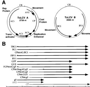
Plasmid constructs.Infectious clones of the A and B components of ToLCV (32) were used to generate the virus constructs used in this study. The genome organization of ToLCV and a schematic representation of the virus constructs used in this study are shown in Fig. 1, and detailed descriptions and methods of construction of each of the plasmids are summarized in Table 1. Partial
head-to-tail dimers made from these constructs were used to infect Nicotiana
benthamianaplants andN. tabacumBY2 protoplasts.
Protoplast and plant inoculations.N. benthamianaplants (2-week-old seed-lings grown in Magenta boxes) and protoplasts isolated from suspensions of BY2 cells were infected with viral DNAs as described earlier (32, 33). Protoplasts were collected from cultures 48 h postinoculation for DNA isolation, immunoprecipi-tation reactions, and Western blot analysis. Plants were scored for symptoms, and the newly formed upper leaves were collected for Southern blot analysis 22 to 25 days following inoculation. To study the local and systemic movements of
* Corresponding author. Mailing address: ILTAB, Division of Plant Biology-BCC206, The Scripps Research Institute, 10550 North Torrey Pines Rd., La Jolla, CA 92037. Phone: (619) 2906. Fax: (619) 784-2994. E-mail: [email protected].
1609
on November 9, 2019 by guest
http://jvi.asm.org/
the virus expressing green fluorescent protein (GFP) (8), bottom leaves of 4-week old seedlings (10 plants per construct) were inoculated. Inoculated leaves and noninoculated upper leaves were observed at 3-day intervals for 15 days under a fluorescence microscope for the detection of fluorescence emitted by GFP. In all experiments that involved plants, wild-type B-component DNA, which is essential for systemic spread and symptom development, was included.
Southern blotting.Total DNA was isolated from protoplasts (28) and plants (11), electrophoresed in 1% agarose gels (without ethidium bromide), and trans-ferred to Hybond nylon membranes (Amersham, Arlington Heights, Ill.) by standard protocols (41). Hybridization reactions were performed with a
ran-domly primed32P-labeled A-component-specific probe (the 900-bpAflII-PstI
fragment containing open reading frames [ORFs] for AC1, AC2, and AC3). The amounts of viral ssDNA and dsDNA (supercoiled, linear, open circular, and dimeric forms) were quantitated by exposing the Southern blots to storage phosphor screen plates and determining counts on a PhosphorImager (Molec-ular Dynamics, Sunnyvale, Calif.). The ssDNA form was confirmed by its sus-ceptibility to S1 and mung bean nucleases (33). In the absence of ethidium bromide, the supercoiled viral DNA form migrates ahead of the ssDNA form.
Immunoprecipitation and Western blotting.For immunoprecipitation reac-tions, protoplasts infected with the virus A component expressing the CP66: 6G:g5 protein tagged with the Flag epitope (FCP66:6G:g5) (Table 1) were lysed by use of a hand-held Polytron with Nonidet P-40 (NP-40) buffer (50 mM Tris-HCl [pH 7.5], 1% NP-40, 0.15, 0.25, 0.50, 0.75, or 1.0 M NaCl) or radioim-munoprecipitation assay (RIPA) buffer (50 mM Tris-HCl [pH 7.5], 150 mM NaCl, 1% NP-40, 0.5% deoxycholate, 0.1% sodium dodecyl sulfate) containing a cocktail of protease inhibitors (Boehringer Mannheim Biochemicals, Indianap-olis, Ind.). Cell debris was removed by centrifugation at 4°C for 10 min at
15,0003g. Lysates were immunoprecipitated with Flag monoclonal
anti-body M2 covalently linked to agarose (Sigma, St. Louis, Mo.). Immune com-plexes were washed four times with NP-40 or RIPA buffer and once with Tris-buffered saline (50 mM Tris-HCl [pH 7.5], 150 mM NaCl). Half of each sample was heated in Laemmli sample buffer, fractionated by SDS-polyacrylamide gel electrophoresis (13% acrylamide), and transferred to a polyvinylidene difluoride membrane (Schleicher & Schuell, Inc., Keene, N.H.). Immunoprecipitated pro-tein was visualized with anti-Flag antibody M2 by use of enhanced chemilumi-nescence-Western blot reagents (Pierce, Rockford, Ill.). The remaining half of each immune complex collected by this procedure was used for isolating viral
DNA. Whole-cell protein extracts for direct Western blotting were prepared by
boiling the protoplast pellets with an equal volume of 23Laemmli sample buffer.
Immunofluorescence.Protoplasts transfected with viral constructs were cul-tured on chamber slides (Nalge Nunc, Rochester, N.Y.) for 48 h, fixed with 3% paraformaldehyde in PBSEM (50 mM phosphate [pH 6.95], 150 mM NaCl, 5
mM EGTA, 5 mM MgSO4) for 30 min, and permeabilized with 100% methanol
at220°C for 10 min. The cells were washed two times with PBSEM containing
0.5% Tween 20 for 30 min each time. CP66:6G:g5 protein tagged with the Stag epitope (CP66:Stag:6G:g5) (Table 1) was detected with the S protein coupled to fluorescein isothiocyanate (FITC) (Novagen, Madison, Wis.). The 15-amino-acid-long Stag peptide was inserted after Arg66 of CP to construct the CP66: Stag:6G:g5 protein. Flag epitope-tagged BV1, T7 epitope-tagged BC1, CP, and
b-glucuronidase (GUS) (Table 1) were detected with anti-Flag antibody M2
(Sigma), anti-T7 tag antibody (Novagen), anti-CP antisera (33), and anti-GUS
antisera (59-39, Boulder, Colo.) diluted 1:100 in phosphate-buffered saline,
re-spectively. After incubation with the primary antibody for 1 h at 30°C, the cells were washed as before and incubated with FITC- or rhodamine-conjugated immunoglobulin G (Pierce) at a dilution of 1:100. The cells were mounted in Fluoromount G (Electron Microscopy Sciences, Fort Washington, Pa.) and viewed with a Nikon fluorescence microscope or an Olympus confocal micro-scope (for detecting T7 epitope-tagged BC1 protein).
RESULTS
[image:2.612.155.460.72.367.2]ToLCV expressing g5p or CP66:6G:g5 protein accumulates ssDNA to wild-type levels in protoplasts.Our earlier work with ToLCV showed that viral CP and AV2 are not required for virus replication in protoplasts, whereas AV2 is required for efficient movement in plants (33). CP is not essential for sys-temic movement and symptom development in ToLCV. How-ever, mutations in the CP sequence caused a marked decrease in ssDNA accumulation in N. bentamianaand tomato plants and in BY2 protoplasts while increasing dsDNA accumulation in protoplasts. Virus that contained mutations in AV2 plus CP FIG. 1. Genome organization and schematic representation of constructs of ToLCV used in this study. (A) Genome organization of ToLCV showing the ORFs and their functions. CR, common region for both components. (B) Linear physical map of AV2 and CP regions of ToLCV with nucleotide positions and relevant restriction enzyme sites (bottom). The positions of different gene replacements are shown above the linear map. Note that the gene replacements shown are not to the scale.
Descriptions of the constructs are given in Table 1.
on November 9, 2019 by guest
http://jvi.asm.org/
behaved like AV2 mutant virus in plants (i.e., poor virus move-ment and very mild symptoms) and like CP mutant virus in protoplasts (i.e., decrease in ssDNA and increase in dsDNA accumulation).
Here we investigated the effects of g5p fromE. coliphage M13 (40) on the replication of ToLCV. Each of the mutations is described in Table 1 and Fig. 1. The AV2 ORF and the overlapping 59portion of the CP ORF were replaced with g5p, and its effect on virus replication in protoplasts was assayed. In these experiments, protoplasts were inoculated with the wild type or mutants as described below. Surprisingly, the modified A component, designated g5AV22CP2, led to the
accumula-tion of ssDNA to the same levels as did the wild-type A com-ponent (Table 2 and Fig. 2, lanes 1 and 3). However, dsDNA accumulation was high (three- to sixfold higher than wild-type levels) and similar to the accumulation in the presence of mutations in CP (Table 2 and Fig. 2, lanes 2 to 4). Infection by virus in which the g5p gene was mutated to prevent its trans-lation (g52AV22CP2) (Table 1) behaved like virus infections
with A-component mutants AV22CP2and CP2(Table 2 and
Fig. 2, lane 4).
Since AV2 is required for efficient virus movement in plants, we made another construct in which g5p was fused to CP at Arg66 without affecting the AV2 ORF (CP66:g5) (Table 1). The CP66:g5 virus A component also led to the accumulation
of ssDNA, but to lower levels than did g5AV22CP2(Table 2
and Fig. 2, lane 6). To address the possibility that the N-terminal 66 amino acids (aa) of CP interfered with the ability of g5p to bind DNA, a linker of six glycine residues was intro-duced between Arg66 of CP and g5p to separate the CP do-main from the g5p dodo-main (CP66:6G:g5). The addition of the linker restored the ability of the CP66:6G:g5 virus A compo-nent to accumulate ssDNA to levels comparable to those of g5AV22CP2(Table 2 and Fig. 2, lane 7). A control construct
in which the g5p portion of the fusion protein was not trans-lated (CP66:g52) failed to accumulate ssDNA (Table 2 and
Fig. 2, lane 8). That the ability of the virus A component expressing the CP66:6G:g5 protein to accumulate ssDNA was not due to the N-terminal 66 aa of CP was suggested by the facts that the virus A component expressing g5p alone accu-mulated ssDNA and the virus A component expressing CP66: 6G:BC1 (see below) or CP66:6G:AV2 (data not shown) failed to accumulate ssDNA.
[image:3.612.51.550.74.449.2]Geminiviruses replicate in the nucleus (1, 29), so it is likely that in order to cause the accumulation of ssDNA, the CP66: 6G:g5 and g5 proteins must be present in the nucleus. To immunolocalize the CP66:6G:g5 fusion protein in protoplasts, we inserted the Stag epitope between Arg66 of CP and the glycine linker (CP66:Stag:6G:g5) (Table 1). At 48 h after in-fection, protoplasts were fixed and subjected to reactions with TABLE 1. Description and method of construction of viral DNAs used in this study
Construct Description and method of construction
AV22CP2...A double mutant of AV2 and CP in which the Met1 codon of AV2 was changed to a termination codon and the Arg66 codon of CP was frameshifted. The mutant was described earlier as M1te/R66fr (33).
g5AV22CP2...A 264-bp sequence coding for g5p from the bacteriophage M13mp18 vector was amplified by PCR (10 cycles) and cloned between theAflIII (nt 125) andStyI (nt 479) sites, resulting in the replacement of the AV2 ORF and overlapping 59CP ORF sequences with the g5p gene.
g52AV22CP2...A negative control for the g5AV22CP2construct in which the Met1 codon of g5p was mutated to a termination codon. CP2...A mutant of CP made by end filling and religation at the uniqueStyI site (nt 479), causing a frameshift at the Arg66
codon and termination after amino acid (aa) 69. The mutant was described earlier as R66fr (33).
CP66:g5...A 264-bp sequence coding for g5p from the M13mp18 vector was amplified by PCR (10 cycles) and cloned between the
StyI (nt 479) andSphI (nt 836) sites, resulting in the fusion of the g5p sequence to the Arg66 codon of CP.
CP66:6G:g5 ...Similar to CP66:g5, except that an oligonucleotide coding for 6 glycines was inserted between the codons for Arg66 of CP and Met1 of g5p.
CP66:g52...A negative control in which the Arg66 codon of CP66:g5 was frameshifted.
CP66:Stag:6G:g5...Similar to CP66:6G:g5, except that a sequence coding for the 15-aa Stag peptide epitope (KETAAAKFERQHMDS [23]) was inserted after the Arg66 codon of CP. The Stag epitope was inserted to immunolocalize the CP66:6G:g5 protein in protoplasts by use of S protein coupled to FITC.
FCP66:6G:g5...A sequence coding for the 9-aa Flag peptide epitope (MDYKDDDDK [19]) was added before the Met1 codon of CP66: 6G:g5 and cloned betweenAflIII (nt 125) andSphI (nt 836). The AV2 ORF was deleted. The Flag epitope was added to immunoprecipitate the CP66:6G:g5 protein from protoplasts by use of anti-Flag antibody.
CP66:GUS ...A 1,806-bp DNA fragment coding for GUS (21) was PCR amplified (10 cycles) and cloned between theStyI (nt 479) and
HindIII (nt 1041) sites of the A component. TheHindIII site was created at the codon for Tyr251 of CP (15 bp before the termination codon [33]), facilitating the replacement of the CP sequence with other sequences.
GUSAV22CP2...A 1,869-bpNcoI-EcoRI DNA fragment coding for GUS was cloned between theAflIII (nt 125) andHindIII (nt 1041) sites of the A component after blunt ending theEcoRI site on the GUS gene and theHindIII site on the A-component DNA.
GFPAV22CP2...A 717-bpNcoI-BamHI DNA fragment coding for GFP (S65C, M153T, V163A [37]) was cloned between theAflIII (nt 125) andSphI (nt 836) sites of the A component after blunt ending theBamHI site on the GFP gene and theSphI site on the A-component DNA.
BV1AV22CP2...An 849-bp sequence coding for BV1 from the B component of ToLCV was amplified by PCR (10 cycles) and cloned be-tween theAflIII (nt 125) andHindIII (nt 1041) sites of the A component.
FBV1AV22CP2...Similar to BV1AV22CP2, except that the sequence coding for the 9-aa Flag peptide was added before the Met1 codon of BV1. The Flag epitope was added to immunolocalize the BV1 protein in protoplasts by use of anti-Flag antibody. BC1AV22CP2...An 882-bp sequence coding for BC1 from the B component of ToLCV was amplified by PCR (10 cycles) and cloned
be-tween theAflIII (nt 125) andHindIII (nt 1041) sites of the A component.
TBC1AV22CP2...Similar to BC1AV22CP2, except that the sequence coding for the 11-aa T7 epitope (MASMTGGQQMG [24]) was added before the Met1 codon of BC1. The T7 tag epitope was added to immunolocalize the BC1 protein in protoplasts by use of anti-T7 tag antibody.
CP66:6G:BC1...A 900-bp sequence coding for 6 glycines and BC1 from the B component of ToLCV was amplified by PCR (10 cycles) and cloned between theStyI (nt 479) andHindIII (nt 1041) sites.
BC12...B-component DNA in which a frameshift mutation of BC1 was created by deletion of the 39overhang and religation at thePstI site (nt 2075). The mutant was described earlier as BC1M (33).
on November 9, 2019 by guest
http://jvi.asm.org/
S protein coupled to FITC. The CP66:Stag:6G:g5 protein as well as wild-type CP (detected with anti-CP antisera) were localized to the nucleus (Fig. 3A and B). When GUS was produced as a fusion protein with the N-terminal 66 aa of CP (CP66:GUS), GUS (detected with anti-GUS antisera) was also localized to the nucleus (Fig. 3C). This result indicated that the N-terminal 66 aa of CP contains a nuclear localization signal. We also determined if g5p contains a nuclear localization sig-nal by fusing the g5p coding sequence to the GUS coding sequence at the N terminus. The g5:GUS fusion protein (ex-pressed in the g5:GUSAV22CP2virus A component) (Table
1) and the unfused GUS protein (expressed in the GUSAV22
CP2virus A component) (Table 1) remained in the cytoplasm
(Fig. 3D and E), suggesting that g5p has no nuclear localiza-tion signal. It is possible that g5p may have entered the nucleus in a passive manner, as its size (9.7 kDa) is smaller than the permeability barrier of the nuclear envelope (12).
Movement of ToLCV expressing CP66:6G:g5 protein is im-paired in plants.N. benthamianaplants were inoculated with selected virus constructs to determine the effect of g5p on virus spread. In these studies, the B component was coinoculated with the A component ontoN. benthamianaseedlings. As ex-pected, plants inoculated with A-component mutant AV22CP2,
g5AV22CP2, or g52AV22CP2plus the B component showed
very mild or no symptoms, and all inoculated plants accumu-lated low levels of viral DNA (Table 2). A previously reported ToLCV mutant (33) that did not produce CP but produced AV2 (CP2) resulted in severe disease symptoms and wild-type
levels of dsDNA in systemic infections (Table 2). Surprisingly, plants inoculated with the virus expressing the CP66:6G:g5 protein showed very mild or no symptoms, even though the virus contained an intact AV2 gene (Table 2). These plants accumulated low levels of viral DNA, similar to plants inocu-lated with the AV22CP2 virus (Table 2). Plants inoculated
with the virus expressing the CP66:g5 protein (which accumu-lated ssDNA to a lower level than CP66:6G:g5 virus in proto-plasts) showed mild symptoms and accumulated moderate lev-els of dsDNA. We also considered the possibility that the impaired movement of the virus expressing g5p was due to possible toxic effects of g5p. We did not detect any differences in protoplast viability or in the appearance of plant leaves inoculated with wild-type virus or virus expressing g5p that might suggest toxicity of g5p.
We next examined the cell-to-cell and long-distance move-ment of ToLCV expressing the CP66:6G:g5 protein by using
green fluorescent protein (GFP) as a visible marker for virus movement. Plants were inoculated with A-component DNA expressing GFP in place of AV2 and CP (GFPAV22CP2)
alone or coinoculated with A-component DNA of the wild-type, CP66:6G:g5, or CP66:g52construct. GFPAV22CP2
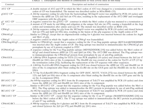
vi-FIG. 2. Replication of ToLCV constructs in infected BY2 protoplasts. South-ern blot analysis was performed as described in Materials and Methods. The viral constructs used for infecting protoplasts are shown above the lanes. Protoplasts were inoculated with A-component DNA alone (lanes 1 to 11) or coinoculated
with A- and B-component DNAs (lanes 12 to 15). Each lane contained 4mg of
[image:4.612.52.551.80.193.2]DNA prepared from protoplasts (single transfection). Viral DNA was detected with a radioactively labeled probe from A-component DNA. The positions of supercoiled (sc), single-stranded (ss), linear (li), and open circular (op) viral DNA forms are indicated. Note that the positions of supercoiled and other viral DNA forms in lane 11 are shifted upward due to the larger size of the CP66: 6G:BC1 construct. wt, wild type.
TABLE 2. Effect of g5p on the replication and movement of ToLCV inN. tabacumprotoplasts andN. benthamianaplants
Virus
Protoplast inoculations Plant inoculations
ssDNAa dsDNAa No. of plants
inoculated Symptomtype ssDNAb dsDNAb
Wild type 100 100 20 Severe 100 100
AV22CP2 ,1 (0–0.03) 506 (427–584) 10 Very mildc 0.3 (0.05–0.5) 11 (9.6–17) g5AV22CP2 102 (79–133) 409 (349–573) 20 Very mildc 0.6 (0.1–2.7) 15.2 (6.2–49.2) g52AV22CP2 7 (5–12) 384 (210–779) 20 Very mildc 0.1 (0.0–0.2) 5.7 (0.0–11.4)
CP2 5 (2–7) 241 (148–369) 20 Severed 4.3 (2.6–6.5) 102 (65–139)
CP66:g5 17 (8–27) 442 (345–576) 20 Mild 2.2 (0.8–4.2) 30.6 (15.3–55.1)
CP66:6G:g5 118 (34–234) 517 (133–784) 30 Very mildc 0.9 (0.4–1.7) 10.9 (5.5–14.7)
CP66:g52 9 (3–14) 424 (179–789) 20 Severed 4.0 (1.8–6.1) 139.7 (56.0–197.7)
aThe values represent the average amount (range) of A-component DNA in five independent protoplast transfections per mutant. Protoplasts (;106) were
transfected with 2mg of A-component DNA and 40mg of herring sperm DNA. Viral DNA was quantitated on Southern blots with a PhosphorImager (Molecular
Dynamics). Values are relative to those for the wild type, which was assigned a value of 100.
bThe values represent the average amount (range) of viral DNA in 12 inoculated plants per virus construct, except for AV22CP2, for which the values represent
the average in four plants. Each plant was inoculated with 0.5mg of A-component DNA and 0.5mg of wild-type B-component DNA, which is essential for viral
movement and symptom development. Values are relative to those for the wild type, which was assigned a value of 100.
cMany plants did not show symptoms.
dSevere symptoms like those in plants inoculated with the wild-type virus but without intense chlorosis.
on November 9, 2019 by guest
http://jvi.asm.org/
[image:4.612.333.521.359.633.2]rus was expected to move inefficiently in plants, as it does not carry AV2; it was expected to move efficiently when comple-mented by another virus carrying AV2. GFP could not be detected in plants by day 3 postinoculation, but it was present on inoculated and upper leaves by day 6 in the majority of the plants inoculated with GFPAV22CP2plus wild-type
A-com-ponent viruses or GFPAV22CP2plus CP66:g52viruses (Fig.
3H and I; only data on plants inoculated with GFPAV22CP2
plus CP66:g52viruses are shown). The virus expressing GFP
continued to spread to upper and newly emerging leaves in
these plants (Fig. 3J and K). GFP was observed in veins, the mesophyll, and epidermal cells and was present in large areas of the leaves in plants inoculated with GFPAV22CP2 plus
CP66:g52 viruses. In contrast, GFP was restricted to small
spots on the inoculated leaves of most of the plants inoculated with GFPAV22CP2 or GFPAV22CP2plus CP66:6G:g5
vi-ruses (Fig. 3L and M; only data on plants inoculated with GFPAV22CP2 plus CP66:6G:g5 viruses are shown). These
[image:5.612.79.531.69.516.2]plants also showed GFP staining in some adjacent and newly emerging leaves, mostly restricted to veins (Fig. 3N, O, and P).
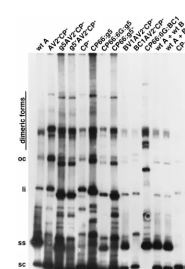
FIG. 3. Indirect immunofluorescence of proteins expressed in protoplasts (A to G) and fluorescence of GFP expressed in plants (H to P). Protoplasts were transfected, and antigens were visualized with different primary antibodies and FITC- or rhodamine-conjugated secondary antibodies. GFP fluorescence in plants was monitored every 3 days for 15 days, and the area shown in each panel corresponds to a leaf area measuring 2.5 by 2.5 mm. (A) Protoplast infected with CP66:Stag:6G:g5 virus and stained with S protein coupled to FITC. (B) Protoplast infected with wild-type virus and stained with anti-CP antisera. (C) Protoplast infected with CP66:GUS
virus and stained with anti-GUS antisera. (D) Protoplast infected with g5:GUSAV22CP2virus and stained with anti-GUS antisera. (E) Protoplast infected with
GUSAV22CP2virus and stained with anti-GUS antisera. (F) Protoplast infected with FBV1AV22CP2virus and stained with anti-Flag antibody. (G) Protoplasts
infected with TBC1AV22CP2virus and stained with anti-T7 tag antibody. Note that two cells are shown in this micrograph. (H and I) Inoculated leaf (H) and
systemically infected leaf (I) of a plant infected with GFPAV22CP2and CP66:g52viruses 6 days postinoculation (dpi). (J and K) Inoculated leaf (J) and systemically
infected leaf (K) of a plant infected with GFPAV22CP2and CP66:g52viruses 15 dpi. (L and M) Inoculated leaf (L) and systemically infected leaf (M) of a plant
infected with GFPAV22CP2and CP66:6G:g5 viruses 6 dpi. (N to P) Inoculated leaf (N) and systemically infected leaves (O and P) of a plant infected with
GFPAV22CP2and CP66:6G:g5 viruses 15 dpi.
on November 9, 2019 by guest
http://jvi.asm.org/
These results indicated that the expression of g5p in place of CP decreased the efficiency of virus systemic movement.
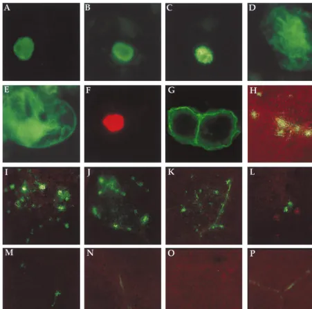
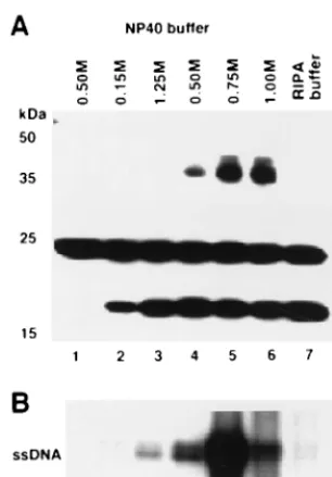
In vivo binding of CP66:6G:g5 protein to viral DNA. The accumulation of viral ssDNA in protoplasts inoculated with the virus A component expressing g5p or CP66:6G:g5 protein sug-gested that g5p binds to ssDNA. To test this possibility, we inoculated protoplasts with the virus A component expressing the Flag epitope-tagged CP66:6G:g5 protein (FCP66:6G:g5) (Table 1), immunoprecipitated the Flag epitope-tagged CP66: 6G:g5 protein with anti-Flag antibody, and characterized the viral DNA that coimmunoprecipitated with the CP66:6G:g5 protein by Southern blotting. The immunoprecipitations were performed under different salt (1% NP-40 buffer with 0.15 to 1.0 M NaCl) conditions and in the presence of 0.1% sodium dodecyl sulfate, 0.5% deoxycholate, and 1% NP-40 detergents (RIPA buffer) to assay the affinity of binding. The Flag epitope-tagged CP66:6G:g5 protein was immunoprecipitated under all of the buffer conditions tested; the amount of protein immunoprecipitated increased with increasing salt concentra-tion (Fig. 4A). The amount of coimmunoprecipitated ssDNA increased up to a 0.5 M salt concentration and decreased at higher concentrations (Fig. 4B), indicating that the g5p-ssDNA complex was destabilized in buffer that contained 1 M salt. Immunoprecipitation in RIPA buffer also resulted in a reduced amount of precipitated ssDNA (Fig. 4B). These results showed that g5p bound to viral ssDNA and that 1 M salt (in NP-40 buffer) dissociated g5p from the viral ssDNA.
Role of BV1 and BC1 movement proteins in the spread of ToLCV.The above results indicate that the CP66:6G:g5 pro-tein is localized to the nucleus and binds stably to ToLCV virus
DNA in vivo and that ToLCV expressing CP66:6G:g5 does not move efficiently in plants. The inefficient movement of ToLCV expressing the CP66:6G:g5 protein may have been due to an interference of g5p with the function of the BV1 or BC1 movement protein of ToLCV. In squash leaf curl virus (SLCV), BV1 (referred to as BR1 in SLCV) but not BC1 (referred to as BL1 in SLCV) binds to ssDNA in vitro (34). BR1 and BL1 of SLCV interact with each other in a cooper-ative manner; in protoplasts, BR1 localizes to the nucleus in the absence of BL1 but localizes to the cell periphery in the presence of BL1 (42, 43). Both BV1 and BC1 are required for the systemic spread and symptom development of ToLCV (33). To determine if BV1 and BC1 of ToLCV have functions similar to those of BR1 and BL1 of SLCV, we immunolocal-ized BV1 and BC1 of ToLCV and examined their ability to complement the viral ssDNA accumulation of CP mutants. For these experiments, the BV1 and BC1 genes were fused to sequences coding for the Flag epitope tag and the T7 epitope tag, respectively, and inserted in place of AV2 and CP in the A component (FBV1AV22CP2and TBC1AV22CP2) (Table 1).
In protoplasts inoculated with the FBV1AV22CP2construct,
the BV1 protein accumulated in the nucleus (detected with anti-Flag antibody) (Fig. 3F), while in protoplasts inoculated with TBC1AV22CP2, the BC1 protein was localized to the cell
periphery (detected with anti-T7 tag antibody) (Fig. 3G). Ex-pression of the BV1 protein in place of the AV2 and CP pro-teins (BV1AV22CP2) also led to the accumulation of ssDNA
by the A-component virus (Table 3 and Fig. 2, lane 9). The binding affinity of the BV1 protein tagged with the Flag epi-tope for viral DNA in protoplasts inoculated with FBV1AV22
CP2DNA was determined by immunoprecipitation reactions
similar to those shown in Fig. 4. The binding affinity of the BV1 protein for viral ssDNA was similar to the binding affinity of the CP66:6G:g5 protein for viral ssDNA (data not shown). In contrast to results obtained with virus A component expressing BV1, A-component virus expressing BC1 in place of AV2 and CP (BC1AV22CP2) did not accumulate ssDNA (Table 3 and
Fig. 2, lane 10). Since the BC1 protein was localized to the cell periphery, we fused BC1 to the N-terminal 66 aa of CP (CP66: 6G:BC1) to direct it to the nucleus. Virus A component ex-pressing the CP66:6G:BC1 protein also did not accumulate ssDNA (Table 3 and Fig. 2, lane 11), showing that the BC1 movement protein may not bind to viral ssDNA or that the binding affinity may not be strong enough to result in the accumulation of ssDNA. These results show that BV1 is local-ized to the nucleus in the absence of BC1 and that BV1 binds to viral ssDNA in vivo.
In plants inoculated with the ToLCV A component
contain-FIG. 4. In vivo binding of g5p to ToLCV DNA. (A) Flag epitope-tagged CP66:6G:g5 protein expressed in protoplasts was immunoprecipitated with anti-Flag antibody coupled to agarose after lysis of protoplasts in NP-40 buffer containing different concentrations of NaCl (shown above lanes 1 to 6) or RIPA buffer (lane 7), and the immunoprecipitated protein was detected on a Western blot with anti-Flag antibody (lanes 2 to 7). Lane 1 contained protein immuno-precipitated from protoplasts transfected with wild-type virus as a control. The
protein band present in all lanes at ;24 kDa is the light chain of anti-Flag
antibody used for immunoprecipitations. The immunoprecipitated CP66:6G:g5 protein was detected at two different molecular masses corresponding to mono-meric and dimono-meric forms. Positions of molecular mass markers are indicated in kilodaltons on the left. (B) Viral ssDNA that coimmunoprecipitated with the Flag epitope-tagged CP66:6G:g5 protein was detected on a Southern blot with
32P-labeled A-component DNA as a probe. Lanes 1 to 7 were given the same
[image:6.612.96.249.66.286.2]treatments as in panel A.
TABLE 3. Complementation by BV1 and BC1 movement proteins of the accumulation of ToLCV ssDNA in protoplastsa
A component B component ssDNA dsDNA
Wild type None 100 100
BV1AV22CP2 None 86 (50–121) 230 (119–195) BC1AV22CP2 None 2 (1–3) 224 (162–288) CP66:6G:BC1 None 5 (1–10) 214 (180–267)
Wild type Wild type 100 100
Wild type BC12 84 (66–100) 82 (66–98)
CP2 Wild type 4 (3–6) 164 (128–198)
CP2 BC12 5 (3–6) 173 (160–185)
aProtoplasts were transfected with 2mg of A-component DNA with or without
10mg of B-component DNA. Viral DNA was quantitated on Southern blots with
a PhosphorImager. The values represent the average amount (range) of viral DNA in two to five independent transfections, relative to a value of 100 assigned to the wild type.
on November 9, 2019 by guest
http://jvi.asm.org/
[image:6.612.311.552.594.683.2]ing CP66:6G:g5 plus the wild-type B component, the expres-sion of the CP66:6G:g5 protein is controlled by the relatively strong CP promoter. The CP66:6G:g5 protein produced from the A component may outcompete the BV1 protein (expressed from the B component) for DNA binding if the amount of BV1 made under the control of its own promoter is relatively low. We conducted an experiment to determine if BV1, expressed under the control of its own promoter on the B component, can lead to the accumulation of ssDNA. Note that BV1 led to the accumulation of ssDNA when expressed in place of CP on the A component (Table 3). However, very little viral ssDNA accumulated in protoplasts coinoculated with A-component DNA with a mutation in CP (CP2) plus wild-type
nent DNA (i.e., expressing both BV1 and BC1) or B-compo-nent DNA with a mutation in BC1 (BC12) (i.e., expressing
only BV1) (Table 3 and Fig. 2, lanes 12 to 15). The failure of BV1 to cause the accumulation of ssDNA when expressed from the B component appeared to be due to low levels of BV1 protein being made; no BV1 protein was detected in proto-plasts coinoculated with A-component DNA and B-compo-nent DNA expressing Flag epitope-tagged BV1 by immunolo-calization and Western blotting procedures (data not shown). These results show that the B-component promoter driving the expression of BV1 is not as strong as when the gene is ex-pressed from the CP promoter on the A component.
DISCUSSION
Previous work done by our group showed that in the absence of CP, ToLCV failed to accumulate ssDNA but produced lev-els of dsDNA severalfold higher than wild-type levlev-els in pro-toplasts (33). Reduced levels of ssDNA have been observed for other geminiviruses when CP is not produced (5, 27, 49, 52). This observation raised the question as to whether the accu-mulation of ssDNA is due solely to encapsidation by CP or whether CP has some additional role in viral replication. We tested these possibilities by expressing a nonspecific ssDNA binding protein in place of CP and monitoring the accumula-tion of ssDNA to determine if it could serve as a substitute for CP in this putative function. g5p fromE. coliphage M13 was chosen because of its small size (9.7 kDa) and lack of any enzymatic function in DNA replication. The role of g5p in the replication of M13 and other filamentous phages has been extensively studied (36), and its structure has been determined (45). g5p binds newly formed viral ssDNA tightly, coopera-tively, and in a sequence-independent manner and protects it from degradation byE. colinucleases (7, 31, 40).
In this report, we demonstrated that g5p can bind to ToLCV ssDNA in plant cells and that ToLCV expressing g5p or g5p fused to the N-terminal 66 aa of CP can accumulate ssDNA to wild-type levels. The binding of g5p to viral ssDNA in vivo was similar to the binding of g5p to M13 ssDNA in vitro (2). Although g5p compensated for the lack of CP by causing an increase in the accumulation of ToLCV ssDNA, it did not reduce the amount of dsDNA to wild-type levels. BV1 move-ment protein (when expressed in place of CP) also behaved like g5p in that it did not down-regulate dsDNA to wild-type levels. If CP regulates the levels of ssDNA and dsDNA by depleting the ssDNA available for conversion to dsDNA, the expression of g5p or BV1 could be expected to result in normal amounts of dsDNA. The fact that it did not suggests that CP may have a direct role in regulating viral replication, possibly by inhibiting minus-strand synthesis or by regulating gene ex-pression. The CP of alfalfa mosaic virus, a virus with a plus-strand ssRNA genome, has been shown to play a direct role in the regulation of plus- and minus-strand RNA syntheses (10).
The alfalfa mosaic virus CP was found in tight association with the viral RNA polymerase and inhibited minus-strand synthe-sis while stimulating plus-strand synthesynthe-sis. Recent results ob-tained with SLCV suggest that CP acts to signal the switch from viral dsDNA replication to ssDNA replication or to se-quester virion ssDNA from replication pools without fully en-capsidating it (25a). Purification of geminivirus replication complexes is needed to directly assess the role of CP in repli-cation.
Why do plants infected with a virus encoding the CP66: 6G:g5 protein show very mild symptoms and accumulate low levels of viral DNA when infected protoplasts accumulate high levels of viral DNA? One likely possibility is that by binding to viral ssDNA, g5p affects virus movement by interfering with the function of the BV1 movement protein. BV1 of ToLCV was localized to the nucleus in infected protoplasts and bound to viral ssDNA in vivo; BC1 was localized to the cell periphery and did not complement viral ssDNA accumulation, even when it was directed to the nucleus as a fusion to the nuclear local-ization signal of CP. Recent studies on the roles of BR1 and BL1 in SLCV movement have shown that BR1 is localized to the nucleus, binds to ssDNA in vitro, and functions as a nuclear shuttle protein (34, 42). BL1 of SLCV is localized to the cell periphery in protoplasts and is associated with endoplasmic reticulum-derived tubules in developing phloem cells of sys-temically infected pumpkin seedlings (20, 43, 51). Based on these results, a model for SLCV was proposed in which BL1-containing tubules serve as a conduit for the transport of BR1 and its associated viral ssDNA from one cell to another (51). Studies with tomato golden mosaic virus have shown that BR1 interacts with viral ssDNA in vivo and that BR1 and BL1 have distinct and essential roles in cell-to-cell movement as well as systemic movement (22). It is likely that ToLCV uses a similar strategy in moving from cell to cell. The poor movement of ToLCV that produces the CP66:6G:g5 protein may be due to reduced binding of BV1 to viral ssDNA. It should be noted that BV1 did not lead to the accumulation of ssDNA of the A component that lacked CP when BV1 was expressed under the control of its own promoter from the B component. In plants coinoculated with the A component producing CP66:6G:g5 plus the A component producing GFP, GFP staining was most-ly restricted to small areas on both inoculated and systemicalmost-ly infected leaves, showing an overall reduction in the efficiency of viral movement rather than specific interference with cell-to-cell spread or long-distance movement.
In contrast to the model presented for the movement of SLCV, a different model was proposed for bean dwarf mosaic virus in which BC1 binds to dsDNA and moves it through plasmadesmata by increasing their size exclusion limit (30). Interference with ToLCV movement due to binding of g5p to viral ssDNA suggests that in this virus, ssDNA moves from cell to cell. Our results also suggest that the expression of g5p in transgenic plants may afford a novel way of controlling gemi-niviruses and that such resistance may be effective against all geminiviruses.
ACKNOWLEDGMENTS
We thank Sondra Lazarowitz and Hal Padget for critically reading the manuscript.
This work was supported by financial assistance from the U.S. Agency for International Development (grant DAN-4197-A-00-1126-00); Maharashtra Hybrid Seeds Company, Jalna, India (grant 5-98378); and Institut Franc¸ais de Recherche Scientifique pour le De´veloppement en Coope´ration (ORSTOM), Paris, France.
on November 9, 2019 by guest
http://jvi.asm.org/
REFERENCES
1.Accotto, G. P., P. M. Mullineaux, S. C. Brown, and D. Marie.1993. Digitaria streak geminivirus replicative forms are abundant in S-phase nuclei of
in-fected cells. Virology195:257–259.
2.Anderson, R. A., Y. Nakashima, and J. E. Coleman.1975. Chemical modi-fications of functional residues of fd gene 5 DNA-binding protein.
Biochem-istry14:907–917.
3.Boulton, M. I., J. Steinkellner, J. Donson, P. G. Markham, D. I. King, and J. W. Davies.1989. Mutational analysis of virion-sense genes of maize streak
virus. J. Gen. Virol.70:2309–2323.
4.Briddon, R. W., and P. G. Markham.1995.Geminiviridae, p. 158–165.In
F. A. Murphy (ed.), Virus taxonomy. Sixth report of the International Com-mittee on Taxonomy of Viruses. Springer-Verlag, Vienna, Austria. 5.Briddon, R. W., J. Watts, P. G. Markham, and J. Stanley.1989. The coat
protein of beet curly top virus is essential for infectivity. Virology172:628–
633.
6.Brough, C. L., R. J. Hayes, A. J. Morgan, R. H. A. Coutts, and K. W. Buck.
1988. Effects of mutagenesisin vitroon the ability of cloned tomato golden
mosaic virus DNA to infectNicotiana benthamianaplants. J. Gen. Virol.69:
503–514.
7.Cavalieri, S., K. Neet, and D. Goldthwait.1976. Gene 5 protein of
bacterio-phage fd: a dimer which interacts co-operatively with DNA. J. Mol. Biol.102:
697–711.
8.Chalfie, M., Y. Tu, G. Euskirchen, W. W. Ward, and D. C. Prasher.1994.
Green fluorescent protein as a marker for gene expression. Science263:802–
805.
9.Davies, J. W., and J. Stanley.1989. Geminivirus genes and vectors. Trends
Genet.5:77–81.
10. De Graaff, M., M. R. Man in’t Veld, and E. M. Jaspars.1995.In vitro
evidence that the coat protein of alfalfa mosaic virus plays a direct role in the regulation of plus and minus RNA synthesis: implications for the life cycle of
alfalfa mosaic virus. Virology208:583–589.
11. Dellaporta, S. L., J. Wood, and J. B. Hicks.1983. A plant DNA
miniprepa-ration: version II. Plant Mol. Biol. Rep.1:19–21.
12. Dingwall, C., and R. A. Laskey.1986. Protein import into the cell nucleus.
Annu. Rev. Cell Biol.2:367–390.
13. Elmer, J. S., L. Brand, G. Sunter, W. Gardiner, D. M. Bisaro, and S. G. Rogers.1988. Genetic analysis of the tomato golden mosaic virus. II. The product of the AL1 coding sequence is required for replication. Nucleic
Acids Res.16:7043–7060.
14. Frischmuth, T., and J. Stanley.1993. Strategies for the control of
geminivi-rus diseases. Semin. Virol.4:329–337.
15. Gardiner, W. E., G. Sunter, L. Brand, L. S. Elmer, S. G. Rogers, and D. M. Bisaro. 1988. Genetic analysis of tomato golden mosaic virus: the coat protein is not required for systemic spread or symptom development. EMBO
J.7:899–904.
16. Hanley-Bowdoin, L., J. S. Elmer, and S. G. Rogers.1990. Expression of functional replication protein from tomato golden mosaic virus in transgenic
tobacco plants. Proc. Natl. Acad. Sci. USA87:1446–1450.
17. Hanley-Bowdoin, L., J. S. Elmer, and S. G. Rogers.1989. Functional expres-sion of the leftward open reading frames of the A component of tomato
golden mosaic virus in transgenic tobacco plants. Plant Cell1:1057–1067.
18. Harrison, B. D.1985. Advances in geminivirus research. Annu. Rev.
Phyto-pathol.23:55–82.
19. Hopp, T. P.1986. Protein surface analysis: methods for identifying antigenic
determinants and other interaction sites. J. Immunol. Methods88:1–18.
20. Ingham, D. J., E. Pascal, and S. G. Lazarowitz.1995. Both bipartite gemi-nivirus movement proteins define viral host range, but only BL1 determines
viral pathogenicity. Virology207:191–204.
21. Jefferson, R. A.1987. Assaying chimeric genes in plants: the GUS gene
fusion system. Plant Mol. Biol. Rep.5:387–405.
22. Jeffrey, J. L., W. Pooma, and I. T. D. Petty.1996. Genetic requirements for local and systemic movement of tomato golden mosaic virus in infected
plants. Virology223:208–218.
23. Kim, J. S., and R. T. Raines.1993. Ribonuclease S-peptide as a carrier in
fusion proteins. Protein Sci.2:348–356.
24. Krek, W., M. E. Ewen, S. Shorodka, Z. Arany, W. G. Kaelin, and D. M. Livingston.1994. Negative regulation of the growth-promoting transcription
factor E2F-1 by a stably bound cyclin A-dependent protein kinase. Cell78:
161–172.
25. Laufs, J., W. Traut, F. Heyraud, V. Matzeit, S. G. Rogers, J. Schell, and B. Gronenborn.1995.In vitrocleavage and joining at the viral origin of repli-cation by the replicator initiator protein of tomato yellow leaf curl virus.
Proc. Natl. Acad. Sci. USA92:3879–3883.
25a.Lazarowitz, S.Personal communication.
26. Lazarowitz, S. G.1992. Geminiviruses: genome structure and gene function.
Crit. Rev. Plant Sci.11:327–349.
27. Lazarowitz, S. G., A. J. Pinder, V. D. Damsteegt, and S. G. Rogers.1989. Maize streak virus genes essential for systemic spread and symptom
devel-opment. EMBO J.8:1023–1032.
28. Mettler, I. J.1987. A simple and rapid method for minipreparation of DNA
from tissue cultured plant cells. Plant Mol. Biol. Rep.5:346–349.
29. Nagar, S., T. J. Pedersen, K. M. Carrick, L. Hanley-Bowdoin, and D. Rob-ertson.1995. A geminivirus induces expression of a host DNA synthesis
protein in terminally differentiated plant cells. Plant Cell7:705–719.
30. Noueiry, A. O., W. J. Lucas, and R. L. Gilbertson.1994. Two proteins of a
plant DNA virus coordinate nuclear and plasmodesmal transport. Cell76:
925–932.
31. Oey, J., and R. Knippers.1972. Properties of the isolated gene 5 protein of
bacteriophage fd. J. Mol. Biol.68:125–138.
32. Padidam, M., R. N. Beachy, and C. M. Fauquet.1995. Tomato leaf curl geminivirus from India has a bipartite genome and coat protein is not
essential for infectivity. J. Gen. Virol.76:25–35.
33. Padidam, M., R. N. Beachy, and C. M. Fauquet.1996. The role of AV2 (“precoat”) and coat protein in viral replication and movement in tomato
leaf curl geminivirus. Virology224:390–404.
34. Pascal, E., A. A. Sanderfoot, B. M. Ward, R. Medville, R. Turgeon, and S. G. Lazarowitz. 1994. The geminivirus BR1 movement protein binds
single-stranded DNA and localizes to the cell nucleus. Plant Cell6:995–1006.
35. Polston, J. E., and P. K. Anderson.1997. The emergence of
whitefly-trans-mitted geminiviruses in tomato in the western hemisphere. Plant Dis.81:
1358–1369.
36. Rasched, I., and E. Oberer.1986. Ff coliphages: structural and functional
relationships. Microbiol. Rev.50:401–427.
37. Reichel, C., J. Mathur, P. Eckes, K. Langenkemper, C. Koncz, J. Schell, B. Reiss, and C. Maas.1996. Enhanced green fluorescence by the expression of anAequorea victoriagreen fluorescent protein mutant in mono- and
dicot-yledonous plant cells. Proc. Natl. Acad. Sci. USA93:5888–5893.
38. Rigden, J. E., I. B. Dry, P. M. Mullineaux, and M. A. Rezaian.1993. Mu-tagenesis of the virion-sense open reading frames of tomato leaf curl
gemi-nivirus. Virology193:1001–1005.
39. Rochester, D. E., C. M. Fauquet, J. J. Depaulo, and R. N. Beachy.1994. Complete nucleotide sequence of the geminivirus tomato yellow leaf curl
virus, Thailand isolate. J. Gen. Virol.75:477–485.
40. Salstrom, J., and D. Pratt.1971. Role of coliphage M13 gene 5 in
single-stranded DNA production. J. Mol. Biol.61:489–501.
41. Sambrook, J., E. F. Fritsch, and T. Maniatis.1989. Molecular cloning: a laboratory manual. Cold Spring Harbor Laboratory Press, Cold Spring Har-bor, N.Y.
42. Sanderfoot, A. A., D. J. Ingham, and S. G. Lazarowitz.1996. A viral
move-ment protein as a nuclear shuttle. Plant Physiol.110:23–33.
43. Sanderfoot, A. A., and S. G. Lazarowitz.1995. Cooperation in viral move-ment: the geminivirus BL1 movement protein interacts with BR1 and
redi-rects it from the nucleus to the cell periphery. Plant Cell7:1185–1194.
44. Saunders, K., A. Lucy, and J. Stanley.1991. DNA forms of the geminivirus African cassava mosaic virus consistent with a rolling circle mechanism of
replication. Nucleic Acids Res.19:2325–2330.
45. Skinner, M. M., H. Zhang, D. H. Leschnitzer, Y. Guan, H. Bellamy, R. M. Sweet, C. W. Gray, R. N. H. Konings, A. H. J. Wang, and T. C. Terwillinger.
1994. Structure of the gene V protein of bacteriophage f1 determined by multiwavelength x-ray diffraction on selenomethionyl protein. Proc. Natl.
Acad. Sci. USA91:2071–2075.
46. Stenger, D. C., G. N. Revington, M. C. Stevenson, and D. M. Bisaro.1991. Replicational release of geminivirus genomes from tandemly repeated cop-ies: evidence for rolling circle replication of a plant viral DNA. Proc. Natl.
Acad. Sci. USA88:8029–8033.
47. Sunter, G., and D. M. Bisaro.1992. Transactivation of geminivirus AR1 and BR1 gene expression by the viral AL2 gene product occurs at the level of
transcription. Plant Cell4:1321–1331.
48. Sunter, G., W. E. Gardiner, and D. M. Bisaro.1989. Identification of tomato
golden mosaic virus-specific RNAs in infected plants. Virology170:243–250.
49. Sunter, G., M. D. Hartitz, S. G. Hormudzi, C. L. Brough, and D. M. Bisaro.
1990. Genetic analysis of tomato golden mosaic virus: ORF AL2 is required for coat protein accumulation while ORF AL3 is necessary for efficient DNA
replication. Virology179:69–77.
50. Timmermans, M. C. P., O. P. Das, and J. Messing.1994. Geminiviruses and their uses as extrachromosomal replicons. Annu. Rev. Plant Physiol. Plant
Mol. Biol.45:79–112.
51. Ward, B. M., R. Medville, S. G. Lazarowitz, and R. Turgeon.1997. The geminivirus BL1 movement protein is associated with endoplasmic
reticu-lum-derived tubules in developing phloem cells. J. Virol.71:3726–3733.
52. Woolston, C. J., H. V. Reynolds, N. J. Stacey, and P. M. Mullineaux.1989. Replication of wheat dwarf virus-DNA in protoplasts and analysis of coat
protein mutants in protoplasts and plants. Nucleic Acids Res.17:6029–6041.