Ano Rectal Malformations – A Study
A three year study Aug2008 to July 2011
A Dissertation submitted in partial fulfillment of M.Ch Branch V
(Paediatric Surgery) examination of Dr. M. G. R. Medical University,
Certificate
Certified that the dissertation – entitled “Ano Rectal
Malformations – A Study” is the Bonafide work under taken by Dr.
M. SHANKAR under my guidance and supervision, in the
Department of Paediatric Surgery, Government Rajaji Hospital,
Madurai Medical College, Madurai, during the period of his
Postgraduate residency in M. Ch. Paediatric Surgery from 2008 to
2011.
Dr. A. Athigaman, M.S., M.Ch.,
Professor and HOD, Department of Paediatric Surgery, Madurai Medical College, Madurai.
DECLARATION
I, Dr.
M. SHANKAR
solemnly declare that this dissertation “
Ano
Rectal
Malformations
–
A
Study
” was prepared by me under the
guidance and supervision of Professor and HOD, Department of Paediatric
Surgery, Madurai Medical College and Government Rajaji Hospital,
Madurai between 2008 and 2011.
This is submitted to The Tamil Nadu Dr. M.G.R. Medical
University, Chennai, in partial fulfillment of the requirement for the award
of MASTER OF CHIRURGIE, in PAEDIATRIC SURGERY, degree
Examination to be held in AUGUST 2011.
Place : Madurai
Acknowledgements
The presentation of this dissertation would not have been possible
without the vision, in depth knowledge not only in subject but in computer
also and constant innovative ideas from Prof. Dr. A. Athigaman, M.S.,
M.Ch., Professor and Head of the department. My heartfelt gratitude to
my mentor for his unlimited effort to bring out this dissertation amidst his
busy schedule.
My sincere thanks to Dr. Diraviaraj, M.S.,M.Ch., Associate
Professor, Dr. S. R. Regunandan, M.S.,M.Ch., Associate Professor, for his
valuable Contribution. I thank Dr. B. Hemanth Kumar. M.S., M.Ch, Dr.
Ravi Kumar, M.S., M.Ch and Dr. N. Karuppasamy, M.S., D. LO., M.Ch
Assistant Professors in our department for their constant guidance in the
course of this study.
I am extremely thankful to my colleagues for their contribution to
this dissertation. I empathize with the suffering of my patients and pray
for their well being and acknowledge their cooperation in the post op
follow-up, crucial for the completion of this dissertation.
Finally, I thank my wife Lavanya and kid Neha for their constant
support.
Table of Contents
Introduction
...
1
Review
of
Literature
...
4
Aims
and
objectives
...
28
Patients
and
methods
...
29
Results
...
31
Discussion
...
44
Summary
...
57
Conclusion
...
59
Bibliography
...
Introduction
Ano Rectal Malformations (ARM) represents a complex group of
congenital anomalies resulting from abnormal development of the hindgut,
Allantois and Mullerian duct, leading to incomplete or partial Uro rectal septal
malformations.
ARM is a relatively common congenital cause of intestinal
dysmorphology in the newborn. There are epidemiological differences in the
level and extent of the abnormality. The spectrum of lesions varies from fairly
minor lesions (e.g. Covered Anus) to some of the most complicated and
complex abnormalities Cloaca, Exstrophy and Rectal atresia. The defect may
include many systems - e.g. Curarino's Triad includes CNS and Vertebral
Defects, Exstrophy includes bladder defects. These are a few of the myriad of
presentations. It is one of the most complicated defects to correct and forms the
back bone of Paediatric Surgery, the details of which is the aspiration of every
student applying himself to this field.
ARM forms a significant load on the surgical services, particularly in
developing countries, not only in the emergency situation but also in terms of
long-term corrective procedures. Although there have been major advances in
the management of these children during the last 15 years, these patients still
represent a continuing challenge as a result of the significant reconstructive
problems involved, as well as the fact that a significant number suffer from
sexual, non correctable defects, not to talk of the associated anomalies in later
life.
With development in the Surgical Specialities the management has
improved and what was a certain disaster has now been converted to normal
livelihood, and we now see patients who have married and borne children with a
normal life span.
In our study we have mainly stressed on the demographic profile of the
disease and the bearing and inferences which we can aggregate from them,
which have been well interpolated towards the end along with the appraisal of
Review
of
Literature
Low socioeconomic status
Congenital defects have been associated with various harmful agents to
which the mothers were exposed during the critical period of embryogenesis,
thalidomide being a good example. In a previous study penile agenesis and
congenital sacrococcygeal teratoma in the population were linked with the
frequent use of insecticides. The affected population in the present survey was
largely of low socio-economic status and were most likely to burn and inhale
mosquito repellant coils at night or to fumigate their farms with insecticides.
These agents may affect pregnant mothers, resulting in the possible birth of
malformed babies. Also, it has been documented that teenage pregnancy is a
major problem in our society and in attempts by these young mothers to
terminate their pregnancies they ingest various concoctions. Presumably, the
constituents of some of these medicaments have teratogenic effects. This
hypothesis constitutes a subject for further research. Poverty and ignorance
were noted to be the main factors affecting treatment outcome.1 A concerted
public enlightenment campaign is therefore required.
High Protein diet, Folic and Iron Supplementation has a salubrious effort
Psychosocial burden and poor quality of life
The quality of life of the parents was also affected. The expressed
feelings were areas for concern. Predominant complaints were feelings of
despair, anxiety, depression, futility of life, difficulty in access to health
services, in adequate improper information provided to them, the means of
transport, and their financial conditions rather than the ethical social aspects and
treatment of the child. The last two concerns are important considering the low
socioeconomic status of most parents in the present study. This resulted in
discontinuity of treatment or improper attendance to hospitals. More intensive
family support and motivation may be indicated for those with these individuals
– Family members of child afflicted with ARM. Though a minority group they
have these common impediments - Low socioeconomic status, limited social
support, or high perceived burden. The present study concludes that there is
greater psychosocial burden and poor quality of life among parents of children
with ARM. It is imperative to provide psychosocial support, including
promotion of a clear understanding of the disease to these participants.2
Insurance companies if really desiring improvement of quality of
life of patients should look into this matter and see how the families afflicted by
Associated malformations
In a study by G. R. Boocock and D. Donnai, One hundred and sixty nine
patients with anorectal malformation were studied. There were 108 boys, 60
girls, and one case of intersex. Low malformations were more common in both
sexes. Over half the subjects had associated malformations. These were more
common in the group with high malformations. There was no difference
between the sexes in this respect. A family history of similar malformation was
found in 15 cases (9%). Where anorectal malformation was the only
abnormality in the family an autosomal dominant mode of inheritance was
likely, except in one case where there was consanguinity. Where there were
associated malformations no single mode of inheritance emerged. Multiple
associated malformations may indicate recessive inheritance and subsequent
pregnancies should be regarded as high risk and full antenatal investigative
facilities provided.3
In a study by Boemer et al., it is recommended that all patients with
anorectal malformations should have all necessary investigations to search the
associated anomalies different systems. However large number of patients and
poor primary health care services make us confine to do basic investigations
rather than follow a protocol. So we routinely do Ultrasonography of abdomen,
X-ray spine of all patients. Special investigations for example: Intravenous
Urography, MCUG and fistulogram are done in appropriate cases. Actual
with specialized investigations like CT scan, MRI Urogram, etc., on a routine
basis.4
Smith ED, Saek. M. N. 1988 stated that Plain X ray chest, abdomen and
pelvis can help to diagnose associated anomalies like Vertebral anomalies,
Sacral anomalies, Cardiac anomalies, Esophageal atresia with or without fistula,
Duodenal atresia and other bowel atresias like small bowel or colonic atresia.
Cystic termination of the gut, complications like spontaneous bowel perforation
due to volvulus of sigmoid (distension and gangrene), Iatrogenic perforation due
to perineal exploration in a high anomaly may be noted by these basic studies.5
Embryology
Kluth D, Lambercht W states that exact mechanism of development of
anorectal is still mysterious and controversial. Two main events are thought to
be important for differentiation of cloaca. The conventional view is that down
growth of the Urogenital septum divides cloaca into Urogenital (anterior) and
anorectal (posterior) parts. Controversy does still persist whether the division
takes place by downward frontal septum (Tourneux fold), median fusion of
lateral folds of Rathke or a combination of both. The cloacal membrane is
divided into anal and Uro genital membranes.6,7
The hindgut has a ventral diverticulum called allantois. The dilated cavity
receiving the hindgut proper and allantois is lined by endoderm and is called
membrane. Endodermal cloaca with hindgut proper and allantois are together
surrounded by mesenchyme. At the junction of the hindgut and allantois there is
proliferation of the mesenchyme and endoderm, and as a result a septum
develops called Uro Rectal septum.
Classically the Uro Rectal septum is formed by down growth of
Tourneux’s fold and ingrowth of the lateral folds of Rathke that fuses in the
midline. The septum divides the endodermal cloaca into the dorsal part, which
develops into rectum and anal canal and the ventral part, which develops into
vesico urethral part and urogenital sinus. The septum also divides the cloacal
membrane into the posterior part called the anal membrane and the anterior part
called the urogenital membrane. Perineal body develops at the junction of the
two membranes.6
Chatterjee and Roy proposed that internal cloaca is separated from the
external cloaca by cloacal membrane. The majority of mal formations are due to
the growth failure of the hindgut or agenesis. Arrest of growth takes place at
various levels giving rise to high, intermediate or low malformations.
Classification
In 1970 at symposium on Anorectal malformation at the pediatric surgical
congress in Melbourne based on work done by Smith and Stephens,8 the
Type of Anomaly Female Male
High 1. Anorectal agenesis
A: Rectal atresia B: With fistula
Rectocloacal fistula Rectovaginal /high 2. Rectal atresia
1. Anorectal agenesis A: Rectal atresia
Rectovesical fistula Rectourethral fistula 2. Rectal atresia
Intermediate 1. Anal agenesis
A. Without fistula B. With fistula
Rectovaginal fistula low Rectovestibular fistula 2. Anorectal stenosis
1. Anal agenesis A. Without fistula B. With fistula Rectobulbar fistula
2. Anorectal stenosis
Low 1. At normal anal site
Covered anus – complete Covered anal stenosis 2. At perineal site Anocutaneous fistula Anterior perineal anus 3. At vulvar site Vulvar anus Anovulvar fistula Anovestibular fistula
1. At normal anal site Covered anus – complete Covered anal stenosis 2. At perineal site Anocutaneous fistula Anterior perineal anus
Miscellaneous Anal membrane stenosis
Imperforated anal membrane
Perineal groove Perineal canal
Anal membrane stenosis
Imperforated anal membrane
Perineal groove Perineal canal
Other classifications are
•Ladd and Gross classification 1934.9
•Stephens and Smith1963 classification based on embryological concepts.
•Anomalies based on a simplified Santulli classification.10
•Wingspread Conference classification. 11
Radiology in Anorectal Malformations
Wangensteen and rice12 in 1930 found that Invertogram will not be
useful when the bowel shadow does not reach the distal pouch in situations like
esophageal atresia, duodenal atresia or small bowel atresias. Over distended
small bowel shadows also can give a deceptive picture in the invertogram.
Accurate interpretation of invertogram of the pelvis requires some thought and
planning. The important precautions to be taken while doing an invertogram
are: Invertogram should be taken after 12 to 18 hours of life to allow enough
time for adequate bowel gas to pass to the end of the blind rectum. If the bowel
is not overly distended with air one may wait 4-6 hours before placing a
nasogastric tube for gastric decompression that will permit enough air to pass
down to the gastrointestinal tract. But one should guard against over distension
of bowels because unrelieved massive bowel distension producing sigmoid
volvulus and spontaneous bowel perforation can occur in some babies by 24
hours of life.
Baby should be held vertically upside down for 1 minute before the film
is taken. The baby should be quite and should not cry or move during exposure.
The hips should be slightly extended or kept relatively straight so that the femur
do not obscure the pubic bone. It is very important to obtain a true lateral view
of the pelvis where both the right and left ischium overlie each other exactly and
paste in the buttocks cleft at the level of the external sphincter helps to denote
the cutaneous level of the anus.12
In 1973, a prone crosstable radiograph was recommended as an alternate
to the classic invertogram for diagnosing the level of rectal atresia. The baby is
placed in Prone Jack-knife position for a few minutes. A lateral film is taken
centering on the greater trochanter. An advantage of prone lateral x rays are
easy to perform and gives superior radiographs. Baby is not disturbed much
during the procedure so baby can rest quietly in this position for a longer period.
The chances of aspiration during the procedure are less especially in babies with
Tracheo esophageal fistulae. Dr. K. L. N. Rao, is credited for this lateral shoot
in lieu of Invertogram.
Interpretation of invertogram and prone lateral x rays
This is based on the relationship of the air in the distal blind pouch to the
pubococcygeal line and the ‘i’ point. Pubococcygeal line is the line drawn from
the upper border of the symphysis pubis to the sacrococcygeal junction. ‘P’
point is centre of the boomerang shape of the Os pubis and C’ point is just
caudal to the last (fifth) ossification centre of sacrum. Pubococcygeal line
passes through the upper cresentic margin of the ossified ischium. The cranial
one quarter with caudal three quarters of the ischial shadow. Pubococcygeal
line in babies with sacral agenesis can be developed by projection from the
landmark. In these circumstances the ‘PC’ line lies well caudal to the last
ossified vertebra of the defective sacrum. Ischial line (‘l’ line) is drawn through
the ‘I’ point parallel to the pubococcygeal line; ‘I’ point is the inferior end of
ischial comma.
In male bladder neck, verumontanum and anterior peritoneal reflection of
the rectum are at ‘pc’ line. In female external os of the cervix is located at ‘pc’
line. Bulb of urethra in male is located at the level of ischial line. The urethral
orifice is lies caudal to the “I’ point. In Anal agenesis and in rectourethral
fistula and gas shadow reaches the ischial line and this is the lowest point of the
levator in these deformities. In female the ‘I’ line corresponds to the upper limit
of the perineal body and the level of triangular ligament.
Interpretation of invertogram :
In high or supra levator anomaly the blind pouch ends at or above the
pubococcygeal line. In intermediate anomalies are in the rectum ends between
pubococcygeal line and ischial point. In low or translevator anomaly air in the
blind pouch is below the ‘I’ line.
There are several fallacies in the interpretation of invertogram. Gas
shadow at a much higher level than expected may be due to contraction of the
puborectalis muscle while taking pictures. Gas shadow may not be smooth and
rounded due to active contraction of the puborectalis or meconium in the distal
meconium. Gas shadow at a level lower than expected may be due to the child
straining excessively or due to excessive pressure on the abdomen.
Some intermediate lesions will appear to have a gas shadow below the ‘I’
point, occasionally in a male child with an intermediate anomaly with
Rectobulbar fistula the pouch may be filled with gas and lie below the ‘i’ point
thus simulating a translevator anomaly.
In female babies with vaginal fistula the invertogram may not reveal the
level of blind pouch because of escape of gas and meconium through the fistula.
Presence of a ‘Beak’ anteriorly may identify the level of the fistula. But the
‘Beak’ may be present in some cases even without fistula. Gas in the vagina or
a low lying loop of small bowel may mimick a low lesion. Invertogram may
show air in the bladder which in a female indicate rectovesical fistula and in a
male may be rectovesical or rectourethral fistula.12
Murugasu et al and Carmin et al in 1972 described percutaneous
injection of soluble contrast material through the perineum into the distal pouch
may be useful in demonstrating the distal end of the bowel and in outlining a
fistulous connection to the genitor-urinary tract. Direct injection of contrast
through a fistula or anus is useful in defining the level of an anorectal stenosis,
anovestibular or rectovestibular fistula and useful in distinguishing between
rectobulbar and rectourethral fistula. This information can be obtained from
Shopfner CE in 1965 demonstrated that micturating cystourethrography is
performed at an early stage which may find the site if any of a rectourethral or
anourethral fistula. Even if the fistula is not visualized there are usually some
characteristic angulations or tenting at the site of the fistula or a telltale streak
from the urethra directed posteriorly near the ischial spine. It may document an
associated urinary abnormally especially vesico-ureteric reflux. Can
demonstrate rare deformities like rectourethral fistula without rectal or anal
agenesis and duplication of urethra.14
Intravenous pyelography:
It helps to assess the structure and function of the upper urinary tract.
Very useful in demonstrating renal dysplasia or agenesis.14
Colostogram
:
Generally performed before the child goes home or 2 to 3 week after
operation or during the early follow up period, around 2 to 3 months of life.
This confirms the level of anomaly suspected by invertogram. It may also
outline the fistula or may show a ‘beak’ at the site of fistula. It can give
valuable information about the length of the distal loop so that a crucial decision
about need for combined abdominal approach can be planned in very high
anomalies.13
Ultrasound scan of pelvis
Ultrasonography proves to be very valuable in identifying the associated
genitourinary abnormalities. The level of the distal pouch can be accurately
outlined by careful interpretation of ultrasound. Gas in the distal pouch and full
bladder are prerequisite for a proper interpretation. The advantages of
ultrasound are simplicity, accuracy, availability in most of the centers now and
absence of radiation hazard.15,16
Computerized axial tomography (CAT)
It is very useful in the initial assessment of neonates with anorectal
anomaly and also can give helpful indications for or against ‘Re-do’ procedures.
CAT can help in demonstrating the site and development of pelvic musculature
(sphincter muscle complex). CAT is useful in identifying sacral and spinal
abnormality, spina bifida occults, sphincter muscle complex deficiency,
(hypoplastic sphincter muscle complex). The conventional axial views best
demonstrate the levator sling and its relation to neorectal placement. Coronal
views give a better estimate of the bulk of the sphincter muscle complex (SMC).
CAT helps to assess the state of SMC and to define the eccentric position of the
rectum in postoperative patients with problems.17
Magnetic resonance imaging (MRI)
It is the newest diagnostic tool that is capable of generative images of
tissues than CAT scan. The anatomical relationship of the most distal portion of
the bowel to the muscles of continence can be directly visualized with good
definition. Impacted meconium in the distal pouch serves as an excellent
contrast agent because of high lipid content. The fistula can be accurately
identified, lipomas in the sacral spine and other bony abnormalities are well
demonstrated and it is believed that they should be corrected before any
reconstruction of the rectum (if they are present) so that optimal innervation of
muscle will be preserved.
MRI will give important details regarding the length and caliber of the
cloacal channel, level of confluence of the urinary, genital and intestinal tracts.
MRI is very useful in demonstrative lesions of the spinal cord such as tethered
spinal cord or neoplasm, sacral agenesis and thoracolumbar spinal anomalies.
Genitourinary tract abnormalities are very clearly outlined by MRI. MRI will
be taking an important place in evaluating the level and other abnormalities
associated with anorectal malformations in future when the technique is more
readily available.18
Procedures and Complications
Stephens proposed Sacroperineal or Sacro-Perineo-Abdominal
Rectoplasty that through a short sacrococcygeal incision, the plane of the
puborectalis is defined by right angled forceps pressing against a metal sound in
the sacral or abdominal incisions, and threaded down through the sling, where it
is anastomosed to skin flaps. The essential puborectalis is defined, there is no
extrarectal dissection that might interfere with the bladder nerve supply; the
anus is skin lined for sensation; and the procedure may be completed by the
sacral route alone (for intermediate anomalies) or the abdominal route (if further
bowel mobilization is required in high anomalies). The advantages are stresses
essential puborectalis, no extrarectal dissection of fistula, permits tapering of
bowel, skin lined anus, suitable for reoperations. Disadvantages are
puborectalis definitions are ‘blind’, access to fistula closure limited, does not
define external sphincter.
One should not forget, however, that despite these difficulties, in the
context of the time of its introduction, Stephens procedure revolutionized the
treatment of anorectal anomalies and formed the basis of every advance since
then. The results still stand in the forefront of reported series. 68% of patients
with high and intermediate anomalies being continent in the series of Stephens
and Smith.19
Swenson and Donnellan proposed Abdominoperineal Rectoplasty. This
procedure is essentially the original Rhoads operation, in which there is no
sacral exposure. However, Swenson and Donnellan, appreciating the concepts
of Stephens, endeavored to define the correct plane through the puborectalis
sling from the abdominal route, and feeding the neorectum through this sling to
if necessary). The difficulty is in defining the puborectalis from the abdominal
route (where it is hidden behind the bladder and the pelvic fascia), the dissection
may involve interference with pelvic parasympathetic outside the rectum and
the external sphincter is not defined. The advantages are attempts to define
puborectalis, adequate mobilization and tapering, excision of Megarectum. The
disadvantages are difficult to define puborectalis, involve pararectal dissection,
damage to urethra and nerves and does not define external sphincter. 20
Stephens – Kiesewette – Rehbein proposed Sacroabdominoperineal
Rectoplasty with submucosal Resection, utilizing the concepts of Romualdi
avoided any dissection outside the rectum by bringing the neorectum down
inside a demucosed sleeve of the original rectum. This step was grafted on to
the Stephens sacral approach. Certainly all pararectal tissues are kept intact.
Again, however, the puborectal is definition is blind; potential afferent nerve
receptors in the rectal mucosa are excised and again little cognizance was given
to any potential external sphincter component. Further, the submucosal sleeve
dissection necessitates an abdominal laparotomy in every case. Advantages are
stresses essential puborectalis, no extrarectal dissection, permits tapering of
bowel, accurate identification of fistula and by-pass inert megarectum. The
disadvantages are Puborectalis definition is ‘blind’, sacrifices potential afferents
from rectum, and does not define external sphincter.21
Mollards Anterior Perineal Rectoplasty approached the sphincter
the new anus. A plane to the fistula and to the puborectalis is thus opened up
immediately behind the urethra (or vagina), with positive identification of both
fistula and levator being visualized. The abdominal portion of the operation is
by the submucosal sleeve dissection technique of Rehbein as previously
mentioned.22
The procedure has the merit of direct identification of the
puborectalis, and limited recognition of the external sphincter component, but
like the Kiesewetter-Rehbein procedure requires an abdominal mobilization for
its performance.22
De Vries and Pena’ Posterior sagittal Anorectoplasty with a keen
awareness of the muscles necessary for control, reintroduced the perineal
approach to the rectum. In this the dissection is aided by electrostimulation of
all muscle fibers, commencing with precise definition of the maximum
confluence of external sphincter components at the proposed anal site. Each
muscle is divided in the sagittal plane, including through the combined deep
external sphincter puborectalis complex, thus affording a wide access to divide
a fistula under vision, mobilize and taper the terminal rectum, and then
reconstitute all muscle elements accurately around the neorectum in precisely
the correct anatomic position. The exposure is so wide, through an incision
from sacrum to anterior perineum that the majority of lesions (even high level
anomalies) can be dealt with, without abdominal exposure. There is no doubt
is accurately positioned with respect to these muscles and damage to the urethra
is minimized by the wide exposure of the fistula.23
The author’s criticism, however, relates to the concept of deliberately
dividing surgically the entire muscular complex on which eventual continence
depends, especially the deep portion (deep external sphincter puborectalis)
which is the essential muscle of continence. Further anxieties are the excessive
degree of bowel tapering that is advocated and the direct anastomosis of bowel
to skin without skin flaps and for both reasons experience is proving that this
can result in some stenosis.23
Yokoyama proposed Abdomino Extended Sacroperineal Rectoplasty and
his colleagues, using electro stimulation and perineal exposure combine some
elements of the De Vries – Pena sagittal approach with the Rehbein abdominal
submucosal dissection. Although the approach through the perineum does
define the puborectalis complex, it does not divide it. The approach to the
fistula is via the abdomen, and essentially recognizes a potential internal
sphincter in the terminal rectum at the fistula through which the neorectum is
tunneled. The external sphincter is identified but not divided. This procedure
has the following advantages of no extra rectal dissection, preserves
internal sphincter, no disturbance to puborectalis at rectal wall, and utilities
external sphincter. Disadvantages of Limited access to fistula, placement
Perineal Recto plasty:
This procedure is utilized in all intermediate lesions, and (as Pena
demonstrates) can be used in many high lesions, such as the standard forms of
rectoprostatic urethra fistula and high recto vaginal fistula. Only in the very
high lesions or in complex anomalies may it be necessary to open the abdomen
.
Perineal Abdomino Perineal Recto plasty:
In some high lesions, insufficient mobilization of the rectum can be
achieved by the perineal route. The operation commences in the perineum, with
the same exposure as above, and the preservation of the deep sling of the muscle
complex, which is defined by a Penrose drain. The patient is then placed in the
lithotomy position. The abdomen is then opened and the bowel mobilized; the
rectal pouch is preserved and by submucosal sleeve dissection, the fistula is
divided from within, the neorectum, tapered if necessary, is brought down
within the rectal sleeve to the base of the pouch through which a hole is made to
identify the Penrose drain, preserving any circular muscles of the rectal pouch
(Hokoyama). From the perineum the tapered bowel is then pulled down
through the undamaged sling, and the anoplasty completed by the Nixon
Surgical operations for low anorectal anomalies
Cut back operation
First described by Browne (1951) favoured by most British surgeons, this
simple operation is recommended by some authors as The procedure of choice
in all low anomalies, especially in females, in the neonatal period. One
blade of a pair of straight blunt-tipped scissors is placed in the ectopic bowel
orifice, and directed backwards, under the skin, strictly in the midline, to
approximately the position of a normal anus. Closing the blades and cutting the
intervening tissue between the skin and posterior rectal wall completes the
operation. The cut surfaces can be opposed with 4-0 Vicryl or a similar suture,
or the raw edges can be left open, as healing is usually rapid. The blade of the
scissors must not advance too deeply into the rectum as the encircling fibers of
the puborectalis muscle will be divided and subsequent consistence affected.
The cosmetic result with no perineal body is unacceptable to many
cultures, and these patients often come back for a secondary transplant of the
anus into a more normal position.26 The functional outcome of the cutback
operation should be satisfactory
.
Anoplasty:
The advantage, when compared with the repositioning operation, is that
the anterior wall remains in close contact with the vaginal and it is not possible
the anus as shown by contraction of the external sphincter can be identified. An
inverted ‘V’ or ‘U’ incision is made over the proposed site of the anus and a
posterior subcutaneous flap raised, identifying and leaving behind the obvious
‘striated muscle complex’ fibres of the contracting external sphincter. A
midline skin incision is made from the opening of the fistula on the perineum to
the apex of the inverted ‘V’ incision. The posterior wall of the rectum is
defined and cleared of any muscle fibres, cranially, in the median plane, for a
distance of about 2 cm from the fistula. When the posterior wall is free, an
incision is made, again strictly in the midline, to enlarge the orifice.
The apex of the ‘V’ skin flap is now turned into the gap in the posterior
rectal wall and sutured into position with 4-0 Vicryl or similar sutures. The base
of the ‘V’ flap must be broad and posterior enough to widen the anal opening to
a size of between 10 and 12 mm in diameter. The ‘V’- ‘V’ anoplasty is
applicable to both male and female infants with low anorectal anomalies and
proves a more satisfactory operation than the simple cutback operation
.
27Posterior transposition:
This operation is reserved for use in female infants with a low lesion –
either an anovesibular or an anocutaneous fistula. The anal transplant operation
is not used in the anocutaneous fistula in the male. The operation can be carried
out in the newborn period, without a diverting colostomy. It is preferable, from
Position is Supine for this operation. The anal site is identified with a
stimulator. A racquet incision is made around the fistula and continued in the
midline posteriorly towards the anal site. The incision is deepened into the
posterior rectal wall. The identification of the wall of the rectum is aided by a
Foley catheter placed in the fistula which inflation of the balloon. By gentle
traction on the catheter the rectum can be pulled down, better defined and
dissected off the vagina. The opening of fistula is identified and 4-8 traction
sutures are placed around the orifice. It is better to err on the side of sacrificing
the vaginal wall rather than the rectum, a these vaginal tears will heal
spontaneously whereas damage to the rectum may result in a recurrent fistula.
Great care should be taken in preserving and dissecting the whole full thickness
of the bowel well together with the ‘anus’ or ‘fistula’ as a rudimentary internal
sphincter is present at the tip in the normal anatomical position.
The dissection is facilitated if the posterior aspect is freed first leaving the
difficult plane of separation between the vagina and rectum to the last. The
striated muscle complex is divided only as far as is necessary, the puborectalis
is not cut and the rectum is freed only enough to reach the perineum. Too much
mobilization may lead to subsequent prolapse of the rectum. Once the rectum is
completely freed, the anterior aspect of the wound, between the vagina and the
A modification is Pott’s procedure in which the skin and muscle bridge is
maintained and the Rectum rerouted into this. Though, technically difficult it
results in a good out come as no structure is divided.
COMPLICATIONS
In a Study by Bliss DP included 355 patients (245 boys and
110 girls) ranging in age from 12-36 months. PSARP was performed in all the
patients. There were 195 boys with rectourethral fistula and 95 girls with
genitourinary tract fistula while 5 girls had cloacal malformations. Operative
and postoperative mortality was 9/355 (2.5%). Early functional results were
good in 30%, fair in 45% and poor in 25% patients. Chronic constipation and
anal stenosis was found in 99 and 35 patients respectively. Mucosal prolapsed
with perineal itching was present in 60 patients. Recurrent UTI and orchitis was
found in 5 and 3 patients respectively. Urethral stricture and urethral
diverticulum was found in two cases each, while redosurgery was performed in
two patients. The incidence of high and intermediate anorectal anomalies was
more in male babies. The PSARP procedure is safe with good functional results
in terms of faecal continence In the female patients, rectovestibular fistula
(46%) and in the males, perineal fistula and imperforate anus without fistula,
each with equal frequency (28%) were the most common anomalies. The mean
SR in the study group was 0.72 + 0.04. SR was 0.67 + 0.03 among patients who
soiling, and 0.67 +0.02 in patients who had postoperative fecal incontinence.
The most common complication following PSARP was soiling (44.9%) and
then constipation and fecal incontinence in order of frequency. None of the
cases developed urinary incontinence or other urinary complications after
PSARP. Seventy-three percent of the patients had voluntary bowel movements
(VBMs) and 51% were totally continent. Although the PSARP has a negligible
complication rate, the success and outcome of the surgical correction in view of
the bowel function depend on the development of the sacral nerves. SR reflects
the sacral bone development and can be easily calculated by a pelvic AP film.
Considering the lower SR in patients suffering from postoperative soling and
fecal incontinence, in comparison to the normal group (0.77), SR could be used
as a prognostic index to predict the probability of achieving total continence
following PSARP.28
A study by Langemeijer RA, there are few follow-up studies
comparing posterior sagittal anorectoplasty (PSARP) with conventional
procedures for patients with anorectal malformations (ARM). The authors have
examined retrospectively postoperative anorectal function of patients with ARM
treated with PSARP compared with those treated with conventional methods.
Anorectal function in 23 patients with high and intermediate type anorectal
malformations (PSARP group), who underwent PSARP more than 4 years
previously, were assessed by Kelly's clinical scoring system and objective
intermediate type cases; control group), who underwent other conventional
surgical procedures. Results: Using Kelly's clinical scoring system, scores of the
PSARP group compared with the control group were good in 48% versus 21%,
fair in 48% versus 58%, and poor in 4% versus 21%, respectively. Barium
enema studies suggested better anorectal sphincteric function in patients with
high anorectal malformation in the PSARP group. Magnetic resonance imaging
(MRI) studies showed more correct placement of the rectum through the striated
muscle complex in the PSARP group at the I-line level. Manometric studies
showed no difference in maximum resting pressure, anal canal length, and the
incidence of anorectal reflex between the two groups. The favorable results of
MRI and barium enema studies can be explained by direct visualization of the
striated muscle complex with the aid of electrical stimulation as well as no
harmful effects of amputation of the sphincter muscle in PSARP. However,
manometric studies suggest anorectal function in patients with high and
intermediate anorectal malformations is limited even after PSARP. Long-term
postoperative follow-up with adequate bowel management is required for all
Aims
and
objectives
• To study the Epidemiology of Anorectal Malformation.
• A demographic survey of the presenting cases.
• Follow up of the cases and their outcome.
• To study different surgical procedures to correct Anorectal
Malformations with regards to it complications.
Patients
and
methods
INCLUSION CRITERIA
All cases of ARM admitted in GRH, Madurai during the period from
September 2008 to March2011, completed all the stages of surgical procedures
with in this period and with the follow up of 6 months.
EXCLUSION CRITERIA
Nil
STUDY PERIOD
September 2008 to March 2011.
PLACE OF STUDY CONDUCTED
Dept. of Paediatric Surgery, Govt. Rajaji Hospital, Madurai.
METHODOLOGY
All babies with ARM patients admitted to GRH are first stabilized. In
newborn male child, child is kept under observation for16 to 24 hours. The child
is subjected to through clinical examination. If there is clinical evidence of
perineal fistula, bucket handle deformity or mid line raphe fistula the child is
subjected to Anoplasty. Child is followed with anal dilatation. If in first 24
hours if there is meconuria and flat bottom the child is subjected to colostomy.
Then at 6 months child is taken for definitive procedure (PSARP) done. If no
If on invertogram low ARM is diagnosed child is subjected to Anoplasty. If
high or intermediate anomaly, child is subjected to colostomy and followed at 6
months with definitive procedure. In all cases Colostogram is taken before
definitive procedure. After 3 months colostomy closure was done. In newborn
female child, child is kept under observation for16 to 24 hours. The child is
subjected to through clinical examination. If there is clinical evidence of
cutaneous fistula, the child is subjected to Anoplasty. Child is followed with
anal dilatation. In case of anovestibular fistula or vulvar anus/fistula posterior
transposition is done at 6 months and followed with anal dilatation. In case of
cloacal anomaly, colostomy is done, followed by definitive procedure
(PSARVUP) at 6 months. If no clinical evidence of fistula then invertogram is
taken at 16 to 24 hours. If on invertogram low ARM is diagnosed child is
subjected to Anoplasty. If high or intermediate anomaly, child is subjected to
colostomy and followed at 6 months with definitive procedure. In all cases
associated anomalies are diagnosed during first month of life by subjecting to
investigations.
OUTCOMES MEASURED
1 Mortality
Results
Table 1 : Socioeconomic status
Income <RS12000/Annum > RS12000/Annum
Number of
patients
90 0
In our study all patients are in a < 12000/Annum income group, no
patients belong to > 12000/Annum income group. All the patients where in our
study were low socio-economic status.
SOCIO ECONOMIC STATUS
100% 0%
Table 2 : Sex Ratio
Gender Our study Associated
anomalies
Male 55% 67%
Female 45% 33%
In our study, the male: female ratio associated with ARM is almost equal,
with a 55:45 male: female ratio. In the same study, males were said to have a
2:1 incidence of associated anomalies than girls.
SEX RATIO
55% 45%
Table – 3 : Types of Fistula in ARM
No. Type of fistula Our study
Male % Female %
1 Rectourethral fistula 30
2 Rectocloacal fistula 5
3. Recto Vesical fistula 10 2
4. Recto vaginal fistula 15
5. ARM – No Fistula 2 2
6. Anterior Perineal anus 12
7. Anovestibular Fistula 29
8. Vulvar Anus 7
9. Complete covered Anus 10 7
10 Anocutaneous fistula 28 12
11. Rectal Atresia 2 2
12. Pouch colon 2
13. Rectovestibular fistula 5
In our study of 90 cases, most common type of fistula in male is
rectourethral fistula (30%) and in females it is anovestibular fistula
( 29%). Cloacal anomaly (5%), Recto vaginal (15) and malpossitioned anus –
Anterior perineal anus (12%), Anovestibular fistula (29%), Recto vestibular
fistula (5%) and Vulvar anus (7%) found exclusively in females.ARM without
[image:37.612.157.471.125.464.2]Rectovesical fistula found more in males (10%) than in females (2%).
Rectal atresia which is common in south India is found in equal distribution in
male and female (2%).
Types of Fistula in ARM
30
0 10
0 2 0 0 0
10 28 2 2 0 0 5 2 15 2 12 29 7 7 12 2 0 5 0 5 10 15 20 25 30 35 R e c to u re th ra l fi stul a R e c to c loa c a l fi st ul a Re cto v e sic a l fi st ul a R e c to v egi na l fi s tua AE M – N o Fi st ul a An te ri o r P e ri nea l anu s Ano v e s ti bul ar Fi st ul a Vul v ar Anu s Co m p le te co ve re d A n u s Ano c u tan eou s fi stul a R e c tal Al ger ia Pow e r c o lour Re ct o v e s ti bul ar fi s tul a
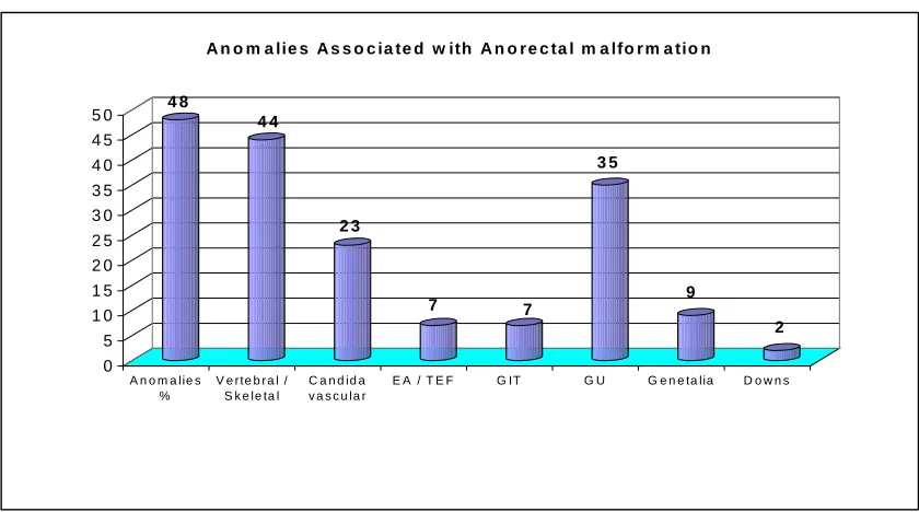
Table – 4 : Anomalies Associated with Anorectal malformation
Type of anomalies
Our study
Anomalies % 48
Vertebral / Skeletal 44
Cardio vascular 23
EA / TEF 7
GIT 7
GU 35
Genitalia 9
Downs 2
In our study of 90 patients , 48%(44) patients has associated anomalies
out of which most common association is Vertebral/ skeletal anomalies(44%)
followed by Urological anomalies(35%), Cardiovascular(23%), Genitalia(9%),
GIT(7%), EA/TEF(7%) and Downs syndrome(2%)
4 8 4 4 2 3 7 7 3 5 9 2 0 5 1 0 1 5 2 0 2 5 3 0 3 5 4 0 4 5 5 0
A n o m a lie s %
V e rte b r a l / S k e le ta l
C a n d id a v a s c u la r
E A / T E F G IT G U G e n e ta lia D o w n s
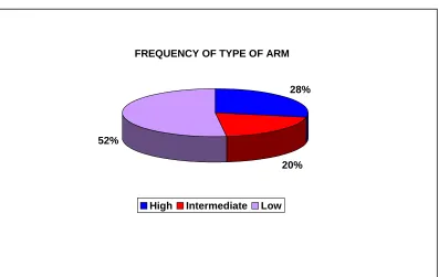
Table – 5 : Type of Anomaly
Male Female Total
High 17 10 27
Intermediate 12 4 16
Low 19 28 47
Total 48 42 90
In our study most common anomaly in female is Low anomaly(28) and in
males is High ( High and Intermediate) - 28 cases
FREQUENCY OF TYPE OF ARM
28%
20% 52%
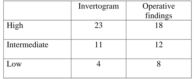
[image:40.612.103.500.410.661.2]Table - 6 : Correlation between invertogram and operative finding
Invertogram Operative
findings
High 23 18
Intermediate 11 12
Low 4 8
In our study out of 23 cases diagnosed as high by invertogram 18 were
confirmed to be high. In the same way11 cases diagnosed as intermediate by
invertogram 12 were confirmed to be intermediate by operative finding and 4
cases diagnosed as low by invertogram 8 were confirmed to be low by operative
finding
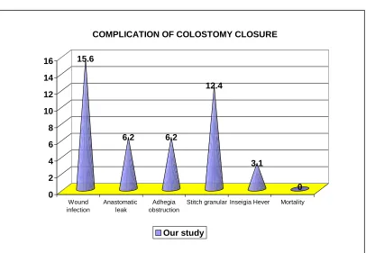
Table – 7 : Complications of colostomy
Complications Our study
Skin excoriation 21
Prolapse 14
Bleeding 12
Obstruction 5
Wound infection 2
Retraction 5
Revision Rate 5
[image:41.612.214.414.462.674.2]In our study 39 colostomies was done out of which 23 were pelvic
colostomy and 16 were transverse colostomy. 21% patients had skin
excoriation, 14% had prolapsed of colostomy, 12 % had bleeding from
colostomy, 5 % had intestinal obstruction and 2% had wound infection
.
21
14
12
5
2
5 5 5
0 5 10 15 20 25
Skin excoriction
Prolapse Bleeding Obstruction Wound infection
Retraction Revision pattern
Mortality
Table – 8 : Complication of colostomy closure
Complication
rate
Wound infection
15.6
Anastomotic leak
6.2
Adhesive obstruction
6.2
Stitch granuloma
12.4
Incisional hernia
3.1
Mortality -
Wound Infection is the most common complication of colostomy
closure(15.6) followed by Stitch granuloma(12.4), Anastomotic leak and
Adhesive obstruction(6.2) and Incisional hernia(3.1)
15.6
6.2 6.2
12.4
3.1
0 0
2 4 6 8 10 12 14 16
Wound infection
Anastomatic leak
Adhegia obstruction
Stitch granular Inseigia Hever Mortality
COMPLICATION OF COLOSTOMY CLOSURE
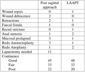
Table – 9 : Complications of posterior sagittal approach & Lap. Assisted approach
Post sagittal approach
LAAPT
Wound sepsis 0 1
Wound dehiscence 2 0
Retractions 2 1
Faecal fistula 1 0
Rectal stricture 0 1
Anal stenosis 1 2
Mucosal prolapsed 3 1
Redo Anorectoplasty 3 1
Redo Anoplasty 1 2
Laparotomy needed 11
Continence Good Fair Poor
45 33 22
48 32 20
A comparison of laparoscopic assisted (LAARP) and posterior
sagittal (PSARD) anorectoplasty in the outcome of intermediate and high
anorectal malformation, There is no significant difference in complication rate
between two procedures
[image:44.612.164.462.143.395.2]
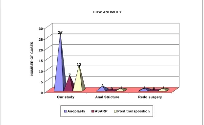
Table – 10 : Low Anomalies
Type of procedure
Number of cases in our
study
Complications Stricture Redo
Anoplasty 27 2 1
ASARP 7
Posterior transposition
12 1 1
In our study totally 3 procedures are done for Low Anorectal
Malformations. We have done Anoplasty in 27 patients, ASARP in 7 patients,
and Posterior transposition in 12 patients. Out of 2 Anoplasty which went in for
stricture, Redo Anoplasty was done in 1 patient. 1 Posterior transposition went
in for stricture which was subjected to redo Posterior transposition.
27
7 12
2
0 1 1 0 1
0 5 10 15 20 25 30
NUMBER
OF
CASES
Our study Anal Stricture Redo surgery LOW ANOMOLY
[image:45.612.109.526.438.693.2]
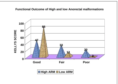
Table – 11 : Functional Outcome of High and low Ano rectal malformations
Kelly Score Good Fair Poor
High ARM 47 33 20
Low ARM 85 13 2
Functional outcome of Low ARM in our study was Good in 85, Fair in
13, Poor in 20 patients and for High ARM it is Good in 47, Fair in 33 and Poor
in 20 patients.
47 85
33
13 20
2 0
20 40 60 80 100
KELLYS SCO
R
E
Good Fair Poor
Functional Outcome of High and low Anorectal malformations
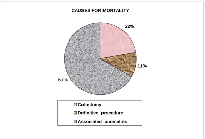
Table – 12 : Mortality
Cause for Mortality Number of cases
Colostomy 2
Definitive procedure 1
Associated anomalies 6
Total 9
Total mortality in our study is 9. Two patients due to colostomy,1 due to
definitive procedure and 6 due to associated anomalies
CAUSES FOR MORTALITY
22%
11%
67%
Colostomy
Definitive procedure
Discussion
Table
1
:
Socioeconomic status
Income
Group
Our study A. E. Archibong, et al.1
Less than Rs. 12000/annum
Mores than Rs. 12000/annum
Less than Rs. 12000/annum
More than Rs. 12000/annum
Number of
patients
90 _ 125 30
The incidence of ARM in affluent patients with income group
>1200 is very meager and practically nil. I understand from my senior
colleagues who practice outside that ARM predominantly affects lower
socioeconomic status patients. According to A. E. Archibong low socio
economic group patients who were exposed to various harm full agents
such as poverty, ignorant are also affected with higher incidence of
[image:48.612.106.517.135.255.2]ARM. In his study also ARM is very low in affluent patients.
Table 2 : Incidence
1ST YEAR 2ND YEAR 3RD YEAR
Number of Cases of ARM
36 30 24
The incidence of ARM is decreasing. Previously in the yester
years we were having a heavy workload because of these congenital
diseases. Now, the number of cases has come down. Reasons probably
may be due to the better nutritional status and better ante natal care which
our people enjoy. Further the Ultra Sound may help in discerning there
afflicted babies leading to spurning of such fetuses which suffer from
malformation disorders. This is reflected in this study as evinced by the
decreasing incidence noted through the years.
Table – 3 : Sex Ratio
Gender Stephens30 Endo et al31 Our study
Male 57% 57% 55%
Female 43% 43% 45%
In our study, the male : female ratio associated with ARM is almost
equal, with a 55 : 45 male : female ratio. Our results are almost similar to the
study of Stephens and Endo et al. In these study also male female ratio is
almost equal. There is no difference in incidence of ARM in both the sex group
Table – 4 : Types of Fistula in ARM
No Types of fistula Our study Stephens & Smith19
Male % Female % Male % Female %
1 Rectourethral fistula 30 36
2 Rectocloacal fistula 5 5
3. Rectovesical fistula 10 2 6 5
4. Rectovaginal fistula 15 19
5. ARM – No Fistula 2 2 8 4
6. Anterior Perineal anus 12 4 17
7. Anovestibular Fistula 29 18
8. Vulvar Anus 7
9. Complete covered Anus 10 7 10 4
10 Anocutaneous fistula 28 12 25 18
11. Rectal Atresia 2 2
12. Pouch colon 2
13. Rectovestibular fistula 3
Though ARM is distributed equally in both the sex, in concurrence with
study by Stephen and Smith Anocutaneous fistula has male preponderance and
ectopically placed anus has female preponderance. Certain types of ARM such
as Cloacal anomaly, recto vaginal fistula and Anovestibular fistula are exclusive
for females. In our study there are 2 cases of rectal atresia 1 in male and other in
female, which is not seen in study by Stephen and Smith. Rectal atresia is a
rare anomaly according to Stephen and Smith but its incidence is high in
Southern parts of India as stated by Prof. T. Dorairajan. Pouch colon is other
stated by Prod. Wakhlu. This shows Geographic variation in incidence of
ARM.
[image:51.612.110.516.190.378.2]Table – 5 : Frequency of type of ARM
Authors High % Intermediate% Low %
Our study 28 20 52
Cook32 28 23 51
Stephens19 46 54
Chen33 20 47 33
Endo et al31 26 11 57
In our study Low anomaly is most common occurring anomaly followed
by High and Intermediate anomaly, which is also shown by other studies –
Cook et al and Endo et al. Difference in incidence shown by Chen et al and
Stephen et al is because of difference in classification of ARM used by them.
We used International Classification of ARM.
Table – 6 : Anomalies Associated with Anorectal malformation
Our study Ratam34 Smith25 Kiesewetter35
Anomalies % 48 58 61 54
Vertebral / Skeletal 44 41 26 6
Cardio vascular 23 10 9 7
EA / TEF 7 6 4 9
GIT 7 9 8 4
GU 35 39 25 40
Genitalia 9
Downs 2
Almost half of the cases with ARM has associated anomalies (48%) and
it is the most common cause of death in ARM patients ( 6 out of 9 mortalities in
our study). It is recommended that all patients with anorectal malformations
should have all necessary investigations to search the associated anomalies
different systems. Urinary anomalies were high in both sexes in high ARM.
Patients with urogenital anomalies require careful assessment and timely
intervention for better out come.
However large number of patients and poor primary health care services
make us confine to do basic investigations rather than follow a protocol. So we
routinely do Ultrasonography of abdomen, X-ray spine of all patients,
echocardiogram and neurosonogram. Special investigations for example:
Actual incidence of urogenital anomalies may be higher if thoroughly
[image:53.612.148.466.189.386.2]investigated.
Table 7 : Correlation between invertogram and operative finding
Invertogram Operative
findings
High 23 18
Intermediate 11 12
Low 4 8
Total 38 38
.
The overall sensitivity of invertogram in detecting type of anomaly is
low. In our study out of 23 cases diagnosed as high by invertogram 18 were
confirmed to be high, four were low and one intermediate, totaling five. In
these five cases, four of them would have been subjected to colostomy because
of the wrong vagaries of the Invertogram. So, we would like to stress, that
though Invertogram is being done as a routine, clinical assessment is the
ultimate parameter for judgment. Cases with epithelisal pearls, bucket handle
deformity are pathognomic of low anomaly and are treated with perineal
suggested high sensitivity and specificity with MRI. So through clinical
examination is needed if possible MRI for diagnosis of type of ARM
[image:54.612.146.481.235.473.2]( e.g. ; avoids 3 staged procedure for misdiagnosed Low type ARM).
Table – 8 : Comparison between Pelvic and Transverse colostomy
No. Pelvic
colostomy
Transverse colostomy
% %
1. Skin excoriation 17.3 31
2. Prolapse 13 18.7
3. Bleeding 8.6 18.7
4. Obstruction - 12.5
5. Wound infection 4.3 -
6. Retraction 4.3 6.2
7. Redo 4.3 6.2
8. Mortality - 12.5
The common complication of colostomy is skin excoriation 17.3% in
pelvic colostomy and 31 % in transverse colostomy. Similarly all the other
complications such as Prolapse, Bleeding, Obstruction, Wound Infection,
Retraction, and Mortality are more in with Transverse colostomy than with
Pelvic colostomy. In addition to the above complications Transverse colostomy
have additional complications such as electrolyte imbalance and
when compared to Transverse colostomy which was also in accordance with
study by Chandramouli .Sigmoid colostomy should be performed whenever
possible except in situations of very high anomaly where surgeon suspects that
distal bowel won’t be sufficient for further pull through procedures. Close
attention to technical details, principles of stomal care, and proper parental
instruction should minimize morbidity. Concluding that while transverse
colostomy is surgeon friendly for subsequent procedures, pelvic colostomy is
[image:55.612.154.469.387.578.2]patient friendly for maintenance and lesser complication rate.
Table – 9 : Complication of colostomy closure
Our study Chandraemouli36
Wound infection 15.6 12.6
Anastomotic leak 6.2 7.1
Adhesive
obstruction
6.2 5.2
Stitch granuloma 12.4 10.5
Incisional hernia 3.1 2.6
Mortality - 1.8
In our study wound infection occurred in 5 cases(15.6%). Incisional
hernia in 1 case in which wound infection was very severe. Anastomotic leak
occurred in 2 cases and were managed conservatively. Adhesive obstructions in
closure reduces morbidity and mortality of colostomy. Hence, it is
recommended that Post PSARP patients should have their colostomy closure
within three months to obviate complications of the pulled through bowel.
Frequently we see patients coming years after the primary procedure. On our
part we should motivate and give dates with in three months for the patients at
the time of discharge after PSARP.
Table – 10 : Complications of posterior sagittal & Lap. Assisted approaches
Our study C. Devos, M. Arnold et al37
Complications Post sagittal
approach
LAAPT Post sagittal
approach
LAAPT
Wound sepsis 0 1 0 2
Wound dehiscence 2 0 2 0
Retractions 2 1 1 1
Faecal fistula 1 0 1 0
Rectal stricture 0 1 0 1
Anal stenosis 1 2 1 3
Mucosal prolapse 3 1 3 2
Redo Anorectoplasty 3 1 3 0
Redo Anoplasty 1 2 1 0
Laparotomy needed 11
Continence Good Fair Poor 45 33 22 48 32 20 48 30 30 43 30 25
A comparison of laparoscopic assisted (LAARP) and posterior
sagittal (PSARP) anorectoplasty in the outcome of intermediate and high
[image:56.612.81.544.328.599.2]assisted (LAARP) and posterior sagittal (PSARP) anorectoplasty but have
specific associated problems. The increased association of anal stenosis in the
LAARP procedure might be due to the fact that the perineum is not as
extensively opened as in PSARP, leading to a smaller fashioned anoplasty.
However, PSARP group showed a high number of patients needing
management for both prolapse. Although a long 'learning curve', with
laparoscopic surgical techniques, extending to all participating staff and even
equipment maintenance. Both the LAARP and PSARP procedures can
successfully treat ARM with comparable outcomes. It appears that LAARP is
optimal for high ARMs that would otherwise require a laparotomy to facilitate
adequate mobilization.
We suggest that were sacro abdominal pull through is contemplated a lap
assisted PSARP would be of value as it obviates the need for laparotomy and it
Table – 11 : Complications of posterior sagittal approach & Lap. Assisted approach
Post
sagittal
approach
LAAPT Chi
square
value
Wound sepsis
0
1
0.663
Not Significant
Wound dehiscence
2
0
0.961
Not Significant
Retractions 2
1
0.663
Not
Significant
Faecal fistula
1
0
0.604
Not Significant
Rectal stricture
0
1
0.663
Not Significant
Anal stenosis
1
2
0.469
Not Significant
Mucosal prolapsed
3
1
0.645
Not Significant
Redo
Anorectoplasty
3 1
0.645
Not
Significant
Redo Anoplasty
1
2
0.469
Not Significant
Laparotomy
needed
11 0
0.122
Not
Significant
Continence
Good
Fair
Poor
45
33
22
48
32
20
Table – 12 : Functional Outcome of High Ano rectal malformationsKelly Score
Good
Fair
Poor
Our
Study
47 33 20
Stephen and Smith
1956 32 12
Trustler &
Willkinson
3826 20 54
Partridge and
Gough
3933 43 24
[image:58.612.122.503.488.692.2]The continence scoring of our procedure ( Posterior sagittal approach) for
high and intermediate ARM is better compared to other traditional procedures
studied in by different authors Trustler & Willkinson, Partridge and Gough and
Taylor.
[image:59.612.123.504.252.392.2]
Table – 13 : Functional Outcome of Low Ano rectal malformations
Kelly Score Good Fair Poor
Our Study 85 13 2
Stephen and Smith19 83 15 2
Trustler &
Willkinson38
80 20 -
Partridge and Gough39 86 11 3
The functional outcome patients treated with low ARM is good. This is
also substantiated by other studies by Stephen and smith, Partridge and Gough,
Trusteler and Willkinson. This is due to less complexity of the defect and good
sphincter muscle complex development. 5 patients had fair out come out of
which 4 did not turned up for regular dilatation. 1` patient had anal stenosis due
to ischemia for which redo surgery was done with poor out come. A typical
problem in treatment of low anomaly is anal stenosis which can be prevented by
Table 14 : Mortality
Causes Number of
patients
Colostomy 2
Defining procedure 1
Associated Anomalies 6
Total 9
The most common cause of death in ARM patients is Associated
Anomalies. Severe forms of anomalies are associated more often with high
ARM). It is recommended that all patients with anorectal malformations should
have all necessary investigations to search the associated anomalies different
systems. Next common cause of death in our study is due to colostomy. These
cases presented very late and had a morbid pre-operative picture itself. Early
Summary
I have studied the present series of 90 patients who were admitted with
ARM and underwent various surgical procedures. Analyzing them, the
following summary were drawn
1) All the patients where in our study were low socio-economic status.
The incidence of ARM in affluent patients with income group >1200 is very
meager and practically nil
2) The male: female ratio associated with ARM is almost equal, with a
55:45 male: female ratio.
3) Low anomaly is common in females and high and intermediate
anomaly in males. Geographic variation in incidence of ARM in case of rectal
atresia and pouch colon. Rectal atresia which Madurai has a higher incidence is
also