0022-538X/97/$04.00
1
0
Copyright © 1997, American Society for Microbiology
Effects of Mutations in the Exo III Motif of the Herpes Simplex
Virus DNA Polymerase Gene on Enzyme Activities,
Viral Replication, and Replication Fidelity
YING T. HWANG,
1BU-YUAN LIU,
2† DONALD M. COEN,
3AND
CHARLES B. C. HWANG
1*
Department of Microbiology and Immunology, Medical College, State University of New York, Syracuse, New York 13210
1;
School of Dentistry, National Taiwan University, Taipei, Republic of China
2; and Department of Biological
Chemistry and Molecular Pharmacology, Harvard Medical School, Boston, Massachusetts 02115
3Received 14 February 1997/Accepted 8 July 1997
The herpes simplex virus DNA polymerase catalytic subunit, which has intrinsic polymerase and 3
*
-5
*
exonuclease activities, contains sequence motifs that are homologous to those important for 3
*
-5
*
exonuclease
activity in other polymerases. The role of one such motif, Exo III, was examined in this study. Mutated
polymerases containing either a single tyrosine-to-histidine change at residue 577 or this change plus an
aspartic acid-to-alanine at residue 581 in the Exo III motif exhibited defective or undetectable exonuclease
activity, respectively, yet retained substantial polymerase activity. Despite the defects in exonuclease activity,
the mutant polymerases were able to support viral replication in transient complementation assays, albeit
inefficiently. Viruses replicated via the action of these mutant polymerases exhibited substantially increased
frequencies of mutants resistant to ganciclovir. Furthermore, when the Exo III mutations were incorporated
into the viral genome, the resulting mutant viruses displayed only modestly defect in replication in Vero cells
and exhibited substantially increased mutation frequencies. The results suggest that herpes simplex virus can
replicate despite severely impaired exonuclease activity and that the 3
*
-5
*
exonuclease contributes substantially
to the fidelity of viral DNA replication.
Replicative DNA polymerases, which are the central
en-zymes for DNA replication, play an important role in the
fidelity of DNA replication. Aside from selecting the correct
nucleotides for incorporation during DNA synthesis, most
rep-licative DNA polymerases are associated with 3
9
-5
9
exonucle-ase activities that proofread misincorporated nucleotides.
DNA replication by mutant polymerases with either impaired
or defective 3
9
-5
9
exonuclease is less accurate by 1 to 3 orders
of magnitude in many in vivo and in vitro systems (reviewed in
references 18 and 42).
The replicative polymerase of herpes simplex virus (HSV)
consists of a catalytic subunit (Pol) and a processivity subunit,
UL42 (27, 31). Because of the variety of molecular, genetic,
pharmacological, and biochemical tools that can be applied to
its study, this enzyme is an excellent model system for studying
polymerase functions in mammalian cells. The Pol subunit of
HSV polymerase contains intrinsic 3
9
-5
9
exonuclease activity
(39, 40, 48), which maps to the N-terminal half of the
polypep-tide (58). Within this region, HSV Pol contains three segments
that align with conserved, homologous segments in other
poly-merases called Exo motifs (2) (Fig. 1). In a variety of other
polymerases, residues within these motifs are known to be
critical for 3
9
-5
9
exonuclease activities (2, 5, 13, 14, 19–21, 25,
30, 41, 50–54). For example,
368Asp (Exo I),
471Asp (Exo II),
581Asp (Exo III), and
577Tyr (Exo III) in HSV Pol correspond,
respectively, to residues
355Asp,
424Asp,
501Asp, and
497Tyr of
the 3
9
-5
9
exonuclease domain of
Escherichia coli
DNA
poly-merase I (Pol I). In Pol I, these residues are critical for
exo-nuclease activity due to effects on metal ion binding (the Asp
residues) or catalysis per se (
497Tyr) (13, 14, 20). In another
example, mutation of the residue in bacteriophage T4 DNA
polymerase that corresponds to
424Asp of Pol I results in an
enzyme that retains polymerase activity yet lacks detectable
exonuclease activity. Bacteriophage containing this mutation
exhibit markedly increased mutation frequencies (21).
For HSV Pol, there is only limited information regarding the
importance of the Exo motifs for various
pol
functions,
includ-ing replication fidelity. Gibbs et al. (25) demonstrated that
certain Exo II mutations were lethal to the virus and abolished
polymerase activity. The effects of the mutations on
exonucle-ase activity were not reported; however, recently, an Exo II
mutation was shown to cause defects in both exonuclease and
polymerase activities in vitro (41). It has been suggested that
exonuclease activity is essential for viral replication, based on
the failure to obtain recombinant viruses containing certain
Exo mutations, including one that had no detectable effect on
polymerase activity (30).
In this study, we constructed two mutant Pols containing
alterations within the Exo III motif to examine their effects on
the enzyme activities, viral replication, and the fidelity of DNA
replication. These mutations were not lethal in vivo, despite
severely impaired 3
9
-5
9
exonucleases. Furthermore, the
mu-tants exhibited drastic increases in mutation frequencies which
could be the result of altered exonuclease activity.
MATERIALS AND METHODS
Cells and viruses.Vero (American Type Culture Collection), DP6 (49), and Pol A5 (see below) cells were grown and maintained as described (35). HSV type
1 (HSV-1) strain KOS, the polnull mutant HP66, and the mutant viruses
generated in this study (see below) were propagated as described previously (49). Plasmids.To construct eukaryotic expression plasmids of the HSV-1polgene, a 3.8-kbpBclI-KpnI (partial digestion) fragment which contains the entire HSV-1
polopen reading frame was isolated from plasmid pDP4 (49) and cloned into the
BamHI andKpnI sites of pGEM3Zf(1) (Promega) to obtain pGEM3-pol. A
3.8-kbpXbaI fragment was then isolated from pGEM3-pol and cloned into the
XbaI site of the eukaryotic expression vector pSVK3 (Pharmacia) and theNheI site of the baculovirus expression vector pBlueBac (Invitrogen) to obtain
pSVK-* Corresponding author. Phone: (315) 464-8739. Fax: (315) 464-7680.
E-mail: [email protected].
† Present address: Endocrine Unit, Massachusetts General
Hospi-tal, Boston, MA 02114.
7791
on November 9, 2019 by guest
http://jvi.asm.org/
pol and pBlue-pol, respectively. The insertedpolgene in plasmid pSVK-pol was under the control of the simian virus 40 (SV40) promoter/enhancer and could be
transcribed by T7 RNA polymerase in vitro (37). The polgene inserted in
pBlue-pol was under the control of the polyhedrin promoter (56). The orienta-tion of the insertedpolgene in these two clones was confirmed by restriction digestion.
The protocol of transformer site-directed mutagenesis (Clontech Laborato-ries, Inc.) and the recipient double-stranded DNA of pGEM3-pol were used for obtaining specific mutations within thepolgene. The following oligonucleotides
were used. G3HM (59-GCATGCACGCGTGAGTATTCT-39), which converts
theHindIII site within the multiple cloning site to aMluI site, was used as the
selective primer to identify mutated clones; 577/581 (59-CAGGGAAT[G/C]CC
TGTATGCAGTA[G/C]CTCGCC-39), which contains degenerate nucleotide(s)
at codons 577 and 581, and 441/443 (59-CATCTCGAAT[G/A/T/C]CGCTG[G
/A/T/C]CGAATTC-39), which contains degenerate nucleotides at codons 441
and 443, were used as targeted primers for site-directed mutagenesis. Clones resistant toHindIII digestion were selected and sequenced to identify mutated sequences within the corresponding region.
The 2.3-kbpBglII-NotI fragment containing each mutatedpolgene was then cloned into pSVK-pol and pBlue-pol to replace the corresponding wild-type 2.3-kbp fragment. Each clone was then sequenced by using the fmol DNA sequencing system (Promega) to confirm that only the desired mutation was
found in the corresponding 2.3-kbp fragment. The primers from thepolgene
corresponding to nucleotides 546 to 562 (546R), 754 to 770 (754R), 1010 to 1027 (1010R), 1190 to 1209 (1190R), 1566 to 1585 (1566R), 1593 to 1577 (1593L), 1643 to 1665 (1643R), 1984 to 2002 (1984R), 2156 to 2138 (2156L), 2429 to 2446 (2429R), 2745 to 2765 (2745R), 2810 to 2791 (2810L), and 3109 to 2990 (3109L), where the A nucleotide of the first ATG codon of thepolgene is defined as nucleotide 1, were used for sequencing analyses.
Recombinant baculovirus, protein expression, and purification.Baculovirus expression plasmids containing recombinantpolgenes were cotransfected with linearized viralAutographa californicanuclear polyhedrosis virus DNA by using a transfection module kit (Invitrogen). Recombinant viruses were selected by their ability to form blue plaques in the presence of 5-bromo-4-chloro-3-indolyl-b-D-galactopyranoside (X-Gal) and purified by the manufacturer’s protocols. Recombinant virus was used to infectSpodoptera frugiperdaSf9 cells, and ex-pressed proteins were then purified as described previously (58), with modifica-tions. Briefly, 23109Sf9 cells were infected with recombinant virus for 72 h.
Infected cells were harvested, washed with phosphate-buffered saline, and resus-pended in 30 ml of hypotonic buffer (20 mM HEPES-NaOH [pH 7.6], 0.5 mM
MgCl2) containing a protease inhibitor (0.5 mM phenylmethylsulfonyl fluoride).
After incubation on ice for 10 min, infected cells were disrupted by Dounce homogenization. Cellular debris was removed by low-speed centrifugation, and the resulting supernatant was clarified by centrifugation at 15,000 rpm for 30 min. About 10 ml of buffer A (20 mM HEPES-NaOH [pH 7.6], 0.5 mM dithiothreitol, 0.5 mM phenylmethylsulfonyl fluoride, 5mg of leupeptin per ml, 4mg of pep-statin A per ml, 10% glycerol) containing 0.05 M NaCl was added to the clarified supernatant, which was then batch absorbed in approximately 25 ml of phospho-cellulose P11 (Whatman) for 1 h at 4°C. The absorbed P11 was loaded into a column. After being washed with 50 ml of buffer A containing 0.05 M NaCl, the column was eluted with a linear 160-ml gradient of 0.05 to 1 M NaCl in buffer A. Pol-containing fractions were identified by Western blotting by using Pol-specific antiserum, pooled, and batch absorbed in 5 ml of preequilibrated hydroxylapatite resin for 30 min at 4°C. The absorbed hydroxylapatite resin was loaded into a column and washed with 25 ml of buffer A containing 1 M NaCl followed by 25 ml of buffer A containing 0.05 M NaCl. Pol was eluted with 25 ml of buffer A containing 0.05 M NaCl and 50 mM potassium phosphate. The Pol-enriched fractions were then absorbed to 2 ml of preequilibrated single-stranded DNA agarose for 30 min at 4°C. The absorbed agarose was loaded into a column and washed with 15 ml of buffer A containing 0.05 M NaCl. Pol was eluted with a linear 25-ml gradient of 0.05 to 1 M NaCl in buffer A. Fractions of purified Pol, which were determined to be apparent homogeneity as indicated by analysis of a sodium dodecyl sulfate (SDS)-polyacrylamide gel stained with Coomassie blue (Fig. 2), were then concentrated in a Centricon 100 (Amicon), diluted with buffer A to remove excess salt, and stored at280°C.
Polymerase and exonuclease activity assays.Polymerase activity was analyzed by the ability of Pol to incorporate [a-32P]dCTP into activated calf thymus DNA
as described previously (48). The 50-ml reaction mixture included approximately
1 or 10 pmol of Pol, 25mg of activated DNA, 60mM each dATP, dTTP, and
dGTP, 50mM dCTP, and 5mCi of [a-32P]dCTP (3,000 Ci/mmol) in reaction
buffer P [20 mM Tris-HCl, 0.1 mM EDTA, 40mg of bovine serum albumin per
ml, 4% glycerol, 3 mM MgCl2, 5 mM dithiothreitol, 150 mM (NH4)2SO4]. The
reaction mixture was incubated at 37°C for 30 min, and the reaction was
termi-nated by adding 200ml of 0.1 M sodium pyrophosphate–10 mM EDTA; 500ml
of 10% trichloroactic acid was then added, and the mixture was incubated on ice for 15 min. Precipitated DNA was filtered through a Whatman GF/C glass filter, washed with 5% trichloroacetic acid containing 0.1 M sodium pyrophosphate, and then washed with ethanol. The filters were dried, and the radioactivities were measured in a liquid scintillation counter.
39-59 exonuclease activity was analyzed by using a 59-32P-labeled 16-mer
FIG. 1. Homology sequences of the Exo III motif shared among diverse DNA polymerases. The top line is a schematic of the HSV Pol polypeptide with the locations of regions of sequence similarity shared among other DNA polymerases. Regions I to VII (25, 35, 57, 59) anddregion C (10, 61), which overlaps the Exo III motif, are shown as empty boxes; the Exo I, II, and III motifs (2–5) are shown as the dark boxes; the Exo I9segment, which was originally assigned as the Exo I motif (2, 3), is also shown as a dark box. The numbers above the line refer to amino acid (a.a.) residues of HSV Pol. Sequence alignment of the Exo III motif of HSV Pol and several other DNA polymerases is shown below the line. Numbers refers to amino acid residues. Highly conserved amino acids are shaded. The mutated amino acids of the two Exo III mutants in this study, Y7 and YD12, are indicated at the bottom. The following polymerases are presented:E. coliDNA Pol I (Pol I) (38), bacteriophage T4 Pol (55), DNA polymerasedofSaccharomyces cerevisiae[d(Sc)] (6), human DNA Pold[d(Human)] (9), bovine DNA Pold[d(Bovine)] (61), and Pols of adenovirus type 2 (Adeno-2) (26), bacteriophagef29 (60), HSV (24), human cytomegalovirus (CMV) (7), Epstein-Barr (EBV) (1), varicella-zoster virus (VZV) (12), and vaccinia virus (17).
on November 9, 2019 by guest
http://jvi.asm.org/
(59-CCGGGGGGGAGGCGCC-39) oligonucleotide (5,000 cpm/0.14 pmol/1.25
ng) hybridized to a 25-mer (59-GGAAGCTTGGGCGCCTCCCCCCCGG-39)
oligonucleotides to form a primer-template. The primer-templates were incu-bated with different amounts of the wild-type, Y7, and YD12 HSV-1 Pols in buffer P, Klenow fragment of Pol I (New England Biolabs, Beverly, Mass.), and
Sequenase version 2 (a T7 DNA polymerase deficient in 39-59 exonuclease
activity; U.S. Biochemical) in reaction buffer supplied by the manufacturer in a total volume of 10ml for 5 min at 37°C. The reaction was stopped by quenching with 4ml of loading buffer and analyzed on a 20% denatured acrylamide gel containing 8 M urea. The 39-59exonucleolytic activity, indicated by the reduction in size of the labeled oligonucleotide, was detected by autoradiography. The integrated band intensity was quantified with a PhosphorImager, and the relative activity of each mutant was compared to that of the wild-type Pol.
Construction of new HSV DNA polymerase-expressing cell lines.The poly-merase-expressing cell line DP6 (49) was used for experiments at the beginning of this study. For unknown reasons, however, this cell line diminished in its ability
to support the replication of polnull mutants. Therefore, new
polymerase-expressing cell lines were constructed by using pSVK-pol and the method of Marcy et al. (49). A total of about 150 G418-resistant clones were obtained from two independent experiments, and 11 independent clones were able to
comple-ment the growth of the HSV-1polnull mutant, HP66 (49). Five were further
analyzed by Southern blot, and each cell line was found to contain integrated
HSV-1polDNA (44). The Pol A5 cell line, which contains two copies of the
integratedpolgene, was used in this study.
Complementation assay.Complementation assays were performed by trans-fection of either the wild-type or mutatedpolexpression plasmids into Vero cells
by the DEAE-dextran transfection method and superinfection with HP66pol
null mutants as described previously (16). In some experiments, LipofectAmine transfection reagent (Life Technologies) was also used to transfect DNA into Vero cells according to the manufacturer’s protocol. The progeny viruses har-vested from each sample were titered on DP6 or Pol A5 cells (permissive for HP66 replication) and also on nonpermissive Vero cells, in which any plaques
formed should represent recombinants between HP66 and thepolgene in
com-plementing cells. Complementation efficiencies were calculated as the ratio [(ti-ter on DP6 or Pol A5 cells2titer on Vero cells)mutant]/[titer on DP6 or Pol A5
cells2titer on Vero cells)wild type]3100.
Construction of Exo III recombinant viruses.Marker transfer experiments were performed as described previously (8, 46), using infectious HP66 DNA, individual Exo III mutant plasmid DNAs, and Pol A5 cells to obtain recombinant viruses. Recombinant viruses were purified twice as white plaques on Pol A5 cells in the presence of X-Gal. Recombinant viruses were amplified in Pol A5 cells and examined by sequencing as described below. Those recombinants containing desired mutations were plaque purified twice further on Vero cells, and the existence of the desired mutations was confirmed by sequencing.
To examine whether recombinant viruses contained the Y7 or YD12 mutation, DNA fragments containing the Exo III region were PCR amplified from crude virion DNAs of recombinant viruses by using primers 1190R and 2156L and the method described previously (34). The resulting DNAs were sequenced by using primer 1643R.
Single-step growth analysis.To compare the abilities of recombinant Y7 and YD12 viruses to replicate in Vero cells with that of the wild-type KOS strain, about 23105Vero cells were infected with each virus with a multiplicity of
infection of 5 PFU per cell. At 24 h postinfection, virus progeny was harvested and titered on Vero cells.
Measurement of mutagenesis frequency.Thetkmutagenesis method origi-nally developed by Hall et al. (29) was used to measure the frequency of acyclovir (ACV)- or ganciclovir (GCV)-resistant mutants with the following modifications. For analysis of mutants in an HP66 background, the polymerase protein required for the growth of HP66 viruses in Vero cells was transiently provided by the transfection of either wild-type or mutantpolexpression plasmids. HP66 progeny viruses were then titered on Pol A5 cells. The relative mutation frequency of each virus stock was determined by the ratio of titers with and without 20mM GCV. Each GCV resistant mutant was isolated, amplified, and further confirmed by its ability to grow on Pol A5 cells in the presence of GCV. When recombinant viruses were used, three independent plaques of Y7 and YD12 recombinant virus were each amplified in 53104Vero cells. The mutation frequencies of these
independent samples were determined by the ratio of titers of viruses in the
presence and absence of 50mM ACV as described previously (33). Ten
inde-pendent virus stocks for each recombinant were then prepared by the inoculation of only 5 PFU of a virus stock which exhibited the lowest mutation frequency (Table 5) into approximately 53104Vero cells. The mutation frequency of each
independent virus stock was then determined by the ratio of titers in the presence
and absence of 50mM ACV.
RESULTS
Previous studies (25, 30, 41) of HSV mutant Pols with
alter-ations in Exo motifs were inconclusive regarding the
impor-tance of exonuclease activity for viral replication and
replica-tion fidelity. To address these quesreplica-tions, we used site-directed
mutagenesis to construct three mutated
pol
genes, Y7, YD12,
and DE35. Mutant Y7 (Y577H) contains a single change at
codon 577 (a change from TAC to CAC), and YD12 (Y577H/
D581A) contains mutations at codons 577 and 581 (the same
change at Y7 plus a change from GAC to GCT at codon 581),
both of which are highly conserved amino acids within the Exo
III motif, with
577Tyr and
581Asp of HSV Pol corresponding to
497Tyr and
501Asp of Pol I (Fig. 1). Mutant DE35 (D441A/
E443A), which contains mutations at codons 441 (change from
GAC to GGC) and 443 (change from GAA to GCA), was also
constructed, because residues 441 and 443 resembled residues
368 and 370 of the Exo I motif and indeed were originally
proposed as the conserved residues of the Exo I motif (3, 4)
(Fig. 1). However, this mutant was not able to complement the
replication of HP66 virus in Vero cells (see below). Attempts
to purify the mutant protein from recombinant
baculovirus-infected Sf9 cells were not successful because of its insolubility
(37). This mutant, therefore, served as a negative control for
this study.
Effects of Exo III mutations on polymerase and 3
*
-5
*
exo-nuclease activities.
To examine the effects of these mutations
on polymerase and exonuclease activities, recombinant
bacu-loviruses harboring the mutated Y7 and YD12
pol
genes were
constructed and plaque purified. Recombinant viruses BV-Y7
and BV-YD12 and the virus containing the wild-type HSV-1
pol
gene, BBP-3, were used to infect Sf9 cells. Proteins
ex-pressed from these recombinant baculoviruses were then
pu-rified to apparent homogeneity, as indicated by analysis on
SDS-polyacrylamide gels (Fig. 2). When the polymerase
activ-ities were examined by their abilactiv-ities to incorporate
nucleo-tides into activated calf thymus DNA, Y7 and YD12 mutant
Pols exhibited polymerase activity indistinguishable from and
only about 20% less than that of the wild-type Pol, respectively
(Table 1). Therefore, single or double mutations at amino
acids 577 and 581 in Y7 and YD12 had at most modest effects
on polymerase activity as measured by this assay.
Mutant Pols were also examined for 3
9
-5
9
exonuclease
ac-tivity by using primer-templates containing 5
9
-end-labeled
primers as substrates. Exonucleolytic products were resolved
by denaturing polyacrylamide gel electrophoresis and
quanti-fied as described in Materials and Methods. Note that some
material (
;
2%) migrated on the gels at a position
;
1 base
smaller than full-length primer even with no enzyme added
(Fig. 3, lane 1). This value was subtracted for the purposes of
quantification.
[image:3.612.387.487.70.175.2]The 3
9
-5
9
exonuclease-proficient Klenow fragment of Pol I
was included as a positive control, and its ability to degrade the
3
9
-end nucleotides and form a ladder of bands is shown in Fig.
FIG. 2. Coomassie blue-stained gel of purified Pols. Wild-type (wt), Y7, and YD12 Pols expressed in Sf9 cells infected with recombinant baculovirus were purified as described in Materials and Methods. For each Pol, 10ml of the peak fraction from the single-stranded DNA agarose column was analyzed separately on SDS–7.5% polyacrylamide gels which were stained with Coomassie blue. Positions of molecular weight markers (lane M) are shown at the left.on November 9, 2019 by guest
http://jvi.asm.org/
3 (lanes 2 and 3). Similarly, wild-type HSV Pol exhibited
sub-stantial 3
9
-5
9
exonuclease activity (lanes 4 and 5). In contrast,
the Y7 mutant Pol exhibited very little exonuclease activity
(Fig. 3, lanes 6 and 7), which when quantified corresponded to
only 2% that of wild-type Pol (Table 1). The YD12 mutant Pol
was even more impaired and exhibited no detectable activity
above the background of the assay (Fig. 3, lanes 8 and 9; Table
1). Thus, this mutant Pol is at least 50-fold impaired for
exo-nuclease activity. Results with YD12 were comparable to the
results with the 3
9
-5
9
exonuclease-deficient version of T7 DNA
polymerase (Sequenase), which also failed to detectably cleave
the labeled primers (Fig. 3, lanes 10 and 11). Similar results
were obtained for each enzyme when the same primer was
used as a substrate in the absence of template or when an
oligonucleotide with a stable hairpin structure was used as the
substrate (37). Therefore, the Y7 and especially the YD12
mutant were severely impaired for 3
9
-5
9
exonuclease activity.
Characterization of Exo III mutants for
pol
function in
tran-sient transfection assays.
Complementation assays were
ap-plied to examine the ability of mutant Y7 and YD12
pol
genes
to support viral replication. A wild-type
pol
gene and a
pol
gene
containing mutations at codons 441 and 443 (DE35) were
included as positive and negative controls, respectively. The
various
pol
genes were cloned into expression vector pSVK3,
which contains an SV40 promoter for transient expression of
Pol upon transfection into Vero cells. Results of six
indepen-dent experiments using two different transfection methods and
two different complementing cell lines for analyzing progeny
viruses recovered from Vero cells are shown in Table 2. When
no plasmid was used, little or no progeny virus was recovered
(Table 2, experiments I and III). In all assays, both mutants Y7
and YD12 increased the yield of
pol
null mutant, HP66,
sub-stantially more than did the no-plasmid control (greater than
50-fold), albeit less efficiently than the wild-type Pol. In
con-trast, mutant DE35 failed to complement HP66 in
nonpermis-sive Vero cells. Similar efficiencies were obtained when
differ-ent batches of plasmid DNAs and HP66 stocks were used.
Interestingly, relatively higher complementation efficiencies of
the Exo III mutants were obtained when progeny viruses were
titered on DP6 cells (Table 2); this may reflect a decrease of
the ability of Pol A5 cells to support mutant virus replication.
Replication fidelity of the Exo III mutant Pols in transient
experiments.
To examine the effects of Exo III mutants on the
fidelity of DNA replication, we performed a modified
tk
mu-tagenesis assay to examine the frequency of GCV-resistant
mutants, which are likely to be
tk
mutants (29, 33). The
estab-lished
tk
mutagenesis assay (29, 33) requires the use of
recom-binant viral stocks. At the earlier stages of this study, such
recombinant viruses were not available. Therefore, Pol
re-quired for DNA replication was transiently provided by
trans-fection of each mutant Pol expression plasmid in order to
complement the growth of HP66 in Vero cells. By this
ap-proach, the frequencies of drug-resistant mutants transiently
induced by these Exo III mutants and the wild-type Pol were
determined by the ratio of the titers of the HP66 progeny
viruses in the presence or absence of GCV (33).
Using this modified mutagenesis assay, the mutation
fre-quency of HP66 was about 3.5
3
10
25if the wild-type Pol was
provided in
trans
to induce the replication of HP66 virus
(Ta-ble 2, experiments V and VI). This mutation frequency
in-duced by the wild-type Pol is in the lower end of the range
reported when the wild-type KOS virus was used (29, 33).
Mutants Y7 and YD12 induced 20- to 80-fold-higher mutation
frequencies (Table 2, experiments V and VI). Therefore, in
this assay, both Y7 and YD12 mutants exhibited mutator
phe-notypes (29).
Isolation of Exo III recombinant viruses.
To construct
re-combinant viruses containing Exo III mutations, infectious
HP66 DNA was cotransfected with Y7 or YD12 plasmid DNA.
Recombinant viruses were isolated from the ensuing progeny
by screening for white plaques on Pol A5 cells in the presence
of X-Gal (49). About 0.6% of progeny viruses from each
trans-fection experiment formed white plaques. Two of 18 Y7 white
plaques derived from two independent transfections of Y7
DNA and 2 of 20 YD12 white plaques derived from two
inde-pendent transfections of YD12 DNA contained the desired Y7
or YD12 mutation. This result implied a marker transfer
fre-TABLE 1. Polymerase and exonuclease activities of the wild-type
and mutants Y7 and YD12 Pols
Pol Polymerase activity
a(cpm)
Exonuclease activity,b 1 pmol (%)
1 pmol 10 pmol
Wild type
4.9
3
10
5(100)
4.1
3
10
6(100)
100
Y7
4.8
3
10
5(97)
4.0
3
10
6(99)
2
YD12
4.2
3
10
5(84)
3.3
3
10
6(79)
0
None
3.2
3
10
3(0)
3.2
3
10
3(0)
0
aAnalyzed by using 25mg of activated calf thymus DNA. One or 10 pmol of HSV Pol was used to determine the polymerase activity. The amounts of32
P-labeled dCTP incorporated were measured in a liquid scintillation counter. The counts per minute incorporated by the wild-type Pol minus the counts per minute found with no enzyme added is defined as 100%. The relative activities of the different polymerases (percentages) are shown in parentheses.
[image:4.612.57.299.91.164.2]bAssayed by using a 59-labeled 16-mer primer and a 25-mer template as described in Materials and Methods. Two microliters of each reaction mixture was loaded on a 20% denaturing acrylamide gel containing 8 M urea. The 39-59 exonuclease activity, indicated by a reduction in size of the labeled oligonucle-otide, was detected by autoradiography (Fig. 2). The integrated band intensity of smaller species was quantified with a PhosphorImager, the band intensity found in the absence of enzyme was subtracted (;2% of the intensity of full-length primer), and the relative activity of each mutant Pol was compared to that of the wild-type Pol (defined as 100% for 1 pmol of Pol used).
FIG. 3. Autoradiograph of an exonuclease activity assay. The 16- and 25-mer double-stranded primer-templates were used as substrates for the exonuclease assay as described in Materials and Methods. 39-59 exonuclease activity was demonstrated by its ability to cleave the 39-end base from the 59-end-labeled primers and form the ladder of bands on the gel. Lane 1, no enzyme added (primer-template alone); lanes 2 and 3, Klenow fragment, 0.5 and 0.05 U, respectively; lanes 4 and 5, wild-type HSV Pol, 0.1 and 1 pmol, respectively; lanes 6 and 7, mutant Y7 Pol, 1 and 0.1 pmol, respectively; lanes 8 and 9, mutant YD12 Pol, 1 and 0.1 pmol, respectively; lanes 10 and 11, Sequenase, 1.3 and 0.13 U, respectively. The relative activity of each mutant was compared to that of the wild-type Pol (Table 1).
on November 9, 2019 by guest
http://jvi.asm.org/
[image:4.612.108.249.443.634.2]quency comparable to those obtained with plasmid containing
wild-type DNA or nonlethal mutation (22, 47). The mutation
containing viruses were plaque purified once further on Pol A5
cells. However, 4 of 10 plaques derived from a Y7 plaque that
contained the desired mutation now contained wild-type
pol
sequences, which could be the result of recombination between
mutant viruses and the
pol
gene in Pol A5 cells.
Because of these results, we were concerned that further
plaque purification and propagation on Pol A5 cells might give
rise to wild-type virus that could confound further experiments.
We therefore tested the abilities of the Y7b and YD12a
re-combinants that contained the desired Exo III mutations to
form plaques on Vero cells. As shown in Table 3, both mutants,
which were previously propagated only on Pol A5 cells, readily
formed plaques on Vero cells, albeit with efficiencies two- to
threefold less than that of KOS. Therefore, these Y7b and
YD12a recombinants were plaque purified twice further on
Vero cells to obtain Y7b.v and YD12a.v, respectively. Again,
the mutated Exo III sequence within these purified
recombi-nants were confirmed by sequencing. The plating efficiencies of
these four-time-plaque-purified recombinants were not
mean-ingfully different from those of the original recombinants that
had been isolated from Pol A5 cells. This result indicated that
there was little or no selective pressure on the Exo III mutants
during passage in Vero cells. We also measured the yields of
Y7b.v and YD12a.v viruses on Vero cells in a single-step
growth experiment. As shown in Table 4, the virus yields of
these two mutants were only
;
3-fold less than that of KOS.
These results suggest that the Exo III mutations are not lethal
to viral replication, although they do appear to reduce plaque
formation and yield modestly.
[image:5.612.56.553.82.363.2]Replication fidelity of the Exo III recombinant viruses.
With
these recombinant viruses available, a
tk
mutagenesis assay
(29, 33) was performed to examine the fidelity of DNA
repli-cation. In an initial experiment, we tested two independent Y7
recombinant viruses and two independent YD12 recombinants
from the first round of plaque purification (see above).
Re-markably, one isolate of each mutant was resistant to 100
m
M
GCV due to the mutation in the
tk
gene (37). Based on this
TABLE 3. Plating efficiencies of recombinant Y7 and YD12 viruses
Virusa Titer (PFU/ml) on: Plating efficiency
(%)b
Pol A5 Cells Vero cells
Y7b
1.5
3
10
67.5
3
10
550
YD12a
2.2
3
10
68.5
3
10
539
Y7b.v
4.0
3
10
62.1
3
10
653
YD12a.v
2.2
3
10
61.0
3
10
645
KOS
5.7
3
10
75.5
3
10
797
aRecombinant viruses Y7b and YD12a were plaque purified twice on Pol A5
cells and confirmed by sequencing to contain the expected mutation(s). These recombinants were then further purified twice on Vero cells to obtain Y7b.v and YD12a.v. These isolates were confirmed by sequencing to contain the expected mutation(s).
[image:5.612.318.556.597.677.2]bDetermined as the ratio of the titers on Vero cells and Pol A5 cells.
TABLE 2. Complementation assays and mutation frequencies transiently induced by Exo III mutants
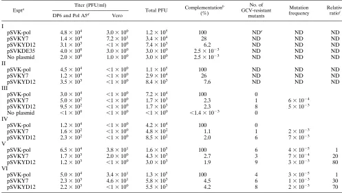
Expta Titer (PFU/ml) Total PFU Complementationb
(%)
No. of GCV-resistant
mutants
Mutation frequency
Relative ratioc
DP6 and Pol A5d Vero
I
pSVK-pol
4.8
3
10
43.0
3
10
01.2
3
10
5100
ND
eND
ND
pSVKY7
1.4
3
10
47.2
3
10
13.4
3
10
428
ND
ND
ND
pSVKYD12
3.1
3
10
3,
1
3
10
07.4
3
10
36.2
ND
ND
ND
pSVKDE35
4.0
3
10
03.0
3
10
03.0
3
10
02.5
3
10
23ND
ND
ND
No plasmid
2.0
3
10
01.0
3
10
03.0
3
10
02.5
3
10
23ND
ND
ND
II
pSVK-pol
4.5
3
10
4,
1
3
10
01.1
3
10
5100
ND
ND
ND
pSVKY7
1.2
3
10
4,
1
3
10
02.9
3
10
426
ND
ND
ND
pSVKYD12
3.5
3
10
3,
1
3
10
08.4
3
10
37.6
ND
ND
ND
III
pSVK-pol
3.0
3
10
4,
1
3
10
07.2
3
10
4100
0
pSVKY7
5.0
3
10
2,
1
3
10
01.7
3
10
32.3
1
6
3
10
24pSVKYD12
9.5
3
10
2,
1
3
10
01.7
3
10
32.3
8
5
3
10
23No plasmid
,
1
3
10
0,
1
3
10
0,
1
3
10
0,
1.4
3
10
230
IV
pSVK-pol
1.2
3
10
4,
1
3
10
04.2
3
10
4100
0
pSVKY7
1.6
3
10
2,
1
3
10
04.8
3
10
21.1
1
2
3
10
23pSVKYD12
2.3
3
10
2,
1
3
10
08.5
3
10
22.0
6
7
3
10
23V
pSVK-pol
6.5
3
10
43.8
3
10
11.6
3
10
5100
6
4
3
10
251
pSVKY7
1.7
3
10
32.0
3
10
04.3
3
10
32.7
3
7
3
10
2420
pSVKYD12
1.2
3
10
3,
1
3
10
03.0
3
10
31.9
9
3
3
10
2380
VI
pSVK-pol
5.0
3
10
43.4
3
10
11.3
3
10
5100
4
3
3
10
251
pSVKY7
2.3
3
10
34.6
3
10
15.8
3
10
34.5
6
1
3
10
2330
pSVKYD12
2.2
3
10
3,
1
3
10
05.5
3
10
34.2
8
2
3
10
2370
aThe DEAE-dextran-DNA transfection technique was applied in experiments I to IV, and LipofectAmine transfection reagent was used in experiments V and VI. bThe complementation efficiency of the wild-type Pol for the growth of HP66 in Vero cells was defined as 100% in each experiment. The relative ability of each mutantpolto complement the growth of HP66 in nonpermissive Vero cells was compared to that of the wild-typepol(pSVK-pol) and expressed as the percentage. cMutants were selected by their resistance to 20mM GCV, and mutation frequencies were determined as described in Materials and Methods. The increased rates of the mutation frequencies were determined as the ratio of the mutation frequency of each mutant Pol to that of the wild-type Pol from the same experiment.
dHSV-1 polymerase-expressing cell lines DP6 and Pol A5 were used in experiments I and II and experiments III to VI, respectively, for complementation assays. eND, not determined.
on November 9, 2019 by guest
http://jvi.asm.org/
finding and the relatively high mutation rates obtained from
transient experiments (Table 2), we modified the mutagenesis
assay as described in Materials and Methods. Three plaques of
GCV-sensitive Y7b.v virus and three plaques of GCV-sensitive
YD12a.v virus were isolated and amplified in Vero cells. Each
stock was examined for the mutation frequency (Table 5). Ten
independent virus stocks were then prepared from one of three
parental stocks which had the lowest
tk
mutation rate (Table
5). Only
;
5 PFU was inoculated into Vero cells to avoid
preexisting
tk
mutants (29). Both the Y7 and YD12 Exo III
mutants exhibited an average mutation rate of 4.1
3
10
22(range from 2.6
3
10
23to 24%) and 1.7
3
10
22(range from
1.4
3
10
23to 8.4%), respectively. In contrast, on average the
mutation frequencies of three wild-type KOS stocks analyzed
in this study were only 5
3
10
25. These results correspond to
an
;
800- and
;
300-fold increases of mutation rates for Y7 and
YD12 mutant Pols, respectively, in comparison to that of the
wild-type Pol.
DISCUSSION
In this study, we examined the effects of mutations within the
Exo III motif of the HSV-1 DNA polymerase on polymerase
and exonuclease activities, viral replication, and replication
fidelity. The Exo III mutations were not lethal, despite their
severe effects on exonuclease activities. This represents the
first report, to our knowledge, of HSV
pol
mutants with
se-verely impaired 3
9
-5
9
exonuclease activity and of the effects of
altered exonuclease activity on the fidelity of DNA replication
in virus-infected mammalian cells.
Effects of Exo III mutations on HSV Pol enzyme activities.
Our results demonstrated that two Exo III mutants are
se-verely impaired for exonuclease activity yet retain substantial
polymerase activity in vitro (Table 1). Consistent with these
results, Ku
¨hn and Knopf (41) found that a mutation of
577Tyr
to phenylalanine (similar to the Y577H mutation in Y7) and a
mutation of
581Asp to alanine (identical to one of the
muta-tions in YD12) decreased exonuclease activity 6- and 16-fold,
respectively, while decreasing polymerase activity less than
30%. Our results are also consistent with those obtained with
Exo III mutants in other systems. In the case of Pol I,
497Tyr
and
501Asp are critical for catalytic activity and metal ion
bind-ing of the 3
9
-5
9
exonuclease (13, 14, 20), respectively; our
results and those of Ku
¨hn and Knopf (41) are consistent with
similar roles for the corresponding residues
577Tyr and
581Asp
of HSV-1 Pol.
A continuing issue in studies of polymerases is the relative
independence of different enzymatic activities. The results
re-ported here and elsewhere (30, 41) in which various Exo
mu-tations severely impair the exonuclease activity of HSV Pol
with little effect on polymerase activity can be simply
inter-preted to mean that the two enzyme activities are functionally
independent. However, the validity of this interpretation
de-pends on whether the in vitro assays of polymerase activity
reflect all aspects of that activity. The in vitro polymerase
assays used in this and other (30, 41) studies of HSV Exo
mutants that appear to retain polymerase activity have mainly
entailed short extensions across gapped or single-stranded
templates. Even with this assay, Exo III mutants had displayed
modest reduction in polymerase activity (reference 41 and this
report). It also remains possible that these HSV Exo mutations
have effects on other aspects of polymerase activity such as
processivity. Along these lines, an Exo III mutation caused a
defect in the strand displacement activity of
f
29 DNA
poly-merase (54).
Interestingly, the Exo III motif is contained within
d
region
C, a region that is found in DNA polymerase
d
and
ε
and
certain viral polymerases (Fig. 1). Mutations both upstream
and downstream of Exo III in
d
region C can confer drug
resistance in vitro and/or in infected cells (23, 41, 43) are thus
presumed to affect the binding of deoxynucleoside
triphos-phate and PP
i, which the drugs mimic. For at least some of the
mutants, enzyme kinetic studies support this presumption (15,
32). Similarly, preliminary data indicate that our two Exo III
mutant Pols are resistant to phosphonoacetic acid (37) in vitro.
This finding suggests that Exo III mutations not only alter the
exonuclease activity but also influence the interactions
be-tween polymerase and PP
i. Regardless, given that certain Exo
TABLE 4. Single-step growth analysis
Virus Virus yield in PFU/ml
(% of wild-type yield)
KOS ... 2.3
3
10
7(100)
Y7b.v... 7.0
3
10
6(30)
YD12a.v... 7.0
3
10
6 [image:6.612.314.553.331.638.2](30)
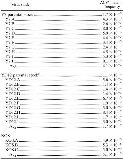
TABLE 5. Mutation frequencies of the
tk
genes induced by
the Exo III recombinant viruses
Virus stock ACVrmutation
frequency
Y7 parental stock
a...
1.7
3
10
23Y7.A... 4.3
3
10
23Y7.B ... 2.6
3
10
23Y7.C ... 8.0
3
10
22Y7.D... 5.9
3
10
23Y7.E ... 4.4
3
10
23Y7.F ... 3.4
3
10
23Y7.G... 2.4
3
10
21Y7.H... 4.5
3
10
23Y7.I ... 5.3
3
10
22Y7.J ... 9.1
3
10
23Avg ... 4.1
3
10
22YD12 parental stock
b...
1.1
3
10
23YD12.A ... 5.6
3
10
22YD12.B... 1.4
3
10
22YD12.C... 1.4
3
10
23YD12.D ... 1.4
3
10
23YD12.E... 6.7
3
10
23YD12.F ... 1.9
3
10
23YD12.G ... 3.0
3
10
23YD12.H ... 8.4
3
10
22YD12.I ... 1.7
3
10
23YD12.J... 3.0
3
10
22Avg ... 1.7
3
10
22KOS
cKOS.A ... 4.9
3
10
25KOS.B... 5.3
3
10
25KOS.C... 5.0
3
10
25Avg ... 5.1
3
10
25aThe Y7 isolate having the lowest mutation frequency of three independent isolates (the other two isolates had frequencies of 3.131023and 3.231023)
was used to prepare 10 independent stocks and assayed as described in Materials and Methods.
bThe YD12 isolate having the lowest mutation frequency of three indepen-dent isolate (the other two isolates had frequencies of 1.931023and 6.73
1023) was used to prepare 10 independent virus stocks and assayed as described
in Materials and Methods.
cThree independent KOS stocks were prepared and assayed for mutation
frequency. The mutation frequency of the wild-type KOS strain is within the range observed in previous studies (28, 32).
on November 9, 2019 by guest
http://jvi.asm.org/
II mutations can affect polymerase activity, even when
mea-sured using activated DNA templates (25, 41), caution should
be exercised in ascribing the importance of specific conserved
motifs to one enzymatic function, but not another.
Is the exonuclease activity of HSV Pol essential for viral
replication?
Although the Exo III mutations reduced
exonu-clease activity at least 50-fold, they were not lethal. They did
reduce the ability of
pol
plasmids to complement the
replica-tion of a
pol
null mutant (Table 2) and resulted in a modest
reduction in plating efficiency (Table 3) and virus yield (Table
4) in Vero cells. (The greater effect of mutations on replication
in transient complementation assay may be due to
incom-pletely appropriate expression of the
pol
gene under the
con-trol of an SV40 promoter.) Thus, one interpretation of our
results is that the exonuclease activity of HSV Pol is not
es-sential for polymerase function in vivo. However, it is possible
that the YD12 Pol contains a small amount of exonuclease
activity, which is beyond the sensitivity of our assays, and/or
that the exonuclease activity may function to at least a limited
extent in infected cells, for example, in the presence of other
replication proteins. Nevertheless, the mutator phenotypes of
Y7 and YD12 strongly suggest that exonuclease activity is
substantially impaired in infected cells. Thus, our results argue
that HSV can replicate even when exonuclease activity is
se-verely impaired.
Our results contrast with those of Hall et al. (30), who were
unable to recover HSV mutants containing various Exo
muta-tions including Y581A, which is included in YD12 and which
evidently exerts a more modest effect on exonuclease activity
than does YD12 (41). Hall et al. (30) raised the possibility,
among several, that 3
9
-5
9
exonuclease activity was required for
HSV replication to prevent intolerable increases in mutation
frequency. One possible explanation for the previous failure to
recover Exo mutants is the necessity of using a complementing
cell line. The Exo mutations studied here did reduce the
rep-lication of HSV and would likely have been difficult to recover
without the use of complementing cells. It could be argued that
these mutants may have acquired additional mutation within
the
pol
gene or the other replicative genes which may suppress
the effects of the Exo III mutations. Although we cannot rule
out this possibility completely, we know that these mutants are
recovered in complementing cells at a frequency comparable
to those of other nonlethal mutations (22, 47) and are able to
form plaque on Vero cells without previously having been
passaged on them. Moreover, we have not found any other
change at the amino acid level within the entire open reading
frame of the
pol
gene of the Y7b.v mutant (37). Further study
is required to determine whether there is a specific
require-ment for 3
9
-5
9
exonuclease activity for HSV replication.
Effects of exonuclease activity on the fidelity of DNA
repli-cation.
3
9
-5
9
exonuclease activities associated with replicative
polymerases have proofreading functions to improve
replica-tion fidelity. Mutant polymerases with defective exonuclease
activity can increase the mutation frequency up to 3 orders of
magnitude (18, 42). We found that Y7 and YD12 Pols
in-creased mutation frequencies 20- to 80-fold in transient
repli-cation assays (Table 2) and 300- to 800-fold in assays of
re-combinant viruses. The difference in the magnitudes of the
increases between the two assays is probably due to the
tran-sient assay accumulating mutations during one replication
cy-cle, whereas the recombinant virus assay accumulates
muta-tions over many replication cycles. The increases in mutation
frequency induced by the Exo III mutant Pols can be readily
explained by their altered exonuclease activities. To our
knowl-edge, we have provided the first evidence of a contribution of
exonuclease proofreading activity to replication fidelity in
virus-infected mammalian cells. It will be interesting to determine
the effects of the mutations on the removal and extension of
mispaired primer termini. Ku
¨hn and Knopf (41) found that the
D581A mutation permitted extension of mispaired primer
ter-mini.
Fifteen years ago, Hall and Almy (28) found that a mutation
closely linked to the HSV
ts
C7
pol
mutation in a
ts
1derivative
of
ts
C7 confers a mutator phenotype. The
ts
C7 mutation maps
within the 5
9
portion of the
pol
gene (11) that encodes the
N-terminal half of Pol that is sufficient for exonuclease activity.
It is therefore tempting to speculate that the mutator
pheno-type observed by Hall and Almy (28) was due to an
exonucle-ase defect.
Several virus stocks of Y7 and YD12 exhibited extremely
high mutation frequencies, up to 24%, for both recombinants
(Table 5). (Fluctuations in mutation frequencies were
ob-served, as expected [45].) It is possible that these high
frequen-cies were due to preexisting drug-resistant mutants that might
have been inoculated in the assay; however, this seems unlikely
as only 5 PFU was inoculated from stocks containing only
;
1
mutant per 1,000 PFU. It is also conceivable that such high
frequencies might reflect the evolution of even more
muta-genic viruses from the Y7 and YD12 mutants. Regardless, we
speculate that high mutation frequencies could have clinical
significance. Although the mutants reported here exhibit some
defects in replication, it is possible that mutator strains of HSV
can cause disease, particularly in immunocompromised
pa-tients. Such strains might be especially likely to give rise to
drug-resistant mutants upon antiviral therapy. It may be of
interest to examine HSV strains associated with clinical drug
resistance for their mutation frequencies.
ACKNOWLEDGMENTS
Part of this work was performed in the Department of Oral
Medi-cine and Diagnostic Science, Harvard School of Dental MediMedi-cine,
Boston, Mass.
This work was supported by NIH grants DE10051 (C.B.C.H.) and
AI19838 (D.M.C.).
REFERENCES
1.Baer, R., A. T. Bankier, M. D. Biggin, P. L. Deininger, P. J. Farrell, T. J. Gibson, G. Hatfull, G. S. Hudson, S. C. Satchwell, C. Seguin, P. S. Tuffnell, and B. G. Barrell.1984. DAN sequence and the expression of the B95-8
Epstein-Barr virus genome. Nature (London)310:207–211.
2.Bernad, A., L. Blanco, J. M. Lazaro, G. Martin, and M. Salas.1989. A conserved 39-59exonuclease active site in prokaryotic and eukaryotic DNA polymerases. Cell59:219–228.
3.Blanco, L., A. Bernad, M. A. Blasco, and M. Salas.1991. A general structure
for DNA-dependent DNA polymerases. Gene100:27–38.
4.Blanco, L., A. Bernad, M. A. Blasco, and M. Salas.1991. A general structure
for DNA-dependent DNA polymerases. Gene108:165. (Erratum.)
5.Blanco, L., A. Bernad, and M. Salas.1992. Evidence favouring the hypoth-esis of a conserved 39-59exonuclease active site in DNA-dependent DNA polymerases. Gene112:139–144. (Letter.)
6.Boulet, A., M. Simon, G. Faye, G. A. Bauer, and P. M. J. Burgers.1989. Structure and function of theSaccharomyces cerevisiae CDC2gene encoding
the large subunit of DNA polymerase III. EMBO J.8:1849–1854.
7.Chee, M. S., A. T. Bankier, S. Beck, R. Bohne, R. Brown, R. Cerny, T. Horsnell, C. A. Hutchinson III, T. Kouzaridies, J. A. Martignetti, E. Preddie, S. C. Satchwell, P. Tomlinson, K. M. Weston, and B. G. Barrell.1990. Analysis of the protein-coding content of the sequence of human cytomeg-alovirus strain AD169. Curr. Top. Microbiol. Immunol.154:125–169. 8.Chiou, H. C., S. K. Weller, and D. M. Coen.1985. Mutations in the herpes
simplex virus major DNA binding protein gene leading to altered sensitivity to DNA polymerase inhibitors. Virology145:213–226.
9.Chung, D. W., J. Zhang, C.-K. Tan, E. W. Davie, A. G. So, and K. M. Downey. 1991. Primary structure of the catalytic subunit of human DNA polymerase
dand chromosomal location of the gene. Proc. Natl. Acad. Sci. USA88:
11197–11201.
10. Coen, D. M.1996. Viral DNA polymerases, p. 495–523.InM. L. Depam-philis (ed.), DNA replication in eukaryotic cells. Monograph 31. Cold Spring Harbor Laboratory, Cold Spring Harbor, N.Y.
on November 9, 2019 by guest
http://jvi.asm.org/
11. Coen, D. M., D. P. Aschman, P. T. Gelep, M. J. Retondo, S. K. Weller, and P. A. Schaffer.1984. Fine mapping and molecular cloning of mutations in the herpes simplex virus DNA polymerase locus. J. Virol.49:236–247. 12. Davison, A. J., and J. E. Scott.1986. The complete DNA sequence of
varicella-zoster virus. J. Gen. Virol.67:1759–1816.
13. Derbyshire, V., P. S. Freemont, M. R. Sanderson, L. Beese, J. M. Friedman, C. M. Joyce, and T. A. Steitz.1988. Genetic and crystallographic identifica-tion of essential groups in the 39-59exonucleolytic site of DNA polymerase. Science240:199–210.
14. Derbyshire, V., N. D. F. Grindley, and C. M. Joyce.1991. The 39-59 exonu-clease of DNA polymerase ofEscherichia coli: contribution of each amino acid at the active site to the reaction. EMBO J.10:17–24.
15. Derse, D., K. F. Bastow, and Y.-C. Cheng.1982. Characterization of DNA polymerases induced by a group of herpes simplex virus type 1 variants selected for growth in the presence of phosphonoformic acid. J. Biol. Chem. 297:10251–10260.
16. Digard, P., W. R. Bebrin, K. Weisshart, and D. M. Coen.1993. The extreme C terminus of herpes simplex virus DNA polymerase is crucial for functional interaction with processivity factor UL42 and for viral replication. J. Virol. 67:398–406.
17. Earl, P. L., E. V. Jones, and B. Moss.1986. Homology between DNA polymerases of poxvirus, herpesviruses, and adenoviruses: nucleotide se-quence of the vaccinia virus DNA polymerase gene. Proc. Natl. Acad. Sci. USA83:3659–3663.
18. Echols, H., and M. F. Goodman.1991. Fidelity mechanism in DNA replica-tion. Annu. Rev. Biochem.60:477–511.
19. Foury, F., and S. Vanderstraeten.1992. Yeast mitochondrial DNA mutators with deficient proofreading exonucleolytic activity. EMBO J.11:2717–2726. 20. Freemont, P. S., J. M. Friedman, L. S. Beese, M. R. Sanderson, and T. A. Steitz.1988. Cocrystal structure of an editing complex of Klenow fragment
with DNA. Proc. Natl. Acad. Sci. USA85:8924–8928.
21. Frey, M. W., N. G. Nossal, T. L. Capson, and S. J. Benkovic.1993. Con-struction and characterization of a bacteriophage T4 DNA polymerase de-ficient in 39-59exonuclease activity. Proc. Natl. Acad. Sci. USA90:2579– 2599.
22. Gibbs, J. S.1990. A functional dissection of herpes simplex virus DNA polymerase. Ph.D. thesis. Harvard University, Cambridge, Mass. 23. Gibbs, J. S., H. C. Chiou, K. F. Bastow, Y. C. Cheng, and D. M. Coen.1988.
Identification of amino acids in herpes simplex virus DNA polymerase in-volved in substrate and drug recognition. Proc. Natl. Acad. Sci. USA85: 6672–6676.
24. Gibbs, J. S., H. C. Chiou, J. D. Hall, D. W. Mount, M. J. Retondo, S. K. Weller, and D. M. Coen.1985. Sequence and mapping analyses of the herpes simplex virus DNA polymerase gene predict a C-terminal substrate binding domain. Proc. Natl. Acad. Sci. USA82:7969–7973.
25. Gibbs, J. S., K. Weisshart, P. Digard, A. DeBruynkops, D. M. Knipe, and D. M. Coen.1991. Polymerization activity of ana-like DNA polymerase requires a conserved 39-59exonuclease active site. Mol. Cell. Biol.11:4785– 4795.
26. Gingeras, T. R., D. Sciaky, R. E. Gelinas, J. Bing-Dong, C. E. Yen, M. M. Kelley, P. A. Bullock, B. L. Parsons, K. E. O’Neil, and R. J. Roberts.1982.
Nucleotide sequences from the adenovirus-2 genome. J. Biol. Chem.257:
13475–13491.
27. Gottlieb, J., A. I. Marcy, D. M. Coen, and M. D. Challberg.1990. The herpes simplex virus type 1 UL42 gene product: a subunit of DNA polymerase that functions to increase processivity. J. Virol.64:5976–5987.
28. Hall, J. D., and R. E. Almy.1982. Evidence for control of herpes simplex virus mutagenesis by the viral DNA polymerase. Virology116:535–543. 29. Hall, J. D., D. M. Coen, B. L. Fisher, M. Weisslitz, S. Randall, R. E. Almy,
P. T. Gelep, and P. A. Schaffer.1984. Generation of genetic diversity in herpes simplex virus: an antimutator phenotype maps to the DNA polymer-ase locus. Virology132:26–37.
30. Hall, J. D., K. L. Orth, K. L. Sander, B. M. Swihart, and R. A. Senese.1995. Mutations within conserved motifs in the 39-59exonuclease domain of herpes simplex virus DNA polymerase. J. Gen. Virol.76:2999–3008.
31. Hernandez, T. R., and I. R. Lehman.1990. Functional interaction between the herpes simplex-1 DNA polymerase and UL42 protein. J. Biol. Chem. 265:11227–11232.
32. Huang, L., K. Kumura, C. B. C. Hwang, A. Gehring, and D. M. Coen. Unpublished results.
33. Hwang, C. B. C., and H. J. Chen.1995. An altered spectrum of herpes simplex virus mutations mediated by an antimutator DNA polymerase. Gene 152:191–193.
34. Hwang, C. B. C., B. Horsburgh, E. Pelosi, S. Roberts, P. Digard, and D. M. Coen.1994. A net11 frameshift permits synthesis of thymidine kinase from a drug-resistant herpes simplex virus mutant. Proc. Natl. Acad. Sci. USA 91:5461–5465.
35. Hwang, C. B. C., K. L. Ruffner, and D. M. Coen.1992. A point mutation within a distinct conserved region of the herpes simplex virus DNA poly-merase gene confers drug resistance. J. Virol.66:1774–1776.
36. Hwang, C. B. C., and E. J. Shillitoe.1990. DNA sequence of mutations induced in cells by herpes simplex virus type-1. Virology178:180–188. 37. Hwang, Y. T., and C. B. C. Hwang.1996. Unpublished results.
38. Joyce, C. M., W. S. Kelley, and N. D. F. Grindley.1982. Nucleotide sequence of theEscherichia coli polAgene and primary structure of DNA polymerase I. J. Biol. Chem.257:1958–1964.
39. Knopf, C. W.1979. Properties of herpes simplex virus DNA polymerase and characterization of its associated exonuclease activity. Eur. J. Biochem.98: 231–244.
40. Knopf, C. W., and K. Weisshart.1990. Comparison of exonucleolytic activ-ities of herpes simplex virus type-1 DNA polymerase and DNase. Eur. J. Biochem.191:263–273.
41. Ku¨hn, F. J. P., and C. W. Knopf.1996. Herpes simplex virus type 1 DNA polymerase. Mutational analysis of the 39-59exonuclease domain. J. Biol.
Chem.271:29245–29254.
42. Kunkel, T. A.1988. Exonucleolytic proofreading. Cell53:837–840. 43. Larder, B. A., S. D. Kemp, and G. Darby.1987. Related functional domains
in virus DNA polymerases. EMBO J.6:169–175.
44. Liu, Y.-Y., and C. B. C. Hwang.1996. Unpublished results.
45. Luria, S. E., and M. Delbru¨ck. 1943. Mutations of bacteria from virus sensitivity to virus resistance. Genetics28:491–511.
46. Marcy, A. I., C. B. C. Hwang, K. L. Ruffner, and D. M. Coen.1990. Engi-neered herpes simplex virus DNA polymerase point mutants: the most highly
conserved region shared amonga-like DNA polymerases is involved in
substrate recognition. J. Virol.64:5883–5890.
47. Marcy, A. I., and D. M. Coen.1989. Unpublished results.
48. Marcy, A. I., P. D. Olivo, M. D. Challberg, and D. M. Coen.1990. Enzymatic activities of overexpressed herpes simplex virus DNA polymerase purified from recombinant baculovirus-infected insect cells. Nucleic Acids Res.18: 1207–1215.
49. Marcy, A. I., D. R. Yager, and D. M. Coen.1990. Isolation and character-ization of herpes simplex virus mutants containing engineered mutations at the DNA polymerase locus. J. Virol.64:2208–2216.
50. Morrison, A., J. B. Bell, T. A. Kunkel, and A. Sugino.1991. Eukaryotic DNA polymerase amino acid sequence required for 39-59 exonuclease activity. Proc. Natl. Acad. Sci. USA88:9473–9477.
51. Reha-Krantz, L. J., and R. L. Nonay.1993. Genetic and biochemical studies of bacteriophage T4 DNA polymerase 39359exonuclease activity. J. Biol.
Chem.268:27100–27108.
52. Reha-Krantz, L. J., S. Stocki, R. L. Nonay, E. Dimayauga, L. D. Goodrich, W. H. Konigsberg, and E. K. Spicer.1991. DNA polymerization in the absence of exonucleolytic proofreading:in vivoandin vitrostudies. Proc. Natl. Acad. Sci. USA88:2417–2421.
53. Simon, M., L. Giot, and G. Faye.1991. The 39-59exonuclease activity located in the DNA polymerasedsubunit ofSaccharomyces cerevisiaeis required for accurate replication. EMBO J.10:2165–2170.
54. Soengas, M. S., J. A. Esteban, J. M. Lazaro, A. Bernad, M. A. Blasco, M. Salas, and L. Blanco.1992. Site-directed mutagenesis at the Exo III motif off29 DNA polymerase; overlapping structural domains for the 39-59 exonuclease and strand-displacement activities. EMBO J.11:4227–4237.
55. Spicer, E. K., J. Rush, C. Fung, L. J. Reha-Krantz, J. D. Karam, and W. H. Konigsberg.1988. Primary structure of T4 DNA polymerase. J. Biol. Chem. 263:7478–7486.
56. Vilard, J., M. Lalumiere, T. Vernet, D. Briedis, G. Alkhatib, D. Henning, D. Levin, and C. Richardson.1990. Synthesis of the membrane fusion and hemagglutinin proteins of measles virus, using a novel baculovirus vector containing theb-galactosidase gene. J. Virol.64:37–50.
57. Wang, T. S.-F., S. W. Wong, and D. Korn.1989. Human DNA polymerasea: predicted functional domains and relationships with viral DNA polymerases.
FASEB J.16:14–21.
58. Weisshart, K., A. A. Kuo, C. B. C. Hwang, K. Kumura, and D. M. Coen.1994. Structural and functional organization of herpes simplex virus DNA poly-merase investigated by limited proteolysis. J. Biol. Chem.269:22788–22796. 59. Wong, S. W., A. F. Wahl, P.-M. Yuan, N. Arai, B. E. Parson, K.-I. Arai, D. Korn, M. W. Hunkapiller, and T. S.-F. Wang.1988. Human DNA polymer-aseagene expression in cell proliferation dependent and its primary struc-ture is similar to both prokaryotic and eukaryotic replicative DNA
poly-merases. EMBO J.7:37–47.
60. Yoshikawa, H., and J. Ito.1982. Nucleotides sequence of the major early region of bacteriophage f29. Gene17:323–335.
61. Zhang, J., D. W. Chung, C.-K. Tan, K. M. Downey, E. W. Davie, and A. G. So.1991. Primary structure of the catalytic subunit of calf thymus DNA polymerased: sequence similarities with other DNA polymerases. Biochem-istry30:11742–11750.