'
I Ill
.
1 ,l
I!:
11l
I:'
'
t I• I II
IJ I~
i I
I<
I '
I , ,,
11
I
I
I
I
~I
THE ROLE OF PLASMINOGEN ACTIVATORS IN NEOPLASIA
AND INFLAMMATORY LESIONS OF THE HUMAN INTESTINE
SIM PHENG SIEW
A THESIS SUBMITTED FOR THE DEGREE OF
DOCTOR OF PHILOSOPHY
TO THE AUSTRALIAN NATIONAL UNIVERSITY
April, 1986
Statement
Acknowledgements
Summary
Abbreviations
CHAPTER 1
1 . 1
1 .2
1 .3
- GENERAL INTRODUCTION
- BIOCHEMISTRY OF PLASMINOGEN ACTIVATORS
- TYPES OF ACTIVATORS OF PLASMINOGEN
1.2.1 Urokinase-type plasminogen
activator: purification and
occurrence of u-PA
1.2.2 Molecular and enzymatic properties
of u-PA
1.2.3 Proenzyme of u-PA
1.2.4 Amino acid sequence, cDNA and genes
of u-PA
1.2.5 Tissue-type PA
- ENZVMATIC ASSAYS AND DETECTION METHODS
1.3.1 Plasminogen as substrate
1.3.2 Enzymatic assays with synthetic PA
substrate
1.3.3 Active site titration
1.4
1.5
1 .6
1 .7
1 .8
methods
- OCCURRENCE AND FUNCTION OF U-PA IN
NON-NEOPLASTIC CONDITIONS
1.4.1 Tissue invasion and degradation in
the normal organism
1.4.1.1 Ovulation
1.4.1.2 Implantation
1.4.1.3 Mammary gland involution
1.4.1.4 Inflammation
1.4.2 Plasminogen activator in non-neoplastic
pathological conditions
- OCCURRENCE AND FUNCTION OF PLASMINOGEN
ACTIVATOR IN NEOPLASIA
1.5.1 Plasminogen activator and transformed
cells
1.5.2 Functions of plasminogen activator in
neoplasia
- ROLE OF THE PLASMIN SYSTEM IN THE INVASIVE
AND METASTATIC PROPERTIES OF NEOPLASIA
- SUBCELLULAR DISTRIBUTION OF PLASMINOGEN
ACTIVATOR
- SIGNIFICANCE & AIMS
21
22
22
22
23
24
25
26
28
29
32
33
CHAPTER 3
CHAPTER 4
CHAPTER 5
CHAPTER 6
CHAPTER 7
MONOCLONAL ANTIBODIES INHIBITORY TO
HUMAN PLASMIN: DEFINITIVE DEMONSTRATION
OF A ROLE FOR PLASMIN IN ACTIVATING THE
PROENZYME OF UROKINASE-TYPE PLASMINOGEN
ACTIVATOR
PROENZYME CONTENT OF UROKINASE-TYPE
PLASMINOGEN ACTIVATOR IN COLORECTAL
CARCINOMAS AND ADENOMATOUS POLYPS:
QUANTITATIVE EXPRESSION AND RELATIONSHIP
TO DEGREE OF INVASION
65
88
CORRELATION AND QUANTITATION OF PROENZYME 1 0 7
OF UROKINASE-TYPE PLASMINOGEN ACTIVATOR
IN INFLAMMATORY BOWEL DISEASE
SUBCELLULAR FRACTIONATION OF HUMAN
INTESTINAL MACROPHAGES: EVIDENCE FOR A
L YSOSOMAL NON-SPECIFIC ESTERASE NOT
FOUND IN MONOCYTES
GENERAL DISCUSSION
120
ID
II I
i
11
I
I:
!
I
'
t
I
'
I
I
..J 1,
I:
I!
11
1,
VI
APPENDIX I 158
II
I
1.' I
I/
1'
I
Ill
II
11
el l'
It
I
'
I I
....
'
'
l
STATEMENT OF ORIGINALITY
The work embodied in this thesis is original and was carried out by myself and contains
no material previously published or written by another person, except when due
reference is made in the text of the thesis.
D
II
I
la
'
I ' ,,
I "
11 1
,1 ,
~·'
VIII
ACKNOWLEDGEMENTS
I would like to thank my supervisors, Prof. William Doe and Dr David Fayle,
whose constant guidance, unfailing optimism and encouragement were an essential part
of this work.
Many other individuals have been of great assistance to me, in particular the
numerous members of the staff of the Department of Medicine and Clinical Science:- Dr
Ross Stephens for assistance and advice in assays of plasminogen activators; Ors William
Allan and David Hume for their help in macrophages studies.
Mr Stuart Butterworth and his staff in the Photography Department of John
Curtin School Of Medical Research for help in producing the figures and photographs in
this thesis and for publications; Dr Dennis Shaw of Physical Biochemistry Department
for amino acid sequencing; the staffs of the Animal Breeding Establishment and Wing E
animal house for provision and care of animals.
I am also grateful to the operating staffs and surgeons of Woden Valley, Royal
Canberra and Calvary Hospitals and in particular the gastroenterologists, Ors Anthony
Davies and Anthony Clarke for provisions of numerous specimens which were an
essential part of this work. Thanks are also due to all the pathologists, especially Dr
Jocelyn Farnsworth for detailed and careful histological review of the specimens used in
this study.
A special friend, Miss Thelma Beltran for her constant encouragement and
patience in the preparation of this thesis.
The support of the Australian National University Postgraduate Scholarship is
I ID . !,
,
' '" I I 11 I II I ' I I i IXSUMMARY
Despite numerous reports of the physiological importance of the role of
proteolysis in vivo, it has been difficult to find correlations between the events observed
and the presence of particular proteinases. The plasminogen-plasmin system, however,
is important in the degradation of the extracellular matrix glycoproteins and the
plasminogen activators are implicated as mediators of a proteolytic cascade which
mammalian cells may employ to degrade the protein macromolecules of their immediate
environment.
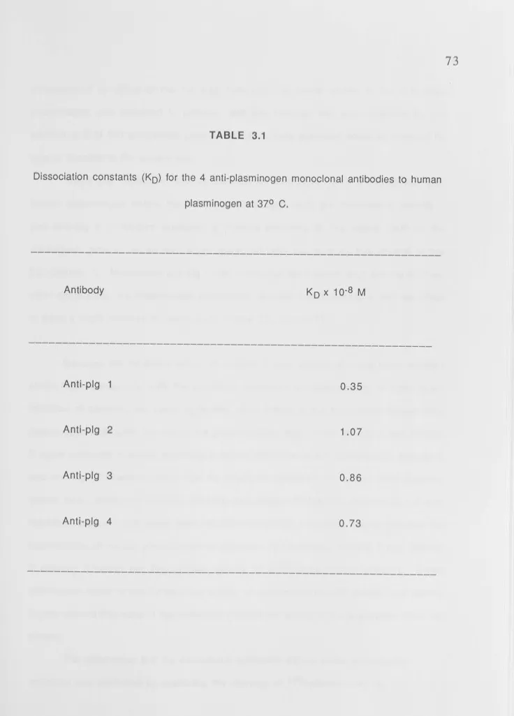
Four monoclonal antibodies raised against purified human plasminogen were
characterized for their effects on the activation of plasminogen and on three enzymic
properties of plasmin viz:- (a) thioesterolysis, (b) fibrinolysis, (c) conversion of
high molecular weight human urokinase to its low molecular weight form.
None of the monoclonal antibodies inhibited plasminogen activation by urokinase.
The monoclonal antibodies characterized in this study fell into three groups. The first
group represented by anti-pig 1 inhibited (a), (b) and (c), while anti-pig 2, inhibited
activities (a), (b) and (c) to varying degrees and in addition formed complexes with
plasmin which were highly stable to sodium dodecyl sulphate. Anti-pig 3 and anti-pig 4
inhibited activity (c), but not (a) or (b).
Selective use of these monoclonal antibodies demonstrated unequivocally that
plasmin mediates the activation of the proenzyme form of urokinase type plasminogen
activator. Besides their use in affinity chromatography, therefore, these antibodies are
valuable for defining the role of plasmin in the mechanisms of extracellular matrix
degradation as shown in their use in Chapters 4-6.
I I ,, I C D I I ii I I I 11 Ii
I :
X
found to be increased significantly in both colorectal carcinomas (n=20) and the
premalignant adenomatous polyps (n=27) when compared to autologous normal mucosa.
Activator content was also found to be increased in adenomatous polyps and autologous
normal mucosa removed from familial polyposis coli patients.
The urokinase-type plasminogen activator was shown by a new monoclonal
antibody technique to be present mainly as the proenzyme form.
For colon cancers, there was a significant correlation between their grade
according to the Dukes' classification and the amount of proenzyme present ( P < 0.05).
Although a similar trend was evident, no significant correlation was, however, observed
for total plasminogen activator content and the Dukes' grading.
For adenomatous polyps, no significant correlation was observed between enzyme
content and the size or degree of dysplasia. However, plasminogen activator expression
was found to be increased in the upper top or apical third of the polyps relative to the
basal third or stalk.
These results suggest that the expression of increased levels of human
plasminogen activator of 52,000 daltons proenzyme in dysplastic colon epithelial cells
correlates with the extent of invasion. The polyp studies suggest that expression of
human plasminogen activator of 52,000 daltons is a pre-requisite for invasion.
The study of the plasminogen activator content of colonic mucosa therefore offers
a useful biochemical correlate of epithelial cell transformation.
The quantitative esterolytic assay incorporating monoclonal antibodies inhibitory
to plasmin (chapter 3) was also used to elucidate the role of human plasminogen
activator of 52,000 daltons in inflammatory bowel disease. The levels of proenzyme and
active forms of human plasminogen activator of 52,000 daltons were compared in
mucosa from patients with active inflammatory bowel disease (n=13), from
I Ill I i I I, I I I In II I I :,
I
I u xi(n=8). Preincubation of tissue homogenates with monoclonal anti-pig 1 and anti-pig 3
antibodies prevented activation of the proenzyme by plasmin.
The level (absorbance 412 nm) of total human plasminogen activator of 52,000
daltons enzyme activity (active + proenzyme) was markedly increased in active
inflammatory bowel disease homogenates (1 .1 O + 0.39), compared to those found in
remission (0.48 + 0.17) and in uninvolved mucosa (0.45 + 0.20; P < 0.01 ). Assays of
proenzyme levels showed a similar highly significant increase in active mucosa (0.80 ±.
0.27) compared to remission (0.25 + 0.15) and uninvolved mucosa (0.23 + 0.15; P <
0.01 ). In actively diseased mucosa, 70°/o of the total human plasminogen activator of
52,000 daltons activity was present in the proenzyme form requiring extracellular
cleavage by plasmin. These results suggest that human plasminogen activator of 52,000
daltons may be involved in the pathogenesis of tissue injury in inflammatory bowel
disease by establishing the presence of a recognised pathway of inflammatory injury
which is selectively and substantially enhanced in actively diseased tissue.
Subcellular fractionation of human intestinal macrophages was performed to
define the localisation of the plasminogen activators and other serine hydrolase. Human
intestinal macrophages, isolated from lamina propria and purified by centrifugal
elutriation were disintegrated by nitrogen cavitation. The membranes were separated by
equilibrium buoyant density using isopycnic centrifugation on a sucrose gradient. The
subcellular membranes were localized using marker enzymes characteristic for plasma
and intracellular membranes.
Mn 2 +-stimulated leucine 2-napthylamidase was identified as a plasma
membrane enzyme.
Analysis of membrane fractions identified a membrane bound esterase not
detected in blood monocytes and exhibiting the same density and release characteristics
btained. Unlike monocytes, the esterase activity was partially resistant to 40 mM
sodium fluoride. The different properties of the human intestinal macrophages
alpha-naphthyl esterase to that of the blood monocytes may have important significance in the
light of recent evidence that alpha-naphthyl esterases are involved in the spontaneous
cytotoxicity of monocytes toward tumour cells (see discussion on chapter 6).
Arylsulphatase C, an endoplasmic reticulum marker was detected in much denser
fractions than that reported in monocytes.
Plasminogen activator of the urokinase-type was detected on the Golgi and plasma
membranes fractions in human intestinal macrophages. The plasminogen activator was
found to exist predominantly as proenzyme requiring plasmin for proteolytic activation.
The findings that the plasminogen activator is membrane associated and exists mainly as
proenzyme in the plasma and Golgi membrane fractions might have important biological
implications and suggest a site of enzyme activity which may optimise their
effectiveness.
The analysis of human intestinal macrophages plasma and intracellular
membranes at high purity represents a valuable approach to elucidating the changes that
PA
u-PA
t-PA
HPA66, HPA52, HPA36
mPU
Ploug Unit
CTA Pig Mr 180 HIM~ OFP
sos
SOS-PAGEEOTA
NaOH NaClOMEM
ABBREVIATIONS- plasminogen activator
- urokinase type plasminogen activator
- tissue type plasminogen activator
human plasminogen activator of
66,000; 52,000 and 36,000 daltons
respectively
milliploug unit
- unit of urokinase activity equivalent
to approx. 1.4 CTA units
Committee on Thrombolytic Agents
- plasminogen
- molecular weight
- inflammatory bowel disease
- human intestinal macrophages
diisopropylfluorophosphate
- sodium dodecyl sulphate
polyacrylamide gel electrophoresis in
the presence of sodium dodecyl
sulphate
- ethylenediaminetetraacetic acid
- sodium hydroxide
- sodium chloride
FCS
Pen
Gent
Strep
NPGB
EACA
BSA
PBS
thymidine
foetal calf serum
- penicillin
- gentamycin
streptomycin
p-nitrophenyl p'-guanidino-benzoate
- E-caproic-n-acid
- bovine serum albumin
I
','I
'"
r
j
I ,
I
I
I
I '
CHAPTER 1
GENERAL INTRODUCTION
PLASMINOGEN ACTIVATORS
SINCE the first observations by the pioneers of tissue culture on the
liquefaction of clotted blood and the enhancement of fibrinolysis in vivo and in vitro in
the 18th and 19th centuries, supporting experimental evidence accumulated until it
promptoed Fisher (1925) to postulate about the proteolysis caused by cultured Rous
sarcomas:-1
"We could imagine that the destructive process of the sarcoma cells in vivo would be as follows. First the stroma binding the cells together is liquefied. Consequently, reorganisation cannot take place as long as the lytic agent, the sarcoma cells, is present and because the fixed cells are deprived of their fibrin stroma".
Twenty years after his postulate, Fisher reported in 1946 that proteolysis
caused by cultured cancer cells appeared to be due to their activation of an inactive
proenzyme present in serum. It was left to Goldhaber et al., (1947), to suggest that the
cancer cells released an enzyme which activated profibrinolysin to fibrinolysin. This
activating enzyme is now widely known as plasminogen activator (PA), a serine protease
which catalyses the conversion of inactive plasminogen to the active plasmin.
1 . 1 . BIOCHEMISTRY OF PLASMINOGEN ACTIVATORS
In order to appreciate the possible implications of the role of plasminogen
activators in tumorigenesis, cancer invasion and metastasis, an understanding of the
'!
'
ID
'
I.
I
Christensen (1945) and Kaplan (1944; 1946) both demonstrated the
existence of an inactive proenzyme, designated profibrinolysin or plasminogen which
could be activated to the active protease fibrinolysin or plasmin. This was soon to be
followed by demonstration of the presence of substances able to activate plasminogen in
tissues and tissue extracts (Astrup & Permin 1947; Astrup & Stage 1952; Albreschten
1957 a,b), urine and blood (Williams 1951; Astrup & Sterndorff 1952; Sobel et al.,
1952; Lewis & Ferguson 1951; Mullertz 1953; Sherry et al., 1959).
Human plasminogen, an inactive proenzyme, is present in abundant amounts
, in the extracellular fluid. Its native form is a single chain glycoprotein with a Mr of
about 92,000 (Collen, 1980), containing about 2°/o carbohydrate and with
NH2-I:
1111 1,
terminal glutamic acid (termed Glu-plasminogen). Glu-plasminogen is readily
converted proteolytically to modified forms with NH2-terminal lysine, valine or
methionine (Wallen & Wiman, 1970; 1972) and the former therefore being termed
Lys-plasminogen with Mr about 8000 lower than its native form. These conversions
occur by hydrolysis of the Arg 67 - Met 68, Lys 76 - Lys 77 or Lys 77 - Val 78
peptide bonds. The complete plasminogen molecule consists of either 790 or 791 amino
acids (Sottrup - Jensen et al., 1978a; Wiman 1978), containing 24 disulfide bridges as
well as five homologous triple loop structures or "Kringles" (Sottrup-Jensen et al.,
11 1978b).
'
I
I
In vitro activation of Glu-plasminogen to plasmin by plasminogen activator
in a purified system occurs about 20 times more slowly than activation of
Lys-plasminogen (Claeys & Vermylen 1974; Wallen & Wiman 1975; Thorsen et al., 1974;
Lijnen & Collen 1982) but in either case, Lys-plasmin is formed. However, activation
I
11 of the fibrinolytic system in vivo occurs by direct cleavage of the Arg 560 - Val 561I
'
I
I
I
'
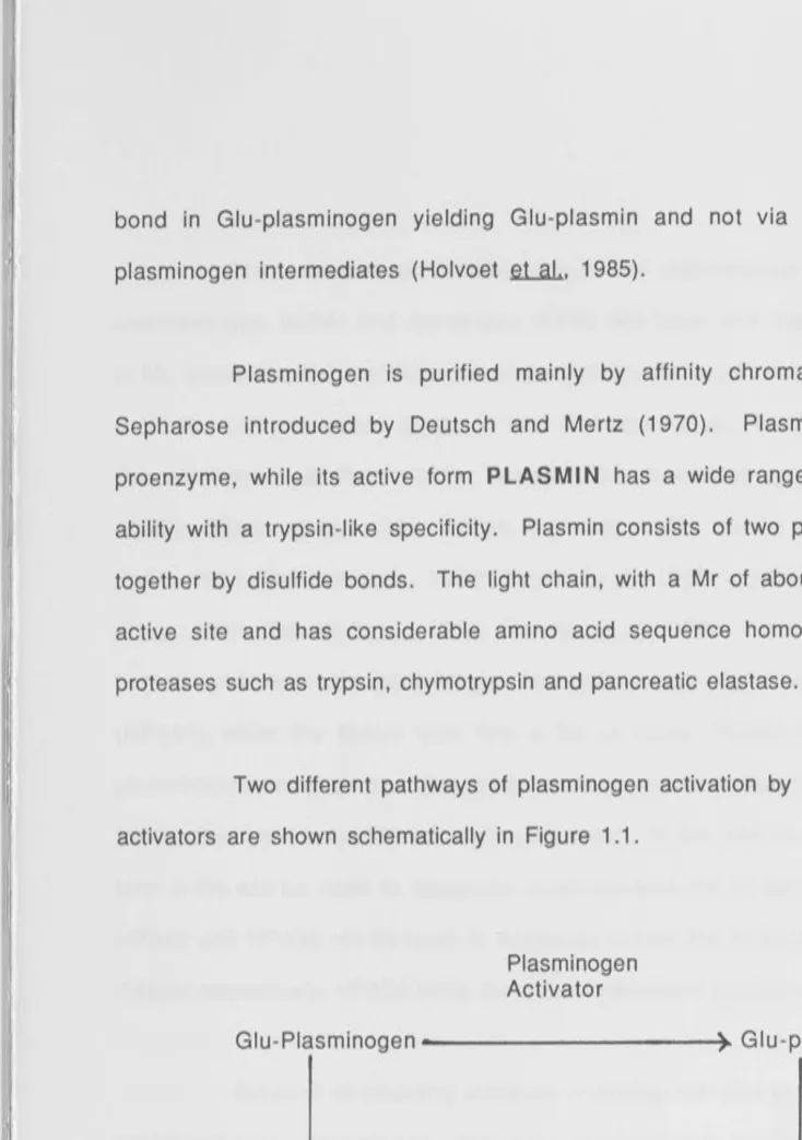
bond in Glu-plasminogen yielding Glu-plasmin and not via formation of the
Lys-plasminogen intermediates (Holvoet et al., 1985).
3
Plasminogen is purified mainly by affinity chromatography with
lysine-Sepharose introduced by Deutsch and Mertz (1970). Plasminogen is an inactive
proenzyme, while its active form PLASMIN has a wide range of general proteolytic
ability with a trypsin-like specificity. Plasm in consists of two polypeptide chains held
together by disulfide bonds. The light chain, with a Mr of about 25,000, contains the
active site and has considerable amino acid sequence homology with other serine
I
1, proteases such as trypsin, chymotrypsin and pancreatic elastase.
1,
11 11
11
•
Two different pathways of plasminogen activation by vertebrate plasminogen
activators are shown schematically in Figure 1.1.
Plasminogen Activator
Glu-Plasminogen --- - - ~ ~ Glu-plasmin
Plasm in
Plasminogen Activator
~~
,~
Lys-Plasminogen - - - ~ ~ Lys-Plas
Figure 1 .1. Activation of Plasminogen by Plasminogen Activator.
1 . 2 TYPES OF ACTIVATORS OF PLASMINOGEN
[image:17.1133.28.762.39.1082.2]The existence of 2 different types of plasminogen activators, designated
urokinase-type (u-PA) and tissue-type (t-PA) has been well documented. They differ
in Mr, immunological reactivity, genotype and are the products of different genes (Aoki
& Kaulla 1971; Unkeless et al., 1974b; Christman et al., 1975; Dano & Reich 1978;
Granelli-Piperno & Reich 1978; Astedt 1979; Vetterlein et al., 1979; Dano et al.,
1980b; Rijken et al., 1980; Roblin & Young 1980; Wilson et al., 1980; Rijken &
Collen 1981; Gunzler et al., 1982b; Kaltoft et al., 1982; Schaller et al., 1982; Steffens
et al., 1982; Edlund et al., 1983; Nielsen et al., 1983; Pennica et al., 1983). The
human urokinase-type plasminogen activator has a Mr of about 50,000 - 52,000
(HPA52) while the tissue type has a Mr of about 70,000 (HPA66). Tissue-type
plasminogen activator has also been termed extrinsic plasminogen activator (Collen
1980) although this term is less commonly used. In this and the following chapters, the
term u-PA will be used to designate urokinase-type PA of any species while HPA66,
HPA52 and HPA36 will be used to designate human PA of 66,000, 52,000 and 36,000
daltons respectively; HPA36 being the active breakdown product of HPA52.
Because of mounting evidence (including our own studies) implicating mainly
urokinase-type plasmingen activator involvement in the role of malignant
transformation, this literature review will be mainly concerned with u-PA and a brief
mention oft-PA whenever it is deemed necessary.
1 . 2. 1 Urokinase-Type Plasminogen Activator:- Purification and Occurence of u-PA
The presence of a high concentration of u-PA in human urine has for many
some has been produced by cultures of kidney cells. More recently, recombinant
urokinase has been produced after cloning of the human gene (Heyneker
et al.,
1983b;Verde
et al.,
1984), and this method will inevitably become the major commercialprocedure.
Purification of u-PA to homogeneity from urine by a tedious sequence of
precipitations and conventional chromatography steps was first described by Lesuk
.et
.aL.,
(1965) and Whiteet al.,
(1966). Since then, purification involving the use ofaffinity chromatography, especially the use of immobilized monoclonal antibodies, has
been widely used (Kaltoft
et al.,
1982; Nielsenet al.,
1982; Herion & Bollen 1983;Vetterlein & Calton 1983;). The purification of u-PA from human blood plasma and
serum, human seminal plasma and from hyperplastic and malignant prostate tissue has
also been reported. Because only very low concentration of u-PA are found in
conditioned culture fluid of several types of cell lines, even after treatment of the cells
with agents enhancing u-PA production e.g. phorbol esters (Goldfarb & Quigley 1980),
urine is still the main source for the purification of u-PA. However, recently,
Grondahl-Hansen
et al.,
(1985), using a one-step procedure with monoclonal antibodyimmobilized on Sepharose, have been able to purify human u-PA to apparent
homogeneity from the conditioned culture fluid of a human glioblastoma cell line.
As mentioned above, u-PA, or a plasminogen activator with similar
properties, has been found in a number of biological fluids or tissue extracts (Kucinski
et al.,
1968; Granelli-Piperno & Reich 1978; Danoet al.,
1980b; Astedtet al.,
1977;Rijken
et al.,
1981; Astedt 1978; Shakespeare & Wolf 1979; Tissotet al.,
1982;Wijngaards
et al.,
1982; Wunet al.,
1982a; Casslenet al.,
1981; Oshiba & Ariga1983; Markus
et al.,
1980, 1983; Camioloet al.,
1984; Nakamuraet al.,
1984;sites are indicated by arrows. The A-chain of the human u-PA contains a "kringle"
structure in its C-terminal part. The N-terminal of the B-chain contains the sequence
lle-lle-Gly -Gly. The A-chain has Arg or Phe as its C-terminal amino acid. Nucleotide
sequence around the cleavage site is Arg-Phe-Lys-lle-Gly-Gly. Activation of
prourokinase is effected by cleavage of a single bond resulting in the formation of A and B
chains linked by a disulphide bridge. The Ser, His and Asp residues forming the active
site in u-PA are marked with stars. Plasmin also cleaves urokinase A-chain at residues
Lys 135-Lys 136 removing the kringle and N-terminal region, leaving only 21-23 residues linked to the B chain (Adapted from Heyneker et al., 1983a).
[image:20.1117.72.748.32.1059.2](a)
Plasmin
C
Prourokinase
+
~ C
A chain
Active uroklnase
Low molecular weight urokinase
(b)
B chain
lmmunocytochemical studies of normal mouse tissues has localized u-PA to a
variety of cell types, in particular connective tissue cells with a fibroblast-like
morphology (Larsson
et al.,
1984) and occurring in high frequency in the laminapropria of the gastrointestinal tract. No other cell types, other than fibroblast-like
cells displayed u-PA in the gastrointestinal tract. These fibroblast-like cells may be
macrophages, although a definitive answer would require double staining, possibility
using a range of known macrophage surface markers.
U-PA is also produced in a number of primary cultures established from
tissues and cells of neoplastic origin.
1.2.2. Molecular and Enzymatic Properties of U-PA
Urokinase-type plasminogen activator has a Mr of about 50,000 - 52,000
and occurs in a single polypeptide chain form as an inactive proenzyme, while the
two-chain form is an active enzyme (Nielsen
et al.,
Skriveret al.,
1982; Wunet al.,
1982b;Eaton 1984). The heavy B chain (Mr about 30,000) and the light A chain (Mr about
20,000) have apparent Mr's varying somewhat between species (Christman & Acs
1974; Unkeless
et al.,
1974b; Holmberget al.,
1976; Aasted 1981; Nielsenet al.,
1982; Skriver
et al.,
1982; Wunet al.,
1982b; Sumi & Robbins 1983; Sudol & Reich1984) and are held together by one disulfide bridge (Sumi & Robbins 1983; see also
Fig. 1.2).
Amino acid sequence studies of human 2 chains u-PA have shown that the B
chain contains a heavy chain identical to that of the two chain 50,000 Mr forms and a
I I Id I: I• I•
50,000 Mr form. These findings demonstrate that the B chain (Mr about 30,000) is
formed from the native 50,000 Mr form by proteolytic conversion (Gunzler
et
al.,1982a). This conversion probably occurs during storage and purification due to
proteases present in urine and other biological fluids (Soberano
et
al., 1976b). Thestudies described in Chapter 3 showed that this conversion is mediated through plasmin
1
1 and not by autocatalysis.
!
I I I
" '
11 ii 1,
ii I I l I ....
U-PA is a glycoprotein (Mclellan
et
al., 1980; Steffenset
al., 1982) withan isoelectric point of between 8.4 and 9.7. However, the exact value varies according to
the species from which the enzyme originated and between different reports on u-PA
from the same species (Christman & Acs 1974; Soberano
et
al., 1976a; Danoet
al.,1980a; Nobuhara
et
al. 1981; Miwaet
al., 1982).Although plasminogen is the only well-documented protein substrate for
u-PA, studies by Quigley
et
al., (1980) and Keski-Oja & Vaheri (1982) have suggestedthe existence of cellular non-plasminogen substrates. U-PA also hydrolyzes a variety of
arginine and enzyme esters and amides and other low Mr substrates (Kjeldgaard & Ploug
1957; Ascenzi
et
al., 1982; Lottenberget
al., 1981; Friberger 1982).The high degree of substrate specificity shown by u-PA is probably related to
the presence of a specific binding pocket (Schoellman
et
al., 1982). U-PA also exhibitsto some degree species specificity with respect to the plasminogen substrate (Reich,
1975).
Inhibition of the esterolytic activity (Landmann & Markwardt 1970) and
plasminogen activating activity (Christman & Acs 1974; Unkeless et al., 1974b; Dano &
1a
I
II ,,
Ii
II ::
reagent for serine proteases diisopropylfluorophosphate (DFP) has indicated that these
activators are serine proteases (Walsh and Wilcox, 1970).
The heavy B chain (Mr about 30,000 - 253 residues) carries the active site
residues as demonstrated by affinity labelling with peptidyl choloromethyl ketone
inhibitors (Ong et al. 1976) or [3H] DFP (Christman & Acs 1974; Unkeless et al.,
1974b; Nielsen et al., 1982; Skriver et al., 1982; Wun et al., 1982b), followed by
SDS-polyacrylamide gel electrophoresis under reducing conditions.
The strong homology of the active site residues of u-PA with trypsin and the
plasmin B-chain, is also illustrated by the fact that several compounds inhibiting u-PA
11 also inhibit plasmin (Dano & Reich 1975, 1979). But as expected from the highly
I f I ' I
.
:I
I
1
•
n
different substrate specificities of u-PA and plasmin, some inhibitors have different
affinities for the two (Walton 1967; Dano & Reich 1975, 1979; Summaria et al.,
1975).
1. 2. 3 Proenzyme of u-PA
There are several examples of serine proteases which are released from cells
as inactive proenzymes which are converted to the corresponding active enzymes by
limited proteolysis (for a review, see Neurath & Walsh 1976).
Studies by Bernik et al. (1974) and Nolan et al., (1977) suggested the
presence of a latent form of u-PA in the conditioned medium from cultured cells, but
definitive identification of an inactive u-PA proenzyme could not be achieved due to the
lack of sufficient purified protein. Since then several authors (Sumi et al., 1982,
Nielsen
et al.,
1982) have demonstrated a one-chain inactive polypeptide form of u-PAboth in human and murine systems. This question is answered definitively by
experiments using monclonal antibodies inhibitory to plasmin as reported in Chapter 3.
ihe identification of proenzyme form of HPA52 has led to several proposed regulatory
implications (see Chapters 4-6).
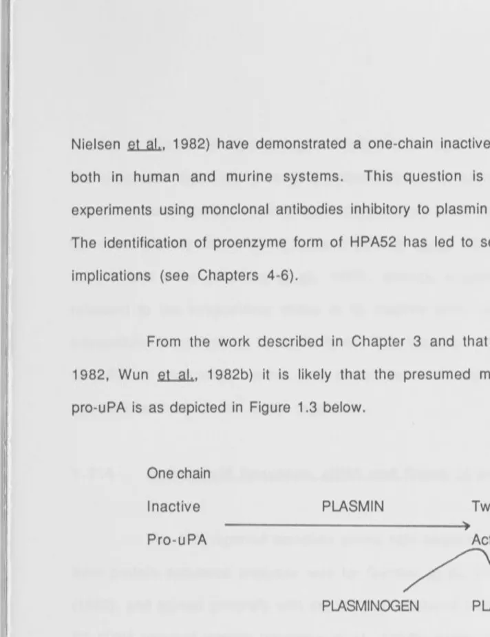
From the work described in Chapter 3 and that of others (Nielsen
et al.,
1982, Wun
et al.,
1982b) it is likely that the presumed mechanism of activation ofpro-uPA is as depicted in Figure 1.3 below.
One chain
Inactive
Pro-uPA
PLASMIN
PLASMINOGEN
Two chains
Active PA
PLASMIN
PROTEINS PEPTIDES
Fig. 1.3 Cascade reaction leading to u-PA initiated extracellular proteolysis.
Pro-urokinase (Pro-u-PA) is an inactive single chain precursor molecule which
is converted to an active two-chain polypeptide (Mr 20,000 and 30,000)
linked by a single disulfide bridge, by cleavage of the Lys 158-lle 159
peptide bond. The active u-PA activates plasminogen to plasmin by cleavage
[image:25.1133.28.740.37.961.2]a
11
I.
' II
Ii
Ii
I
I : ' .!
I
m
I
....
10
The above reports, together with the findings that plasminogen activators of
the urokinase type are present predominantly in proenzyme form in the colonic
adenomatous polyps and carcinomas, in inflammatory bowel disease (see Chapters 4 and
5), extracellularly in a murine tumour (Skriver
et
al. 1984) and in various murinetissues and urine (Kielberg
et
al., 1985), strongly suggest that u-PA is generallyreleased to the extracellular milieu in its inactive form, requiring plasmin mediated
extracellular proteolysis to its active form. This inactive form is the predominant form
of u-PA in intracellular stores and comprises a sizeable fraction of the u-PA in
extracellular fluids in the intact organism.
1 . 2. 4
Amino Acid Sequence. cDNA and Genes of u-PA
The first reported complete amino acid sequence of human two-chain u-PA
from protein sequence analyses was by Gunzler et al., (1982b) and Steffens et al.,
(1982), and agreed generally with the structure deduced from the base sequence of
u-PA cDNA reported recently (Heyneker
et al.,
1983b; Verdeet al.,
1984). Nagamine.ru
.a.1.,
(1984) also recently provided the first evidence of close homologies betweenporcine and human u-PA as determined from the nucleotide sequence of the porcine u-PA
gene.
The sequencing of the cDNA for both u-PA and its comparison with that of
t-PA has definitively established that each activator is the product of different genes as
previously speculated.
The single structural gene for urokinase has been reported to be present on
human chromosome 6, and nucleotide sequencing of genomic libraries indicated the
presence of up to 13 introns (Kucherlapati et al., 1978, Holmes et al., 1985).
u-1
I
i
I
I'
•
I'
Ii
I~ i
'
r;
::I
PA DNA on Southern blots and in situ with a cDNA probe localized the human u-PA gene
to the long arm of chromosome 10.
1 . 2. 5 Tissue Type Plasminogen Activator
11
Tissue-type plasminogen activator (t-PA) can now be isolated quite readily
from various sources and indeed milligram quantities had been purified from the tissue
culture media of Bowes human malignant melanoma (Rijken & Collen 1981, Wallen fil
.aL.,
1 983) .Although the Mr to be expected from the nucleotide sequence of the
corresponding cDNA of t-PA is 59,000, the Mr as determined by SDS-polyacrylamide
gel electrophoresis is 66,000 daltons. Glycosylation of the t-PA molecule could account
for this apparent discrepancy (Mclellan
et
al., 1980).Like u-PA, t-PA exists in two forms, a one-polypeptide chafn form and a
two-polypeptide chain form, the two chains in the latter, which is an active enzyme,
being held together by disulfide bridges (Binder
et
al., 1979; Rljken & CoUen 1981;Rijken
et
al., 1979; Aasted 1980; Wallenet
al., 1981, 1982, 1983; Ranbyet
al.,1982· Nielsen
et
al., 1983). Howeverl unlike u-PA, conflicting results as to theenzyme actrvity of the one-chain polypeptide form have been reported (Rljken & Collen
1981; Wallen
et
al., 1981, 1982, 1983; Ranby 1982; Rijkenet
al., 1982; Andreasenet
al., 1984· Ichinoseet
al., 1984). Using monoclonal antibodies against human t-PAand a linear regression analysis using 3[H]-DFP labell[ng, Andreasen
et
al., (1984)reported that the single chain t-PA represents a fibrlnolytically inactive proenzyme;
while Rljken
et al..
(1982) using a 1251-labelled plasminogen conversion assaythat the one chain from of both human and porcine t-PA appeared to have considerable
activity (Rijken & Collen, 1981; Wallen et al., 1981, 1982, 1983; Randy 1982).
Irrespective of whether it is accepted that the one chain form of t-PA is
partly or completely inactive proenzyme, it could be converted by plasmin to a two chain
form by the cleavage of an Arg 274-lle bond, resulting in A and B chains linked by a
single disulphide bond (Edlund et al., 1983; Pennica et al., 1983; Jornvall et al., 1983;
Wallen et al., 1983). The B chain with a Mr of between 28,000 - 33,000 comprises
the c-terminal region of the enzyme and is homologous with trypsin, plasmin and u-PA
B chains and carries the active centre residues. An increase in amidolytic activity as a
result of this bond cleavage (which does not affect the PA activity) has been used as an
assay to measure the proportions of one and two-chains forms. The A-chain of human
t-PA has a Mr of 37,000 - 40,000 with carbohydrate content differences or slight
proteolytic modification at the N-terminal residues probably accounting for the small
degree of heterogeneity (Wallen et al., 1983; Pennica et al., 1983). Whereas the
A-chain of human u-PA contains one triple-loop kringle structure, the A-A-chain of human
t-PA contains two triple loop kringle structures similar to those found in urokinase,
plasminogen and prothrombin. Closer to the N-terminus, t-PA, like u-PA, contains a
cysteine-rich domain like that found in blood coagulation factors IX and X and epidermal
growth factor (Banyai et al., 1983; Pennica et al., 1983). However, controversy still
persists as to whether the fibrin and lysine binding properties of t-PA reside in one or
both kingles since these same properties are also exhibited by plasminogen, where they
reside in the kringle structures (Banyai et al., 1983). Unlike u-PA, the one and two
chain forms of t-PA and plasminogen both bind to Sepharose-bound L-lysine, arginine
and to fibrin. Comparison of the four lysine-binding kringles (Reich 1978b) of
Ill
Ii
II
.
::Ii
i
'
r
...
fibrin-binding properties exhibited by urokinase kringle (Gunzler
et
al., 1982a;Pennica
et
al., 1983, Collen 1980).As for u-PA, t-PA is a highly specific enzyme and plasminogen is the only
known protein substrate. However, t-PA differs from u-PA in having very strong
affinity for fibrin (Thorsen
et
al., 1972; Rijken & Collen 1981 ), an affinity that haseven been used for purification of the enzyme by Wallen
et
al., (1982). Fibrin has alsobeen found to strongly stimulate plasminogen activation by t-PA (Camiolo
et
al., 1971;Wallen 1978; Hoylaerts et al., 1982; Ranby 1982; Radcliffe 1983; Suenson et al.,
1984).
Using Bowes melanoma cells, sufficient specific mRNA was synthesized to
allow complete gene cloning of t-PA (Opdenakker et al., 1982; Edlund et al., 1983; Ny
et al., 1984; Pennica et al., 1983). Like u-PA, t-PA is found in a number of places and
is particularly prominent in the endothelial regions of tissues (Cano
et
al., 1985).1 . 3 ASSAYS AND DETECTION METHODS FOR PLASMINOGEN ACTIVATORS
Because of the findings that u-PA is generally released from producer cells as
an inactive proenzyme form requiring conversion to its active counterpart by limited
proteolysis involving plasmin (see Chapter 3), methodological approaches in the
enzymatic detection and quantitation of u-PA will therefore be highly influenced by the
trace amounts of plasmin that are usually present in the plasminogen preparation used
in the assay. Detection and quantitation of plasminogen activator activity could be
further complicated by the presence of inhibitors of PA and plasmin since even minute
amounts of plasmin inhibitors could strongly inhibit the assay when u-PA is present in
its proenzyme form.
I I
I
'I
The increased interest during the past decade in PA involvement in
fibrinolysis, tumorigenesis, inflammatory responses and the expression of hormonal
regulation has led to a rising interest in the development of sensitive and precise
methods for the specific assay of PA. The optimal assay method for PA obviously depends
on the purpose for which it is to be used.
ENZYMATIC ASSAYS AND DETECTION METHODS
1 . 3. 1
Plasminogen as Substrate
In these assay methods where plasminogen is used as the substrate, the PA
activity is measured either directly by the amount of plasmin molecules formed or
indirectly through the enzymatic activity of the plasmin generated, taking advantage of
the amplification involved.
By the use of specific inhibitory antibodies against t-PA or u-PA, it is
possible to distinguish between t-PA and u-PA since antibodies against the respective PA
inhibit activation of plasminogen catalysed by that activator but not that catalysed by the
other type (Kucinski et al.. 1968; Aoki 1974; Astedt & Holmberg, 1976; Vetterlein
.e.1
.al...,
1979; Mackie et al., 1981; Corasanti et al. 1980; Rijken & Collen, 1981; Dano fil.al...,
1980b; Kaltoft et al., 1982; Berger & Tuttle, 1983; Nielsen et al. 1983; Ossowski& Reich 1983).
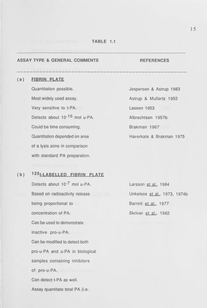
Table 1.1 shows the common types of assays used to detect and quantitate PA
activity using plasminogen as
[image:30.1130.27.736.37.932.2]II: L ' !, I I '
TABLE 1.1
ASSAY TYPE & GENERAL COMMENTS
(a) FIBRIN PLATE
Quantitation possible.
Most widely used assay.
Very sensitive to t-PA.
(b)
Detects about 1 o-16 mol u-PA.
Could be time consuming.
Quantitation depended on area
of a lysis zone in comparison
with standard PA preparation.
1251-LABELLED FIBRIN PLATE
Detects about 1 o-7 mol u-PA.
Based on radioactivity release
being proportional to
concentration of PA.
Can be used to demonstrate
inactive pro-u-PA.
Can be modified to detect both
pro-u-PA and u-PA in biological
samples containing inhibitors
of pro-u-PA.
Can detect t-PA as well.
Assay quantitate total PA (i.e.
REFERENCES
Jespersen & Astrup 1983
Astrup & Mullertz 1952
Lassen 1953
Albrechtsen 1957b
Brakman 1967
Haverkate & Brakman 1975
Larsson
et al.,
1984Unkeless
et al.,
1973, 1974bBarrett
et al.,
1977Skriver
et al.,
1982 [image:31.1130.32.741.24.1077.2]active plus proenzyme).
Not suited for kinetic studies.
Several modifications based
on these method e.g. [3H]
labelled fibrin.
( c) CASEINOLYTIC
Casein used as a substrate
for plasmin.
Based on absorbance.
Sensitivity 2 x 1 o-15
mol for u-PA.
Quantitation possible but
measures only total PA.
Has low sensitivity for t-PA.
( d) SYNTHETIC PLASMIN SUBSTRATES
Also based on absorbance.
Quantitation possible.
Measured total PA but can be
modified using monoclonal
antibodies to quantitate either
pro-u-PA and active u-PA. (See
Chapter 4 & 5).
Specific for u-PA in the absence
of added fibrin.
Synthetic substrate commonly
Markus et al., 1980
Kline 1971
Kline & Reddy 1977
Lottenberg et al., 1981
Friberger 1982
used are S-2251 (H-D-Val-Leu
-Lys-p-nitroanilide) and
thiobenzyl benzyloxycarbonyl
-L-lysinate (ZLS).
Can be used for kinetic and
binding studies.
Sensitivity 2 x 1 o-1 7 mol u-PA.
( e) DIRECT PLASMINOGEN CONVERSION
PA activity estimated directly by
( f )
the number of native plasminogen
molecules converted to
2-polypeptide chains plasmin
under reducing conditions.
Measured total PA only and can
used to demonstrate pro-t-PA.
Measurement of u-PA also
possible.
Useful for kinetic sudies and
studies of inhibition of plasminogen
activation.
FIBRIN OVERLAY
Distinction amount different types
of PA can be obtained by use of
specific inhibitory antibodies.
Quantitation not possible.
Reddy & Markus 1972
Dano & Reich 1975, 1979
Eaton & Baker 1983
Lucas et al., 1983
Suenson et al., 1984
Mussoni et al., 1984
Rijken et al., 1980
Ljunger et al., 1983
Soreq & Miskin 1981
Assay influenced by inhibitors of
PA and plasmin.
Can be used to detect proenzyme.
Several modifications based on
this assay. Inferior to
immuno-assay when used to detect PA
in tissues.
( g) ZYMOGRAPHIC DETECTION OF PA
Can differentiate types of PA.
Assay based on diffusion of PA
into gel containing plasminogen
and fibrin leading to lysis zone.
However, assay semi-quantitative.
Effect of protease inhibitors
minimal.
1 o-14 mol of u-PA & t-PA
detectable.
Modification of this method with
125-labelled casein which detects
about 1 o-18 mol u-PA.
Can be modified to detect and
demonstrate PA inhibitors and
proenzyme forms of PA
particularly u-PA.
Granelli-Piperno
& Reich 1978
Dano et al., 1 980b
Miskin & Soreq 1981 b
Erickson et al., 1984
Although plasminogen is the only well-documented protein substrate for
plasminogen activators, the ability of the enzymes to hydrolyze amide and ester
derivatives of arginine and lysine in certain small peptides is being used in some assay
methods.
The conversion of N-acetyl-L-lysine methyl ester into N-acetyl-L-lysine
and methanol is one example. The methanol generated is then oxidized to formaldehyde
which when mixed with chromotropic acid can be quantitated spectrophotometrically
(White & Barlow 1970). In amidolytic assays, tripeptides derivatized with
p-nitroaniline at the C-terminal amino acid (Friedman et al., 1977; Claeson et al., 1978;
Lottenberg et al., 1981; Friberger 1982) are commonly used. Hydrolysis of the
p-nitroanilide bond with the release of p-nitroaniline can then be measured
spectro-photometrically.
In particular, such assays have been used to measure the conversion of
proactivators to the active enzymes (Wun et al., 1982b; Andreasen et al., 1984) but
their use in biological samples is limited by their non-specificity in cleavage by
proteases other than PA.
11
3. 3 Active-Site Titration
Active-s i e titration assays can only be used for the analysis of purified
preparations of PA sin ce the activ e-s i e titrants are not specific for PA. Rijken and
Co llen (1981) used incorporation of [3 ] DFP , an irreversible inhibitor of the active
si e of serin e pro eases as a measure of the numbers of active sites in the preparation of
incorporation, together with SDS-polyacrylamide gel electrophoresis to quantitate the
relative amouns of one and two chain forms of human t-PA in preparations containing
both; while Some no
et
al., (1982) used a combination of the inhibitor andchromatography to quantitate the relative amouns of low and high Mr u-PA.
1 . 3. 4
Immunological Assay and Detection Methods
The specificity of antigen-antibody interaction, coupled with the recent
production of monoclonal antibodies (McAbs) against both types of human PA and
plasmin (Kaltoft
et
al., 1982; Herionet
al., 1981, 1983; Nielsenet
al., 1983,Vetterlein & Calton, 1983; Salerno
et
al., 1984, and Chapter 3) is likely to enhance thespecificity of assays employing antibodies, provided that inhibitors of PA do not compete
with antibodies for binding.
Immunological assays may be superior since enzymatic assays for PA can
often be prejudiced by the presence of interfering inhibitors of proteolytic enzymes in
many biological fluids. These may interfere through direct inhibition of PA or through
inhibition of the plasmin formed indirectly in coupled plasminogen activation-plasmin
assays. Furthermore inhibitors of plasmin complicate the measurement of proenzymes
due to interference with the plamin catalysed activation of proenzymes.
Commonly used immunological assays using both polyclonal and
monoclonal antisera against PA
are:-( i ) Radioimmunoassays (Astedt
et
al., 1975, 1981; Vetterleinet
al., 1980,Urden & Blomback 1984, Wun
et
al., 1982a; Huberet
al., 1984) with a( i i )
( i i i )
( i V)
(v)
1 . 4
Two-Site lmmunoradiometric Assay (Holmberg et al., 1982; Rijken et al.,
1983) Sensitivity 2 x 1 o-15 mol for both u-PA and t-PA.
Enzyme-Linked lmmunosorbent Assay (ELISA) - Sensitivity 2 x 1 o-15 mol
for both human u-PA and t-PA. (Bergsdorg et al., 1983, Rijken et al.,
1984, Matsuo et al., 1983, Herion et al. 1983.)
lmmunocytochemical Detection Although not really an "assay", nevertheless,
the recent availability of strong and specific antibodies (polyclonal and
monclonal) have seen their use in the localisation of murine u-PA and human
u-PA and t-PA in tissues by immunofluorescence and
peroxidase-antiperoxidase staining (Dano et al., 1982, Markus et al., 1983, Kristensen
et al., 1984, 1985; Larsson et al., 1984; Nakamura et al., 1984; Salerno
.et.
aL.,
1984.)lmmunoblotting This technique employed both SOS-PAGE and the transfer of
separated proteins onto nitrocellulose paper followed by detection with
antibodies for specific proteins. This has been used as a screening method for
McAb production against t-PA (Nielsen et al., 1983), as a chain specificity
assay of McAb against u-PA (Salerno et al., 1984) and for distinguishing
between one-chain pro-u-PA and two chain active u-PA in impure biological
products (Kielberg et al., 1985; Skriver et al., 1984).
OCCURRENCE AND FUNCTION OF u-PA IN NON-NEOPLASTIC
The correlation of in vivo and in vitro studies of inflammatory processes and
normal invasive or remodelling events, whether regulated temporally or/and
hormonally have suggested the functional significance and occurrence of PA in
non-neoplastic conditions and likely involvement of u-PA in extra-cellular proteolysis. In
all the biological processes, the event and the associated extracellular proteolysis and
tissue matrix degradation have been found to be coordinately controlled.
1.4.1 TISSUE INVASION AND DEGRADATION IN THE NORMAL ORGANISM
1.4.1.1
Ovulation
The ovum contained in the ovary follicle is enmeshed in several layers of
cells and basement membrane. The presence of plasminogen in the follicular fluid and
the temporal correlation between PA production and disruption in ovulation provided
unambiguous evidence that PA is involved in the tissue degradation of the follicle wall
(Strickland 1978; Beers et al., 1975; Strickland & Beers 1976; Beers 1975).
Granulosa cells when stimulated with gonadotrophins in vivo and in vitro at
physiological levels that induce ovulation have been shown to produce significant levels
of PA. It is postulated that the plasmin generated by the activation of plasminogen
present in the follicular fluid by gonadotropin-induced PA disrupts the follicular wall
and digests, with the help of other proteases like collagenase, the extracellular matrix
leading to the eventual release of the ovum (Rohrlich & Rifkin 1979).
Implantation of the fertilized egg into the uterine wall involves the invasion
of the trophoblast through the uterine epithelium and the underlying basement
membrane and into the stromal tissue of the endometrium (Kirby 1965, Schlafke &
'
Enders 1975). The work of Strickland et al., (1976) with the in vitro
pre-implantation of mouse blastocysts was later confirmed by Sherman (1980) and Kubo
.et.
.al.:.,
(1981 ), who also found that the trophoblast cells expressed cell-associated PA as well as secreting the enzyme.This activity was detectable only from days 6-10, during which time the
mouse trophoblast was invasive in vivo, and was unrelated to the emergence of the
embryo from the zona pellucida.
1 . 4. 1 . 3 Mammary Gland Involution
Involution of the mammary glands after cessation of lactation is a classic
example of tissue remodelling. The findings of Ossowski et al., (1979) indicated a
positive temporal correlation between the onset of involution and the concomitant
increase in the level of PA secretion by glandular epithelium. Various hormones such as
hydrocortisone, aldosterone, prolactin and/or oxytocin which prevented the
postlactational involution in vivo, also prevented PA increase in the mammary gland
tissue. Similarly, factors such as epidermal growth factor or insulin which promoted
glandular degeneration in organ cultures of mammary gland fragments induced high PA
secretion especially after prolactin potentiation. U-PA but not t-PA was detected in the
extracts of involuting mammary gland. Recent immunocytochemical studies by Larsson
et al., (1984) confirmed previous indirect evidence that the u-PA was produced by the
epithelial cells and also the lack of immune reactive staining in the non-involuting
1.4.1 .4 Inflammation
The possible role(s) of PA and plasmin in inflammation has been of
longstanding interest. Macrophages are derived from precursors in the bone marrow
and circulate in the blood as monocytes and ultimately take up specific locations in
tissues as macrophages. In sites of inflammation, these macrophages can become
'activated' to kill micro-organisms and tumour cells. However, in chronic
inflammatory lesions, these cytocidal activities can be directed against the host to
mediate tissue injury. Both in vivo and in vitro studies of the response of macrophages
and polymorphonuclear leukocytes to inflammatory and inhibitory stimuli suggest the
some effects of these agents influence the ability of the phagocytic cells to regulate the
synthesis and secretion of plasminogen activators, collagenase and elastase. The findings
reported by Unkeless et al., (1974a) and Gordon et al., (1974) that unstimulated
murine macrophages produced PA of barely detectable levels, while cultured
thioglycollate-stimulated murine macrophages produced and secreted PA of 1 O times the
resting amount, had renewed interest in the role of proteolytic proteases in
inflammation. Since then, a number of agents have been found to either stimulate or
inhibit the production/secretion of PA in macrophages. Exposure of macrophages to
lymphokines (Klimetzek & Sorg 1977; Nogueira et al., 1977; Vassalli & Reich 1977;
Gordon 1978; Gordon & Cohn 1978; Gordon et al., 1978; Greinder et al., 1979),
asbestos (Hamilton et al., 1976), interferon (Hovi et al., 1981), colony-stimulating
factor derived from cultured murine cells (Lin & Gordon 1979), Concanavalin A, and
phorbol myristate acetate, a potent irritant, inflammatory agent and tumour promoter
(Vassalli et al., 1977; Neumann & Sorg 1983) have all shown ability to stimulate the
production of PA several-fold. PA synthesis and secretion by stimulated macrophages
glucocorticoids, pharmacological agents like colchicine, vincristine and compounds
affecting cyclic nucleotide metabolism and cholera toxins (Hamilton
et
al., 1976;Vassalli
et
al., 1976, 1977, 1980; Neumann & Sorg 1983). Similar findings havebeen reported for polymorphonuclear leukocytes, with respect to PA stimulation by
concanavalin A and phorbol myristate acetate and inhibition by glucocorticoids
(Granelli-Piperno
et
al., 1977).All these observations pointed to the physiological importance of PA and led to
the proposal that PA plays a role in inflammation and, in particular, in the migration of
inflammatory cells to the sites of inflammation as well as subsequent plasmin formation
by macrophages in the recruitment phase of the inflammatory response in vivo (Reich
1978a).
Since inflammation often involves degradation of the injured tissue, the u-PA
released by macrophages and polymorphonuclear leukocytes may contribute to this
process, with the help of other proteases, by extracellular degradation of proteins in a
manner parallel to that proposed for cancer cells (see Section 1.5.2 in this Chapter}.
Hence, it should be noted that the secretion of elastase, collagenase and other serine
proteases by stimulated macrophages and not by unstimulated macrophages have also
been well documented (Unkeless·
et
al., 1974a; Werb & Gordon 1975a,b; Werbet
al.,1980; Wahl
et
al., 1974, 1975).1 . 4. 2 PLASMINOGEN ACTIVATOR lN NON-NEOPLASTIC PATHOLOGJCAL
CONDITIONS
The possible implications of proteolytic enzymes, in particular PA and
pathological conditions in which PA has been implicated include the following, some of
which involve inflammation and/or tissue
degradation:-Allergic vasculitis (Toki et al., 1982), Xeroderma pigmentosum (Miskin &
Ben lshai, 1981 ), rheumatoid arthritis (Berger, 1977; Meats et al., 1980 Hamilton &
Slywka 1981; Hamilton et al., 1982), pemphigus (Hashimoto et al., 1982; 1984),
experimental murine leprosy (lzaki et al., 1983), chronic inflammatory bowel disease
(Doe & Dorsman, 1982; see Chapter 5), corneal ulceration (Berman et al., 1980),
diabetes mellitus (Almer et al., 1975), demyelinating diseases (Cammer et al., 1978;
1981 ), chronic sinusitis (Kosugi et al., 1982) and protein-losing gastroenteropathy
(Kondo et al., 1976).
1 . 5 OCCURRENCE AND FUNCTION OF PLASMINOGEN ACTIVATOR IN
NEOPLASIA
Neoplastic cells display a diverse range of biochemical and physiological
changes in comparison to their normal counterparts. Invasiveness and metastatic
potentials are two features commonly associated with neoplastic cells. Destruction of the
surrounding tissues and extracellular matrix that are normally 'cemented' together, by
lytic enzymes, has been proposed to account for the invasiveness of tumour cells. One
such lytic enzyme proposed is PA. It should, however, be stressed that in the following
discussion, the attribution of definitive roles to PA and plasmin in various aspects of
neoplasia/malignancy in NO WAY implies that these functions are specific to
cancer-related events. Hydrolysis of the elaborated extracellular matrix that normally would
serve as a barrier to the movement of tumour cells, would presumably allow migration
of these cells and hence invasion. This view has been supported with ultrastructural
(Markus et al., 1984; Camiolo et al., 1984; Skriver et al., 1984) where intensive
staining and hence localisation of PA (u-PA) have been observed in areas with invasive
growth and degradation of normal tussue. By contrast, no demonstrable staining has been
observed in the larger parts of tumours where no invasion and degradation of normal
tissue were occuring.
Early demonstration of PA in intact tumours using fibrin overlay methods
most probably measured t-PA since fibrin stimulates t-PA activity and furthermore, no
distinction was made between u-PA and t-PA. Recently, the production of high affinity
specific antibodies has enabled the visualisation of the occurrence and distribution of
u-PA in human colon adenocarcinomas and melanoma cells (Markus et al., 1983; 1984)
and in transplantable murine Lewis lung tumours (Skriver et al., 1984). In the study
by Skriver et al., (1984), these authors also noted that some immunoreactivity was
localized in the cytoplasm of the tumour cells, often with a perinuclear localization.
However, most of the immunoreactivity appeared to be localised extracellularly or near
to the cell membrane.
To date, the occurrence of u-PA in benign tumours and of t-PA in malignant
and benign tumours in immunocytochemical studies has yet to be demonstrated.
Extractable u-PA enzymatic activity has also been reported in human lung,
colon and breast cancer (Corasanti et al., 1980; Markus et al., 1980; Evers et al.,
1982), aspirates from patients with endometrial carcinomas (Niklasson et al., 1981 },
virus-induced mammary tumours and chemically induced rat mammary tumours
(Mira-y-Lopez et al., 1983). PA of non-determined type has also been demonstrated
So far we have mentioned the occurrence of PA in intact tumours and tumour
extract. PA has also been reported to occur in explants and cell cultures of neoplastic
origin. PA of the urokinase type has been found in cultured cells from an ovarian
carcinoma (Astedt & Holmberg 1976), in conditioned medium of tissue cultures derived
from endometrial carcinomas (Astedt et al., 1978; Svanberg & Astedt 1979), mouse and
rat mammary tumours (Mira-y-Lopez et al., 1983) and a number of cells lines of
neoplastic origin (Hisazumi et al., 1977; Wu et al., 1977; Dano et al., 1980a, 1982;
Naito et al., 1980, 1982; Rossman & Troll, 1980; Wilson et al., 1980, 1982, 1983;
Harvey et al., 1982; Shyamala & Dickerman 1982; Strickland et al., 1983; Azzarone
.e.t
.al.:.,
1983; see also Chapter 3).1 . 5. 1
Plasminogen Activator and Transformed Cells
Cellular transformation that occurs spontaneously in culture (Sanford et al.,
1954), or by infection with oncogenic viruses (Termin & Rubin, 1958) or
carcinogenic chemicals (Chen & Heidelberger, 1969) is believed to be closely related to
malignancy in vitro.
PA has been shown to be elevated in primary turmour cells, cells
transformed by both oncogenic viruses and chemical carcinogens and in a number of
turmorigenic cell lines (Unkeless et al., 1974a, 1973; Ossowski et al., 1973; Rifkin
.e.t
al,
1974; Goldberg 1974; Pollack et al., 1975). While there are a number ofexceptions (Gallimore et al., 1977; Jones et al., 1975; Rifkin & Pollack 1977;
Rosenthal et al., 1978; Wolf & Goldberg 1976), there is strong evidence in many cases
that PA production often accompanies or precedes malignant transformation. This
relationship has been most thoroughly studied in Rous sarcoma viruses (RSV), where