TO ASSESS THE EFFECT OF LOW LEVEL LASER
THERAPY ON SENSITIVITY OF TEETH PREPARED
FOR FIXED PARTIAL DENTURE
– A RANDOMIZED CONTROL TRIAL
A Diss ertation sub mitted
in parti al fulfil men t of th e requirements
for the degree of
MASTE R O F DENT AL SURGE RY
BRANCH – I
PROSTHO DO NTICS AND CROWN & BRIDGE
THE TAMIL NADU D r. M.G.R. ME DICAL UNIVERS ITY
CHENNAI- 600032
ADHIPARASAKT HI DENTAL COLLEGE AND HOSPITAL
MELMARUVATHUR – 603319.
DE PART MENT O F PROSTHO DO NTICS AND CROWN & BRIDGE
CERTI FICATE
This is t o certi f y t hat Dr. S. VINOTH KUMAR, P ost Graduat e student (2015 -2018 ) in the Departm ent of Prosthodonti cs and crown &
bridge , Adhiparasakthi Dent al Coll ege and Hos pit al, M elm aruvathur – 603319, has done this dissertation titled "TO ASSES S THE
EFFECT O F LO W LEVEL LASE R T HERAPY O N SE NSITIVIT Y
O F TEETH PRE PARE D FOR FIXE D PARTI AL DENTURE – A
RANDO MI ZE D CO NTROL T RI AL " u nder our di rect gui dance and supervision in parti al ful film ent of the regul ati ons l ai d down b y the Tami lnadu Dr. M .G.R Medi cal Uni versit y, C hennai – 600032 for M DS., (Branch - I) (P rosthodonti cs and C rown & Bridge ) degree exam inati on.
Dr. S. THILL AINAYAGAM MDS.,
Principal
Guide
Dr. N. VENKATES AN MDS.,
Professor d Co-Guide
ACKNOWLEDGEMENT
First and f or emost I would li ke to thank God. Y ou have gi ven me
the power to beli eve in myself and pur sue my dreams. I could never
have done this without the f aith I have i n you, the Al might y.
Wi th submis sive ambition, I aspir e t o r egis ter my Gr atitude to
my r es pect ed Mentor Dr. A. S . RAMESH M.D.S ., Prof ess or and Head,
and my guide Dr. N. VE NKATES AN M DS., Depar tment of
Prost hodonti cs , Adhiparasakthi Den t al coll ege and Hospit al, for his
inspir ing guidance, invaluable counsel and encouragement throughout
the cours e of t he s tudy. T his work w ould not have s een the li ght of t he
day wi thout his aff ectionat e and compassionat e counselling, whi ch
repos ed by confidence in mys elf t o undertake the challenges in the
study.
My s incere t hanks t o Dr.S .Thill ainayagam MDS., Our beloved
Principal, Adhiparasakthi Dent al Coll ege and Hos pital ,
Mel maruvathur f or providi ng me wit h t he opportunity to utilize the
facil ities of the college.
I t hank our Cor res pondent Dr.T.Ram esh, M D., for his vital
encouragement and s upport.
I am extr emely thankful to my t eachers Dr.Prabhu MDS . Reader,
Dr.Ki rubakaran MDS., Senior l ect urer, Dr.Raghun anth an MDS .,
Senior l ectur er , Dr.Karthi k, MDS., seni or l ecturer, Dr.Ram esh
constant encouragement and ti mely help r ender ed throughout t his
study.
I warml y acknowl edge my seni ors and my Juniors, for their hel p and support .
A special menti on of thanks to all m y patients for t heir cons ent , co-oper ation and par ticipation in thi s st udy.
I would li ke to thank and dedi cat e thi s valuabl e wor k to my
parents and uncl e & aunt y Mr. T. Panneer sel vam & Geetha P for
thei r suppor t, and my dear wi fe Dr. V. Niveth a for her unconditi onal
love and care. I thank the aut hors of all t he books and arti cl es which I
ref erred and to all my coll eagues and relatives . If by over sight any
acknow ledgement s have been omitt ed I of fer my si ncer e apolo gy.
Dr. S. Vin oth ku mar
DECLARATION
TIT LE OF THE DISS ERTAT ION
To assess the Effect of low level laser therapy on sensitivity of teeth prepared for fixed partial denture – A Randomized Control Trial.
P LACE OF THE STUDY Adhi paras akt hi Dental C oll ege and Hospital, Melm aruvathur -603319.
DUR AT ION OF THE COURS E
3 Years
NAM E OF THE GUIDE Dr.N.Venkat es an, M DS.
NAME OF C O -GUIDE Dr. A.S.R am esh, M DS.
I hereb y decl are that no part of t he dis sert ation will be utilized for gaining fi nancial assi stance or an y prom otion wit hout obt aining prior permi ssion of the P rinci pal , Adhi paras akthi Dent al col lege and Hospital, Me lmaruvathur -603319. In addition, I decl are that no part of this work will be publi shed either in print or in elect ronic m edia without the guides knowledge who have been act ivel y involved in diss ert ati on. The aut hor has the ri ght to res erve for p ubl ish work s olel y with the permiss ion of t he princi pal , Adhiparas akthi Dental college and Hospital, M elm aruvathur -603319.
Co-guid e Guide & H ead of Dep artmen t
ABSTRACT
Ai m of th e s tudy: The stud y was conduct ed to evaluat e the effect of low l evel laser t herap y on s ensi tivit y of t eeth prepared for fix ed parti al denture.
Materi als and methods: Thirt y pati ents from the Departm ent of
Prost hodonti cs Adhiparas akt hi dent al coll ege and hospit al Melm aruvathur, Tamilnadu, who requi red conventi onal fi xed parti al denture were sel ect ed in random for the s tud y purpos e. Out of either of the abutm ents , one was s elect ed using Block Random izati on met hod and put i n the t est group. The ot her abutment was put i n t he Cont rol group. S o of the 60 abutments, 30 were control group, and 30 were in the t est group. Toot h preparati on was done b y conventional met hod. Aft er t ooth preparation sensiti vit y of both the abutm ent teeth was recorded using Vi sual Analogue S cal e (VAS). Then low l evel l as er therap y was appli ed to t est group abut ment and placebo therap y was applied to control group. Aft er l aser therap y again sensiti vit y of bot h the abutm ent t eet h was recorded using vi sual anal ogue scal e (VAS ).
Con clusion : Accordi ng to res ults of thi s stud y Low level l as er therap y
is effective in t reat ment of sensitivit y t eeth prepared for fi xed parti al denture .
Key words :
TABLE OF CONTENTS
S. NO . TO PIC PAGE
NO.
1. INTRODUCTI ON 1
2. REVIE W O F LITE RAT URE 7
3. MATE RI ALS AND METHO DS 11
4. RESULTS 17
5. STATISTI CAL ANALYSIS 19
6. DISCUSSIO N 20
7. SUMMARY 29
8. CONCLUS ION 30
9 LIMIT ATIO NS O F THE STUDY 31
10. RE FE RENCES 32
LIST OF PICTURES
Fig.
NO
TITLE PAGE
NO
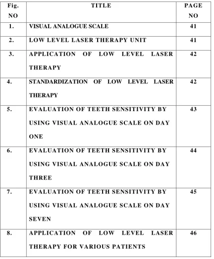
1. VISUAL ANALOGUE SCALE 41
2. LOW LEVEL LASE R THE RAPY UNIT 41
3. APPL ICATIO N O F LO W LEVEL LASER
THERAPY
42
4. STANDARDIZATION OF LOW LEVEL LASER
THERAPY
42
5. EVAL UAT ION O F TE ETH SENSIT IVI TY B Y
USING VISUAL ANALO GUE SCALE O N DAY
ONE
43
6. EVAL UAT ION O F TEETH SENSIT IVI TY B Y
USING VISUAL ANALO GUE SCALE O N DAY
THREE
44
7. EVAL UAT ION O F TEETH SENSIT IVI TY B Y
USING VISUAL ANALO GUE SCALE O N DAY
SEVEN
45
8. APPL ICATIO N O F LO W LEVEL LASER
THERAPY FO R VARIOUS PATIE NTS
[image:9.595.84.517.133.656.2]LIST OF TABLES
Table
No TITLE
Page
No
1. Visual analogue scale scores 49
2. Descrip tive S tatis ti cs. 51
3. Mann -Whi tney Test to comp are val ues b etw een
Groups .
52
4. Wilcoxon Sign ed Ranks T es t to compar e valu es
betw een Before and After laser.
52
5. Fri ed man test for rep eated measu res to comp are
before las er values betw een 1, 3 and 7 Days .
52
6 Bonferron i adjus ted Wilcoxon Signed Ranks tes t to
compare values b etween p airwise ti me points .
53
7. Fri ed man test for rep eated measu res to comp are
after las er values b etw een 1, 3 and 7 Days.
53
8. Bonferron i ad jus ted Wil coxon Sign ed Ranks tes t to
compare values b etween p airwise ti me points .
LIST OF CHARTS
Chart
No TITLE
Page
No
1. Mean valu es in T est Group . 54
2. Mean valu es in Con trol Grou p . 54
3. Before and after laser th erapy mean values in
test & control grou p.
Introduction
1
INTRODUCTION
Dentin h ypers ensit ivit y is defined as an exaggerated res pons e to a stimul us that usual l y caus es no respons e in a health y t ooth( 1 , 2 ).Denti n h ypersensiti vit y i s a comm on probl em for whi ch t here was no effective soluti on. Abel characterized t he denti n h ypers ensi tivit y as a short , sharp pain arisi ng from exposed dentine i n res ponse to stim uli t ypi call y evaporative, t actil e, or chemical an d that cannot be des cribed as an y other form of dent al pathol ogy( 3 ). It is preval ent among t he large port ion of indi viduals aged 30 -40 yrs. The most comm on factors responsibl e for dent in h ypersensiti vit y are abrasi on, caus ed by tooth brushing wi th inadequa te i nt ensit y and abfraction, caus ed b y tooth flexion ass ociat ed with ill -di rect ed occlus al forces , parafunctional habits or occl us al di sequilibrium and erosion, as an effect of aci ds in the oral cavit y and anatom ic predi sposit ion, due to structural defi cie nc y in the cement enam el j unction and cavit y preparations in teeth with vit al pul p that expos e the dentin and Tooth preparation for fixed parti al dentures would al so causes sensitivi t y of prepared teeth.
Introduction
2
together with other caus es it l eads to loss of dental enamel in the cervical area and cons equentl y l eads t o dentin h ypers ensit ivit y. The erosive agents are probabl y responsibl e for st arti ng the sensit ivit y due to the opening of the dentinal tubul es. The noci cepti ve stimulus comm onl y report ed i n majorit y of the cas es i s that of cold, fol lowed b y the m echani cal s t imulus of tooth brushing and the chemi cal sti mulus of diet with a hi gher concentrati on of s ugar.18% of the adult popul ation suffer from thi s problem, and the hi gher i nci dence occurs in prem ol ars, foll owed b y t he cani nes , incisors and m olars and the vestib ul ar area is most comm onl y affect ed( 5 ).
Introduction
3
plas ma proteins and sali va constit uents, as well b y active m echanis ms such as deposit of intra canalicul ar cr ystalline m at eri al and secreti on of prot ei n m at eri al from the int erior of t he tubul es , thereb y diminishing dentinal perm eabili t y and sensiti vit y. However, h ypers ensi tivit y sometim es remai ns i n spit e of the effective blocking of the tubules , suggesti ng that other mechanisms cont ribute to nerve act ivati on i nst ead of or in addit ion t o the h ydrod ynami c m echani sm. Thi s parti all y explai ns the l arge sensitivi t y variati on of exposed dentin and furthermore, nerve activati on ma y result in the rel ease of neuropeptides from the activat ed nervous terminations and, cons equent l y induce neurogeni c inflammation. The s ym ptoms of dent in h y pers ensi tivit y up to a cert ain poi nt would be s elf -sust ainable.
Introduction
4
These t reatm ents are performed to bl ock t he h ydrod ynamic mechanism b y t ransmitting stimuli to the dent in, and closing the dentinal tubul es( 7 ). Man y treatm ents wit h topi cal products i n form of dental cream, m out hwash, and varnishes have been offered t o t he popul ati on i n an att empt sol ve the probl em. Man y l iteratures supports the appli cation of dentin adhesive( 8 ), propolis app li cation( 9 ) and the us e of si lver nit rate sol ution on cervical denti n, whi ch were considered effective for reducing denti n h ypersensiti vit y( 1 0 ).
Introduction
5 Low level las er therapy
Low l evel l as er therap y ( LLLT) was i niti all y utiliz ed i n dentis t r y to accelerate wound healing, mini mize pain, and t o reduce infl amm ator y responses. Low l evel las ers have been widel y investi gat ed due to thei r lower cost s compared wi th ot her l as ers, and thei r simpl ici t y of use. The first low -l evel laser int roduced was heli um - neon (He -Ne), which com bined a gas eous mixture to produce a wavelength in the visible light spectrum (λ = 632.8 nm) and low power
output (ranging from 5 to 30 mW). Since the wavel ength produced b y He-Ne l as er was hi ghl y abs orbed b y soft tiss ues, its penetration was limited.
Introduction
6
Review of Literature
7
REVIEW OF LITERATURE
1. Aldo Brugnera et al (2000) di d a st ud y on las er t herap y i n the treatm ent of dent al h ypersensiti vit y and concl uded t hat , LLLT used wi th appropri at e treatm ent paramet ers is effective in treati ng dentin h ype rs ensit ivit y as it quickl y reduces pain and maint ai ns a prolonge d pain free st at us in 91.27% of the 1102 cases studi ed. (WALT, speci al millinneum editi on 2000,l as er therap y vol 12,16 -21.)
2. Karen C risti na et al(2003) di d a stud y on, low l evel l as er therap y for dent in h ypersens itivit y and concl uded that t he treatm ent with Ga-Al-As l as er was effective for reducing denti n H ypers ensit ivit y.( C i encOdont ol Bras 2003 out./dez.; 6 (4): 17 -24.)
3. Analuci a Marsi lio et al (2003) di d a st ud y on, effect of cli nical applicati on of the Ga -Al-As l as er i n t he t reatm ent of dentin h ypersensiti vit y and concl uded that, t he initi al DH was reduced aft er treatm ent wi th the low level Ga -Al-As laser. The di fference bet ween ini tial dentin h ypers ensi tivit y and dentin h ypersensiti vit y at 60 da ys pos t -t reatment was st ati sti call y si gni fi cant.( J ournal of Clini cal Las er Medici ne & Surger y Volum e 21, Number 5, 2003).
Review of Literature
8
decades of developm ent up to the present time.(Aust rali an Dental J ournal 2003;48: 3)
5. Ana R aquel Benetti et al (2004) did a st ud y on las er t herap y f or dentin h ypers ensitivit y; a cri tical apprai sal and concl uded t hat , the avail abl e evidence i s not consist ent , and thus cannot prove the effi cac y of l as er therap y i n the m anagem ent of h ypers ensit ive dentin.(J oral l as er appli cat ions 2004; 4:271 -278.)
6. Ahm et Eralp et al (2006) di d a st ud y on, a clinical i nvesti gat ion of low l evel l as er irradiati on on h ypers ensi tive denti n and concl uded t hat the action mechanism of low output l as ers are unclear, t he results of the st ud y indicat ed that t he y were ver y effective i n denti n h ypersensiti vit y and resist ant to m echanical forces and chemical irri tat ion.( Cil t: 30, Sa yı: 2, Sa yfa: 94 -99, 2006).
7. Aless andra Buhl er et al (2012) did a lit erat ure revi ew on dentin h ypersensiti vit y – etiology , t reatm ent possi biliti es and ot her rel at ed factors and concluded that, the adhesive s yst ems are one of t he mos t effect i ve cli nical treatm ents and the l as ers are expect ed to pl a y an import ant role in treating dentin h ypersensiti vit y.(World Journal of Denti str y,J an -Mar 2012; 3(1)60 -67).
Review of Literature
9
com bination wit h NaF gel.( Int ernational J ournal of Denti str y Volum e 2012, Arti cl e ID 858950, 8 pages ).
9. Mohamm ad Asnaashari et al (2013) di d a stud y on appl icati on of low l evel l as ers in denti str y(endodonti c) and concluded t hat , l ow level l asers are ver y effecti ve in dent al applicati ons .(J ournal of Las ers i n M edical S ciences Vol ume 4 Number 2 Spring 2013). 10. Mohamm ad Ali Ansari (2013) did a st ud y on m echanisms of
las er -ti ssue i nt eraction: 11.tiss ue therm al properti es and concl uded that ,cancer pati ent s consum e less ox ygen an d gai n heat at a hi gher rat e than the non -cancer pati ent s.( J ournal of Las ers i n M edical S ciences Vol ume 4 Number 3 Summ er 2013). 11. Rola et al (2014) did a stud y on t he us e of low level energy l aser
radi ation in basi c and clini cal research and concl uded that , LLLT holds promis e as a novel supportive t ool in the treatm ent of wounds and chroni c pai n s yndrom es .( AdvClinExp M ed 2014,23, 5, 835– 842)
12. Vartika Kathuri a et al (2015) did a st ud y on low level l as er therap y; a panacea for oral m aladi es and concluded t hat , LLLT can prove to be an effective t reatm ent m odalit y for vari ous oral mal adi es provided t hat the clini ci an t akes proper t rai ning and adopts necess ar y safet y meas ures.( Laser Therap y 24.3: 215-223) 13. Eloss ais Andre Afi f et al (2015) did a stud y on, in vivo
Review of Literature
10
dentin h ypers ensit i vit y after t hree s essi ons and s tat ist ical si gni fi cant di fferences were found between the groups . Group 1 (Des ens ibiliz e) present ed 90% effi cienc y and Group 2 (Whit ening Laser II) 95% effi ci enc y.(S cienti fi c J ournal of Dentist r y Vol. 2:4 Jul -Aug 2015.)
14. Snehal A, Naik et al (2016) did a cri tical revi ew on , las er therap y in the m anagem ent of denti n h ypers ensitivi t y and concl uded that laser therap y is effecti ve.(Univers al Res earch J ournal of Denti str y Sept -Oct 2012, Vol 2 , Issue 3.)
Materials and Methods
11
MATERIALS AND METHODS
Clini cal case s el ecti on:
Thi rt y pati ents from the Departm ent of Prosthodont ics, Adhi paras akt hi D ental C ollege and Hospi tal M elm aruvathur, Tami lnadu, who required conventi onal fixed parti al dent ure were sel ected i n random for t he st ud y purpos e.
Inclusi on criteria for abu tmen t s el ection
Subject s who requires fixed parti al prosthesis
Subject s with vit al t eeth
Subject s with adequate crown root rati o
Subject s with good peri odont al health
Exclus ion criteria for abutment s electi on
Subject s with non -vi tal t eeth
Subject s with i nadequat e crown root rat io
Subject s with poor peri odont al health
Note: Periodontal health to be assessed using Russel‟s
periodont al Index.
Evaluation of s ensi tivity of teeth :
Materials and Methods
12
regist ered on a fill ing sheet whi ch had the Visual Anal ogue S cale [VAS](fi gure 1).
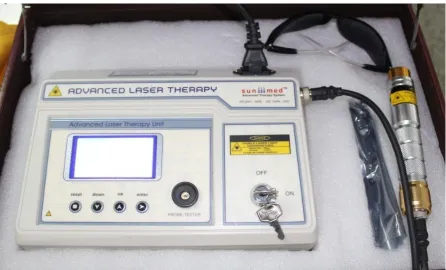
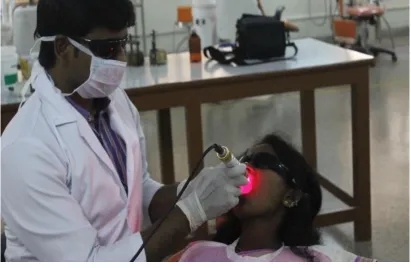
Low level las er therapy uni t:
The laser unit used was: Class ШB laser [sun med] and the probe
has Ga-Al-As diode that emit s coherent l ight at the RED wavel engt h of 685 nm. It deli vers maximum energy of 3 joule/cm ² within one minute (fi gu re 2).
Precauti ons and S afet y features t o be foll owed:
1. During l as er appli cat ion bot h the patient and t he operator should wear the prot ect ive goggl es.
2. Before l as er appli cation the operator should ens ures t hat all the connecti on and power s uppl y to the l as er unit was kept read y 3. Place the las er probe perpendi cul ar t o the prepared t eeth for
effective i rradiat ion.
4. Focus the l as er probe on parti cular surface to be treat ed, so t hat unneces sar y irradi ati on of t he adj acent s urfaces were prot ect ed. 5. During l as er thera p y laser beam should not pass es t hrough an y
met als.
6. During the l as er appl ication the probe tip shoul d st a y in t he same positi on for speci fi ed tim e period.
Materials and Methods
13 Procedu re:
Toot h preparati on was done b y conventi onal m ethod. It implies that the tooth preparati on procedures for all t ype of fixed parti al dentures are comm on, but onl y t he amount of toot h reducti on vari es. So the amount of tooth reduct ion for all t ypes of fixed part ial dent ures have mini mum of 1 mm, therefore aft er tooth preparati on patient m a y feel the s ensitivi t y of prepared tooth. Of the two abutm ents , one abutment was s elect ed b y B LOC K RANDOM IZAT ION METHOD and incl uded i n the t est group. The other abutment acts as t he cont rol . Tot all y out of 60 abutments, 30 abut ment s were grouped into t est group, anot her 30 abutments i n t he cont rol group.
BLOCK RANDOMI ZATIO N METHO D:
Materials and Methods
14
sizes. Confounding distort s the stat i stical validit y of stati sti cal inferences about caus e and effect. The failure to control for confounding m a y i nfl at e t ype 1 error and erroneous l y l ead to the concl usion that a putati ve risk factor i s causal l y ass ociat ed wit h t he out com e under st ud y (false positive fi nding). A chance run of parti cipant s to a particul ar st ud y group also m a y occur under a sim pl e random izati on s cenario. This can l ead to bias, for exam pl e, i f the initi al partici pants i n the trial are heal thier than the l at er ones( 2 4 ). Blocked r andomizat ion offers a simpl e means to achi eve bal ance bet ween stud y arm s and t o reduce t he opport unit y for bias and confounding.
Materials and Methods
15
chosen( 2 7 ). Sel ection bias ma y be reduced b y using random block sizes and keeping the investi gator bl ind to t he size of each block.
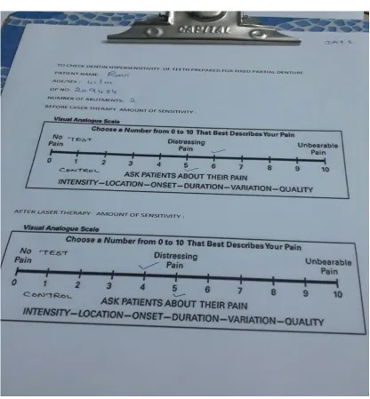
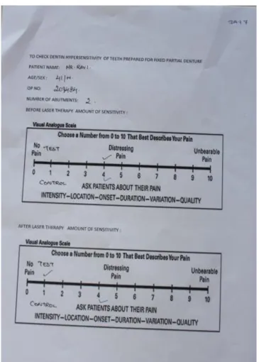
Testing for S ensi ti vity:
Once the t ooth preparati on procedure was com pleted, t hen sensitivi t y of t est group abutm ent t eeth was recorded b y usi n g visual analogue scal e (VAS ) before appli cation of l as er on s el ect ed abutment teeth. S ensi tivit y degree of prepared abutment was t est ed t hrough t he probe b y running t he probe ti p on prepared teeth s urfaces from gi ngi val to occlus al or incis al as pect wit h gent le finger press ure. Bas ed on t he subj ective answer of the pati ent , scores from 0 to 10 were attribut ed, thes e val ues were regis tered on a filling s heet whi ch had t he Visual Analogue S cale [VAS] (fi gure 1). The perceived dis com fort for each tooth was gr aded for each of stim uli b y using a 10cm VAS, labell ed at the two extremes with „no pain‟ at the zero extreme and with „unbearable pain‟ at the 100mm extreme.
Application of L as er:
Materials and Methods
16
Low l evel l as er therap y was perform ed on s elect ed t es t group abutment t eeth i n non cont act continuous mode wi t h 12 joules / cm² energy for 4 mi nut es (fi gure 4). The procedure was repeat ed aft er da y three and da y seven for all t hirt y pat ients and t he s core were t abulat ed.
Placeb o:
The procedure for pl acebo group i s sim il ar to t hat of t est group. The onl y difference being that the pl acebo abutm ent was exposed t o point er li ght without las er energy. The subject thinks t hat both the teeth are treated. Then the subject is eval uat ed for denti n h ypersensiti vit y and the results were recorded (fi gure 5).
Results
17
RESULTS
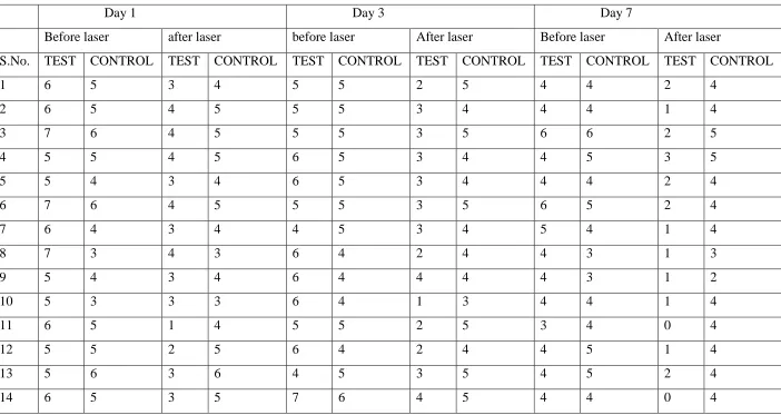
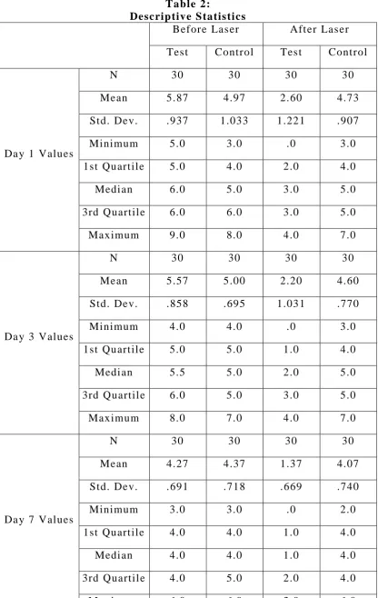
Table 1 s hows the VAS s cores of the tooth h ypersensiti vit y of the subj ects. Tabl e 2 shows t he des cri ptive st atisti cs of t he data.
On da y 1 before laser therap y t he mean s core for t es t group was 5.87 wit h a minim um score of 5.0 and a maximum s core of 9.0 and the placebo s howed a m ean s core of 4.97 wi th a mini mum s core of 3 and a maximum s core of 8.0. Aft er laser therap y the mean score for t est group was 2.60 with minimum s core of 0.0 and a m aximum s core of 4.0 and the pl acebo s howed a m ean s core of 4.73 with minim um s core of 3.0 and a m aximum s core of 7.0.(chart1&2)
On da y 3 before laser therap y t he mean s core for t es t group was 5.57 wit h a minim um score of 4.0 and a maximum s core of 8.0 and the placebo s howed a m ean score of 5.00 wi th a minim um score of 4.0 and a m aximum score of 7.0. Aft er l as er therap y t he m ean s core for t est group was 2.20 with minimum s core of 0.0 and a m aximum s core of 4.0 and the pl acebo s howed a m ean s core of 4.60 with minim um s core of 3.0 and a m aximum s core of 7.0.(chart 1&2)
Results
18
Statistical Analysis
19
STATISTICAL ANALYSIS
Discussion
20
DISCUSSION
Dentin h ypers ensitivit y (DH) is a comm on probl em fol lowed by tooth preparation for fixed parti al denture. H ypers ensitivit y is an abnorm al response of the exposed vit al denti n to therm al, chemical, or tactil e st imuli . This condition m a y affect patients of a ll ages, and both genders are equal l y affect ed. Al though different t heori es have been propos ed for the mechanism involved in denti n h ypersensitivi t y etiology, recent studies gave support to Brannstrom‟s hydrodynamic
theor y( 2 8 ).According to this a stimul us appli ed to open tubul es denti n increases the fl ow of dentinal tubular flui d, wit h mechani cal deform ation of the nerves locat ed into t he inner ends of the t ubul es or in the outer l a yers of the pulp( 2 9 ). T ype A delt a fibers are suppos ed to be res ponsibl e for denti n sensitivit y bei ng probabl y activat ed b y t he h ydrod ynami c proces s( 3 0 ).
There are so m an y t reatm ent of choice are avail abl e for treat ing dentin h ypersensiti vi t y, whi ch i ncl udes des ensi tizing agents, and us e of las ers. Grossm an l isted the requi rem en ts for an ideal dentine des ensitizing agent as: rapi dl y acting with long t erm effects, non -irritant to pulp, pai nles s and eas y to appl y, and shoul d not stain the tooth.
At home des ensi ti zi ng th erapy
These “at home” desensitizing agents include toothpaste s,
Discussion
21
cit rat e), sodium fluoride, strontium chl oride, dibasi c sodi um citrat e, formaldeh yde, s odi um mono -fl uorphos phate and s tannous fluori de. Potas sium s alts act by diffusion along t he dentinal tubules and decreasi ng t he exci t abili t y of t he int ra dent al nerve fibers b y blocki ng the axonicaction( 3 2 , 3 3 ). Vari ous clini cal st udi es have s hown the effi cac y of pot as sium s alts i n con trolling t he dentin h ypers ensitivit y( 3 4 , 3 5 ). It has been shown that toothpast es contai ning 5% pot assi um ni trate and 0.454%stannous s i gnifi cantl y reduced the dentin h ypers ensitivi t y. Also, toothpast es contai ning pot ass ium nitrate and fluorides have been shown to reduce post -bl eachi ng sens it ivit y( 3 6 , 3 7 ). The des ensi tizing toothpastes s houl d be us ed wit h the help of a toothbrush wit h soft brist les. P ati ent s s hould be advi sed t o us e mi nim al amount of wat er to prevent t he dilut ion of the active agent . Along wi th the des ensi tizing toothpastes, mouthwashes and chewing gums cont aini ng potas sium nitrat e, s odi um fluoride or pot assi um cit rat e are also recomm ended( 3 1 ). The results of “at-home” desensitizing therapy should be reviewed
aft er ever y 3 –4 weeks. If there is n o rel ief in dentin h ypers ensi tivit y, “in-office” therapy should be initiated.
In-office d es ensi ti zi ng agen ts :
Discussion
22 Fluorides
Discussion
23
expos ed dent ine( 4 4 ). Fluorosili cat es act by form ation of preci pitat es of calci um phosphat es from sal iva. Am m onium hexa fluorosi licat e has been used as a des ensitizing agent . It can pres e nt a conti nuous effect of dentinal tubul e occl usion via precipit at ion of a mixture of cal ci um fluori de and fluoridat ed apatit e( 4 5 , 4 6 ). If the precipitat e is predom inantl y composed of fluori dated apatit e, it can form stabl e cr ys t als depos ited deep i nside th e dent inal tubules( 4 5 , 4 6 ). Thes e cr ys t als are res ist ant to removal from the action of sal iva, brus hing or action of diet ar y substances .
Oxalates
Discussion
24
dentinal perm eabilit y. This can al so be foll owed b y coveri ng the expos ed surface wi t h a dent al adhesive( 4 9 ).Potassium oxal at e can l ead to gast ric i rrit ation. Therefore, it should not be us ed wit h a tra y wi th prol onged pl acement. Varnishes are common l y us ed us eful in -offi ce measures to t reat dentin h ypersensiti vit y. Copal varnis h can be applied to cover t he expos ed dentinal surface. But its effect i s for short term and is not recom mended for long term managem ent of denti n h ypersensiti vit y( 5 0 ). To improve it s effi cac y, removal of sm ear l a yer is advocat ed. Als o, the varnishes can act as a vehi cl e for fluoride. The fluori de varnishes can be aci dul at ed to increas e the penet rat ion of ions( 5 0 ).
Adhes ive materials
Discussion
25
some dentin bonding agents have been introduced in the m arket wit h the s ol e purpose of t reati ng dentin h ypers ens itivit y. Gl uma Desensitiz er (HeraeusKulz er, Hanau, German y) cont ains h y dr ox yl et h yl methacr yl ate (HEM A), benz alkonium chl ori de, gl ut eraldeh yde and fluori de. Glut eral deh yde causes coagul ation of the prot ei ns inside t he dentinal tubules( 5 4 ). It react s with the s erum al bumi n in the dentinal flui d, causi ng it s precipit ation. HEMA forms deep res inous tags and occludes the denti nal tubul es( 5 4 ). Glum a has s hown promis ing results in the cli ni cal t rials( 5 4 , 5 5 ).
Bioglass
Biogl ass was devel oped to st imul at e the form ation of new bone( 5 6 ). It is us ed in ort hopedi cs to cover the impl ant s to prom ot e union between impl ant and bone( 5 6 , 5 7 ). It has been used i n dentist r y t o fill up the oss eous defect s duri ng peri odont al surger y( 5 8 ). It has been report ed t hat a formulat ion of biogl as s can promot e i nfi lt rat ion and rem ineraliz ation of dentinal tubul es( 5 9 ). The basic com ponent is silica, whi ch acts as a nucl eation sit e for precipit ation of cal cium and phos phat e. SEM anal ys is has s hown that biogl ass applicati on forms an apatit e l a yer, whi ch occludes t he dentinal tubul es( 5 9 ). The use of biogl ass in m anagement of denti n h ypers ensit ivit y has been shown b y some products s uch as NovaMin (NovaM in Technology Inc., FL, USA).
Portl and cement
Discussion
26
h ypersensiti vit y( 3 1 ). It hel ps to occl ude t he dent inal t ubul es b y rem ineraliz ation.
Cas ein ph osphor p eptide –amorphous calciu m phosph ate
Recentl y, milk protein casein has been used to devel op a rem ineralizing agent (GC Tooth Mousse). The cas ein phosphor pepti de (CPP) cont ains phosphos er yl sequences whi ch get attached and stabilized wi th amorphous cal ci um phosphat e (ACP )( 6 0 ). The stabilized CPP–ACP prevents t he di ssol ution of cal cium and phosphat e ions and maint ai ns a supers aturated solut ion of bio availabl e cal cium and phos phat es( 6 0 ). Vari ous st udi es have shown that CPP – ACP can effectivel y remineral ize the enam el subs urface lesions( 6 1 , 6 2 ). B y virt ue of its remineralizing capacit y, it has al so been propos ed b y the manufacturers that it can also help in prevention and t reatm ent of dentin h ypers ensit ivi t y.
Laser
Discussion
27
Most of t he t reatm ents tested have ai med t o bl ock exposed dentinal t ubules , but none of thes e t reat ments has produced consist entl y effective or l ong l asting resul ts. S o to overcome this problem, low l evel l as er t herap y was t ri ed in the m anagem ent of dent in h ypersensiti vit y fol lowed b y toot h preparation fo r fixed partial denture. It is necess ar y to consider the s everi t y of t he denti n h ypersensiti vit y before using the l aser and the effi ci enc y of the us e of las er for denti n hypersensitivi t y t reatment is hi gher t han ot her methods .
Discussion
28 Pres ent s tudy:
In thi s stud y Ga -Al-As l aser was us ed t o treat s ensitivit y of t eeth prepared for fixed parti al dent ure. In da y one aft er l as er irra diat ion most of t he t est group samples showed sli ght reducti on in s ens itivit y of prepared t eeth, but i n control group there was no si gni fi cant reduction in s ensit ivit y of prepared t eet h (tabl e 2). On da y t hree, after l as er applicati on there was no change i n the control group s ampl es but in test s ampl es t here was much bett er reduction in s ensi tivit y of prepared teeth when compared to da y one (t abl e 3). In da y s even after laser applicati on again control group sampl es showed no change in amount of sensiti vit y and i n test group sampl es showed great reduction in sensitivi t y when compared t o da y three(tabl e 3). So i n t est group aft er las er appli cation sensitivi t y of prepared teeth gradual l y decreas ed i n da y b y da y appoi ntm ents .
Summary
29
SUMMARY
The analysis and values obtained from the clinical study can result in the following findings;
On day one, application of low level laser therapy on sensitivity tooth prepared for
the fixed partial denture resulting in immediate relief.
On da y three, aft er l as er appli cation there was much bett er
reduction i n s ens iti vit y of prepared teeth when compared to da y one.
On da y s even, aft er laser applicati on i t showed great reduction i n
sensitivi t y when compared to da y three. So in t est group aft er l as er applicati on s ensi tivit y of prepared t eeth graduall y decreased in da y b y da y appointm ent s .
On comparing da y one, thr ee and seven control group showed no
Conclusion
30
CONCLUSION
Limitations of the Study
31
LIMITATIONS OF THE STUDY
We st andardiz ed the values of mW, j oul es/minut e, mode of
applicati on, and the durati on period for all the subj ects . Effect of change of t hese param eters on sensitivi t y of the tooth can be further expl ored.
Obj ective t ype of data requi res rather than subjecti ve dat a. This
stud y was bas ed on subj ective evaluati on of the pati ent. More robust resul ts can be seen i f more s ubjective eval uation was done.
Long term foll o w up studi es are requi red further to conclude the
References
32
REFERENCES
1. Fl ynn J , Gall owa y R, Orchands on R. The inci dence of “hypersensitive” teeth in the west of Scotland. J Dent 1985; 13
(3): 230 -6.
2. Bis sada NF. S ym ptom atol ogy and clini cal feat ures of h ypersensiti ve t eet h. Arch Oral Bi o l 1994; 39 (12): 31S -32S. 3. Abel I. Stud y of h yp ersensiti vit y t eet h and a new therapeuti c aid.
Oral S urg Oral Med Oral P at hol 1958; 11 (5): 491 -5 .
4. Chris tens en GJ . Desensi tizati o n of cervi cal tooth st ructure. J Am Dent Assoc 1998; 129 (6): 765 -6.
5. Wichgers TG, Emert R L, Denti n h yp ersensiti vit y. Gen Dent 1996; 44: 225 -230.
6. Branst rom M, Li nden LA, As trom A. The h ydrod ynami cs of the dental tubul e and of pulp fluid: a dis cus si on of its s i gni fi cance in rel ati on to dentinal s ensi tivit y. C aries R es 1967;1:310 -317.
7. Add y M, West N. Etiology, mechani s ms, and m anagem ent of dentine h ypersensiti vit y. CurrOpi nPeriodontol 1994; 71 -7.
8. Ide M, Morel AD, Wilson R F, As hley FP . The rol e of a dentinebondi n g agent in reducing cervi cal denti ne s ensitivi t y. J ClinPeriodontol 1998; 25 (4) 286 -90.
References
33
10. Tou yz LZ, Stern J . H ypers ensit ive denti nal pain att enuati on with pot assium ni trat e. Gen Dent 1999; 47 (1): 42 -5.
11. Aun CE, Brugnera Junior A, Vil la RG. Rai o las er: hipers ensibil idade denti nari a. Avali aç ao clíni ca de paci entes port adores de hipersensibili dade dentinari a cujos dentes foram tratados com rai o Laser Héli o -Neon. Rev Ass Paul Ci r Dent 1989; 43 (2): 65 -8.
12. Lan WH, Li u HC. Treatment of denti n h ypers ensit ivit y b y Nd: YAG l as er. J Clin Med S urg 1996 ; 14 (2) 89 -92.
13. Gutknecht N, Morit z A, Derecks HW, Lam pert F. Treatm ent of h ypersensiti ve t eeth using neod ym ium: yttrium -alum inum -garnet las ers: a compari son of the use of various sett ings in an in vivo stud y. J Clin Las er Med Surg 1997; 15 (4):171 -4.
14. Perei ra J C. Hiperest esi a dentinari a as pectos clini cose form as de tratam entos . M axi -Odonto: dent isti ca 1995; 1 (2): 1 -24.
15. Midda M, R enton -Harper P. Las er in dentist r y. Br Dent J 1991, 170 (9): 343 -6 .
16. Eduardo CP, Cecchi ni RC, C ecchini SC M. The usage of las er i n dentist r y [ Abst ract 0506 -13]. Ph ys M ed Biol 1994; 39 (1): 139. 17. Benedi centi A. Manual e di laser terapi a del cavooral e. Cast ello:
Maggioli 1982. 159 p.
References
34
19. Furuoka M, Yokoi T, Fukuda S, Usuki M, Matsuo S , Tani guchi K, et al. Effects of GaAlAs l as er diode in treatm ent of H ype rs ensit ive dentine. Fukuoka Shi kaDai gakuGakkai Zas shi 1988; 15 (1): 42 -8.
20. Pass arel i Neto A. C ontribui çao ao es tudo da apli caçao do Soft las er no t rat am ent o da hipersensibi l idade dentinaria. Sao Bernardo do C am po; 1998. [Di ss ert açao de M est rado em Dentisti ca R est auradora – Faculdade de Odontol ogi a, Universidade M etodi sta de S ao P aul o].
21. Gers chm an J A, R uben J , Gebart -Eagl emont J . Low l evel las er therap y for denti nal tooth h ypersensitivi t y. Aust Dent J 1994; 39 (6): 353 -7.
22. Groth E.B. Cont ribuiçao para o est udo d a apli caçao de l as er de baixapot enci a de GaAl As no t rat am ent o da hipersensibilidade dentinári a. Sao P aulo; 1993. [Di ss ert açao de M est rado em Dentísti ca – Faculdade de Odontologi a da Universi dade de S ao Paulo].
23. Kimura Y, Wil der -S mith P, Yonaga K, Mats umot o K. Treat ment of denti ne h ypers ens itivit y b y lasers: a review. J ClinPeriodontol 2000 Oct.; 27 (10): 715 -21.
24. Matts , J . Lachin, J . Properti es of permut ed -block randomizat ion in clini cal t ri als. Control Clin.T rials1988, 9, 327 -344.
References
35
McKinl a y, S., Eds.; Wolters Kl uwer: P hiladel phi a, PA, US A, 2009; Chapt er 3, p. 27.
26. Lachi n, J . Properti es of simple randomi zation i n cli nical t ri als. Control Clin. Tri al s 1988, 9, 312 -326.
27. Efron, B. Forci ng a sequenti al experi ment to be bal anced. Biomet rika1971, 58, 403.
28. B. Matthews and N. Vongsavan, “Interactions between neural and hydrodynamic mechanisms in dentine and pulp,” Archi vesof
Oral Biology, vol . 39, no. 1, pp. S87 –S95, 1994.
29. M. Brannstr¨om, “A hydrodynamic mechanism in the transmi ssion of pai n -producing st imuli through denti ne,” in
Sensor y Mechanis m in Denti ne, D. J . Anderson, Ed., pp. 73 –
79,P ergamon, Oxford, UK, 1963.
30. T. C. C. G. P . Ladalardo, A. Pinhei ro, R. A. D. C. Cam pos et al,“Laser therapy in the treatment of dentine hypersensitivity,”
Brazi lian Dent al Journal, vol. 15, no. 2, pp. 144 –150, 2004.
31. Orchards on R, Gil li am D. M anaging dentin h ypersensiti vit y. J Am Dent Assoc 2006;137: 9908.
32. Markowitz K, Bilott o G, Kim S. Decreasing intra dent al nerve activit y in the cat wi th potas sium and dival ent cations. Arch Oral Biol 1991; 36: 1 -7.
References
36
34. Hodosh M. A superi or des ensit izer: Pot assium nit rate. J Am Dent Assoc1974;88: 831 -2 .
35. Frechoso SC , M enendez M, Gui sasol a C, Arregui I, Tejeri na J M, Sicili aA. Eval uat ion of the effi cac y of t wo potassium nit rat e bio adhesi ve gels (5% and 10% ) in the treat ment of dentine h ypersensiti vit y: A random ized clini cal tri al. J ClinP eri odontol 2003; 30: 315 -20.
36. Schi ff T, Zhang YP, DeVizio W, Stewart B, Chaknis P, Pet rone ME, et al. Arandomized cli ni cal t rial of the des ensitizi ng effi cac y of three dentifr i ces. Compend Contin Educ Dent S uppl 2000; 27: 4 -10.
37. Sowinski J A, Batti st a GW, P et rone ME, Chaknis P, Zhang YP, DeVizio W,et al. A new desensitizing denti frice: An 8 -week clini cal i nvesti gati on. C ompend Contin Educ Dent S uppl 2000; 21: 11 -6.
38. Paine M L, Slot s J , Rich SK. Fl uoride us e in peri odont al therap y: A review of the li terature. J Am Dent As soc 1998;129: 69 -77. 39. Morris M F, Davis R D, Richardson BW. Clinical effi cac y of two
dentin desensitizing agents. Am J Dent 1999;12:72 -6.
40. Leonard RH J r , Smit h LR, Garl and GE, C apl an DJ . Desensitiz ing agent efficac y during whitening in an at -risk popul ation. J Es thet Restor Dent 2004;16: 49 -55.
References
37
42. Gangaros a LP, Park NH. Pract ical consi derations in iontophoresis of fl uoride for des ensitiz ing dent in. J Prost het Dent 1978;39: 173 -8 .
43. Thras h WJ , Dodds MW Jones DL. The effect of st annous fluoride on dentinal h ypers ensitivit y. Int Dent J 1994;44:107 -18.
44. Morris M F, Davis R D, Richardson BW. Clinical effi cac y of two dentin desensitizing agents. Am J Dent 1999;12:72 -6.
45. Suge T, Kawas aki A, Ishi kawa K, Mat suo T, Ebisu S. Ammoni um hexa fluorosili cate eli c its calci um phos phate precipit ati on and shows continuous denti n tubul e occlus ion. Dent Mat er 2008; 24: 192 -8 .
46. Suge T, Kawas aki A, Is hikawa K, M ats uo T, Ebis u S. Effect of ammonium hexa fl uorosil icat e on denti n t ubul e occl usion for t he treatm ent of dentin hyp ers ensitivi t y. Am J Dent 2006;19:248 -52. 47. Pillon FL, Romani IG, S chmi dt ER. Effect of a 3% pot as s ium
oxalat e t opi cal appl i cation on dentinal hypers ensitivi t y after sub gi ngi val s caling and root pl ani ng. J Periodontol 2004; 75: 14614. 48. Hongpakm anoon W , Vongs ava n N, S oo-ampon M. Topi cal
applicati on of warm oxalat e t o exposed human dentine In vi vo. J Dent R es1999;78: 300.
49. Sauro S, Gandol fi MG, P rat i C, Mongiorgi R. Oxal ate -contai ning ph ytocompl exes as denti ne desensitiz ers: An In vi tro st ud y. Arch Oral Bi ol 2006; 51: 655-64.
References
38
51. Duran I, S engun A. The l ong -t erm effect ivenes s of five current des ensitizing products on cervi cal dentine s ens itivit y. J Oral Rehab2004;31: 351 -6 .
52. Prat i C, C ervell ati F, S anasi V, M ont ebugnoli L. Treatm ent of cervical dentin h ypersensiti vit y with resin adhesives: 4 -week evaluati on. Am J Dent 2001;14:378 -82.
53. Ba ys an A, Lynch E. Treatment of cervi cal sensiti vit y wit h a root seal ant. Am J Dent 2003;16: 135 -8.
54. Dondidall‟Orologio G, Lone A, Finger WJ. Clinical evaluation of the rol eof glut ardi al deh yde in a one -bott le adhesive. Am J Dent 2002; 15: 330 -4 .
55. Dondidall‟Orologio G, Lorenzi R, Anselmi M, Grisso V. Dentin des ensitizing effects of Gl um a Alternate, Hea lt h-Dent Desensitiz er and Scot chbond Multi -Purpos e. Am J Dent 1999; 12: 103 -6 .
56. Hench LL, Splinter RJ , All en WC, Greenl ee TK. Bonding mechanism s at int erface of ceramic prostheti c m ateri als . J Biomed M at er R es S ym p1971; 2:117 -41.
57. Hench LL, P as chall HA. Di rect chemi cal bond of bioactive gl ass -cerami c m at eri als to bone and m us cl e. J Biomed M at er R es S ym p 1973; 4:24 -42.
References
39
59. Fors back AP, Areva S, Salonen J I. Mineralization of denti n induced b y treatm ent wit h bioactive gl as s S53P 4 In vi t ro. Act aOdontolS cand2004;62:14 -20.
60. Re ynolds EC. R emi neraliz ation of enam el subs urface l esi ons b y casein phos phopepti de -st abiliz ed cal cium phosphat e s oluti o ns. J Dent R es1997;76: 1587 -95.
61. Cai F, Shen P, Morgan M V, R e ynolds EC. Rem ineraliz ation of enam el s ubs urface lesions in-sit u by sugar free lozenges contai ning cas ein phosphopeptide – amorphous calcium phos phat e. Aust Dent J 2003; 48:240 -3.
62. Lat a S, Varghes e NO, Varughes e J M. Remineralizati on pot ential of fluoride and am orphous cal cium phosphat e -cas ein phos pho peptideon enam el lesions: An In vitro comparative eval uat ion. J Cons erv Dent2010; 13:42 -6.
63. Kimura Y, Wil der -S mith P, Yonaga K, Mats umot o K. Treat ment of dentine h ypersensitivi t y b y l asers: A review. J ClinPeriodontol 2000;27: 715 -21.
64. McC art h y D, Gill am DG, P arson DJ . In vit ro effects of las er radi ation on denti ne surfaces. J Dent Res 1997; 76: 233.
65. Schwarz F, Arweil er N, Georg T, Rei ch E. Des ens i tizing effects of an Er:YAG l as er on h ypersensiti ve dentine. J Cli nPeriodontol 2002; 29: 211 -5 .
References
40
Data and Photos
41
DAT A AND PHOT OS
Figu re 1:
VIS UA L ANALOGUE SCA LE
Figu re 2:
[image:52.595.113.563.132.288.2] [image:52.595.111.558.390.660.2]Data and Photos
42 Figu re 3:
APP LIC ATION OF LOW LEVEL LASER THERAP Y
Figu re 4:
[image:53.595.113.524.93.360.2]Data and Photos
43 Figu re 5:
EVAL UAT ION O F TEETH SENSIT IVI TY B Y US ING VIS UAL
[image:54.595.109.526.87.537.2]Data and Photos
44 Figu re 6:
EVAL UAT ION O F T EETH SENSIT IVI TY B Y US ING VIS UAL
[image:55.595.122.515.92.660.2]Data and Photos
45 Figu re 7:
EVAL UAT ION O F TEETH SENSIT IVI TY B Y US ING VIS UAL
[image:56.595.134.500.92.603.2]Data and Photos
46 Fi gure 8:
[image:57.595.111.523.158.426.2]Data and Photos
Data and Photos
Data and Photos
49 Table 1:
VISUAL ANALOGUE SCALE SCORES.
Day 1 Day 3 Day 7
Before laser after laser before laser After laser Before laser After laser
S.No. TEST CONTROL TEST CONTROL TEST CONTROL TEST CONTROL TEST CONTROL TEST CONTROL
1 6 5 3 4 5 5 2 5 4 4 2 4
2 6 5 4 5 5 5 3 4 4 4 1 4
3 7 6 4 5 5 5 3 5 6 6 2 5
4 5 5 4 5 6 5 3 4 4 5 3 5
5 5 4 3 4 6 5 3 4 4 4 2 4
6 7 6 4 5 5 5 3 5 6 5 2 4
7 6 4 3 4 4 5 3 4 5 4 1 4
8 7 3 4 3 6 4 2 4 4 3 1 3
9 5 4 3 4 6 4 4 4 4 3 1 2
10 5 3 3 3 6 4 1 3 4 4 1 4
11 6 5 1 4 5 5 2 5 3 4 0 4
12 5 5 2 5 6 4 2 4 4 5 1 4
13 5 6 3 6 4 5 3 5 4 5 2 4
[image:60.842.73.775.146.519.2]Data and Photos
50
15 5 6 3 6 5 5 3 5 4 4 2 4
16 5 4 2 4 6 5 3 4 4 5 1 4
17 6 6 1 5 5 5 2 4 4 4 1 4
18 6 5 3 6 5 5 2 5 4 4 1 3
19 7 6 3 6 5 6 1 5 4 5 2 5
20 7 6 3 5 6 5 0 4 5 3 1 3
21 9 8 4 7 6 7 1 7 5 5 2 6
22 5 4 1 4 7 5 1 6 6 5 1 5
23 5 4 2 4 6 5 1 5 4 4 1 4
24 6 5 1 5 8 6 4 5 4 5 1 4
25 5 4 0 4 6 5 2 5 4 4 1 4
26 6 5 1 5 5 4 2 4 4 4 2 4
27 6 5 0 4 5 5 1 4 4 5 1 5
28 6 5 4 5 5 5 2 5 4 4 1 4
29 5 5 3 5 6 4 1 4 4 5 2 4
Data and Photos
51 Table 2:
Descrip tive S tatis ti cs
Before Las er Aft er Las er Test Control Tes t Control
Da y 1 Values
N 30 30 30 30
Mean 5.87 4.97 2.60 4.73
Std. Dev. .937 1.033 1.221 .907
Minimum 5.0 3.0 .0 3.0
1st Quart ile 5.0 4.0 2.0 4.0
Medi an 6.0 5.0 3.0 5.0
3rd Quartil e 6.0 6.0 3.0 5.0
Maximum 9.0 8.0 4.0 7.0
Da y 3 Values
N 30 30 30 30
Mean 5.57 5.00 2.20 4.60
Std. Dev. .858 .695 1.031 .770
Minimum 4.0 4.0 .0 3.0
1st Quart ile 5.0 5.0 1.0 4.0
Medi an 5.5 5.0 2.0 5.0
3rd Quartil e 6.0 5.0 3.0 5.0
Maximum 8.0 7.0 4.0 7.0
Da y 7 Values
N 30 30 30 30
Mean 4.27 4.37 1.37 4.07
Std. Dev. .691 .718 .669 .740
Minimum 3.0 3.0 .0 2.0
1st Quart ile 4.0 4.0 1.0 4.0
Medi an 4.0 4.0 1.0 4.0
3rd Quartil e 4.0 5.0 2.0 4.0
[image:62.595.108.525.75.734.2]Data and Photos
52 Table 3:
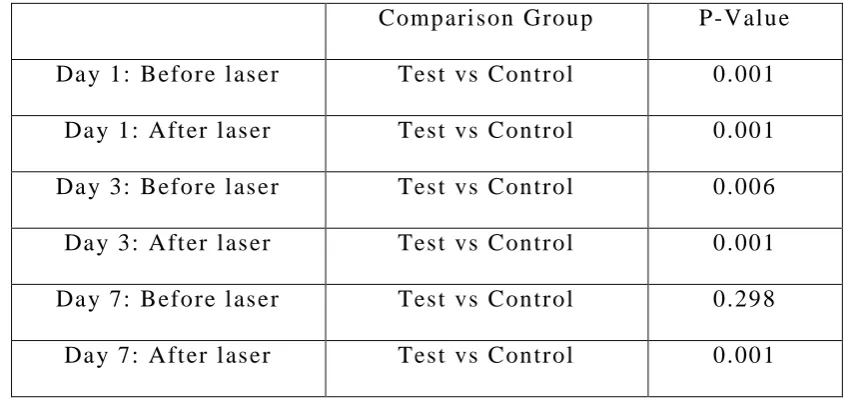
Mann -Whi tney T est to comp are valu es betw een Groups
Compari son Group P-Value Da y 1: Before l as er Test vs Control 0.001
Da y 1: Aft er l as er Test vs Control 0.001 Da y 3: Before l as er Test vs Control 0.006 Da y 3: Aft er l as er Test vs Control 0.001
Da y 7: Before l as er Test vs Control 0.298 Da y 7: Aft er l as er Test vs Control 0.001
Table 4:
Wilcoxon Sign ed Ranks Test to comp are valu es b etween B efore and After las er
Group Da y Compari son group P-Value
Tes t
Da y 1 Before vs After <0.001 Da y 3 Before vs After <0.001 Da y 7 Before vs After <0.001
Control
Da y 1 Before vs After 0.020
Da y 3 Before vs After 0.001
Da y 7 Before vs After 0.007
Table 5:
Fri ed man test for repeated measu res to compare b efore las er values betw een 1, 3 and 7 Days
Group Compari son group (Before laser) P-Value Test Da y 1 vs Da y3 vs Da y 7 <0.001
[image:63.595.102.531.112.313.2] [image:63.595.109.525.393.592.2]Data and Photos
53 Table 6:
Bonferron i ad jus ted Wil coxon Sign ed Ranks tes t to comp are valu es betw een pai rwise ti me poin ts
Group Compari son group (Before laser) P-Value
Tes t
Da y 1 vs Da y3 0.999
Da y 1 vs Da y 7 <0.001 Da y3 vs Da y 7 <0.001
Control
Da y 1 vs Da y3 0.999
Da y 1 vs Da y 7 0.035
[image:64.595.106.526.127.326.2]Da y3 vs Da y 7 0.011
Table 7:
Fri ed man test for repeated measu res to compare af ter las er valu es betw een 1, 3 and 7 Days
Group Compari son group (Aft er l as er) P-Value Tes t Da y 1 vs Da y3 vs Da y 7 <0.001 Control Da y 1 vs Da y3 vs Da y 7 0.001
Table 8:
Bonferron i ad jus ted Wil coxon Sign ed Ranks tes t to comp are valu es betw een pai rwise ti me poin ts
Group Compari son group (Aft er l as er) P-Value
Tes t
Da y 1 vs Da y3 0.660
Da y 1 vs Da y 7 <0.001
Da y3 vs Da y 7 0.017
Control
Da y 1 vs Da y3 0.999
Da y 1 vs Da y 7 0.007
Da y3 vs Da y 7 0.051
[image:64.595.107.525.547.746.2]Data and Photos
54 Chart:1 Chart: 2 5.87 5.57 4.27 2.60 2.20 1.37 0.0 2.0 4.0 6.0 8.0 10.0Day 1 Day 3 Day 7
M ea n v al u e
Mean values in Test Group
Before Laser
After Laser
4.97 5.00
4.37 4.73 4.60
4.07 0.0 2.0 4.0 6.0 8.0 10.0
Day 1 Day 3 Day 7
M e an v al u e
Mean values in Control Group
Before Laser
Data and Photos
55 Chart: 3
5.87 5.57
4.27
2.60
2.20
1.37
4.97 5.00
4.37
4.73 4.60
4.07 0.0 2.0 4.0 6.0 8.0 10.0
Day 1 Day 3 Day 7
M e an v al u e
Before and after laser therapy mean values in
test & control group.
Test Before Laser
Test After Laser
Control Before Laser