COMPARATIVE STUDY OF EFFICACY OF MODIFIED VACUUM ASSISTED CLOSURE DRESSING USING GLOVES VERSUS BETADINE DRESSING IN CHRONIC NON HEALING ULCERIN
GOVT RAJAJI HOSPITAL, MADURAI
Dissertation submitted to
THE TAMILNADU
DR.M.G.R.MEDICAL UNIVERSITY CHENNAI - 600032
With fulfillment of the regulations
For the Award of the Degree of
M.S.GENERAL SURGERY (BRANCH- I) MAY 2019
DEPARTMENT OF GENERAL SURGERY
2
CERTIFICATE
This is to certify that this dissertation entitled COMPARATIVE STUDY OF EFFICACY OF MODIFIED VACUUM ASSISTED CLOSURE DRESSING USING GLOVES VERSUS BETADINE DRESSING IN CHRONIC NON HEALING ULCER IN GRH, Madurai" at Government Rajaji Hospital, Madurai submitted by DR SAKTHEESWARAN R to the faculty of General Surgery, The Tamilnadu Dr.M.G.R. Medical University, Chennai in partial fulfilment of
the requirement for the award of MS degree (Branch I) General Surgery ,is a
bonafide research work carried out by him under my direct supervision and
guidance.
DR.A.M.SYED IBRAHIM M.S.,FAIS
PROFESSOR OF GENERAL SURGERY
DEPT OF GENERAL SURGERY
MADURAI MEDIACL COLLEG
CERTIFICATE
This is to certify that this dissertation entitled COMPARATIVE STUDY OF EFFICACY OF MODIFIED VACUUM ASSISTED CLOSURE DRESSING USING GLOVES VERSUS BETADINE DRESSING IN CHRONIC NON HEALING ULCER IN GRH,
Madurai" at Government Rajaji Hospital, Madurai submitted by
DR SAKTHEESWARAN R to the faculty of General Surgery, The Tamilnadu Dr.M.G.R. Medical University, Chennai in partial fulfilment of
the requirement for the award of MS degree (Branch I) General Surgery ,is a
bonafide research work carried out by him under my direct supervision and
guidance.
DR.S.R.DHAMOTHARAN M.S,FIAGES
PROFESSOR AND HOD OF THE DEPARTMENT
DEPT OF GENERAL SURGERY
MADURAI MEDIACL COLLEGE
4
CERTIFICATE BY THE DEAN
This is to certify that the dissertation entitled " COMPARATIVE STUDY OF EFFICACY OF MODIFIED VACUUM ASSISTED CLOSURE USING GLOVES VERSUS BETADINE DRESSING IN CHRONIC NON HEALING ULCER IN GRH, Madurai"is a bonafide research work done by
DR.SAKTHEESWARAN R, Post Graduate Student ,Department of General Surgery, MADURAI MEDICAL COLLEGE AND GOVERNMENT RAJAJI HOSPITAL, MADURAI, under the guidance and supervision of
DR.A.M.SYED IBRAHIM M.S.,FAIS., Professor Department of Surgery, MADURAI MEDICAL COLLEGE AND GOVERNMENT RAJAJI HOSPITAL,MADURAI.
DATE: PROF. DR. D. MARUTHUPANDIAN MS., FICS., FAIS.,
PLACE: MADURAI. DEAN,
Madurai Medical College,
DECLARATION BY THE CANDIDATE
I declare that this dissertation entitled" COMPARATIVE STUDY OF EFFICACY OF MODIFIED VACUUM ASSISTED CLOSURE USING GLOVES VERSUS BETADINE DRESSING IN CHRONIC NON HEALING ULCER IN GRH, MADURAI" is prepared by me under the direct guidance and supervision of Dr. A.M. SYED IBRAHIM M.S, FAIS,.., Professor, Department of General Surgery, MADURAI MEDICAL COLLEGE AND GOVERNMENT RAJAJI HOSPITAL, MADURAI. This is submitted to The Tamil Nadu DR.M.G.R. Medical University, Chennai, in partial fulfillment of the regulations for the award of MS degree(Branch I) General Surgery course on MAY 2019.
DATE: DR.SAKTHEESWARAN .R
6
ACKNOWLEDGEMENT
I take this opportunity to extend my gratitude and sincere thanks to all those who have helped me complete this dissertation.
I am extremely indebted and remain grateful forever to my guide, Dr. A.M. SYED IBRAHIM M.S FAIS., Professor of Surgery, for his constant able guidance and constant encouragement in preparing this dissertation and during my post-graduate course.
It gives me immense pleasure to express my deep sense of gratitude to my Professor and Head of Department of Surgery,
Dr.S.R.DHAMOTHARAN M.S.,FIAGES.,the person who has mastered the art of surgical skills, for his excellent guidance, encouragement and constant inspiration during my P.G. Course.
I thank the dean of Madurai Medical College and Govt. Rajaji Hospital,
Dr. D. MARUTHUPANDIAN MS., FICS., FAIS for permitting me to conduct this study in the Department of General Surgery of the Govt .Rajaji medical college and Hospital, Madurai.
I extend my sincere thanks to my Post- graduate Colleagues, and Friends, who had helped me in preparing this dissertation.
I must give my sincere thanks to my PARENTS for their moral support, constant encouragement and sincere advices throughout my career.
Last but not the least my heartful thanks to all patients who formed this study group and co-operated wholeheartedly.
DR.R.SAKTHEESWARAN
8
S.NO CONTENTS PAGE NO
1 INTRODUCTION 9
2 REVIEW OF LITERATURE 10
3 AIM OF STUDY 60
4 MATERIALS AND METHODS 61
5 OBSERVATION AND RESULTS 64
6 ANALYSIS OF DATA 77
7 CONCLUSION 83
8 BIBILIOGRAPHY 84
9 MASTER CHART 90
10 ABBREVATIONS 93
11
ETHICAL COMMITTEE APPROVAL LETTER
94
INTRODUCTION
Negative pressure wound therapy or vacuum assisted closure
dressing is a newer non-invasive technique that use controlled
negative pressure, using vacuum-assisted closure (VAC) device. It
helps to promote wound healing by removing fluid from open
chronic wounds, preparing the wound bed for graft or other closure
methods by reducing edema and promoting formation of
granulation tissue. VAC dressing can be used to treat Chronic non
healing ulcers following debridement of infection or amputation,
and in reconstructive soft tissue and osseous procedures.
Vacuum assisted closure dressing has been frequently
10
REVIEW OF LITERATURE
SKIN – ANATOMY
Skin is made of three layers
- Epidermis
- Dermis
- Subcutaneous tissue
Epidermis :
Keratinocytes:
These cells from the basal layer towards the skin surface
losses its water , begin to hard and dies eventually. The dead
keratinocytes forms the outer most protective layer of the
epidermis –stratum corneum which is sloughed off and replaced
regularly.
Melanocytes :
Produces melanin pigment which is responsible for the
colour of skin.
Langerhans cells:
Part of the immune system
Acts as a defense against the pathogens entering the
epidermis
DERMIS:-
It is the thickest layer of the skin. The primary cells present
in this layer is fibroblast , which has collagen and elastin and gives
skin its elasticity and resilience. It is the home of sebaceous gland
and also contains capillaries and langerhans producing lymph
nodes . Sebaceous glands produce sebum and it lubricates the skin
12
SUBCUTANEOUS TISSUE:
It is composed of mainly adipose tissue responsible for the
padding and insulation as well as contains sweat glands and erector
pili muscle. Cutaneous vessels arising from named vessels supplies
a 3-dimensional vascular territory from bone to skin called
angiosomes. Cutaneous vessels anastomosis with nearby cutaneous
CHOKE VESSELS:
Choke vessels plays an important role in flap survival , as
they provide initial resistance to blood flow between tip and base
of the flap .
When the skin flap delaying done ,this choke vessels open up
and provide adequate blood supply to the flap for the survival
FUNCTIONS OF SKIN
The skin has 4 major functions
1) PROTECTION – It provides protection agaist UV radiation ,
mechanical , chemical and thermal injuries. It acts as a
physical barrier to microorganism invasion and prevents
dehydration as it is relatively impermeable.
2) SENSATION – Skin is the largest sensory organ of the body
and has the receptors for touch , pain , pressure and
temperature.
3) THERMOREGULATION – Skin prevents heat loss by
presence of hair and subcutaneous tissue . skin is the major
14
facilitated by sweat evaporation from skin surface and
increased blood flow through the rich vascular network in
the dermis.
4) METABOLIC – Subcutaneous fat tissue contains major
energy store mainly in the form of triglycerides. Vitamin D
is synthesized in skin epidermis and also supplements from
diet.
ANATOMY OF FOOT
A complete anatomical knowledge is essential while
treating the non healing ulcers of the foot and its
complications. Without aggressive management , non
healing ulcer will end up in amputation either minor or major
The foot skeleton consists of tarsal-7 ,metatarsal – 5 and
phalanges -14. Foot is usually divided into three zones
FOREFOOT – metatarsal and phalanges
MIDFOOT – navicular , cuboid and cuneiform
HINDFOOT – talus and calcaneum
SKIN AND SUBCUTANEOUS TISSUES OF THE FOOT
The skin of dorsum of the foot is thinner and less
sensitive while the plantar skin is thicker in weight-bearing
areas such as heel , lateral margin and ball of foot and more
sensitive and not pinchable . The sole of foot consist of thick
stratum corneam and thin dermis. The subcutaneous tissue in
the sole is more fibrous than dorsum of foot. Fibrous septa
divides the sole into fat filled areas making the foot shock
absorbable especially over the sole. The skin of the sole is
hairless and has numerous sweat glands . the entire sole is
more sensitive especially the thinner areas underlying the
arch of foot.
The epidermis is usually transformed into the nail
matrix. It contains three ill defined layers dorsal, intermediate
and ventral layers. It is firmly attached to epithelium of nail
bed .The margin of the nail is overhung by skin fold
predisposing to in growing toe nails.
SKELETON AND FASCIA OF THE FOOT
The skeleton is shaped to form arches and adjust to
16
phalanges. The superficial fascia of the sole is more fibrous
and dense. Thick central part of the plantar fascia forms the
plantar aponeurosis, resembles the palmar aponeurosis but
tough dense and elongated.
The plantar aponeurosis fixes the skin of the sole and
also helps in maintaining the longitudinal arches of the foot.
Compartments of the sole:
Medial compartment:- covered by medial plantar fascia
which is thin and consist of abductor hallucis , flexor hallucis
brevis , tendon of flexor hallucis longus , medial plantar
nerve and vessels.
lateral compartment:- covered by lateral plantar fascia and
consist of abductor digiti minimi and flexor digiti minimi
central compartment – covered by plantar aponeurosis and
consisit of flexor digitorum brevis , tendons of flexor
digitorum longus and flexor hallucis longus , lumbricals and
adductor hallucis and also lateral plantar vessels and nerve
forefoot fourth compartment – interroseous compartment
consist of metatarsal bones , deep plantar and metatarsal
vessels and plantar and dorsal interrosei muscles .
Fifth compartment or dorsal compartment:- it lies
between dorsal fascia and tarsal bones and consists of
extensor hallucis brevis and extensor digitorium brevis ,
MUSCLES AND TENDONS OF THE FOOT
Despite , arranged in layers and compartment ,the muscles act
as a group during the support phase of the stance and in
maintaining longitudinal and transverse arches of the foot. These
muscles become more active during the later phase of walking by
stabilizing the foot for propulsive movement.
MUSCLES OF DIFFERENT COMPARTMENTS
COMPARTMENTS MUSCLES
1ST layer Abductor hallucis , flexor digitorum brevis,abductor digiti minimi
2nd layer Quadrates plantae, Lumbricals
3rd layer Flexor hallucis brevis , adductor hallucis, flexor digiti minimi brevis
20
22
LYMPHATIC DRAINAGE
Superficial lymphatics are numerous in the sole of foot
medial superficial lymphatics are large and accompany great
saphenous vein to vertical group of inguinal nodes. Lateral
superficial lymphatics accompany short saphenous vein and drains
into popliteal nodes.
Deep lymphatics follows main blood vessels ; fibular anterior
tibial, posterior tibial , popliteal and femoral veins into deep
inguinal lymph nodes
ARCHES OF FOOT
These arches used to distribute the weight over the foot and
acts as shock absorbers and spring boards for propelling while
walking , running and jumping etc,,
Longitudinal arch – medial and lateral parts
Medial – is higher and formed by calcaneus , talus , navicular
, three cuneiform and three metatarsals. Talar head is the keystone
in maintaining medial longitudinal arch.
lateral – flat than medial arch .and formed by calcaneus ,
24
transverse arch formed by cuboid , cuneiform ,and bases of
metatarsals
PASSIVE FACTORS FORMING ARCHES OF FOOT:
Shape of the united bones
Four successive fibrous tissue layers
Plantar aponeurosis
Long plantar ligament
Plantar calcaneocuboid ligament
Plantar calcaneonavicular ligament
DYNAMIC FACTORS IN MAINTAINING ARCHES OF
FOOT:
Active bracing of the intrinsic muscles of the foot
Active contraction of muscles with long tendon
o Flexor hallucis and digitorum longus
26
ANATOMICAL PRINCIPLES OF SURGICAL INCISIONS:
Some of the anatomic principles one should keep in mind
while making incisions in the foot
Avoid neuro-vascular injuries.
Avoid incision in weight bearing points.
Always make liberal counter incision.
De-roofing should be liberally done
Always excise the metatarsal head while doing
ULCER
An ulcer is defined as “Any break in the continuity of the
epithelium of the skin or mucous membrane”.
It may be caused by molecular death of the surface epithelium or
traumatic removal of epithelium
CLASSIFICATION OF ULCER:
I. ACCORDING TO DURATION:
ACUTE -less than 2 weeks
CHRONIC – more than 2 weeks
II. CLINICAL:
SPREADING ULCER:
It is an acute painful ulcer . Edges are inflamed , irregular
edematous. Floor is covered with profuse purulent discharge
and slough with surrounding edematous skin. Regional
lymph nodes are enlarged and often tender.
HEALING ULCER :
Edges of healing ulcer is sloping and the floor is covered
with healthy pink granulation tissue with minimal serous
discharge. Regional lymph nodes may or may not be tender
28
show any edema or induration. Three zones are seen in
healing ulcer. Innermost red granulation tissue ; middle
bluish growing epithelium zone and the outermost whitish
fibrosis and scar zone.
CALLOUS OR NON HEALING ULCER:
Pale granulation tissue seen in the floor of the ulcer.
Edges and the surrounding skin show induration. Ulcer doesn’t
show any tendency of healing.
III. PATHOLOGICAL CLASSIFICATION:
NON SPECIFIC ULCERS:
Traumatic
Arterial ulcer due to ischemia
Venous ulcer eg. Varicose veins
Neurogenic ulcer
Martorell’s ulcer
Bazin ulcer
SPECIFIC ULCERS :
Tuberculous ulcer
Syphilitic ulcer
Actinomycosis
Meleny’s ulcer
MALIGNANT ULCERS :
Squamous cell carcinoma
Basal cell carcinoma
Malignant melanoma
30
CLASSIFICATION OF WOUNDS
1. RANK AND WAKEFIELD CLASSIFICATION:
TIDY WOUNDS:
Clean wounds like surgical wound without obvious skin
loss,wounds caused by sharp instruments
Healing is mainly by primary intention
UNTIDY WOUNDS:
Dirty wounds due to
- Avulsion
- Crushing
- Multiple lacerated wounds
- Burns
- Tearing
2.BASED ON TYPE OF WOUNDS:
a) Clean incised wounds:- surgical wounds with clear cut edges
without skin loss comes in this category
b) Lacerated wounds:- wounds with irregular edges , loss of skin
c) Contusion :- wounds without skin loss with minimal tissue
injury and discolouration
d) Closed blunt injury
e) Hematoma
f) Puncture wounds
g) Gunshot wounds
h) Penetrating injuries
3. BASED ON THE THICKNESS OF THE WOUND:
1)Superficial wounds:- loss of skin epidermis only
2) Partial thickness wound :- loss of epidermis and dermis and
only the deep dermis with sweat gland left
3) Full thickness wound :- complete loss of both epidermis and
32
4) Deep wounds:- extend into deep fascia and expose the muscle ,
tendon or bone
CLASSIFICATION OF SURGICAL WOUNDS:
1)Clean wounds- hernia and cardiovascular surgeries
2)clean contaminated :- bowel , biliary and pancreatic surgeries
3) Contaminated wounds:- accidental wounds and acute
abdominal conditions
4) Dirty wounds :- pyocele , abscess drainage , biliary peritonitis
Wounds are classified into two categories – open and closed .
The closed wound happens when a blunt force contusion and
damages the part of the skin . Closed wounds are as dangerous as
an open wound .It is divided into contusion , hematoma , crush
WAGNER classification system of diabetic foot
Grades Features
0 No open ulcers or may have deformity or cellulitis
1 Ulcer - superficial
2 Ulcer deep to joint capsule or tendon
3 ulcer deep with abscess, joint sepsis or osteomyelitis
4 Local gangrene of foot either fore foot or heel
5 Gangrene invoving the entire foot
TEXAS classification system of diabetic foot
Stages Grades
0 1 2 3
A Pre or post
ulcerative
lesions are
completely
epithelized
Superficial
wound are not
involving tendon,capsule,or bone Wound penetrating to tendon or capsule Wound penetrating
to bone or
joint
B Infected Infected Infected Infected
C Ischemic Ischemic Ischemic Ischemic
D Infected
34
NEUROPATHIC ULCER- PATHOPHYSIOLOGY
Increased load to foot
Unaware due to decreased sensation
Focused inflammation of foot
keratin plaques
Tissue breakdown
Cavity as nidus of sepsis
Inactive infection
WOUND HEALING
Wound healing is complicated process to accomplish the
functional and anatomical integrity of the disrupted tissues by
various mechanisms.
TYPES OF WOUND HEALING:
1) HEALING BY PRIMARY INTENTION:
Wounds with clear cut edge with no skin loss such as
surgical incision heal by primary intention. Epithelization rate is
higher and heals quickly
2) HEALING BY SECONDARY INTENTION:
Wounds with large area of skin loss such as contaminated
wounds heal by secondary intention . Heals slowly with
fibrosis and re-epithelization from the wound edges
3) TERTIARY INTENTION:
Wound is initially infected and pus will be present. After
regular cleaning and dressing and controlling the infection
36
PRIMARY AND SECONDARY HEALING
CHARACTERS PRIMARY SECONDARY
CLEANLINESS CLEAN NOT CLEAN
INFECTION NOT INFECTED INFECTED
SUTURES USED NOT USED
MARGINS SURGICALLY CLEAN IRREGULAR
HEALING SMALL
GRANULATION
TISSUE
LARGE
COMPLICATIONS NOT FREQUENT FREQUENT
OUTCOME LINEAR SCAR IRREGULAR
PHASES OF WOUND HEALING
There are four distinct phases of wound healing
Hemostasis
Inflammation
Proliferation
Tissue remodeling
HEMOSTASIS PHASE:
Hemostasis is the process of the wound closed by clotting.
Hemostasis starts when blood leaks out . The first step is blood vessels
constrict to restrict the blood flow. Then the platelets stick together to
seal the break in wall of the blood vessel. Coagulation occurs and
reinforces the platelet plugs with threads of fibrin . The platelets adhere
to the endothelial surface within seconds of the rupture of a blood vessel
wall epithelium . After that, the fibrin strands begin to adhere in about
sixty seconds.
As more fibrin begin to adhere, the blood is transformed from
liquid to gel form through the pro-coagulants and the release of
prothrombin . The formation of a thrombus keeps the platelets and other
38 INFLAMMATORY PHASE:
Inflammation is the second stage of wound healing. It begins within
6-8 hrs and lasts for 5-7 days .It begins right after injury when the
injured blood vessels leak transudate causing localized swelling.
Inflammation controls both the bleeding and also prevents infection.
Macrophages release FGF which is responsible for the angiogenesis.
Polymorphonuclear leukocytes secretes inflammatory mediators and
free radicals after 48 hours . These mediators remove the foreign body ,
clots and bacteria.
PROLIFERATIVE PHASE:
The proliferative phase starts after 7 days and lasts for more than 6
wks. Fibroblast secrets collagen and glycosamines. Proliferation of the
vascular endothelial cells occurs at the wound edges by the cytokines
and growth factors with the help of fibroblasts. Hydroxylysine and
hydroxyproline secreted by various enzymes with the help of vit C , iron
and alpha ketoglutarate.
Proliferative phase has 3 sub-phases
Fibroplasias
Collagen and re-epithelization
i) Neovascularisation:
It is the process of regeneration of new blood vessels. It concurrently occurs with the endothelial cell migration and
proliferation of fibroblast. Transforming growth factor – β ,
fibroblast growth factor and VEGF ( vascular endothelial
growth factor) facilitates the endothelial cell elongation to
cause neovascularization in the granulation tissues. It is
promoted by hypoxic conditions , presence of lactic acid and
reduced oxygen environment.
At the end of this phase ,when the tissues are perfused with
sufficient nutrients the blood vessels no longer needed will
undergo apoptosis
ii) Fibroplasias and tissue formation:
Fibroblasts are important component in the granulation tissue that appears after inflammatory phase. When the
angiogenesis stops newly formed endothelial cell ,
myofibroblast and ECM grows continuously until the wound
40 iii)Collagen deposition :
Fibroblasts are the maximum contributor of collagen synthesis and deposition and increases the strength of the
wound by facilitating the angiogenesis and differentiation of
the connective tissues. The type III collagen and fibronectin
are synthesized between 10 hours and 3 days after injury and
the deposition of collagen peaks between one to three weeks
depends on the wound size.
REMODELLING:
This phase starts by 6 weeks and lasts for upto 2 years . cross linking of the collagen takes place in this phase laeding to the
maturation of the collagen responsible for the tensile strength of the
wound. usually synthesis did not occur after 42 days of wound healing It has 2 sub phases
Epithelization
Contraction i)Epithelization:
Epithelization occurs after formation of granulation tissue in open wound that allows the epithelial cells and keratinocytes to
surface area. keratinocytes alters their shape and become flat and
elongated morphology and thus the cells forms cellular process by
actin and lamellipodia. This migration is usually mediated by lack
of contact inhibition and nitric oxide etc. they utilize the fibrin to
move across the wound site. The epithelial cells formed at the
wound edge act as a base for keratinocyte proliferation.
Keratinocyte migration will continue cells from other wound
edges meet to form contact inhibition which stops migration.
ii) Wound contraction:
contracture is the process of dimunition of the wound surface area that occurs by the centripetal movement of the
wound edge towards centre. It usually starts by approximately 4-5
days after wounding at an rate of 0.6-0.7 mm / day depending on
the type of tissue and wound. This contraction is brought about by
myofibroblast interaction with the ECM. Prolonged contraction
42
FACTORS AFFECTING WOUND HEALING
LOCAL FACTORS SYSTEMIC FACTORS
Hypoxia
Repeated trauma
High bacterial burden
High metalloproteinases
Growth factor deficiency
Cellular senescence
Irradiation
Necrotic tissue burden
Excessive exudates
Corrupt ECM
Diabetes mellitus
Anemia
Malnutrition
Immunodeficiency
Immunosupressants
Age
Obesity
44
COMPLICATIONS OF WOUND HEALING
1. Deficient scar formation: due to improper formation of the granulation tissue
2. Incisional hernias : Dehiscence of scar results from increased pressure from within . Abdominal dehiscence carries a mortality rate of
upto 30 %
3. Ulceration: wound ulcerate because of the inadequate blood supply as in the case of leg ulcers with atherosclerosis or varicose veins.
Repeated trauma due to impaired sensation leads to trophic or
neuropathic ulcer as in Diabetes or leprosy.
4. Excessive scar formation : An exuberant granulation tissue formation results in hypertrophic scar or keloid formation. Both these
scars exhibit abundant irregular collagen bundles with more fibroblast
and capillaries than the scar of same age.
5. Excessive contraction : an exaggeration in the process of contraction leads to severe wound deformity , for eg . Dupuytren’s contracture ,
plantar contracture and the peyronie’s disease of penis. Contracture may
compromise joint movements . contracture in the esophagus or intestine
CAUSES FOR THE FORMATION OF NON HEALING ULCER
Repeated infections
Decreased blood supply
Hypoxia
Loss of sensation
Malignancy
Surrounding area edema
Systemic illness like TB , Diabetes mellitus
Underlying osteomyelitis
46
MANAGEMENT OF THE DIABETIC FOOT:
The foot ulcers in diabetics are generally not a non healing
ulcers but they are mal treated ulcers.
(a) WAGNER’s grade 0 foot:
This grade includes the patients with apparently normal foot
but varying degree of neuropathy or joint deformities. They donot
have an ulcer or infection but are potentially at risk. They need
regular assessment annually. Neuropathy must be examined during
each assessment. The best way to prevent neuropathy or delay it is
to keep glycemic control.
Assessment of vascular status is al mandatory. Absent
pulses in the foot even in the absence of “rest pain or claudication”
indicates a signicant vascular disease and such patients may be
ideal candidates for the vascular reconstruction or angioplasty. The
“at risk” patients have elevated pressure over some points on the
sole of the foot. They need to wear appropriate footwear .Charcot’s
(b) WAGNER’s grade 1 foot:
These patients have presented with either superficial ulcer.
Ulcer occurs either with repetitive low or high pressure at any
pressure point on sole during walking.
Relief of pressure is mainstay of ulcer treatment. An ulcer
will not heal if the patient walks with the ulcer. The various
methods of “off-loading” devices are used. the appropriate
management of vascular disease is needed as in grade 0. Infection
needs debridement and antibiotics as appropriate.
(c) WAGNER grade 2 and 3 foot:
patients who have deep ulcer with or without complications
like osteomyelitis or abscess. These patients requires initial
aggressive debridement. Osteomyelitis must be managed by
debridement or excision of the infected bone.
The patient requires follow up for long duration to advise
appropriate foot wear and education regarding foot care to
48
(d)WAGNER grade 4 and 5 foot:
They require minor or major amputation. Always there is
concomitant vascular occlusive disease. These patients therefore
need appropriate vascular reconstructions. After the ulcer heals
patient needs to wear special footwear for the ipsilateral and
contra-lateral foot. For the major amputees, prosthetic devices
Principles of medical management:
1. Pus from ulcers should be sent for pus culture and sensitivity.
2. Careful monitoring of random and fasting blood glucose levels.
3. Appropriate antidiabetic drugs measures – either insulin or oral
hypoglycemic agents.
4. Broad spectrum antibiotics covering aerobic and anaerobic
organisms should be started at the onset and change to other
antibiotics depending on the culture and sensitivity report.
Principles of the surgical management of diabetic ulcer:
1. Early diagnosis of ulcer and prompt intervention.
2. Correction blood glucose.
3. Complete rest to the injured area.
4. Careful and complete debridement and drainage of all involved
areas at the initial visit itself.
5. Appropriate antibiotics coverage.
6. regular wound care and dressings.
7. Appropriate vascular reconstruction if needed .
8. Careful follow up with podiatric appliances and modified
50
MANAGEMENT OF CHRONIC ULCER
The ideal wound management plan should be less infectious,
cost effective , low exchange of water vapour , allows healing
without delay and avoid damaging the newly formed tissues. Some
disease such as atherosclerosis, DM and conditions such as anemia
, malnutrition and local infections cause delay in wound healing.
Supplementation of appropriate diet and treatment of underlying
disease is necessary to assist in faster wound healing. Infection is
the most common cause for death in the burns injury and accounts
for 50 % of the hospital deaths in burns . so it is essential that the
wound management should be done with sterile and aeptic
conditions.
INVESTIGATIONS:
Basic blood investigations – HB% , blood sugar , creatinine
and urea
Serum proteins and electrolytes
Pus culture and sensitivity
In suspected tuberculosis – Chest x –ray and Mantoux
Edge wedge biopsy if suspicious of malignancy
Venous and the arterial Doppler study – to rule out venous
and arterial insufficiency
ASSESSMENT OF THE ULCER :
Cause of the ulcer should be identified initially
Systemic examination should be done to assess the sensation
, movements , peripheral pulses and lymph node status.
Parameters assessed:
o Location
o Size
o Signs of infection
o Surrounding skin – colour and temperature
o Wound edges- induration or maceration
52
MANAGEMENT OF CHRONIC ULCER:
Adequate debridement of all necrotic tissue (eschar , slough)
is essential before adequate assessment and staging of the ulcer.
There are various methods of debridement which includes sharp
surgical debridement , mechanical debridement , enzymatic and
autolytic debridement. It is continuum from flushing away necrotic
debris with low pressure irrigation upto wide excision.
1.Sharp surgical debridement
The most efficacious method of debridement is sharp surgical
debridement. Debridement of ulcer base upto bleeding is ideal
method of debridement for the patient with non healing ulcer.
2.Mechanical debridement
Mechanical debridement can be accomplished by wet to dry
3.Autolytic debridement
Autolytic debridement with moist dressings is selective and it
liquefies the slough and eschar and also it promotes the granulation
tissue formation.
4.Enzymatic debridement
Enzymes such as collagenase , papain, urea has been used as
debridment agents for eschar and slough. They are costly and labour
intensive method of debridement.
5. Antibiotic
The use of double or triple antibiotics is justified. The
antibiotics used must have broad spectrum coverage of gram
54
TIME APPROACH FOR CHRONIC NON HEALING
ULCER
FALANGA (2004) utilised the work of SCHULTZER et
al (2003) to develop a framework called TIME to provide the
comprehensive approach for wound care. The TIME principle
guidelines helps in approach for the best wound care.
T
• TISSUE MANAGEMENT
I
• INFLAMMATION AND INFECTION
CONTROL
M
• MOIST BALANCE
CLASSIFICATION OF DRESSING :
Dressings are classified into two major categories according to
the usage
1.long term application or skin substitutes:
Further subdivided into :
Temporary : it is applied onto the fresh partial thickness
wound until complete healing of the wound is ensured
Semi permanent – it is applied on the full thickness wounds
until the auto-grafting of the wounds.
2.Short term application :
Dressing usually requires replacement at regular intervals
It is further classified into conventional , synthetic and biological
based on the type of material used in the dressing
A.Conventional:
B. Synthetic :
i) Films
ii) Composite
56
C. Biological :
i) Allograft
ii) Xenograft
iii) Collagen dressing
Within each category , dressing are sub classified into
1. Primary - dressing contact with the ulcer bed
2. Secondary- dressing which covers the primary dressing
3. Island – dressing constructed with adhesive portion outside
and a central absorbent
MANAGEMENT OF INDOLENT NON HEALING ULCERS
a. Collagen
b. Silver colloid dressing
c. dressings with Growth factors
Growth factors are derived from the platelets and bioengineered
NEGATIVE PRESSURE WOUND THERAPY
Vacuum assisted closure dressing(VAC), also called as
negative pressure wound therapy (NPWT) or Micro deformational
wound therapy , brought a revolution in wound care since past 20
years. This method was first coined by Fleischmann et al. in 1993.
In this era of modern wound care, negative pressure therapy
has been routinely used for treatment of wounds and become the
integral part of the treatment plan of the chronic non healing ulcers.
It is used in acute, chronic or complex wounds and has been proven
more efficacious and promotes for faster healing than the
conventional dressing.
The trademark VAC therapy belongs to KCI VAC needed a
sophisticated equipment, specialized foam and drain and also
trained person for the application and maintenance, which is
possible only at high cost settings.
In our study, we used the easily available materials in low
resource setting such as gauze , glove to make the negative
58
FOUR PRIMARY MECHANISMS:
1. Macro deformation
2. Microdeformation near
3. Removal of excess exudates
4. Optimisation of the wound
1. MACRODEFORMATION
It refers to the decrease in the wound surface by shrinkage of
sponge and action of the centripetal forces over the wound surface.
Due to the inherent tension present in the dermis near the wound
and the underlying attachments of different wounds over the
different sites contract wound to different extent.
2. MICRODEFORMATION:
The vaccum transmitted through the interface material acting
over undulated surface of wound produces changes occur in micro
to millimeter range scale. The mechanical forces are transmitted to
the every cell through the ECM and lead to the cell deformation
3. REMOVAL OF EXCESS FLUID
Excess fluid in ECF leads to edema and the deprivement
leads to signs of dehydration. This compartment is drained by the
lymphatics ; abnormality may lead to lymphedema. Excess of fluid
cause delay in the wound healing due to compressive effect over
the tissues. Removal of the excess fluid from the wound will
decrease the compression effect over the microvasculature and
thereby promoting the perfusion and wound healing.
CONTRAINDICATIONS:
Untreated osteomyelitis
Non enteric fistulas
Necrotic tissues along with eschar
Exposed blood vessels
Malignancy
Exposed nerves
Anastamotic sites
60
AIM & OBJECTIVE
To compare the efficacy of the modified vacuum assisted
closure (VAC) dressing using gloves and available resources in a
low resource setting with routine Povidine iodine dressing in
MATERIALS AND METHODS
DESIGN OF THE STUDY: PROSPECTIVE STUDY
STUDY PERIOD : 2019
SAMPLE SIZE : 100
STUDY PLACE : GRH MADURAI
INCLUSION CRITERIA:
• Patients more than 25 years of age
• Diabetic ulcers in the extremities
• Non healing ulcers in the extremities
• Amputation stump ulcer
EXCLUSION CRITERIA
• Malignant wounds
• Wounds with underlying osteomyelitis
• Wounds with sinus or cavity
• Wounds with unstable fracture
62 • Larger wound surface
• Wounds with Exposed blood vessels
• Patients on anticoagulation therapy
METHOD OF COLLECTION OF DATA
Detailed history
Clinical Examination
Dimensions of the ulcer
Rate of granulation tissue
Duration of hospital stay
METHODOLOGY:
• wound debridement
• The wound base was covered with gauze piece placed in two
layers.
• A tube with adequate fenestrations depending on the size of
• The gauze layer is held in place over the wound by applying
thin Opsite.
• An appropriate sized sterile surgical glove is taken and
inserted over the wound covering the extremity
• The tube is brought out from the edge of the glove.
• A piece of Opsite is applied covering entire margin of glove
circumferentially.
• Exit site of tube through the glove is fashioned like T-tailing
using opsite strip to prevent air leakage
• End of the tube connected to Romovac device or 50 cc
syringe and suction is applied
Total ulcer surface area measured initially and the reduction
in the surface area before grafting measured. Area of granulation
tissue covering the ulcer also measured using butter paper. After
grafting on the post op day 5 graft uptake was measured in
percentage. Total duration of the hospital stay was noted . pain in
64
OBSERVATIONS AND RESULTS
The 100 patients admitted in our GRH selected for the study
were divided into two equal and comparable groups. Patients
subjected to modified VAC dressing using gloves were classified
under study group and those who underwent conventional povidone
Graph: Sex wise distribution of patients.
MALE FEMALE
BETADINE DRESSING 34 16
VAC DRESSING 33 17
0 5 10 15 20 25 30 35 40
BETADINE DRESSING VAC DRESSING
66
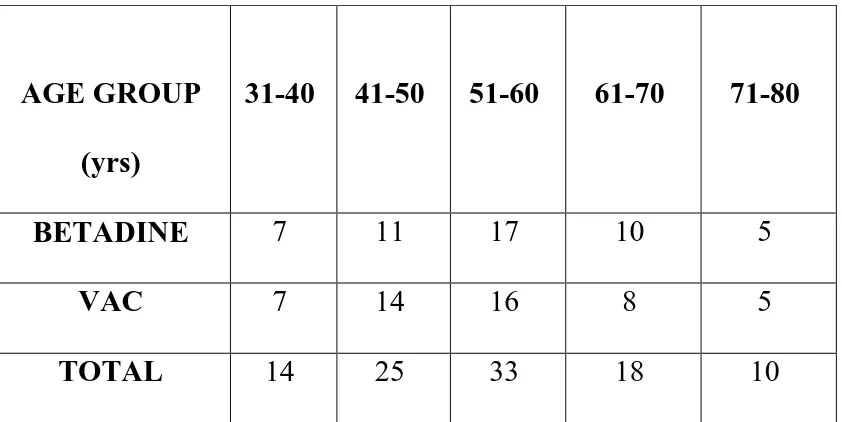
TABLE: Age wise distribution of patients
AGE GROUP
(yrs)
31-40 41-50 51-60 61-70 71-80
BETADINE 7 11 17 10 5
VAC 7 14 16 8 5
TOTAL 14 25 33 18 10
Mean age of Betadine group is 54.42
Mean age of VAC DRESSING group is 54.7
P value is 0.90.Not significant.
GRAPH : Age wise distribution of patients
0 2 4 6 8 10 12 14 16 18
BETADINE DRESSING VAC DRESSING
TABLE: ULCER SURFACE AREA
GROUP NO.OF PTS
MEAN STD. DEVIATION
t VALUE p VALUE
BETADINE 50 38.74 5.73 1.82 0.07
VAC DRESSING
50 40.41 2.85
The mean ulcer area in control group is 38.74+5.73(SD)cm2
and in the study group is 40.41+2.85(SD)cm2. The ulcer area was
measured by using tissue paper.
BETADINE VAC DRESSING
68
TABLE: RATE OF GRANULATION TISSUE FORMATION
GROUP NO
.
MEAN STD.
DEVIATION
t
VALUE
p
VALUE
GRAN
TISSUE
BETADINE 50 36.21 5.99 3.58 0.0005
VAC
DRESSING
50 39.57 2.69
The mean rate of granulation tissue formation in Betadine group is
36.21+5.99(SD) of total ulcer surface area and in VAC DRESSING
The mean rate of granulation tissue formation in Betadine group is
36.21+5.99(SD) of total ulcer surface area and in VAC DRESSING
is 39.57+2.69(SD) of total ulcer surface area.
34 35 36 37 38 39 40
BETADINE VAC DRESSING
GRANULATION TISSUE
70
TABLE: GRAFT UPTAKE AS PERCENTAGE OF ULCER
SURFACE AREA
Assessment of graft uptake was done at the end of POD 5 as
percentage of ulcer surface area. The mean graft uptake in the study
group is 87.6% and in the control group is 56.06 %
GROUP N MEAN SD t VALUE p value
SSG BETADINE 50 56.06 19.05 10.9 0.0000
VAC
DRESSING
Assessment of graft uptake was done at the end of POD 5 as
percentage of ulcer surface area. The mean graft uptake in the study
group is 87.6% and in the control group is 56.06 %
0 10 20 30 40 50 60 70 80 90 100
BETADINE VAC DRESSING
GRAFT UPTAKE
72
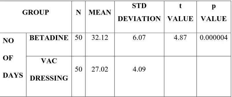
TABLE: DURATION OF HOSPITAL STAY
GROUP N MEAN STD
DEVIATION
t
VALUE
p
VALUE
NO
OF
DAYS
BETADINE 50 32.12 6.07 4.87 0.000004
VAC
DRESSING
50 27.02 4.09
The mean duration of hospital stay in the vacuum group was
27.02 days and in the povidone iodine dressing group was 32.12
The mean duration of hospital stay in the vacuum group was
27.02 days and in the povidone iodine dressing group was 32.12
and the p value was (p<0.000004) which is highly significant.
24 25 26 27 28 29 30 31 32 33
BETADINE VAC DRESSING
DURATION OF HOSPITAL STAY
74
PAIN SCORING
Average pain score in the range of 0 to 10 was 6.96 in the
conventional betadine dressing and it was 3.38 in the study group.
P< 0.001 which is significant reduction in the pain score.
PAIN TEST CONTROL
MEAN 3.38 6.96
SD 1.24 1.41
Average pain score in the range of 0 to 10 was 6.96 in the
conventional betadine dressing and it was 3.38 in the study group.
P< 0.001 which is significant reduction in the pain score.
0 1 2 3 4 5 6 7 8
Betadine Vacuum
MEAN PAIN SCORE
76
The main postoperative parameters noted in the study and control
groups:
- Wound size
- Contracture
- Pain
- Infection
All these parameters are less in the study group when compared to
ANALYSIS OF DATA:
The number of patients studied was 100 and are randomly
divided into study (50) and control group (50).both the study and
control group were matched regarding their age ,sex and there was
no significant difference between the two groups with respect to
age and sex.
The average pain score in the range of 0-10 was 3.38 in the
vacuum dressing and it was 6.96 in the povidone iodine dressing
group.
The mean duration of hospital stay in the vacuum group was
27.02 days and in the povidone iodine dressing group was 32.12
and the p value was (p<0.000004) which is highly significant.
78
The mean split skin graft uptake in the vacuum dressing
group was 87.6% and in the povidone dressing group is 56.06%
and the p value showed highly significant difference in split skin
graft uptake (p<0.0000).
The mean rate of granulation tissue formation in povidone
group is 36.21 of total ulcer surface area and in vacuum dressing
group is 39.57 and the p value was (p<0.0005) which is highly
80
CONCLUSION
Modified VAC dressing significantly reduces the size of ulcer.
Modified VAC dressing improves the rate of granulation tissue
formation.
Modified VAC dressing improves SSG uptake also.
Modified VAC dressing reduces the duration of stay at the
hospital.
Patients undergoing Modified VAC dressing feels lesser amount
of pain when compared with the patients undergoing
conventional wound dressing.
84
BIBLIOGRAPHY
1. Fleischmann W, Strecker W, Bombelli M, Kinzl L. Vacuum
sealing as treatment of soft tissue damage in open fractures.
Unfallchirurg 1993;96:488-92.
2. KCI VAC, 2014. Available from: http://www.kci1.com/KCI1/
educationtraining. [Last accessed on 2014 Nov 11].
3. Danu M, Rosadi S. The simplest modified vacuum assisted
closure to treat chronic wound: Serial case report. J Plastik
Rekonstruksi 2012;1:117-22.
4. Singh M, Singh R, Singh S, Pandey V, Singh D. Vacuum
assisted closure in wound management – Poor man’s VAC. Int J
Plast Surg 2008;6.
5. Kumar P. Exploiting potency of negative pressure in wound
dressing using limited access dressing and suction-assisted
dressing. Indian J Plast Surg 2012;45:302-15.
6. Hemmanur SR, Siddha LV. Role of the surgical glove in
modified vacuum assisted wound healing. Arch Plast Surg
7. Ram S, Vijayan SK, Kini A. T tail configuration of opsite on
suction tubing outlet for modified vacuum assisted closure. Int J
Plast Surg 2012;8.
8. Thompson N, Gordey L, Bowles H, Parslow N, Houghton P.
Reliability and validity of the revised photographic wound
assessment tool on digital images taken of various types of chronic
wounds. Adv Skin Wound Care 2013;26:360-73.
9. Clarkeand RA, Henson PM, editors. The Molecular and Cellular
Biology of Wound Repair. NewYork, NY, USA: Plenum Press;
1988.
10. Stinner DJ, Waterman SM, Masini BD, Wenke JC. Silver
dressings augment the ability of negative pressure wound therapy
to reduce bacteria in a contaminated open fracture model. J Trauma
2011;71:S147-50.
11. Mendez-Eastman S. Guidelines for using negative pressure
wound therapy. Adv Skin Wound Care 2001;14:314-22.
12. Orgill DP, Manders EK, Sumpio BE, Lee RC, Attinger CE,
Gurtner GC, et al. The mechanisms of action of vacuum assisted
86
13. Borgquist O, Ingemansson R, Malmsjö M. The infl uence of
low and high pressure levels during negative-pressure wound
therapy on wound contraction and fluid evacuation. Plast Reconstr
Surg 2011;127:551-9.
14. Saxena V, Hwang CW, Huang S, Eichbaum Q, Ingber D, Orgill
DP. Vacuum assisted closure: Microdeformations of wounds and
cell proliferation. Plast Reconstr Surg 2004;114:1086-96.
15. Hinman CD, Maibach H. Effect of air exposure and occlusion
on experimental human skin wounds. Nature 1963;200:377-8.
16. V.A.C. Therapy Indications and Contraindications. Available
from: http://www.kci1.com/KCI1/indicationsandcontraindications.
PROFOMA
Name of the patient:
Age/sex:
IP No:
D-O-A:
D-O-D:
Duration of hospital stay:
Diabetic status:
Other co-morbidities
Alcoholic/ smoker:
History of presenting illness:
Significant past history:
Examination of ulcer:
Number
Site:
Size:
Shape:
88
Surrounding skin status:
Feautures of malignancy:
Induration :
Vascular status:
Skin/nail :
Palapation of the peripheral Pulses:
Examination of the regional lymph nodes:
Investigations:
Basic blood investigations:
Hb% / TC /DC /ESR:
Random Blood sugar:
FBS/PPBS( if diabetic):
Blood urea / Sr creatinine:
Chest xray :
ECG:
Arterial and venous Doppler study:
X ray of the local part:
Viral markers:
Treatment:
Antibiotics :
Insulin dosage / OHA ( if DM ):
Treatment before Modified VAC dressing:
Modified VAC dressing:
Date of application:
Date of removal:
No of dressings:
Treatment After Modified VAC dressing:
Granulation tissue:
Exudates:
Pain scoring ( VAS scale)
90 MASTER CHART S N o N
AME AGE SEX IP No
TYPE OF D RE SS IN G ULC ER S URFA C E AR EA G RA N UL AT IO N SURF AC E AR EA SS G U PT AKE PA IN S C O RE DURA TI O N OF HOSP IT AL S TAY
1 KARUPPASAMY 40 M 40711 BET 40.5 37.2 45 5 34
2 GOPAL 51 M 31228 BET 42.5 38 30 7 37
3 RAJALAKSHMI 68 F 32622 BET 44.6 41.2 42 8 32
4 CHELLAMMAL 62 F 33722 BET 42.7 41.2 42 9 28
5 MOHAMMED IBRAHIM 50 M 36282 BET 44.6 42.7 39 8 38
6 RAJESH 56 M 34282 BET 41.9 40.2 48 7 30
7 JEYARAJ 52 M 11286 BET 40.9 39.2 42 6 32
8 RAMAR 43 M 12682 BET 38.6 37 39 7 35
9 SELVARAJ 56 M 13928 BET 36.7 35.2 29 5 28
10 PONNAMMAL 60 F 14682 BET 35.4 32.4 21 6 28
11 PANDIYAMMAL 72 F 14662 BET 36.4 33.2 32 6 32
12 RAGURAMAN 40 M 16282 BET 32.2 29.6 43 8 28
13 MUTHU 56 M 15923 BET 42.6 40.1 42 9 26
14 SARAVANAN 50 M 16283 BET 39.7 37.7 40 9 30
15 PANDI 52 M 14862 BET 44.7 43.4 42 6 31
16 DOGLOUS 62 M 15932 BET 38.6 34.5 36 7 32
17 JESURAJA 70 M 17286 BET 37.8 36.2 34 6 36
18 ANSARI 55 M 24216 BET 30.3 28.6 68 5 40
19 AROCKIYASAMY 55 M 32962 BET 29 26.3 28 6 24
20 CHANDRASEKAR 32 M 36282 BET 32.9 30.1 40 7 30
21 KENNETH 40 M 44028 BET 40 37.6 88 9 28
22 JEGANATHAN 42 M 49628 BET 38 34.5 68 8 26
23 FATHIMA BEGAM 62 F 45326 BET 45.6 41.7 76 9 42
24 JANAKI 75 F 32374 BET 43.2 39.6 72 8 24
25 AMMASI 60 M 32962 BET 28.6 27.3 90 7 27
26 KANNAMMAL 72 F 32419 BET 27.2 24.3 92 6 23
27 SELVI 79 F 32454 BET 32.5 30.1 45 7 30
28 BABU 42 M 56286 BET 29.5 26.2 80 5 28
29 KAMALADEVI 70 F 32452 BET 25.3 23.6 92 4 30
30 ANDIRASU 68 M 32334 BET 40.2 36.2 86 3 45
31 GANESAN 79 M 35692 BET 33.7 30.3 84 7 28
32 VEERAPPAN 45 M 54286 BET 45.3 42.6 55 8 24
33 IRULANDI 62 M 56284 BET 30.9 22.4 54 9 32
34 PERIYASAMY 55 M 59322 BET 52.8 46.4 52 6 38
36 VAHITHA 54 F 57126 BET 42.1 41.5 70 8 32
37 SURESH 35 M 58282 BET 38.7 37.1 76 9 30
38 PANDISELVAM 37 M 55396 BET 39.6 37.1 57 7 35
39 MARIYAPPAN 42 M 60962 BET 41.2 40.6 62 6 32
40 CHITTAN 45 M 70132 BET 43.2 42.1 40 5 42
41 MUTHUPANDI 50 M 61146 BET 50.2 49.6 64 7 40
42 MURUGESHWARI 56 F 70411 BET 32.6 30.2 72 7 34
43 JOHN PETER 58 M 69262 BET 43.6 40.7 66 8 30
44 SEKAR 54 M 73211 BET 41.2 40.6 50 6 26
45 SOMASUNDAR 62 M 66636 BET 38.6 33.8 52 7 28
46 NAGALAKSHMI 70 F 70212 BET 36.5 34.2 80 8 24
47 SAKTHIVEL 43 M 71396 BET 40.8 38.9 72 9 32
48 MUTHUSELVI 38 F 76928 BET 38.7 36.8 60 8 36
49 ZAKIR HUSSAIN 42 M 39286 BET 41.2 40.2 44 7 45
50 MUNEESWARI 56 F 32968 BET 40.8 38.6 48 6 52
51 KASIAMMAL 52 M 92616 VAC 45.6 43.7 82 5 28
52 SOKKAN 38 M 34218 VAC 35.4 35.1 84 4 24
53 PUSHPARAJ 50 M 35962 VAC 44.6 43.3 81 3 27
54 RADHAMANI 60 F 101282 VAC 46.7 44.2 86 2 32
55 ARJUNAN 58 M 43211 VAC 42.3 42.1 92 4 31
56 KARUPPASAMY 65 M 44866 VAC 37.1 36.4 94 3 26
57 DHANALAKSHMI 62 F 45962 VAC 39.4 38.8 93 6 22
58 KAMALAKANNAN 55 M 50234 VAC 40.6 39.9 82 4 25
59 PANDIYARAJ 52 F 51432 VAC 35.2 34.6 66 3 36
60 MUTHUMANIKKAM 52 F 102364 VAC 39.7 38.1 76 5 35
61 JOSEPHRAJ 62 M 104928 VAC 42 41.3 89 7 22
62 RATHINAM 64 M 104628 VAC 40.5 39 85 5 28
63 SUSAIRAJ 38 M 76112 VAC 38.1 38 83 4 24
64 CHELLAPANDI 57 M 81132 VAC 42.7 41.8 82 3 25
65 MUTHANDI 52 M 86142 VAC 37.4 36.5 96 4 26
66 SAHUL HAMEED 42 M 78143 VAC 38.4 37.2 99 6 30
67 PONNUTHAI 60 M 98116 VAC 36.7 35.4 100 2 32
68 MUTHALAGU 52 M 86926 VAC 40.6 39.3 92 3 24
69 NALLAN 36 M 86811 VAC 42.5 41.1 87 4 26
70 VADAMALAIYAN 69 M 75282 VAC 43.1 42.8 85 5 20
71 ANANDHAN 40 M 43291 VAC 42.7 41.9 92 4 21
72 KAMALDEVI 32 M 32914 VAC 40.1 39.5 94 2 32
73 RAMAYEE 40 F 56814 VAC 39.4 38.6 81 2 26
74 PALANI 42 F 104328 VAC 38.1 37.8 72 4 27
75 MUTHUSAMY 62 M 45627 VAC 36.8 36.1 90 3 32
76 PERIYATHAMBI 75 M 54328 VAC 43.7 42.1 92 1 28
77 LAKSHMI 60 M 98136 VAC 42.5 41.8 80 2 24
78 SUNDARAM 72 F 99111 VAC 44.6 44.7 86 3 35
92
80 BAKKIYAM 54 F 50162 VAC 37.4 36.8 84 3 22
81 MUTHUSELVI 35 F 55112 VAC 35.2 34.8 92 2 30
82 ALI MOHAMMED 56 M 43149 VAC 43.5 41.9 88 3 26
83 KANDHASAMY 55 M 101862 VAC 41.1 39.6 94 5 24
84 CHITHIRAISELVI 59 F 105282 VAC 39.4 37.6 96 4 28
85 RAMESH 60 M 78963 VAC 41.6 40.2 89 3 26
86 THIYAGARAJAN 53 M 73928 VAC 43.1 42.6 86 2 24
87 SIVAMALAI 36 M 74768 VAC 39.7 39.2 84 3 20
88 MEENAKSHI 42 F 92862 VAC 40.1 39.9 92 2 24
89 GANESAN 58 M 95292 VAC 42.6 41.9 96 3 30
90 MINAR 42 M 102136 VAC 43.6 42.7 100 2 26
91 VELAYUDHAM 44 M 103786 VAC 43.2 42.2 88 2 22
92 SANDHANAM 80 M 109293 VAC 36.5 36.1 90 4 24
93 MUTHUVEERAN 72 M 113962 VAC 37.4 36.6 88 3 26
94 SARAVANAPANDI 66 M 111743 VAC 38.5 38 86 3 28
95 RAMACHANDRAN 55 M 114222 VAC 41.7 40.6 84 2 32
96 VEERAYEE 57 F 111628 VAC 42.8 42.1 93 4 35
97 AROCKIYASAMY 43 M 108323 VAC 36.7 36.1 90 2 30
98 ESWARI 48 F 106628 VAC 43.2 42.6 88 3 26
99 RAVISANKAR 56 M 98999 VAC 40.2 39.8 86 4 27
ABBREVATIONS
VAC : vacuum assisted closure.
D-O-A : date of admission
D-O-D : date of discharge
DM : Diabetes mellitus
BET : Betadine