of antennal and mandibular parasegment
boundaries and is regulated by a pair-rule gene
in the beetle
Tribolium castaneum
Peel
et al.
R E S E A R C H A R T I C L E
Open Access
Tc-knirps
plays different roles in the specification
of antennal and mandibular parasegment
boundaries and is regulated by a pair-rule gene
in the beetle
Tribolium castaneum
Andrew D Peel
1†, Julia Schanda
2†, Daniela Grossmann
2†, Frank Ruge
2, Georg Oberhofer
2, Anna F Gilles
2,
Johannes B Schinko
2, Martin Klingler
3and Gregor Bucher
2*Abstract
Background:TheDrosophilalarval head is evolutionarily derived at the genetic and morphological level. In the beetleTribolium castaneum, development of the larval head more closely resembles the ancestral arthropod condition. Unlike inDrosophila, aknirpshomologue (Tc-kni) is required for development of the antennae and mandibles. However, publishedTc-knidata are restricted to cuticle phenotypes andTc-even-skippedandTc-wingless stainings in knockdown embryos. Hence, it has remained unclear whether the entire antennal and mandibular segments depend onTc-knifunction, and whether the intervening intercalary segment is formed completely. We address these questions with a detailed examination ofTc-knifunction.
Results:By examining the expression of marker genes in RNAi embryos, we show thatTc-kniis required only for the formation of the posterior parts of the antennal and mandibular segments (i.e. the parasegmental boundaries). Moreover, we find that the role ofTc-kniis distinct in these segments:Tc-kniis required for the initiation of the antennal parasegment boundary, but only for the maintenance of the mandibular parasegmental boundary. Surprisingly,Tc-knicontrols the timing of expression of the Hox geneTc-labialin the intercalary segment, although this segment does form in the absence ofTc-knifunction. Unexpectedly, we find that the pair-rule gene Tc-even-skippedhelps set the posterior boundary ofTc-kniexpression in the mandible. Using the mutantantennaless, a likely regulatory Null mutation at theTc-knilocus, we provide evidence that our RNAi studies represent a Null situation.
Conclusions:Tc-kniis required for the initiation of the antennal and the maintenance of the mandibular
parasegmental boundaries.Tc-kniis not required for specification of the anterior regions of these segments, nor the intervening intercalary segment, confirming thatTc-kniis not a canonical‘gap-gene’. Our finding that a gap gene orthologue is regulated by a pair rule gene adds to the view that the segmentation gene hierarchies differ betweenTriboliumandDrosophilaupstream of the pair rule gene level. InTribolium, as inDrosophila, head and trunk segmentation gene networks cooperate to pattern the mandibular segment, albeit involvingTc-knias novel component.
Keywords:Knirps, Head gap gene,Tribolium, Antenna, Mandible, Intercalary segment
* Correspondence:gbucher1@uni-goettingen.de
†Equal contributors
2
Blumenbach Institute of Zoology, Georg-August-University Göttingen, Justus-von-Liebig-Weg 11, 37077, Göttingen, Germany
Full list of author information is available at the end of the article
Background
The insect head is composed of several segments and a non-segmental anterior region. However, the exact seg-mental composition of the insect head has long been a matter for debate [1-7]. The posterior gnathocephalon is made up of the mandibular, maxillary and labial seg-ments that each bear a pair of appendages specialized for feeding. The anterior procephalon consists of anter-ior non-segmental parts and an antennal segment, which is separated from the mandibular segment by an appendage-free segment (the intercalary segment), whose development in insects is significantly delayed, as well as reduced in size.
The genetic mechanisms of head segmentation were first examined in the dipteran fruit fly Drosophila melanogaster [3]. Its gnathal segments are patterned by the trunk segmentation gene cascade, involving mater-nal, gap, pair-rule and segment polarity genes [3,8], while the patterning of the procephalic segments follows a different paradigm [3,9-15]. Whilst segment polarity genes (i.e. en, wg, hh) are involved in establishing these segments, pair-rule genes are not [3,10-15]. Four Dros-ophila head gap genes, orthodenticle (otd), empty spira-cles (ems), buttonhead (btd) and sloppy paired are expressed in broad overlapping domains in the develop-ing anterior head [9,16]. Mutation of these genes leads to classic ‘gap phenotypes’- the loss of all the adjacent segments covered by their domains of expression [9,17]. However, mis-expression studies have shown that only otd affects segment polarity gene expression when expressed in ectopic domains, and only ems, with the help of btd, appears to confer identity to head segments [18-20]. Indeed, second order regulators have been iden-tified that operate at levels in between the head gap genes and segment polarity genes: i.e.collierand cap ‘n’ collar[11,12,21,22].
Drosophila exhibits an evolutionary derived mode of head development, in which the larval head is greatly re-duced and undergoes ‘head involution’ during which head regions are folded into the body cavity [3]. This situation is far from typical for insects and moreover, the reduced and experimentally inaccessible Drosophila lar-val head has limited the comprehensive identification and analysis of insect head development genes for tech-nical reasons [1].
In recent years the red flour beetle Tribolium castaneum has emerged as a powerful genetic insect model system [23] and offers an opportunity to study the genetic and cellular mechanisms underlying the de-velopment of a more insect-typical head [1]. As in Dros-ophila, the Tribolium gnathal segments appear to be patterned using similar mechanisms to those operating in the trunk, including a central role for pair-rule gene homologues [24-30]. In the anterior head, second order
regulators and the segment polarity genes might be rela-tively well conserved between Drosophila and Tribolium [27,28,31-35]. However, clear differences have been identi-fied at the level of the head gap genes, and the maternally provided anterior protein gradients that establish their ex-pression domains [1,36-40]. For example, while the Triboliumhomologue oforthodenticle(Tc-otd) apparently plays a broadly conserved role as a gap gene during head segmentation in Tribolium, it appears to be much more involved in axis formation than itsDrosophilaorthologue [36,41,42]. The expression of the Tribolium homologues ofempty spiraclesandbuttonhead(Tc-emsandTc-btd) is limited to single segment wide domains instead of large overlapping domains in the blastodermal head anlagen. Tc-emsis required to form parts of the antennal segment only and knockdown ofTc-btddoes not lead to a head cu-ticle phenotype at all [36]. This raised the question of what genes might control development of these head regions in Tribolium. Work by Cernyet al.[43] suggests that the an-swer to this question is, at least in part, the single Triboliumhomologue of theDrosophilagenesknirps and knirps-related.
TheDrosophilagenesknirpsandknirps-relatedencode steroid hormone receptor-like transcription factors [44-46]. Ancestrally, the insectknirpsfamily consisted of two genes, eagle and knirps-related, while knirps arose via a recent gene duplication of the knirps-relatedgene in the higher Diptera [47]. At the blastoderm stage, knirpsand knirps-relatedare expressed in almost identi-cal anterior and posterior domains [45,48-50]. Drosoph-ila knirps acts as a canonical gap gene during trunk segmentation [51-53]. In contrast, the anterior mandibu-lar expression domain is not required for head segmen-tation, since segment polarity gene (i.e. engrailed) expression in the head is not affected in embryos that lack both paralogues [49] while a loss of the stomatogas-tric nervous system is observed [49].
Cerny et al. [43] have shown that Tc-knirps (Tc-kni), the single Tribolium homologue of the Drosophila knirps-family paralogues, is also expressed in anterior and posterior domains during early development [43]. However, the Tc-kni posterior domain is shifted anteri-orly relative to its position in Drosophila, and knock-down of Tc-kni does not lead to a canonical gap phenotype in the trunk, but rather minor defects in the posterior abdomen. The anterior expression domain of Tc-kni is largely conserved. In contrast to Drosophila, the anterior domain does play an essential role in head patterning: Knockdown of Tc-kni leads to loss of both antennae and mandibles [43].
correct Tc-wg expression in the intercalary segment. Fi-nally, light abnormalities in the maturation and mainten-ance of the first pair-rule stripe ofTc-eveexpression were observed, where the distance between the first segmental Tc-eve stripe (in the mandibular segment) and the ocular Tc-wg expression domain was reduced in Tc-kni RNAi blastoderms [43]. It has remained unclear, however, whe-ther antennal and mandibular segments are deleted com-pletely and whether the intercalary segment is affected.
In this study we examined the effect of knocking down Tc-kni on a comprehensive set of genes that mark sub regions of the antennal, intercalary and/or mandibular segments. We show that Tc-kniis required for correctly specifying only posterior regions of antennal and man-dibular head segments (i.e. the parasegmental boundar-ies). Interestingly, Tc-kni is essential for the initial specification of the antennal parasegmental boundary, while it is required only for the maintenance of the man-dibular parasegmental boundary. The intercalary seg-ment does not appear to be affected. Unexpectedly, we find that the trunk pair-rule geneTc-even-skippedis re-quired to set the posterior boundary of the mandibular Tc-kni expression. Unlike most RNAi studies, we have good evidence that we were investigating a Null situ-ation due to our finding that antennaless, a Tribolium mutant arising from an EMS screen [54], is a likely regu-latory Null-mutation of Tc-kni. Taken together, we show that Tc-kni is not a head gap gene, since its mutation does not lead to the complete deletion of several adjacent segments. Further, we provide a model, for how head and trunk patterning mechanisms cooperate to pattern the mandibular segment in Tribolium, and how these interactions differ betweenDrosophilaandTribolium.
Results and discussion
EarlyTc-kniexpression prefigures the appearance of the anterior head anlagen
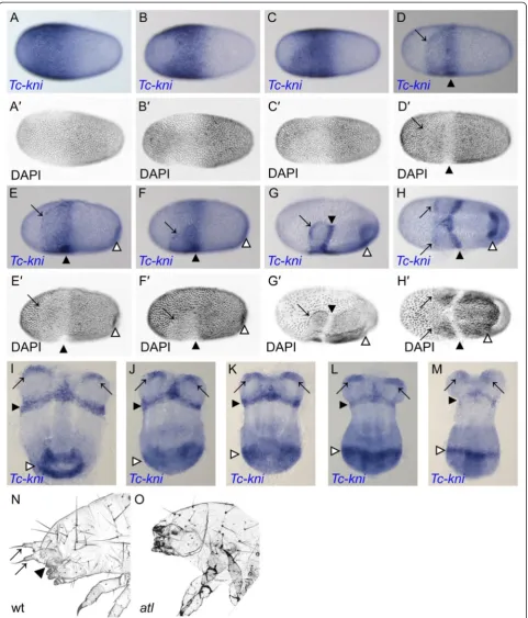
First, we studied in more detail the dynamics of anterior Tc-kni expression because previous studies had focused on posterior aspects [43]. Zygotic Tc-kni expression in the syncytial blastoderm begins as a broad domain cov-ering most of the anterior half of the blastoderm, only absent from a small region around the anterior pole (Figure 1A).Tc-kniexpression then retreats from the an-terior pole, but much more so on the dorsal side of the egg, clearing from regions that will later become the extra-embryonic serosa (Figure 1B) [55]. Tc-kni expres-sion is maintained in the anterior/ventral regions that will become head tissue [55] (Figure 1C). These early shifts in expression occur before embryonic and extra-embryonic tissue can be distinguished at the level of nu-clear morphology (Figure 1A’-C’and Cerny et al. [43]). When this distinction becomes apparent (Figure 1D’-F’), the uniform anterior Tc-kni expression domain splits
into two anterior lateral domains at the boundary between each head lobe and the abutting extra-embryonic tissue (black arrows), and a more posterior stripe marking the future anterior compartment of the mandibular segment (black arrowheads) (Figure 1D-F). These domains are maintained through subsequent blastoderm (Figure 1G-I) and early germband (Figure 1J, K) stages, before fading (Figure 1L, M) and then disappearing completely during mid-germband elongation stages. Hence, the early uni-form domain of Tc-kni expression (Figure 1C) encom-passes the entire future anterior head anlagen; i.e. everything anterior to the parasegment 0/1 boundary.
Antennalessis likely a regulatory mutation at theTc-kni
locus
In most RNAi experiments outside Drosophila, it re-mains unclear to what extent a genetic Null-situation is phenocopied. We aimed at determining the Null situ-ation by studying a genetic mutant.
Antennaless is a homozygous lethal mutation that was recovered from an EMS mutagenesis screen [54]. The cuticular phenotypes of antennaless mutant larvae are highly reminiscent of Tc-kni RNAi phenotypes (Tc-kniRNAi) [43]. Larvae of theantennaless mutant lack antennae and mandibles, and occasionally also display minor abdominal defects, very similar to those previ-ously reported for weakTc-kniRNAilarvae (Figure 1N, O and Cerny et al. [43]). In order to check for defects in dorsal and lateral head tissues which are derived from pre-antennal and intercalary regions, respectively [32,36,56], we scored both sides of 20 antennalesslarval cuticles for the head bristle pattern. All bristles were present, albeit shifted somewhat compared to the wildtype condition in these regions (Figure 1N, O). This is consist-ent with pre-antennal regions and much of the intercalary segment not being affected in the mutant [32,43].
Figure 1Embryonic expression ofTc-kniand theTc-knihead cuticle phenotype. A-M: Expression ofTc-kniduring blastoderm (A-H’) and early germband stages (I-M). See text for a detailed description. PanelsA’-H’show inverted DAPI images of the embryos in panelsA-Hrespectively. Black arrows in panelsD-MmarkTc-kniexpression at the anterior border of one or both head lobes. Black arrowheads in panelsD-Mindicate the stripe of
Tc-kniexpression marking the anterior compartment of the mandibular segment. White arrowheads in panelsE-Mmark the posterior domain ofTc-kni
segment. At 25°C this structure was defective in 79% of the phenotypic larvae examined (n = 43), compared to 44% at 32°C (n = 32).
In offspring from nine independent pairs of heterozy-gous parents, theantennalesscuticle phenotype is found in 25% (n = 364) and 27% (n = 267) of larvae at 25°C and 32°C respectively, levels consistent with a highly pene-trant homozygous lethal mutation. A significant propor-tion of mutant larvae were able to hatch despite their lack of antennae and mandibles; 45% at 25°C (n = 91) compared to only 17% at 32°C (n = 72). The vast major-ity of these larvae died at the L1 stage. Only very rarely did individuals reach the L2 stage. When corrected for a background reduction in hatching rate associated with this shift in temperature (measured from wildtype lar-vae), these data reveal a 19% increase in sensitivity to temperature with regards to hatching in theantennaless mutant background.
In our Tc-kni RNAi experiments head defects were also found to be more severe at 32°C compared to 25°C (see Additional file 1, compare panel B to panel A). The severity of head defects decreased over time post-injection as expected for parental RNAi experiments [57] (see Additional file 1, panel C). However, as for antennaless, we found inverse temperature sensitivity with respect to defects in segments A5-A8 following Tc-kniRNAi(see Additional file 1, compare panel E to panel D). The frequency of abdominal defects also increased with time post-injection from about 20% (n = 10) at day eight injection, to 37% (n = 35) eleven days injection, and to 56% (n = 27) thirty-one days post-injection, before dropping again (For eggs at 25°C; see Additional file 1, panel D). This is not in line with the usual observation that in RNAi experiments phenotypic strength decreases over time and indicates a complex re-lationship between knockdown and phenotype, which we do not fully understand.
Since the antennaless cuticle defects described above are almost identical to the Tc-kniRNAi phenotypes both in terms of physical phenotype and sensitivity to temperature, we suspectedTc-knias the gene affected by the mutation. We therefore independently sequenced the three Tc-kni exons from the genomic DNA of two first instar larvae that had been identified as homozy-gous mutant by their cuticle phenotype. We found that the coding sequence of Tc-kni is not altered in mutant beetles (data not shown).
In order to test the hypothesis that antennaless is a regulatory mutation at the Tc-kni locus, we carried out in situhybridization on embryos from heterozygous mu-tant parents with a mix of probes targeting Tc-kni and Tc-caudalas positive control. We failed to detectTc-kni expression in 15% of the offspring examined (n = 100), whereas Tc-caudal was well stained in the same colour
reaction in all cases (see Additional file 2). This is con-sistent with embryos homozygous for theantennaless al-lele not expressing Tc-kniat levels detectable via in situ hybridization.
Taken together, our results are consistent with the hy-pothesis that antennaless is a regulatory mutation of Tc-kni, and as such we now refer to it asTc-kniatl. How-ever, we cannot rule out thatantennalessis a mutation in a gene that acts rather exclusively upstream of, or is a re-quired interaction partner of,Tc-kni. Further studies will be necessary to confirm our hypothesis by identifying the cis-regulatory regions that are affected by the mutation.
TheTc-kniNull phenotype is revealed by bothTc-kniatl
andTc-kniRNAi
The lack of Tc-kni expression in homozygous Tc-kniatl embryos implies thatTc-knifunction is strongly reduced in the mutant. However, it remains possible that some residual function remains. In order to test whether Tc-kniatl constitutes a complete Null phenotype for Tc-kni, we performed Tc-kni RNAi in theTc-kniatl background, assuming that the added effects should not lead to stronger phenotypes if Tc-kniatl is a Null mutant. The frequency of the Tc-kni phenotype in the pooled off-spring from 30 independent pairs of Tc-kniatl heterozy-gous animals was 11% (n = 149) at 25°C and 17% (n = 82) at 32°C. As expected, after injecting the same fe-males with Tc-kni dsRNA, the frequency of the Tc-kni phenotype strongly increased, to 97% (n = 32; 25°C) and 81% (n = 32; 32°C). Crucially, we did not find evidence for an increase in the severity of the cuticle phenotype: i.e. there were no larvae with phenotypes more severe than those seen in Tc-kniatlorTc-kniRNAialone. Hence, unlike in most RNAi experiments, we can be rather confident that the most severe RNAi phenotypes we observe fully phenocopy a Null situation. Therefore, we use results from both mutant and RNAi embryos for this work.
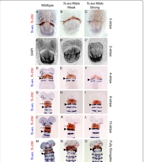
Tc-kniis differentially required for specifying antennal, intercalary and mandibular parasegment boundaries
the anterior median boundary of the Tc-Dfd domain) (Figure 2J-K, N-O, black asterisks).
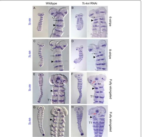
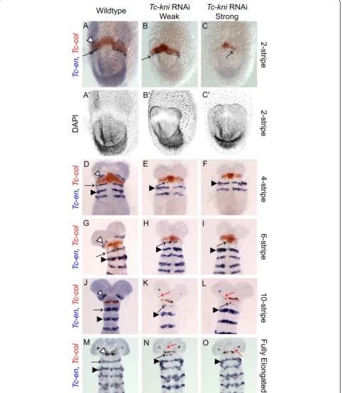
We find that bothTc-en(Figure 2) andTc-hh(Figure 3) expression is absent in the antennal segment in Tc-kniRNAi embryos throughout embryogenesis (marked by white arrowhead in wildtype embryos in Figures 2 and 3). Occasionally,Tc-en and Tc-hhexpression associated with structures at the base of the antenna remains in later germband embryos, despite the clear failure of the an-tenna to form (marked by a red arrow in panels K, L, M, N and O in Figure 2 and panels F and H in Figure 3).
The mandibular stripe of Tc-en expression is initiated in early Tc-kniRNAi germband embryos, albeit abnor-mally; the stripe of expression is broken and in extreme cases only patches of expression are seen (compare panels B and C with panel A in Figure 2; mandibular stripe marked by black arrow). In older Tc-kniRNAi germband embryos,Tc-enexpression associated with the mandibular segment is often missing completely (com-pare panels F, I and O, with panels D, G and M respect-ively in Figure 2), suggesting a failure to properly maintain Tc-en expression in this segment in strong knockdowns of Tc-kni expression. Similarly, Tc-hh ex-pression in the mandibular segment is greatly reduced in young Tc-kniRNAi embryos, but a thin broken stripe is still detected medially in early germbands (compare black arrows in panels B and D to panels A and C re-spectively in Figure 3). Later, this Tc-hhexpression dis-appears in contrast to wildtype (compare panels F and H to panels E and G respectively in Figure 3). Note that Tc-en mandibular expression appears to be more sensi-tive to the loss ofTc-kni expression in lateral regions of the germband (Figure 2E, H, K and N).
Tc-enand Tc-wgexpression in the intercalary segment appears relatively late and at a quite variable time point during mid germband elongation in wildtype embryos. Whilst we cannot completely rule out minor disruptions of intercalary Tc-en/Tc-hh expression initiation and/or maintenance inTc-kniRNAiembryos, we do detect wildtype expression of these genes in late elongation (i.e. post 10 Tc-enstripe embryos) and early fully elongatedTc-kniRNAi embryos (black asterisk in panels K, L, N and O in Figure 2 and black asterisk in panel F in Figure 3).
In conclusion, the disruption ofTc-knifunction leads to the complete failure to initiate segment polarity gene ex-pression in the antennal segment, failure to properly maintain segment polarity gene expression in the man-dibular segment, and most likely has no effect on segment polarity gene expression in the intercalary segment [43].
Molecular markers show thatTc-kniis not a canonical head gap gene
Segment polarity genes mark a posterior portion of each segment. Therefore, we also asked whether the anterior
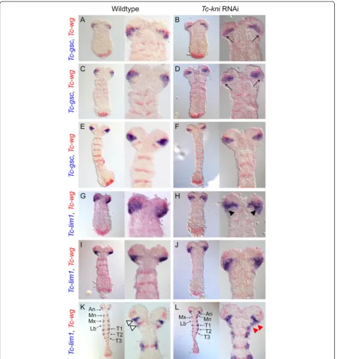
parts of the segments are miss-specified and/or missing in Tc-kniRNAiembryos by analyzing markers for the an-terior regions of head segments.Tc-goosecoid(Tc-gsc) ex-pression [56] initially partially overlaps the ocular Tc-wg domain and extends posterior to it (Figure 4A). Later, this domain widens in lateral regions, forming a wedge shape domain that abuts the antennal Tc-wgstripe as it appears (Figure 4C) but later retracts from it (Figure 4E). Thus,Tc-gscis a marker for posterior ocular and anterior antennal regions in early embryos but later predominantly marks ocular tissue. In early Tc-kniRNAi germband em-bryos, Tc-gsc expression is down-regulated in posterior-lateral regions of the wedge shaped expression domain (black arrows in Figure 4B, D; compare to Figure 4A, C re-spectively). In olderTc-kniRNAiembryos, theTc-gsc expres-sion domain again closely resembles the wedge shape seen in wild-type embryos at these stages (Figure 4F). This indi-cates that the ocular parasegment boundary and anterior parts of the antennal segment are not greatly affected by loss of Tc-kni, apart from some degree of lateral down-regulation ofTc-gscin early germ bands.
Tc-lim1 is a similar marker for the posterior of the ocular segment and anterior compartment of the anten-nal segment [56], albeit shifted slightly posteriorly rela-tive to Tc-gsc. In early embryos, expression is partially overlapping the ocularTc-wgdomain and extending pos-terior to it (Figure 4G). In elongating germbands, Tc-lim1 expression forms a wedge shaped domain that covers all cells between the ocular and antennal Tc-wg stripes (Figure 4I). In the Tc-kniRNAi background, the posterior boundary of this domain is irregular and the domain is somewhat narrower indicating posterior re-duction of the Tc-lim marked tissues (Figure 4H). In wild-type elongating germbands, additionalTc-lim1 mental expression arises that laterally overlaps the seg-mentalTc-wg domains in gnathal and thoracic segments (Figure 4K). At these stages, the ocular-antennal domain ofTc-lim1expression splits into a posterior domain that overlaps the antennal Tc-wg stripe, and an anterior do-main positioned between the ocular and the antennal Tc-wg stripes (Figure 4K; white arrowheads). Expression in the ocular-antennal region remains also inTc-kniRNAi germbands, but the domain is not split as in wildtype (upper red arrowhead in Figure 4L) and appears to be fused with the mandibular domain of lateral Tc-lim1 ex-pression (lower red arrowhead in Figure 4L). In extreme cases this domain is also fused to the maxillaryTc-lim1 ex-pression domain (Figure 4L and discussed below). This confirms that anterior antennal tissue is properly specified while the parasegmental boundaries of the antennal and mandibular segments are not formed correctly.
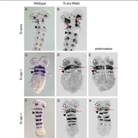
Figure 2Expression ofTc-enandTc-Dfdin wildtype andTc-kniRNAi embryos.The antennalTc-enstripe (marked by white arrowhead in wildtype panelsA, D, G, J, M) is missing in allTc-kniRNAi embryos. The mandibularTc-enstripe (marked by black arrow) forms within theTc-Dfd
to theTc-wgstripe in each segment [36]. Tc-ems expres-sion therefore predominantly marks the anterior portion of each segment. In contrast to wild-type expression, the antennal and mandibularTc-emssegmental domains are fused in Tc-kniRNAi embryos (red arrows in Figure 5B), as seen forTc-lim1. This is consistent with defects being restricted to the posterior compartment of the antennal
segment leading to the fusion of the antennal and man-dibularTc-emsexpression domains.
[image:9.595.58.542.89.554.2]Tc-sloppy-paired-1(Tc-slp-1) is also expressed in a seg-mentally reiterated pattern in the developing Tribolium head (Figure 5C, F and [25,32]). Tc-slp-1 expression do-mains overlap the Tc-wg stripe in each head segment, but also extend further into the anterior compartment,
as well as a little across the parasegmental boundary into the posterior compartment [25,32]. Tc-slp-1 expression therefore predominantly marks a posterior portion of the anterior segment compartment.Tc-slp-1is expressed in a stripe in the antennal and each gnathal head segment in bothTc-kniRNAiand Tc-kniatl embryos (Figure 5D, E and
[image:11.595.59.538.89.566.2]G, H), as in wild-type embryos (Figure 5C, F). However, the antennal stripe often appears to broaden (white arrow-head in Figure 5D, E, H), and the distance between the mandibular and the maxillaryTc-slp-1stripes is often de-creased, completely fusing in lateral regions (red arrows in Figure 5D, E and G, H). These data indicate that overall
Figure 5Expression ofTc-emsandTc-slp-1in wildtype,Tc-kniRNAi andantennalessembryos.PanelsB,D, E, G and Hrepresent documentation of fluorescent FastRed stainings of the respective gene and are therefore shown in grayscale.Tc-emsexpression in the antennal (white arrowhead, panelsA, B) and mandibular (black arrow, panelsA, B) segments is abnormally fused inTc-kniRNAi embryos (red arrows in panelB; compare to panelA). A black arrowhead marks the maxillary domain ofTc-emsexpression in panelsA, B. The antennalTc-slp-1
theTc-slp-1stripes are not dramatically affected byTc-kni RNAi. However, the decreased distance between the man-dibular and maxillary stripes could indicate incorrectly specified tissue in the intervening posterior compartment of the mandibular segment.
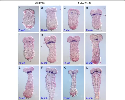
A normal complement of bristles and setae in lateral regions of Tc-kniatl larval heads implies the retention of intercalary segment derived cuticle inTc-kniknockdown embryos (Figure 1 and [32]). SinceTc-labial (Tc-lab) is expressed during embryogenesis throughout the pre-sumptive intercalary segment [58], and is required for its formation [32], we used it as a molecular marker for the presence/absence of the intercalary segment. In Tc-kni RNAi embryos (Figure 6G-L),Tc-lab expression appears prematurely but with similar dynamics as in wildtype (Figure 6A-F). The prematureTc-labexpression domain
first appears medially as a pair of dots on either side of the median mesoderm (Figure 6G; compare to wild-type in Figure 6A). Tc-labexpression then extends into more lateral and medial regions. Expression is not entirely wildtype, as it may form an unusual regular stripe with-out the typical median expansion (compare Figure 6H, L with D, F), or it may be laterally reduced (Figure 6I, J, K). However, in all instances, the stripe was present indi-cating that the intercalary segment is present albeit its morphology appears not to be entirely unaffected by ad-jacent defects.
[image:12.595.59.539.300.685.2]We also examined the expression of another marker for the intercalary segment inTc-kniRNAiembryos, the second order regulator Tc-collier(Tc-col), also called Tc-knot. Tc-colacts downstream ofTc-laband is required for wildtype expression ofTc-enin the intercalary segment [31]. Aside
from a slight delay in the initial appearance of theTc-col expression domain in early embryos (Figure 7 compare B, C with A) we found no evidence of strong disruption of Tc-col expression in aTc-kniRNAi background (Figure 7). Besides, this staining confirmed the identity, and wildtype appearance, of the intercalary spots ofTc-enin late elong-ating and early fully elongated embryos (black asterisk in panels K, L, N and O in Figure 7).
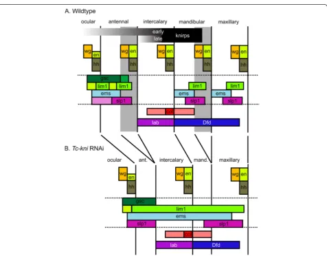
TheDrosophilahead gap genes lead to the loss of en-tire adjacent segments but their Tribolium orthologues do not fit this definition [36]. Similarly, taken together, our data (summarized in Figure 8) show that Tc-kni
[image:14.595.61.538.244.616.2]cannot be considered a canonical head gap gene, be-cause Tc-kni function is only required for the posterior parts of the antennal and mandibular segments (the re-gions shaded grey in Figure 8A). Moreover,Tc-kniis not required for the formation of the intercalary segment, which lies between the affected antennal and mandibular segments. Intercalary segment polarity gene expression is instead likely dependent on a conserved pathway in-volvingTc-lab and Tc-col[31,32]. Overall, our data con-firm that the regulatory networks underlying the establishment of the anterior head segments have di-verged significantly between Tribolium and Drosophila,
Figure 8Schematic diagrams summarizing the expression ofTriboliumhead genes in wildtype andTc-kniRNAiembryos.Changes in
marker gene expression followingTc-kniRNAi are consistent with a failure to specify and form posterior antennal and mandibular segmental regions (marked in grey inA, and deleted inB). Deletion of these regions explains the absence of segment polarity gene expression in antennal and mandibular segments, the deletion of posterior regions of the ocular/antennal domains ofTc-gscandTc-lim1at early stages, the reduced width of theTc-Dfddomain, and the reduction in distance between antennal, mandibular and/or maxillary domains ofTc-lim1,Tc-emsand/or Tc-slp-1. Abnormalities in the expression patterns of the genes bounded by the horizontal doted lines are most apparent in lateral regions of the germband. Additional defects affecting the maxillary segment have to be assumed to explain the fusion of respectiveTc-emsandTc-slpstripes. These could be due to aberrant morphogen signaling of the mandibular parasegment boundary. InTc-kniRNAi, the antennal parasegment boundary is never established while our model assumes that the posterior of the mandibular segment is lost later due to disturbance of the
involving changing roles for knirps, buttonhead and empty spiracleshomologues [36].
TheTc-kniphenotype is due to a failure to correctly specify cell fates
We decided to check whether missing head regions in Tc-kniknockdown and mutant embryos were lost due to a failure to maintain already specified tissue - which might be indicated by high levels of cell death - or through the failure to specify cells to their correct devel-opmental fate. Using TUNEL staining, we did not detect any apoptosis in either the blastoderm (see Additional file 3) or early germband stages (see Additional file 4) in Tc-kniRNAiembryos. In later germband stages there were a few apoptotic cells detectable in the head (as well as the posterior growth zone), but no more than in wild-type embryos, and there was no specific pattern of apoptosis that would indicate the loss of the tissues in question, namely the posterior portions of the antennal and man-dibular segments (see Additional files 3 and 4). Thus, the lack of antennae and mandibles inTc-kniRNAiandTc-kniatl larvae is not due to tissue degeneration, but rather the fail-ure to specify the respective segmental regions.
The posterior border of the anteriorTc-kniexpression domain is regulated byTc-even-skipped
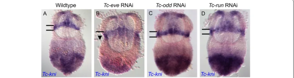
In theTriboliumblastoderm, expression ofTc-kniand the pair rule genes occurs at the same time, opening the possi-bilityTc-knicould be regulated by pair rule genes. Indeed, inTc-eveRNAiblastoderm embryos, we found that the an-terior domain of Tc-kni expression expanded into more posterior regions (Figure 9: compare panel B to panel A). In contrast, expression defects in this Tc-kni mandibular domain were not seen in Tc-oddRNAi or Tc-runRNAi germbands (Figure 9: compare panels C and D with A). This is consistent with the first pair-rule and/or segmental stripe ofTc-eveexpression being required to set the pos-terior boundary of mandibularTc-kniexpression.
Head and trunk segmentation gene networks cooperate to establish the mandibular segment in a different way in
TriboliumandDrosophila
[image:15.595.60.539.558.685.2]In Drosophila, the anterior head gene regulatory net-work (through btd, col and cnc) and the gnathal/trunk regulatory gene network (througheve) cooperate to pat-tern aspects of the mandibular segment (Figure 10A and [11,12]). In this study we provide evidence that this is also true inTribolium. BothTc-eveandTc-kniRNAi lead to the failure to properly maintain mandibular Tc-en stripe expression and as a result to loss of the mandible itself (this study and [24,43]). In our model (Figure 10B) Tc-knicontributes to the activation of mandibularTc-wg expression. This interaction could be direct because the respective expression patterns overlap. Tc-eve in turn could be directly involved in activatingTc-hh and Tc-en within its anteriormost expression domain. Subse-quently, the interactions of the Wnt and hh pathways ensure maintenance of the parasegement boundary. In the absence ofTc-kniexpression,Tc-evestill partially ini-tiates Tc-en/Tc-hh expression in the posterior compart-ment, which leads to partial initiation of Tc-wg in the anterior adjacent cells (through Hedgehog signaling). However, absence of Tc-kni leads to failure to maintain Tc-wg expression, and as a result failure to maintain Tc-en expression in posterior adjacent cells (through Wingless signaling). Interestingly, in Drosophila, the maintenance of en and wg expression in the mandibu-lar segment appears more interdependent in dorsal regions [13]. The same might also be true in Tribolium, since both Tc-en and Tc-wg expression is maintained more often in medial (i.e. ventral) regions in Tc-kni mutant and knockdown embryos. Taken together, it appears to be a conserved feature of in-sects that two systems cooperate in patterning the mandible, but the molecular details of this cooperation have diverged significantly between Drosophila and Tribolium (Figure 10).
Figure 9Tc-kniis regulated byTc-eve.TheTc-knihead expression domain, which covers the anterior compartment of the mandibular segment (bounded by black horizontal lines in panelsA, C, D) extends posteriorly (arrowhead in panelB), in aTc-eveRNAi background, but not inTc-odd
Conclusions
In this study we show that anterior regions of the antennal and mandibular segment, as well as the intervening intercal-ary segment, are correctly specified in the absence ofTc-kni function. Moreover, we identify key differences in the role of
[image:16.595.61.539.85.600.2]Tc-kni in setting up the non-adjacent antennal and man-dibular parasegment boundaries. We identify the pair-rule geneTc-even-skippedas a potential positive regulatory input that enables the initial specification (but not maintenance) of the mandibular parasegmental boundary in the absence
Figure 10The genetic interactions that help establish the PS0/PS1 parasegmental boundary inDrosophilaandTribolium.InDrosophila
ofTc-knifunction. We also present the first evidence that, as inDrosophila, head and trunk gene regulatory networks cooperate to specify the mandibular segment. However, our data, and those of others, point to significant divergence in the molecular interactions involved in mandibular pattern-ing betweenDrosophilaandTribolium.
A recent study of the expression and function of head patterning genes in the hemimetabolous insect Onco-peltus fasciatus suggests that the developmental gene networks operating in Tribolium castaneum more closely resemble the ancestral insect condition, perhaps not surprisingly given the highly derived Drosophila lar-val head [59]. Indeed, recent data from a myriapod sug-gest that the developmental genetic basis of Tribolium larval head development might even closely resemble the ancestral arthropod condition [60,61]. It will be in-teresting to see whether studies on other arthropods re-veal an ancestral role for knirps-family homologues in head segmentation, which would implyemsandbtdhave usurped the role of knirps, and potentially other genes, in the lineage leading toDrosophila.
Methods
Antennalessmutant analysis
Theantennalessmutant line was maintained as described by Berghammer et al. [62] and Maderspacheret al. [54]. The separation of offspring from single pair crosses was performed after three and six days of egg laying at 32°C and 25°C respectively. Cuticle preparations of mutant larvae were prepared as previously described [54]. Genomic DNA was extracted from individual L1 antennaless mutant and wildtype larvae by first removing flour and chorion by bleaching egg collections two times for four minutes in 100% bleach. Embryos were washed three times with extrac-tion buffer (without proteinase K - see below), put individu-ally into 0.5 ml cups and frozen at−20°C. 10μl of extraction buffer was added (25 mM NaCl, 10 mM Tris pH8.0, 1 mM EDTA, 200μg/ml proteinase K) and the embryos macerated with a pipette tip. After 90mins at 42°C, the preparations were shortly spun down and proteinase K was inactivated by three minutes at 95°C. Debris was spun down for two mi-nutes at 14000 rpm and 8μl of supernatant was transferred to new tubes. 4μl were used for a 20μl PCR reaction. The threeTc-kniexons were amplified by PCR using the follow-ing four primer combinations: ex1 fw 5′-ACATTCCC CACCATTGAAATCACA-3′; ex1 rev 5′-GGGTTAAGTT TCTCGGTATTGGGACTA-3′; ex2 fw 5′-CCTGTAATG TGTACAGTCACGAGCAG-3′; ex2 rev 5′-ATTCTTGCA TCGGCCGAAGTTTACGT-3′; ex3A fw 5′-CGGAAGCT CTGTCAAACAATAATCTCA-3′; ex3A rev 5′-TCCAG GAACACCCGCTTGTTGA-3′; ex3B fw 5′-CGCCGACGT TTCTACCTCCTCA-3′; ex3B rev 5′-TCGACGCTAATA GCTGCCATCATC-3′. Sequencing of the PCR products was performed by Macrogen (Korea).
Expression analysis
Fixation of embryos and enzymatic single and double in situ hybridizations were carried out according to established protocols [63]. For double in situ hybridiza-tions Fastred ® (Sigma) was sometimes used (e.g. for Tc-wg) in place of INT/BCIP (Roche 11-681-460-001) [63]. Probes for in situ hybridization were prepared using ei-ther the Digoxigenin RNA Labeling Kit or Fluorescein RNA Labeling Kit (Roche Applied Science, Mannheim) following established protocols and the manufacturers instructions [63]. The DNA templates used for RNA probe production were in some cases (i.e. for probes detecting Tc-ems, Tc-slp-1, Tc-wg, Tc-eve, Tc-gsc, Tc-hh, Tc-lab, Tc-lim1, Tc-odd, Tc-run) produced by PCR-amplifying DNA fragments of the gene of interest from clones using appropriate vector primers (T3, T7, SP6). In other cases (i.e. for probes detecting Tc-en, Tc-col, Tc-Dfd) clones containing a fragment of the gene of interest were used directly as templates following 5′ linearization using an appropriate restriction enzyme. Clones and information are available on request. Follow-ingin situhybridizations, nuclei in blastoderm and early germband embryos were sometimes counterstained using Hoechst 33258 (Additional file 2), Hoechst 33342 (Additional file 3) or DAPI (Figures 1, 2 and 7).
Parental RNAi
Adult or pupal parental RNAi was carried out using established protocols [64]. dsRNA was produced using the T7 and SP6 MEGAscript High Yield Transcription Kits (Ambion). Template DNA was either PCR-amplified using the vector insert flanking primers T7: 5′-TAATACGAC TCACTATAGG-3′and T7-Sp6: 5′-TAATACGACTCACT ATAGGATTTAGGTGAACACTATAGA-3′) or by using a stock of the linearized plasmid. In this case the antisense and sense strands were amplified separately, and later the ssRNA combined in equimolar amounts. A concentration of between 2 and 4.3μg/μl ofTc-knidsRNA was injected in each experiment, since this concentration range has been previously shown to consistently produce fully pene-trantTc-kniRNAi phenotypes [43]. To knockdownTc-eve, Tc-odd and Tc-runt concentrations of between 2 and 3.5μg/μl of dsRNA were used.
TUNEL staining
positive control embryos were washed three time in DNaseI buffer (40 mM Tris–HCl, pH7.5, 0.1 mM dithiothreitol, 6 mM MgCl2), incubated for 30 minutes at 37°C in DNaseI buffer with 0.06 U DNaseI per micro-litre of buffer, before being washed several times in PBT. All embryos were then incubated for 20 minutes in 0.1% sodium borohydride. During incubation, the embryos were gently shaken several times. The sodium borohy-dride was then removed by repeated washing with TdT buffer (140 mM cacodylic acid, 1 mM cobalt chloride, 30 mM Tris–HCl, pH7.2). Embryos were then incubated at 37°C in TdT buffer containing 20μM DIG-UTP and 0.3 U/μl terminal deoxynucleotidyl transferase (TdT) (Sigma). In the case of negative control embryos, the TdT was omitted. All embryos were then washed three times for five minutes in TBST (140 mM NaCl, 2.7 mM KCl, 25 mM Tris–HCl pH7.4, 0.1% Tween-20) at room temperature, before being incubated at 70°C for 20 minutes in TBST. Embryos were then washed three times for five minutes in PBT, before being incubated in PAS (PBT with 10 mg/ml bovine albumine (BSA) and 2% sheep serum) for one hour. Embryos were then incu-bated in PAS with anti-Dig antibody (1:2000) for one hour. This was followed by several washes in PBT for a total of two hours. Embryos were stored overnight at 4° C in PBT before being washed for 30 minutes at room temperature in PBT prior to NBT/BCIP staining. The staining was stopped by repeated washing in PBT and the embryos stored at 4°C in 1 ml of PBT + 125μl of for-maldehyde (37%).
Microscopy
Mostin situhybridization stained preparations were im-aged on an Axioplan 2 photomicroscope (Carl Zeiss Vi-sion GmbH, Jena) using a polarized light (DIC) filter with low Normaski contrast (ImageProPlus, Version 5.0 .2.9, MediaCybernetics). For clear detection of the fluor-escence signal of the Fastred® color reaction the filter set No. 43 (Cy3) from Zeiss, and a mercury vapor lamp HBO100 as a light source, was used. For the detection of Hoechst 33258 and Hoechst 33342 signal the filter set No. 49 from Zeiss (DAPI filter), and a mercury vapor lamp as the light HBO100 source, was used. A few in situhybridization stained preparations were imaged on a Leica MZ16F epifluorescence stereoscope with a DFC300FX digital camera (images in Figures 2 and 7). Larval cuticles were imaged using a confocal laser-scanning microscope (LSM 510, Zeiss) and processed as described [65].
Availability of supporting data
The data set supporting the results of this article is in-cluded within the article (and its additional files).
Additional files
Additional file 1:The sensitivity ofTc-kniRNAi head and abdominal larval phenotypes to temperature.(A, B) Head phenotypes are more severe at higher temperatures. Note the higher frequency of strong head phenotypes (up to ~90% vs. circa 70%) and lower frequency of weak head phenotypes (0% at some time points at 32°C) followingTc-kniRNAi carried out at 32°C compared to 25°C. Larvae exhibiting strong head phenotypes lacked both antennae and mandibles, larvae exhibiting medium phenotypes possessed at least one antenna and larvae exhibiting weak phenotypes exhibited at least one antenna and mandibles. (C) The frequency of larval head phenotypes decreases with time postTc-knidsRNA injection. (D, E) In contrast to head phenotypes, abdominal phenotypes showed an unusual reverse sensitivity with respect to temperature and time followingTc-kniRNAi injection. Abdominal phenotypes were more common followingTc-kniparental RNAi carried out at 25°C compared to 32°C (compare height of blue bars in panelDvs. panelE). Abdominal phenotypes showed the unusual characteristic of increasing in frequency with time postTc-knidsRNA injection.
Additional file 2:Tc-kniis not expressed inantennalessembryos. Wildtype andantennalessblastoderm (panelsA-D) and early germband (panelsE-H) embryos co-stained with a mix ofTc-kniandTc-cadprobes detected with the same colour reaction. A probe againstTc-cadwas used to control against the possibility that the absence ofTc-knisignal in
antennalessembryos was due to technical problems. Blastoderm embryos were stained with Hoechst 33258 (A’-D’) in order to identify similar stage embryos. Inantennalessblastoderm embryos, a block of signal (bounded by white lines in panelsA, C) corresponding to the anterior headTc-kniexpression domain is missing, whereas the posterior domain ofTc-cadexpression is detected. Inantennalessearly germband embryos the anterior mandibular stripe ofTc-kniexpression (black arrowhead in panelsE, G) is missing, whereas the posterior growth zone domain ofTc-cadexpression is detected. Similar experiments using a
Tc-otdprobe as control proved that the posteriorTc-kniexpression domain is also missing inantennalessblastoderm and germband embryos (data not shown). PanelsA-D’:Lateral views, anterior to the left. PanelsE-H:Ventral views, anterior to the top.
Additional file 3:TUNEL stained wildtype andTc-kniRNAi blastoderm stage embryos.DAPI staining (A’-F’) was used to identify wildtype andTc-kniRNAi blastoderm embryos of similar stages. No apoptotic nuclei were observed in wildtype orTc-kniRNAi blastoderm embryos.
Additional file 4:TUNEL stained wildtype andTc-kniRNAi germband stage embryos.Apoptotic nuclei were not observed in wildtype orTc-kniRNAi early germband stage embryos (A-F). A few apoptotic nuclei (arrowheads in panelsG-J) were observed in mid-elongation (G-H), late-elongation (I-J) and fully elongated (K-L) wildtype andTc-kniRNAi germband embryos. However, levels of apoptotic nuclei were no higher in
Tc-kniRNAi germband embryos when compared to controls, and apoptotic nuclei were not concentrated in regions within which the antennal and mandibular segments should or would develop. Note that in some cases (panelsC, E, G,I, N) TUNEL reactions were developed for much longer than needed to detect apoptotic nuclei, leading to background staining. Note that apoptotic nuclei can nevertheless be distinguished from background (e.g. panelsG, I).
Competing interests
The authors declare that they have no competing interests.
Authors’contributions
AP performed experiments, analyzed the data and wrote the paper, DG, JS, FR and GO performed experiments and analyzed the data, JS supervised experiments and analyzed the data, AG drafted the paper, MK identified the
Acknowledgements
We thank Michalis Averof for the use of reagents and laboratory space and Ernst A. Wimmer for support. This work was funded by Deutsche Forschunggemeinschaft DFG (BU-1443/3-1). MK’s screen for segmentation mutants was funded by DFG grant Kl656/2.
Author details 1
Institute of Molecular Biology and Biotechnology (IMBB), Foundation for Research and Technology - Hellas (FoRTH), Nikolaou Plastira 100, GR-70013, Heraklion, Crete, Greece.2Blumenbach Institute of Zoology,
Georg-August-University Göttingen, Justus-von-Liebig-Weg 11, 37077, Göttingen, Germany.3Department of Biology, Friedrich-Alexander University Erlangen, Staudtstrasse 5, 91058, Erlangen, Germany.
Received: 1 April 2013 Accepted: 12 June 2013 Published: 18 June 2013
References
1. Posnien N, Schinko JB, Kittelmann S, Bucher G:Genetics, development and composition of the insect head–a beetle’s view.Arthropod Struct Dev
2010,39:399–410.
2. Scholtz G, Edgecombe GD:The evolution of arthropod heads: reconciling morphological, developmental and palaeontological evidence.Dev Genes Evol2006,216:395–415.
3. Rogers BT, Kaufman TC:Structure of the insect head in ontogeny and phylogeny: a view fromDrosophila.Int Rev Cytol1997,174:1–84. 4. Budd GE:A palaeontological solution to the arthropod head problem.
Nature2002,16:271–275.
5. Schmidt-Ott U, González-Gaitán M, Jäckle H, Technau GM:Number, identity, and sequence of theDrosophilahead segments as revealed by neural elements and their deletion patterns in mutants.Proc Natl Acad Sci USA1994,91:8363–8367.
6. Haas MS, Brown SJ, Beeman RW:Pondering the procephalon: the segmental origin of the labrum.Dev Genes Evol2001,211:89–95. 7. Rempel GJ:The evolution of the insect head: the endless dispute.Quaest
Entomol1975,11:7–25.
8. St Johnston D, Nüsslein-Volhard C:The origin of pattern and polarity in theDrosophilaembryo.Cell1992,68:201–219.
9. Cohen SM, Jurgens G:Mediation ofDrosophilahead development by gap-like segmentation genes.Nature1990,346:482–485.
10. Mohler J:Spatial regulation of segment polarity gene expression in the anterior terminal region of theDrosophilablastoderm embryo.Mech Dev
1995,50:151–161.
11. Crozatier M, Valle D, Dubois L, Ibnsouda S, Vincent A:Collier, a novel regulator ofDrosophilahead development, is expressed in a single mitotic domain.Curr Biol1996,6:707–718.
12. Crozatier M, Valle D, Dubois L, Ibnsouda S, Vincent A:Head versus trunk patterning in theDrosophilaembryo; collier requirement for formation of the intercalary segment.Development1999,126:4385–4394. 13. Gallitano-Mendel A, Finkelstein R:Novel segment polarity gene
interactions during embryonic head development inDrosophila.Dev Biol
1997,192:599–613.
14. Schmidt-Ott U, Technau GM:Expression ofenandwgin the embryonic head and brain ofDrosophilaindicates a refolded band of seven segment remnants.Development1992,116:111–125.
15. Vincent A, Blankenship JT, Wieschaus E:Integration of the head and trunk segmentation systems controls cephalic furrow formation inDrosophila.
Development1997,124:3747–3754.
16. Grossniklaus U, Cadigan KM, Gehring WJ:Three maternal coordinate systems cooperate in the patterning of theDrosophilahead.
Development1994,120:3155–3171.
17. Hülskamp M, Tautz D:Gap genes and gradients–the logic behind the gaps.Bioessays1991,13:261–268.
18. Wimmer EA, Cohen SM, Jäckle H, Desplan C:Buttonheaddoes not contribute to a combinatorial code proposed forDrosophilahead development.Development1997,124:1509–1517.
19. Gallitano-Mendel A, Finkelstein R:Ectopicorthodenticleexpression alters segment polarity gene expression but not head segment identity in the
Drosophilaembryo.Dev Biol1998,199:125–137.
20. Schöck F, Reischl J, Wimmer E, Taubert H, Purnell BA, Jäckle H:Phenotypic suppression ofempty spiraclesis prevented bybuttonhead.Nature2000, 405:351–354.
21. Ntini E, Wimmer E:Unique establishment of procephalic head segments is supported by the identification ofcis-regulatory elements driving segment-specific segment polarity gene expression inDrosophila.Dev Genes Evol2011,221:1–16.
22. Ntini E, Wimmer EA:Second order regulator Collier directly controls intercalary-specific segment polarity gene expression.Dev Biol2011,360:403–414. 23. Brown SJ, Shippy TD, Miller S, Bolognesi R, Beeman RW, Lorenzen MD,
Bucher G, Wimmer EA, Klingler M:The red flour beetle,Tribolium castaneum(Coleoptera): a model for studies of development and pest biology.Cold Spring Harb Protoc2009,2009:pdb emo126.
24. Choe CP, Miller SC, Brown SJ:A pair-rule gene circuit defines segments sequentially in the short-germ insectTribolium castaneum.Proc Natl Acad Sci USA2006,103:6560–6564.
25. Choe CP, Brown SJ:Evolutionary flexibility of pair-rule patterning revealed by functional analysis of secondary pair-rule genes, paired and sloppy-paired in the short-germ insect,Tribolium castaneum.Dev Biol
2007,302:281–294.
26. Choe CP, Brown SJ:Genetic regulation of engrailed and wingless in
Triboliumsegmentation and the evolution of pair-rule segmentation.Dev Biol2009,325:482–491.
27. Farzana L, Brown SJ:Hedgehog signaling pathway function conserved in
Triboliumsegmentation.Dev Genes Evol2008,218:181–192. 28. Brown SJ, Patel NH, Denell RE:Embryonic expression of the single
Tribolium engrailedhomolog.Dev Genet1994,15:7–18.
29. Oppenheimer DI, MacNicol AM, Patel NH:Functional conservation of the
wingless-engrailedinteraction as shown by a widely applicable baculovirus misexpression system.Curr Biol1999,9:1288–1296. 30. Sarrazin AF, Peel AD, Averof M:A segmentation clock with two-segment
periodicity in insects.Science2012,336:338–341.
31. Schaeper ND, Pechmann M, Damen WGM, Prpic NM, Wimmer E: Evolutionary plasticity ofcollierfunction in head development of diverse arthropods.Dev Biol2010,344:363–376.
32. Posnien N, Bucher G:Formation of the insect head involves lateral contribution of the intercalary segment, which depends onTc-labial
function.Dev Biol2010,338:107–116.
33. Economou AD, Telford MJ:Comparative gene expression in the heads of
Drosophila melanogasterandTribolium castaneumand the segmental affinity of theDrosophilahypopharyngeal lobes.Evol Dev2009,11:88–96. 34. Nagy LM, Carroll S:Conservation ofwinglesspatterning functions in the
short-germ embryos ofTribolium castaneum.Nature1994,367:460–463. 35. Kittelmann S, Ulrich J, Posnien N, Bucher G:Changes in anterior head
patterning underlie the evolution of long germ embryogenesis.Dev Biol
2013,374:174–184.
36. Schinko JB, Kreuzer N, Offen N, Posnien N, Wimmer EA, Bucher G:Divergent functions oforthodenticle,empty spiraclesandbuttonheadin early head patterning of the beetleTribolium castaneum(Coleoptera).Dev Biol2008, 317:600–613.
37. Schoppmeier M, Fischer S, Schmitt-Engel C, Löhr U, Klingler M:An ancient anterior patterning system promotes caudal repression and head formation in ecdysozoa.Curr Biol2009,19:1811–1815.
38. Stauber M, Jackle H, Schmidt-Ott U:The anterior determinantbicoidof
Drosophilais a derived Hox class 3 gene.Proc Natl Acad Sci USA1999, 96:3786–3789.
39. Brown S, Fellers J, Shippy T, Dennell R, Stauber M, Schmidt-Ott U:A strategy for mappingbicoidon the phylogenetic tree.Curr Biol2001,11:R43–R44. 40. Fu J, Posnien N, Bolognesi R, Fischer TD, Rayl P, Oberhofer G, Kitzmann P,
Brown SJ, Bucher G:Asymmetrically expressedaxinrequired for anterior development inTribolium.PNAS2012,109:7782–7786.
41. Kotkamp K, Klingler M, Schoppmeier M:Apparent role ofTribolium orthodenticlein anteroposterior blastoderm patterning largely reflects novel functions in dorsoventral axis formation and cell survival.
Development2010,137:1853–1862.
42. Schröder R:The genesorthodenticleandhunchbacksubstitute forbicoid
in the beetleTribolium.Nature2003,422:621–625.
43. Cerny AC, Grossmann D, Bucher G, Klingler M:TheTriboliumortholog of
44. Oro AE, Umesono K, Evans RM:Steroid hormone receptor homologs in development.Development1989,107:133–140.
45. Rothe M, Nauber U, Jäckle H:Three hormone receptor-likeDrosophilagenes encode an identicle DNA-binding finger.EMBO J1989,8:3087–3094. 46. Gerwin N, La Rosée A, Sauer F, Halbritter H, Neumann M, Jäckle H, Nauber
U:Functional and conserved domains of theDrosophilatranscription factor encoded by the segmentation geneknirps.Mol Cell Biol1994, 14:7899–7908.
47. Naggan Perl T, Schmid BG, Schwirz J, Chipman AD:The Evolution of the
knirpsFamily of Transcription Factors in Arthropods.Mol Biol Evol2013, 30:1348–1357.
48. Rothe M, Wimmer EA, Pankratz MJ, González-Gaitán M, Jäckle H:Identical transacting factor requirement forknirpsandknirps-relatedgene expression in the anterior but not in the posterior region of the
Drosophilaembryo.Mech Dev1994,46:169–181.
49. González-Gaitán M, Rothe M, Wimmer EA, Taubert H, Jäckle H:Redundant functions of the genesknirpsandknirps-relatedfor the establishment of anteriorDrosophilahead structures.Proc Natl Acad Sci USA1994,91:8567–8571. 50. Nauber U, Pankratz MJ, Kienlin A, Seifert E, Klemm U, Jäckle H:Abdominal
segmentation of theDrosophilaembryo requires a hormone receptor-like protein encoded by the gap geneknirps.Nature1988,336:489–492. 51. Riddihough G, Ish-Horowicz D:Individual stripe regulatory elements in
theDrosophila hairypromotor respond to maternal, gap and pair-rule genes.Genes Dev1991,5:840–854.
52. Pankratz MJ, Seifert E, Gerwin N, Billi B, Nauber U, Jäckle H:Gradients of
Krüppelandknirpsgene products direct pair-rule gene stripe patterning in the posterior region of theDrosophilaembryo.Cell1990,61:309–317. 53. Clyde DE, Corado MS, Wu X, Pare A, Papatsenko D, Small S:A
self-organizing system of repressor gradients establishes segmental complexity inDrosophila.Nature2003,426:849–853.
54. Maderspacher F, Bucher G, Klingler M:Pair-rule and gap gene mutants in the flour beetleTribolium castaneum.Dev Genes Evol1998,208:558–568. 55. Handel K, Grünfelder CG, Roth S, Sander K:Triboliumembryogenesis: a
SEM study of cell shapes and movements from blastoderm to serosal closure.Dev Genes Evol2000,210:167–179.
56. Posnien N, Koniszewski ND, Hein HJ, Bucher G:Candidate gene screen in the red flour beetleTriboliumrevealssix3as ancient regulator of anterior median head and central complex development.PLoS Genet2011,7:e1002416. 57. Bucher G, Scholten J, Klingler M:Parental RNAi inTribolium(Coleoptera).
Curr Biol 122002,12:R85–R86.
58. Nie W, Stronach B, Panganiban G, Shippy T, Brown S, Denell R:Molecular characterization ofTclabialand the 3′end of theTriboliumcomplex.Dev Genes Evol2001,211:244–251.
59. Birkan M, Schaeper ND, Chipman AD:Early patterning and blastoderm fate map of the head in the milkweed bugOncopeltus fasciatus.Evol Dev
2011,13:436–447.
60. Janssen R, Budd GE, Damen WGM:Gene expression suggests conserved mechanisms patterning the heads of insects and myriapods.Dev Biol
2011,357:64–72.
61. Janssen R, Damen WGM, Budd GE:Expression ofcollierin the premandibular segment of myriapods: support for the traditional Atelocerata concept or a case of convergence?BMC Evol Biol2011,11:50. 62. Berghammer A, Bucher G, Maderspacher F, Klingler M:A system to
efficiently maintain embryonic lethal mutations in the beetleTribolium castaneum.Dev Genes Evol1999,209:382–389.
63. Schinko J, Posnien N, Kittelmann S, Koniszewski N, Bucher G:Single and double whole-mountin situhybridization in red flour beetle (Tribolium) embryos.Cold Spring Harb Protoc2009,2009:pdb prot5258.
64. Posnien N, Schinko J, Grossmann D, Shippy TD, Konopova B, Bucher G: RNAi in the red flour beetle (Tribolium).Cold Spring Harb Protoc2009, 2009:pdb prot5256.
65. Wohlfrom H, Schinko JB, Klingler M, Bucher G:Maintenance of segment and appendage primordia by theTriboliumgene knödel.Mech Dev2006, 123:430–439.
doi:10.1186/1471-213X-13-25
Cite this article as:Peelet al.:Tc-knirpsplays different roles in the specification of antennal and mandibular parasegment boundaries and is regulated by a pair-rule gene in the beetleTribolium castaneum.BMC Developmental Biology201313:25.
Submit your next manuscript to BioMed Central and take full advantage of:
• Convenient online submission
• Thorough peer review
• No space constraints or color figure charges
• Immediate publication on acceptance
• Inclusion in PubMed, CAS, Scopus and Google Scholar
• Research which is freely available for redistribution
![Figure 10 The genetic interactions that help establish the PS0/PS1 parasegmental boundary incompartment (PS1) [24]](https://thumb-us.123doks.com/thumbv2/123dok_us/7983176.203154/16.595.61.539.85.600/figure-genetic-interactions-help-establish-parasegmental-boundary-incompartment.webp)
