the Development of a Viral Disease Model for Multiple Sclerosis
Jinjong Myoung,a* Hyun Seok Kang,aWanqiu Hou,aLiping Meng,aMauro C. Dal Canto,band Byung S. Kima,b
Department of Microbiology-Immunologyaand Department of Pathology,bNorthwestern University Medical School, Chicago, Illinois, USA
Theiler’s virus-induced demyelinating disease has been extensively investigated as a model for persistent viral infection and mul-tiple sclerosis (MS). However, the role of CD8ⴙT cells in the development of disease remains unclear. To assess the role of virus-specific CD8ⴙT cells in the pathogenesis of demyelinating disease, a single amino acid substitution was introduced into the pre-dominant viral epitope (VP3 from residues 159 to 166 [VP3159-166]) and/or a subdominant viral epitope (VP3173-181) of
susceptible SJL/J mice by site-directed mutagenesis. The resulting variant viruses (N160V, P179A, and N160V/P179A) failed to induce CD8ⴙT cell responses to the respective epitopes. Surprisingly, mice infected with N160V or N160V/P179A virus, which lacks CD8ⴙT cells against VP3159-166, did not develop demyelinating disease, in contrast to wild-type virus or P179A virus
lack-ing VP3173-181-specific CD8ⴙT cells. Our findings clearly show that the presence of VP3159-166-specific CD8ⴙT cells, rather than
viral persistence itself, is strongly correlated with disease development. VP3173-181-specific CD8ⴙT cells in the central nervous
system (CNS) of these virus-infected mice expressed higher levels of transforming growth factor, forkhead box P3, interleu-kin-22 (IL-22), and IL-17 mRNA but caused minimal cytotoxicity compared to that caused by VP3159-166-specific CD8ⴙT cells.
VP3159-166-specific CD8ⴙT cells exhibited high functional avidity for gamma interferon production, whereas VP3173-181-specific
CD8ⴙT cells showed low avidity. To our knowledge, this is the first report indicating that the induction of the IL-17-producing CD8ⴙT cell type is largely epitope specific and that this specificity apparently plays a differential role in the pathogenicity of vi-rus-induced demyelinating disease. These results strongly advocate for the careful consideration of CD8ⴙT cell-mediated inter-vention of virus-induced inflammatory diseases.
I
t has been well established that virus-specific CD8⫹T cells playthe most efficient role in eradicating viral persistence from the
infected host. These antiviral CD8⫹ T cells typically produce
gamma interferon (IFN-␥) and/or tumor necrosis factor alpha
(TNF-␣) upon activation and exhibit strong cytolytic function via
the granzyme/perforin and the Fas/FasL systems (8,16). However,
the presence of intereukin-17 (IL-17)-producing CD8⫹T cells,
termed Tc17 cells (versus the conventional IFN-␥-producing Tc1
cells), has recently been detected following viral infection (14,20),
some autoimmune disease lesions (41), and cytokine-derivedin
vitrodifferentiation (19). Interestingly, Tc17 cells produce a dis-tinct set of cytokines, including IL-17, and exhibit a low cytotoxic function. Recent studies indicated that the environments associ-ated with tumor and chronic inflammation promote the
induc-tion of abundant IL-17-producing CD8⫹cells with reduced
cyto-lytic function (5,29). However, the induction mechanisms and
roles of the Tc1 and Tc17 subpopulations in protection or patho-genesis following viral infection remain unclear. Therefore, it
would be important to investigate the roles of these CD8⫹T cell
subtypes in the protection/pathogenesis of virus-induced chronic inflammatory disease and whether induction is dependent on the cognate epitopes following viral infection. Furthermore, there is
growing evidence to suggest the potential involvement of CD8⫹T
cells in the pathogenesis of multiple sclerosis (MS), including the
presence of greater numbers of CD8⫹T cells than CD4⫹T cells in
MS lesions (12,34,51). In this study, we examine these questions
using the Theiler’s murine encephalomyelitis virus (TMEV)-in-duced demyelinating disease model of MS.
SJL/J (SJL) mice infected with TMEV reproducibly develop chronic progressive demyelinating disease, displaying
histopatho-logical similarities to human MS (7, 26,28). Interestingly, the
resistance of C57BL/6 (B6; H-2b) mice to TMEV-induced
demy-elinating disease (TMEV-IDD) is genetically linked to the H-2D
major histocompatibility complex (MHC) class I locus (31,47). In
addition, perforin-deficient (43) or2-microglobin-deficient (44)
mice with a resistant B6 background develop demyelinating
dis-ease, suggesting the possibility that CD8⫹T cells may be
impor-tant in maintaining resistance to TMEV-IDD. More recently, it
was shown that transgenic (Tg) expression of H-2Dbin
suscepti-ble FVB mice (H-2q) rendered them resistant (35). Furthermore,
when the predominant H-2Db-restricted CD8⫹T cells specific for
the VP2 epitope from residues 121 to 130 (VP2121-130) were
toler-ized by infusion with the epitope peptide, resistant H-Db
-trans-genic FVB mice developed demyelinating disease. Therefore,
im-munodominant VP2121-130-specific CD8⫹T cells appear to be
responsible for conferring resistance in B6 mice.
In contrast, little is known about the role of CD8⫹T cells in the
pathogenesis of TMEV-IDD in susceptible strains, such as SJL
(H-2s) mice. Existing evidence, however, suggests that CD8⫹T
cells in susceptible SJL mice play an important role in the protec-tion against TMEV-IDD: class I-deficient SJL mice succumbed to higher persisting viral titers and developed exacerbated
demyeli-nation and clinical disease (2). Epitope-specific CD8⫹T cells in
Received5 July 2012 Accepted3 October 2012
Published ahead of print10 October 2012
Address correspondence to Byung S. Kim, [email protected].
* Present address: Jinjong Myoung, Infectious Diseases, Novartis Institutes for Biomedical Research, Emeryville, California, USA.
J.M. and H.S.K. contributed equally to this article.
Copyright © 2012, American Society for Microbiology. All Rights Reserved.
doi:10.1128/JVI.01733-12
on November 7, 2019 by guest
http://jvi.asm.org/
BeAn strain TMEV-infected SJL mice account for up to 70% of
central nervous system (CNS)-infiltrating CD8⫹T cells (24).
Fur-thermore, virus-specific CD8⫹T cells from susceptible SJL mice
showed activation markers and levels of IFN-␥production similar
to those for resistant B6 mice. However, the number of
virus-specific IFN-␥-producing CD8⫹T cells in susceptible SJL mice
was roughly 3-fold lower than that in resistant B6 mice (32).
Therefore, the overall levels of initial virus-specific IFN-␥
-pro-ducing CD8⫹T cells seem to be critical for maintaining resistance
to TMEV-IDD.
Despite the protective role of TMEV-specific CD8⫹T cells, a
pathogenic role for virus-specific CD8⫹T cells remains possible.
A group of investigators previously reported that perforin is nec-essary for the development of clinical symptoms but not demyeli-nation following infection of resistant B6 mice with the DA strain
of TMEV (37). These results suggest that perforin-mediated
cyto-toxicity by virus-specific CD8⫹T cells may play a role in the
pathogenesis of TMEV-induced clinical disease. However, these studies utilized mice with a resistant genetic background, and con-flicting results were reported, depending on the viral strains
and/or investigators (43,44). We previously showed that
trans-genic SJL mice expressing the P1 region of TMEV-encoding
cap-sid proteins are unresponsive to the predominant VP3159-166
epitope but have intact CD8⫹T cell responses to subdominant
epitopes (40). Interestingly, P1-transgenic SJL mice lacking CD8⫹
T cell responses to the predominant epitope develop less severe delayed clinical disease. These results suggest an interesting
possi-bility that VP3159-166-specific CD8⫹T cells, and not subdominant
epitope-reactive CD8⫹T cells, may play a pathogenic role in the
development of TMEV-induced demyelinating disease.
To further analyze the role of epitope-specific CD8⫹T cells, we
utilized mutant viruses containing a single or double substitution
in the CD8⫹T cell epitope regions of the TMEV genome. We
chose to generate epitope-null viruses rather than to induce im-mune tolerance to the epitopes, as an imim-mune tolerance often accompanies the generation of regulatory cells and/or deviations of immune cells that may complicate the interpretation. SJL mice
infected with the N160V virus, which lacks the CD8⫹T cell
re-sponses to the predominant VP3159-166, remained free of clinical
symptoms. In contrast, mice infected with the P179A virus, which
lacks the CD8⫹T cell responses to the subdominant VP3173-181,
displayed clinical signs comparable to those of wild-type (WT) virus-infected mice. These results strongly suggest that individual
epitope-specific CD8⫹T cells play different roles in pathogenesis
or protection during the development of TMEV-IDD.
Surpris-ingly, CNS-infiltrating CD8⫹T cells specific to VP3173-181
ex-pressed high levels of forkhead box P3 (FoxP3), IL-17, and
trans-forming growth factor1 (TGF-1) mRNA. Furthermore, our
data show that Tc1-like VP3159-166-specific CD8⫹T cells, which
exhibit superior cytolytic function and IFN-␥production,
pro-mote the development of disease, whereas Tc17-like VP3173-181
-specific CD8⫹T cells, which display low cytolytic function, do not.
These results strongly suggest that cytolytic Tc1-like CD8⫹T cells
participate, perhaps together with Th17 cells, in the pathogenesis of virally induced demyelinating disease. Thus, our current
obser-vation reveals an important function of virus-specific CD8⫹T cell
subpopulations associated with protection versus pathogenesis,
which is a critical consideration for CD8⫹T cell-mediated
im-mune intervention.
MATERIALS AND METHODS
Animals.SJL/J mice were purchased from Harlan Sprague-Dawley and housed in the Animal Care Facility of Northwestern University. Six- to 8-week-old female mice were used for all experiments.
Synthetic peptides and antibodies.All peptides used were purchased from GeneMed (GeneMed Synthesis Inc., CA) and used as described pre-viously (24). All antibodies were purchased from BD Pharmingen.
Viruses and cell lines.The wild-type and variant viruses of the BeAn strain of TMEV were generated by site-directed mutagenesis (Fig. 1).
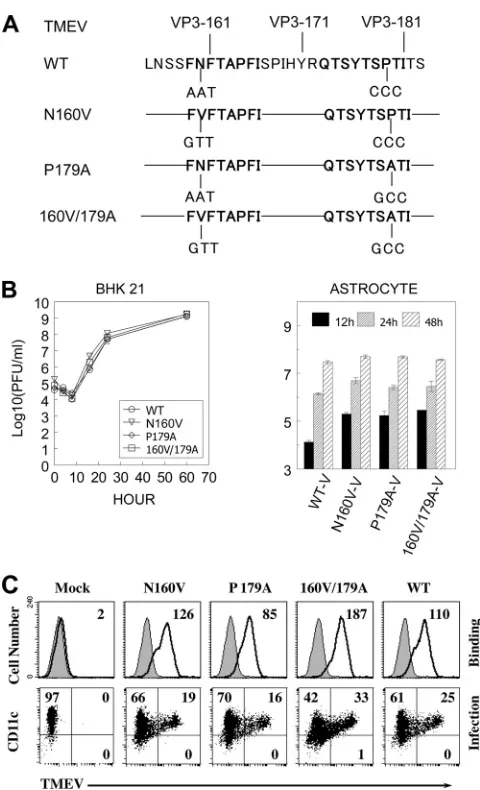
FIG 1Generation and growth properties of cytotoxic T lymphocyte epitope variant viruses. (A) Diagram depicting the site-directed mutagenesis of the predominant and subdominant TMEV BeAn strain CD8⫹T cell epitopes. To eliminate the ability to stimulate epitope-specific CD8⫹T cells, a series of site-specific mutations was introduced into the cytotoxic T lymphocyte epitope regions (VP3159-166and/or VP3173-181) of the TMEV BeAn genome
using a QuikChange kit. (B) Variant viruses replicated comparably to the WT virus in BHK-21 cells and primary mouse astrocytes. BHK-21 cells or primary mouse astrocytes were infected with wild-type and variant viruses harboring a single amino acid substitution (N160V or P179A) or double amino acid sub-stitutions (N160V/P179A). Infectious TMEV titers were assessed at the indi-cated time points from the viral lysates of BHK-21 cell or primary astrocyte cultures by plaque assay. The results of a representative of two separate exper-iments are shown. (C) Virus binding and infection of bone-marrow-derived DCs were determined by flow cytometry using anti-TMEV capsid antibody. The results of one of two similar experiments are shown.
on November 7, 2019 by guest
http://jvi.asm.org/
[image:2.585.300.540.61.456.2]These viruses were propagated and titers were determined in BHK-21 cells grown in Dulbecco’s modified Eagle medium supplemented with 7.5% donor calf serum. For intracerebral (i.c.) infection, 30l virus solution, containing 30⫻106PFU, was injected into the right cerebral hemisphere
of 6- to 8-week-old mice anesthetized with isoflurane. Clinical symptoms of disease were assessed weekly on the following grading scale: grade 0, no clinical signs; grade 1, mild waddling gait; grade 2, severe waddling gait; grade 3, moderate hind limb paralysis; grade 4, severe hind limb paralysis. Reverse transcriptase PCR and real-time PCR.Total cellular RNA was isolated from the brain and spinal cord of infected SJL/J mice using TRIzol reagents (Invitrogen, Carlsbad, CA). First-strand cDNA was syn-thesized from 1g total RNA utilizing SuperScript III first-strand synthe-sis Supermix or Moloney murine leukemia virus reverse transcriptase (Invitrogen, Carlsbad, CA). Primers for control GAPDH (glyceraldehyde-3-phosphate dehydrogenase) and cytokine genes were purchased from Integrated DNA Technologies, as follows: for GAPDH, forward primer AACTTTGGCATTGTGGAAGG and reverse primer ACACATTGGGGG TAGGAACA; for TGF-1, ACAGAGAAGAACTGCTGTGTGC and GT TGTGTTGGTTGTAGAGGGCAA; for IFN-␥, ACTGGCAAAAGGATG GTGAC and TGAGCTCATTGAATGCTTGG; for IFN-, CCCTATGGA GATGACGGAGA and CTGTCTGCTGGTGGAGTTCA; for IL-10, GCC AAGCCTTATCGGAAATGATCC and AGACACCTTGGTCTTGGAG CTT; for IL-18, ACAACTTTGGCCGACTTCAC and GGGTTCACTGG CACTTTGAT; for IL-17A, CTCCAGAAGGCCCTCAGACTAC and AG CTTTCCCTCCGCATTGACACAG; for IL-23, CAAGCGGGACATATG AATCT and CATGGGGCTATCAGGGAGTA; for IL-6, AGTTGCCTTC TTGGGACTGA and TCCACGATTTCCCAGAGAAC; for TNF-␣, GGT CACTGTCCCAGCATCTT and CTGTGAAGGGAATGGGTGTT; for FoxP3, TTGGTTTACTCGCATGTTCGCC and TGGAGGTCAAGGGC AGGGATTG; and for VP1, TGACTAAGCAGGACTATGCCTTCC and CAACGAGCCACATATGCGGATTAC. GAPDH expression served as an internal reference for normalization. Real-time PCR was performed in triplicate.
Plaque assay.Viral titers in infected CNS tissues were enumerated by a standard plaque assay on BHK-21 cell monolayers (27). After methanol fixation, 0.1% crystal violet was used to visualize plaques on the mono-layer.
Viral replication assays.Wild-type or mutant virus was infected at a multiplicity of infection of 10 into BHK-21 cells or primary mouse astro-cytes with rocking and resting for 1 h followed by an extensive wash. Cells were then cultured for various time periods at 33°C as previously de-scribed (3). Both cells and supernatants were harvested and kept at⫺80°C until use. Primary mouse astrocytes were prepared as described elsewhere (42). Briefly, single-cell suspensions were obtained from 0- to 3-day-old neonatal brains and seeded on flasks coated with poly-L-lysine (25g/ml)
(48). After differential shakings at 200 and 250 rpm, adherent astrocytes were collected as previously described (42). The purity of the cell prepa-rations (⬎95% pure) was routinely confirmed by staining with antibodies specific for an astrocyte marker, glial fibrillary acidic protein (Dako, CA). Isolation of CNS-infiltrating MNCs.Mice were perfused with sterile Hanks’ balanced salt solution (HBSS). The excised brains and spinal cords were forced through steel mesh to prepare single-cell suspensions, which were incubated at 37°C for 45 min in 250 mg/ml collagenase type 4 (Wor-thington Biochemical Corp., Lakewood, NJ). CNS-infiltrating mononu-clear cells (MNCs) were then enriched in the 1/3 bottom fraction of a continuous Percoll (Pharmacia, Piscataway, NJ) gradient after centrifu-gation for 30 min at 27,000⫻gas described previously (10).
Intracellular staining of cytokine production.Freshly isolated CNS-infiltrating MNCs were cultured in 96-well round-bottom plates in the presence of relevant or control peptide. Allophycocyanin-conjugated an-ti-CD8 (clone Ly2) or anti-CD4 (clone L3T4) antibody and phycoeryth-rin (PE)-labeled rat monoclonal anti-IFN-␥(XMG1.2) antibody were used for intracellular cytokine staining. Cells were analyzed on a Becton Dickinson FACSCalibur or LSRII cytometer.
Generation of H-2Kstetramers.H-2Kstetramers were generated as
previously described (1). Briefly, H-2Ksand human
2-microglobulin
genes were subcloned into a pET28 bacterial expression vector. Esche-richia coliBL21(DE3) was transformed, and protein expression was in-duced with IPTG (isopropyl--D-thiogalactopyranoside) for 4 to 5 h. The
inclusion body was purified and refolded in the presence of peptides. The soluble monomeric form of peptide-MHC was biotinylated with BirA at room temperature and tetramerized with streptavidin-PE (Invitrogen, Carlsbad, CA).
T cell proliferation assay.Spleen cells from SJL/J mice infected with wild-type or variant viruses were cultured in the presence of peptides for 3 days and then pulsed with 1Ci [3H]thymidine deoxyribose ([3H]TdR)
for 18 h. Cells were harvested, and the levels of [3H]TdR incorporated
were measured using a TopCount liquid scintillation counter (Perkin-Elmer, San Jose, CA). Data are expressed as the mean⫾standard devia-tion (SD) of triplicate samples.
Histopathological analyses. At day 120 postinfection, mice were anesthetized and perfused with 0.1% cold glutaraldehyde in phosphate-buffered saline (PBS). Spinal cords were excised, fixed in 1% OsO4, and
embedded in Epon. Sections were cut at a 1-m thickness, stained with toluidine blue, and analyzed by microscopy (44). Ten different sections of the lumbar region of the spinal cord of individual mice (two mice per experimental group) were assessed. For Luxol Fast Blue (LFB) staining for axonal demyelination and Bielschowsky silver staining for axon loss and damage, spinal cords from mice infected with WT or N160V virus were dissected and fixed in 4% formalin in PBS for 24 h. The tissues were embedded in paraffin and sectioned at 6m. Adjacent sets of 3 sections from each animal were deparaffinized, rehydrated, and stained. Slides were examined using a Leica DMR light microscope, and images were captured using AxioCam MRc camera and AxioVision imaging software. Statistical analysis.Data are presented as the mean⫾SD of either two to three independent experiments or triplicates of one representative from at least three separate experiments. The significance of the differences in the mean values was determined by Student’sttest. The statistical signif-icance of disease incidence was tested by Fisher’s exact test. Clinical scores were analyzed by the Mann-Whitney U test.Pvalues of⬍0.05 were con-sidered statistically significant.
RESULTS
TMEV variants of the CD8ⴙT cell epitopes replicate similarly to WT virus in BHK-21 cells and mouse astrocytes.We previously
showed that CD8⫹T cells reactive to the predominant (VP3159-166)
and subdominant (VP3173-181) epitopes of the TMEV BeAn strain
do not recognize the equivalent epitopes of the closely related TMEV DA strain due to a single amino acid substitution in each
(23). To investigate the role of epitope-specific CD8⫹T cells in the
development of TMEV-IDD, mutant viruses containing a single amino acid substitution or double amino acid substitutions in the
CD8⫹T cell epitopes of the BeAn VP3 protein with the DA
resi-dues were generated by site-directed mutagenesis of an expression
cDNA clone (pSBW) of the BeAn strain (Fig. 1A). Therefore, it
was expected that the variant viruses, encoding a single substitu-tion or double substitusubstitu-tions in the epitope regions of the VP3 protein, would be deficient in the corresponding epitope-specific
CD8⫹T cell responses in the CNS and periphery of virus-infected
SJL mice.
The levels of WT and variant virus replication were first
as-sessed using BHK-21 cells and primary mouse astrocytes (Fig. 1B).
The WT and variant viruses replicated similarly in BHK-21 cells
that were deficient in type I interferon production (Fig. 1B, left).
This result suggests that no stage of the virus life cycle, including virus binding on a cellular receptor(s), viral entry, uncoating of the viral genome, RNA replication, viral coat protein translation,
on November 7, 2019 by guest
http://jvi.asm.org/
and virus assembly, was significantly affected by the introduction of mutations. To further investigate if these viruses replicate sim-ilarly in mouse cells, primary SJL astrocytes were infected with WT and variant viruses. Similar viral replication levels were also
ob-served in astrocytes infected with WT and variant viruses (Fig. 1B,
right). In addition, these viruses displayed no significant differ-ences in their ability to bind to mouse cells, as shown with
den-dritic cells (DCs) (Fig. 1C). Therefore, it is likely that WT and
variant viruses have comparable infection and growth rates in
vitro.
Variant viruses do not induce CD8ⴙT cell responses to the corresponding epitopes.To assess the CD8⫹T cell responses to the corresponding epitopes, susceptible SJL/J mice were infected
with WT and variant viruses. Levels of epitope-specific CD8⫹T
cells in the CNS of infected mice were determined at day 8
postin-fection by assessing the levels of binding of VP3159-166- and
VP3173-181-loaded H-2Kstetramers to CD8⫹T cells (Fig. 2Aand
B). Of the CD8⫹T cells from WT virus-infected mice, 32% were
VP3159-166specific and 11% were VP3173-181specific. In contrast,
less than 3% of the CD8⫹T cells from N160V virus-infected mice
were VP3159-166specific but 24% were VP3173-181specific. These
results suggest that unresponsiveness to the predominant epitope is partly compensated for by an increase in the subdominant VP3173-181-specific CD8⫹ T cell response. However, the lack
(0.5%) of a VP3173-181-specific CD8⫹T cell response in mice
in-fected with the P179A virus was not accompanied by such an
increase in the dominant VP3159-166-specific CD8⫹ T cell
re-sponse. Mice infected with the doubly substituted (N160V/
P179A) virus did not induce the CD8⫹T cells reactive to any of
these epitopes (Table 1).
To further correlate the CD8⫹T cell response with the
produc-tion of key T cell cytokines, IFN-␥production in response to the
epitopes was analyzed by flow cytometry (Fig. 2C). Variant viruses
were similarly unable to induce the corresponding
epitope-spe-FIG 2Epitope-specific CD8⫹T cell responses in mice infected with WT and variant viruses. (A) On the indicated days postinfection, SJL mice (3 mice per group) infected with WT or variant viruses were sacrificed, and isolated CNS-infiltrating MNCs were stimulated with PBS or VP3159-166, VP3173-181, or VP111-20peptide
for 6 h. The proportion of CNS CD8⫹T cells was analyzed using flow cytometry after staining with H-2Ks–VP3
159-166and H-2K s–VP3
173-181tetramers. (B) The
proportion of epitope-specific tetramer-positive CD8⫹T cells (top) and the number of epitope-specific tetramer-positive CD8⫹T cells in the CNS (bottom) are shown. *,P⬍0.05; **,P⬍0.01. (C) The percentage of IFN-␥-producing CD8⫹T cells of the total CD8⫹T cells is shown in the upper right quadrant of each plot. Cells were stained for both CD8 and intracellular IFN-␥for flow cytometric analysis. A representative fluorescence-activated cell sorter analysis plot of three independent experiments is shown. (D) The proportion (top) and number (bottom) of epitope-specific IFN-␥-producing CD8⫹T cells in the CNS of SJL mice during viral infection are shown. The values given are the means of the percentages or numbers of IFN-␥-producing CD8⫹T cells from four independent experiments (mean⫾SD).
on November 7, 2019 by guest
http://jvi.asm.org/
[image:4.585.96.490.67.423.2]cific CD8⫹ T cells producing IFN-␥. For example, the N160V
virus was unable to induce VP3159-166-specific IFN-␥-producing
CD8⫹T cells, in contrast to WT virus. N160V/P179A
virus-in-fected animals did not induce a CD8⫹T cell response to either of
the respective epitopes in the CNS. In addition, the loss of these
epitope-specific CD8⫹T cell responses was not compensated for
by an increase of VP111-20-specific CD8⫹T cells. These patterns of
epitope-specific CD8⫹ T cell responses remained at day 120
postinfection (Fig. 2D).
N160V and N160V/P179A viruses, deficient in the predomi-nant CD8ⴙT cell epitope, do not induce demyelinating disease.
To further investigate how differences in virus-specific CD8⫹T
cell responses in the CNS affect the development of TMEV-IDD, SJL mice (9 to 10 mice/group) were infected with WT or variant
viruses. Mice infected with the WT or P179A virus (⬎75%)
devel-oped clinical disease by 50 days postinfection (Fig. 3A). In
con-trast, the majority of the N160V or N160V/P179A virus-infected
an-imals (⬎80%) did not develop disease. In addition, clinically affected
mice infected with the N160V or N160V/P179A virus showed only
minimal disease severity (P⬍0.01 at 42 days postinfection and
there-after) compared to the mice infected with the WT or P179A virus.
These studies suggest that VP3159-166-specific CD8⫹T cell responses
are critical in disease development (Table 1).
The histopathology of the spinal cord was further examined at 120 days after viral infection and was correlated with disease de-velopment by the epitope mutant viruses. The WT and P179A virus-infected animals exhibited similar severe demyelination in the spinal cord, whereas the N160V and N160V/P179A
virus-in-fected animals remained free of demyelinating lesions (Fig. 3B).
These results are consistent with the clinical disease levels induced by the WT and mutant viruses. To further determine if the level of
axon damages reflects the pathogenic function of VP3159-166
-spe-cific CD8⫹T cells, the integrity of axons in the spinal cord of mice
infected with the WT and N160V virus were compared on the basis of the density of the stains after Bielschowsky silver staining (Fig. 3C). The levels of axonal loss and damage were more
exten-sive in the spinal cord of the WT virus-infected mice (54.2⫾25.4)
than that of the N160V virus-infected mice (133.9⫾28.6) lacking
the VP3159-166-specific CD8⫹T cell response. These data strongly
suggest that the CD8⫹ T cells specific for the predominant
VP3159-166epitope but not those specific for the subdominant P179A
epitope play an important role in the pathogenesis of demyelination in the spinal cord and consequent clinical disease.
Mice infected with the N160V or N160V/P179A virus display higher viral loads in the CNS.To determine if the reduced disease
development in mice infected with viruses to which CD8⫹T cell
responses to the predominant epitope are deficient is associated
with lower levels of viral replication, viral RNA levels in the CNS of
infected mice were assessed using real-time PCR (Fig. 4A).
Inter-estingly, the N160V and N160V/P179A virus-infected mice
lack-ing VP3159-166-specific CD8⫹T cells displayed significantly higher
levels of viral messages in the spinal cord at 8 days postinfection. In contrast, viral message levels in the CNS of the P179A virus-in-fected mice were significantly lower than those in the CNS of the WT virus-infected mice. Levels of infectious virus in the CNS of
these mice were further determined by plaque assay (Fig. 4B).
Roughly 2-log-unit higher infectious viral titers were detected in the brain and spinal cord of N160V virus-infected mice than
WT-infected mice (P⬍0.0005). The N160V/P179A
virus-in-fected mice similarly displayed more infectious virus in the
brain at day 8 postinfection (P⬍0.017). In contrast, 10-fold
fewer infectious viruses were detected in the brain of the P179A virus-infected mice, which induce a significantly higher level of VP3159-166-specific CD8⫹T cells in the CNS than the WT
virus-infected mice (P⬍0.001). The levels of viral load remained
high in both the N160V and N160V/P179A virus-infected mice at day 21 postinfection. However, at day 120 postinfection, infectious viruses were not detectable in the spinal cord of the N160V or P179A virus-infected mice, whereas the levels of viral
load in the brain were largely comparable in all groups (Fig.
4B). These results suggest that VP3159-166-specific CD8⫹T cells
are most efficient in viral clearance and that the loss of the
CD8⫹T cell response results in an elevated viral load at the
early stages of viral infection.
Levels of IFN-␥-producing TMEV-specific CD4ⴙT cells are comparable between WT and variant virus-infected mice.
Epitope-specific CD4⫹T cell levels were also investigated to
ex-amine if the mutations in the CD8⫹T cell epitope regions of the
TMEV genome affect the virus-specific CD4⫹T cell responses in
the CNS (Fig. 5). Proliferative responses of splenic CD4⫹T cells to
viral epitopes were not significantly different between WT and variant virus-infected groups throughout the course of viral
infec-tion (Fig. 5A). Similarly, the levels of virus-specific (combined capsid
and noncapsid epitopes) CD4⫹T cells in the CNS measured by
IFN-␥production were comparable among all experimental groups,
as shown for a representative experiment at 8 days postinfection (Fig.
5B). The proportions and numbers of CNS-infiltrating CD4⫹T cells
at 8 days postinfection were further assessed, and the combined
re-sults of 3 separate experiments are shown inFig. 5C. These results
suggest that there are no significant alterations in the CD4⫹T cell
responses to the variant viruses during the course of infection.
There-fore, the differences in virus-specific CD4⫹T cell responses unlikely
[image:5.585.43.547.79.152.2]resulted in the altered clinical outcome following infection with VP3159-166-modified viruses (Fig. 3).
TABLE 1Mutant viruses and their properties
Virus Reactivity of cytotoxic T lymphocytec
Presence ofa:
Clinical symptoms Demyelination Virus persistenceb
WT VP3159-166, VP3173-181, VP111-20 ⫹⫹⫹ ⫹⫹⫹ ⫹
N160V VP3173-181, VP111-20(VP3159-166) ⫾ ⫾ ⫹⫹
P179A VP3159-166, VP111-20(VP3173-181) ⫹ ⫹⫹⫹ ⫹
N160V/P179A VP111-20(VP3159-166, VP3173-181) ⫾ ⫾ ⫹⫹
a⫹⫹⫹, very high;⫹⫹, high;⫹, moderate;⫾, low. b
Based on viral message levels in the spinal cords at 8, 21, and 120 days postinfection.
cParentheses indicate absence.
on November 7, 2019 by guest
http://jvi.asm.org/
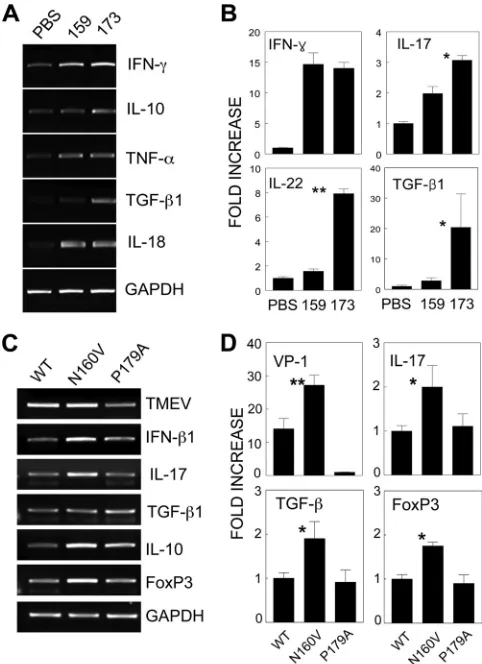
Distinct cytokine genes are expressed in CD8ⴙT cells reac-tive to the dominant VP3159-166epitope versus the subdominant
VP3173-181epitope.It is conceivable that CD8⫹T cells produce a
different set of cytokines depending on epitope recognition, which would consequently result in differential viral clearance and pathogenesis. To examine this possibility, CNS cells from mice
infected with the WT virus were restimulatedin vitrowith
domi-nant and subdomidomi-nant epitope peptides (Fig. 6AandB). Because
it is difficult to isolate the individual epitope-specific CD8⫹T cells
FIG 3Comparison of the clinical disease course in mice infected with either WT or variant viruses. (A) SJL mice (n⫽9 to 10) were infected with WT or variant TMEVs (30⫻106PFU/mouse). Animals were graded for clinical signs
as described in Materials and Methods. The results are expressed as the per-centage (left) or mean (right) clinical score of affected animals on the indicated days postinfection. The statistical significance of disease incidence was tested by Fisher’s exact test. The clinical scores were analyzed by the Mann-Whitney U test. For WT-infected versus N160V- or N160V/P179A-infected mice,P
was⬍0.05 at day 35 postinfection and thereafter; for WT-infected versus P179A-infected mice,Pwas⬍0.05 at day 63 and thereafter; for N160V-infected versus P179A-infected mice,Pwas⬍0.05 at day 49 and thereafter; for N160V- or P179A-infected versus N160V/P179A-infected mice,Pwas not significant at all time points. (B) Spinal cords from SJL mice infected with WT or variant viruses for 120 days were obtained (two mice per group). One-micron-thick sections were prepared and stained with toluidine blue. Ten different sections of the lumbar region of the spinal cord of each individual mouse were graded
for demyelination by microscopy. Results of a representative of three different experiments are shown. (C) Spinal cords of virus-infected mice were stained with Luxol Fast Blue (a, b) or Bielschowsky (c, d). The red boxes (c and d) indicate the areas of the insets. Density comparisons of 10 random areas stained with Luxol Fast Blue showed values of 40.8⫾5.0 for WT virus-infected mice and 117.9⫾32.6 for N160V-infected mice. Density comparisons of 10 random areas stained with Bielschowsky showed values of 54.2⫾25.4 for WT virus-infected mice and 133.9⫾28.6 for N160V-infected mice.
FIG 4Viral persistence in mice infected with WT and variant viruses. Brain and spinal cord homogenates were prepared from SJL mice (n⫽3) infected with WT and variant TMEV. (A) Viral RNA levels in the brain and spinal cord homogenates from mice infected with viruses for 8 days were compared with the fold VP1 expression using real-time PCR. (B) Infectious titers in the tissue homogenates of mice were assessed by plaque assay on days 8, 21, and 120 (d8, d21, and d120, respectively) postinfection. The values given are the mean numbers from two to three independent experiments (mean⫾SD). *,P⬍
0.05; **,P⬍0.01; ***,P⬍0.001; ND, not detectable (below the detection level).
on November 7, 2019 by guest
http://jvi.asm.org/
[image:6.585.302.540.69.415.2] [image:6.585.43.283.74.573.2]from the CNS, we have determined the levels of cytokine messages induced after stimulation with the cognate epitopes compared to those in the unstimulated controls. In contrast to the minimal
level of IFN-␥ in the control cultures with PBS, similarly high
levels of IFN-␥mRNAs were detected upon cognate stimulation
with the VP3159-166and VP3173-181peptides. Therefore, the level of
IFN-␥mRNA appears to reflect the cytokine message levels
pro-duced by the respective epitope-specific CD8⫹T cells.
Interest-ingly, the VP3173-181-specific CD8⫹T cells expressed significantly
higher levels of IL-10, IL-17, IL-22, and TGF-1 genes than the
VP3159-166-specific CD8⫹T cells. These results suggest that the
major T cell populations specific for these two epitopes represent
distinct CD8⫹T cell types. The predominant VP3159-166-specific
CD8⫹T cells appear to represent the typical antiviral CD8⫹T
cells, whereas the subdominant VP3173-181-specific CD8⫹T cells
may represent the recently described Tc17 cell type (14,19,20).
To further delineate the potential mechanism(s) underlying the resistance to the development of demyelinating disease in the
absence of VP3159-166-specific CD8⫹T cells, the levels of T
cell-associated cytokine gene expression in the CNS of WT- and vari-ant virinfected mice were analyzed at 8 days postinfection
us-ing PCR (Fig. 6CandD). The expression of IL-10, IL-17, TGF-1,
and FoxP3 mRNA was noticeably higher in the CNS of the VP3159-166-deficient N160V virus-infected mice, which have
in-creased levels of VP3173-181-specific CD8⫹T cells, than in the CNS
of either the WT virus-infected or VP3173-181-deficient P179A
vi-rus-infected mice displaying high levels of VP3159-166-specific
CD8⫹T cells. These results clearly demonstrate that viral RNA
and IL-17, TGF-1, and FoxP3 mRNA levels are highly elevated in
the absence of VP3159-166-specific CD8⫹T cells. Because the
num-ber of VP3173-181-specific CD8⫹T cells is increased in the absence
of VP3159-166-specific CD8⫹T cells, the elevated cytokine gene
expression in N160V virus-infected mice likely reflects the
cyto-kine profile of VP3173-181-specific CD8⫹ T cells. However, the
presence of intracellular IL-17 was undetectable by flow
cytom-etry, suggesting that the IL-17 levels produced in these CD8⫹T
cells are relatively low (not shown). Nevertheless, the differences in T cell cytokine levels suggest that the predominant
virus-spe-cific CD8⫹T cells in the CNS recognize VP3159-166and that the
deletion of this population results in a drastically altered cytokine
profile of CD8⫹T cells. These results are consistent with the
cyto-kine production levels obtained upon stimulation with the
respec-tive epitope peptides (Fig. 6AandB).
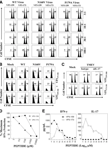
VP3159-166- and VP3173-181-specific CD8ⴙ T cells differ in
their cytolytic function. Because SJL mice infected with the
N160V virus deficient in the VP3159-166epitope do not develop
demyelinating disease, we further examined if differences in the
epitope-dependent cytolytic function of CD8⫹T cells are
associ-ated with disease development. The cytolytic activity of CD8⫹T
cells from SJL mice infected with the WT or the N160V or P179A
mutant virus was assessedin vitroat various effector-to-target cell
ratios (Fig. 7A). The CD8⫹T cells from the WT virus-infected
mice failed to lyse VP3173-181-loaded target cells at an
effector-to-target cell ratio as high as 100:1, whereas these CD8⫹T cells
effec-tively lysed VP3159-166-loaded target cells at an effector-to-target
cell ratio as low as 10:1. Furthermore, the CD8⫹T cells from the
N160V virus-infected mice lacking VP3159-166-specific CD8⫹T
cells could not lyse the target cells. In contrast, the CD8⫹T cells
from the P179A virus-infected mice deficient in VP3173-181
-spe-cific T cells effectively lysed the target cells. These results clearly
indicate that VP3159-166-specific CD8⫹T cells, but not VP3173-181
-specific CD8⫹T cells, exhibit strong cytolytic activityin vitro.
Despite the lack of significant cytolytic function by VP3173-181
-specific CD8⫹T cellsin vitro(Fig. 7A), it is conceivable thatin
FIG 5Reactivity of CD4⫹T cells in mice infected with WT and variant viruses. (A) Splenic cells from mice infected with WT and variant viruses at 8 days postinfection were stimulated for 3 days in the presence of PBS or a structural CD4⫹T cell epitope mix (S; VP1233-250, VP274-86, and VP324-37) and the
predominant nonstructural epitope (NS; 3D21-36). Proliferation levels were assessed by [
3H]TdR uptake. (B) CNS-infiltrating MNCs were stimulated with PBS
or a CD4⫹T cell epitope mix (VP1233-250, VP274-86, VP324-37, and 3D21-36). After 6 h of stimulation, cells were stained for CD4 and intracellular IFN-␥. The
percentage of CD4⫹IFN-␥-producing cells is shown in the upper right corner of each plot. The data are representative of three independent experiments. (C) Epitope-specific IFN-␥production by CNS-infiltrating CD4⫹T cells. The proportion of epitope-specific CD4⫹T cells in the CNS (left) and the number of epitope-specific CD4⫹T cells (right) are shown. The values given are the means of the percentages or numbers from 3 independent experiments (mean⫾SD).
on November 7, 2019 by guest
http://jvi.asm.org/
[image:7.585.85.504.68.275.2]vitrocytotoxicity may not reflect cytolytic function in virus-in-fected mice. To explore this possibility, we further examined the
potential differences in cytolytic functions of CD8⫹T cells in
vi-rus-infected mice using target cells loaded with an equal mixture
of the VP3159-166and VP3173-181epitopes (Fig. 7B). Control and
cognate peptide-loaded target cells labeled with two different in-tensities of carboxyfluorescein succinimidyl ester (CFSE; high and low) were administered to virus-infected mice (3 mice per viral
group) at 8 days postinfection, as previously described (17). The
N160V virus-infected mice (3/3) lacking VP3159-166-specific
CD8⫹T cells failed to lyse the target cells. In contrast, the WT
virus-infected mice and P179A virus-infected mice deficient in VP3173-181-specific CD8⫹T cells efficiently lysed the target cells.
These results clearly indicate that WT virus-infected SJL mice are
able to efficiently clear VP3159-166- and VP3173-181-bearing cells,
suggesting that only VP3159-166-specific CD8⫹T cells are capable
of effectively clearing the target cells in infected animals, which is
consistent with thein vitrocytotoxicity (Fig. 7A).
It has recently been shown that IL-17 inhibits the cytolytic
function of CD8⫹T cells by protecting virus-infected target cells
from apoptosis (17) and that IL-17-producing CD8⫹T cells
dis-play deficient cytolytic functionin vitro(14). Despite the relatively
higher level of IL-17 mRNA in VP3173-181-specific CD8⫹T cells,
we failed to detect intracellular IL-17 by flow cytometry. There-fore, it is conceivable that a relatively small amount of IL-17 in
CD8⫹T cells is sufficient for blocking the cytolysis of target cells.
To examine this possibility, either isotype or anti-IL-17 antibody-treated SJL mice were infected with WT TMEV. Eight days after
infection,in vivocytolytic activity was evaluated in the virus- and
mock-infected mice (Fig. 7C). Interestingly, both the VP3159-166
-and VP3173-181-specific CD8⫹T cells in the anti-IL-17
antibody-treated mice significantly gained cytolytic function. In particular, VP3173-181-specific CD8⫹ T cells in the anti-IL-17
antibody-treated mice displayed strong cytolysis (⬎50%) of target cells,
whereas there was a lack of detectable cytolysis in the isotype an-tibody-treated mice. A similar improvement in the cytolytic
activ-ity of the VP3173-181-specific CD8⫹T cells was also detected in
vitroin the presence of anti-IL-17 antibody (not shown). There-fore, these results suggest that a small amount of IL-17 production
by CD8⫹T cells may efficiently block target cell cytolysis in
cell-cell contact interactions. It is also noteworthy that the cytolytic
levels in virus-infected mice (Fig. 7C) were much lower than those
assessed within vitrocytotoxic assays (Fig. 7A). This may be
at-tributable to the presence of higher IL-17 levels in virus-infected
mice. Taken together, these results strongly suggest that CD8⫹T
cells with strong cytolytic functionin vivoare essential for the
pathogenesis of demyelinating disease, as mice lacking this CD8⫹
T cell population do not develop the disease.
VP3159-166- and VP3173-181-specific CD8ⴙT cells display
dif-ferent avidities for their epitopes and produce distinct cyto-kines.It has recently been shown that T cell activation signals
affect the development of CD4⫹T cell subtypes (11,30). To
fur-ther understand the potential underlying mechanisms of the dif-ferences in cytokine production and cytolytic function between VP3159-166- and VP3173-181-specific CD8⫹T cells, we determined
the functional avidity of T cells to the epitopes by measuring the
levels of IFN-␥and/or IL-17A production upon stimulation with
various concentrations of the cognate peptides (Fig. 7DandE), as
recently described (25). Interestingly, the concentrations of
VP3159-166peptide required to stimulate intracellular IFN-␥
pro-duction over 6 h to the levels similar to those of the VP3173-181
peptide were 30- to 100-fold lower (Fig. 7D). For example, more
than 50 nM VP3173-181peptide versus less than 2 nM VP3159-166
peptide was required to achieve 50% of the maximum IFN-␥
pro-duction. These results indicate that the functional avidity of VP3159-166-specific CD8⫹T cells to the cognate epitope required
to stimulate the initial production of IFN-␥is at least 30-fold
higher than that of VP3173-181-specific CD8⫹T cells. However,
intracellular IL-17 production after 6 h ofin vitrostimulation was
not detectable by flow cytometry (not shown), although the in-crease in IL-17 mRNA production was observed by reverse
tran-scription-PCR (RT-PCR) (Fig. 6). To further determine the
pro-duction of IL-17 by the epitope-specific CD8⫹T cells over a longer
time period, we incubated 1.5⫻106CNS-infiltrating cells from
TMEV-infected SJL mice for 72 h in the presence of the epitope
peptides. The levels of IFN-␥and IL-17 that accumulated in the
culture supernatants were assessed using cytokine-specific en-zyme-linked immunosorbent assays (ELISAs). Although the
pro-duction of IFN-␥was detected in the cultures stimulated with
FIG 6Distinct cytokine profiles of different epitope-specific CD8⫹T cells. CNS-infiltrating MNCs from SJL mice infected with WT virus were restimu-latedin vitroat 8 days postinfection with PBS, VP3159-166, or VP3173-181for 6 h.
The mRNA levels of various cytokines in these cultures were analyzed using either conventional RT-PCR (A) or quantitative real-time PCR (B). The RNA levels of TMEV, FoxP3, and various cytokines in CNS-infiltrating cells of mice infected with either WT or variant viruses were assessed using conventional RT-PCR (C) or real-time PCR (D). Fold increase indicates the relative fold increases after normalization on the basis of GAPDH levels. One representa-tive set from three separate experiments is shown. *,P⬍0.05; **,P⬍0.01.
on November 7, 2019 by guest
http://jvi.asm.org/
[image:8.585.42.283.66.398.2]FIG 7Different cytolytic functions of epitope-specific CD8⫹T cells. (A) Splenocytes from naive SJL mice were loaded with control ovalbumin from residues 323 to 339 (OVA323-339) and viral (VP3159-166or VP3173-181) peptides and labeled with a high and a low concentration of CFSE, respectively. A fixed number of
CFSE-labeled target cells and various numbers of splenic effector cells from mice at 8 days postinfection with WT or variant viruses were cocultured for 60 h at the indicated effector-to-target cell ratios. The numbers in each histogram represent the percentages of cells labeled with low (VP3159-166or VP3173-181) and high
(OVA323-339) concentrations of CFSE. (B) At 8 days after infection,in vivolysis activity was evaluated in virus- and mock-infected mice (n⫽3) that received the
peptide-loaded CFSE-labeled target cells. OVA323-339and viral (both VP3159-166and VP3173-181) peptides were used to load the target cells. Mixtures of control
and viral peptide-loaded target cells were injected into SJL mice (n⫽3) infected with WT, N160V, or P179A virus and mock infected with PBS. Cytolytic profiles of three individual mice are shown. (C) SJL mice were treated with either the isotype (n⫽3) or anti-IL-17 (n⫽3) antibody at days 0 and 7 relative to the time of TMEV infection. At 8 days postinfection,in vivocytolysis activity was evaluated in virus- and mock-infected mice (n⫽3) that received the target cells. (D) CNS-infiltrating cells from the SJL mice infected with TMEV were stimulated with VP3159-166or VP3173-181at different concentrations for 6 h, and intracellular
IFN-␥production was assessed by flow cytometry. The numbers of IFN-␥-producing CD8⫹T cells after stimulation with 2M peptide were considered to be 100% of the epitope-specific IFN-␥-producing cells. (E) CNS-infiltrating cells (1.5⫻106) from SJL mice infected with the TMEV BeAn strain were stimulated
with various concentrations of VP3159-166or VP3173-181peptide for 72 h. The production of IFN-␥and IL-17 in the culture supernatants was measured using the
respective cytokine-specific ELISA.
on November 7, 2019 by guest
http://jvi.asm.org/
[image:9.585.114.472.67.556.2]either the VP3159-166or the VP3173-181peptide, IL-17 production
was detectable only after VP3173-181peptide stimulation (Fig. 7D).
Therefore, it is conceivable that differences in the functional avid-ity toward their cognate epitopes and/or the type of cytokines
produced may affect the consequent cytolytic function of CD8⫹T
cell populations in an epitope-dependent manner.
DISCUSSION
TMEV infection in susceptible strains of mice induces chronic
demyelinating disease mediated by CD4⫹ T cells and
macro-phages (17,26,28). However, epitope-specific CD4⫹T cells can be
protective, depending on the time of availability in conjunction
with viral infection (36). In addition, the pathogenicity of CD4⫹T
cells may also be dependent on the recognizing viral epitopes (21,
54). Therefore, epitope-specific cytotoxic CD8⫹T cells may
sim-ilarly play a protective or pathogenic role depending on the type and/or reactivity of epitopes. We previously demonstrated that
the level of the VP3159-166-specific CD8⫹T cell response in
suscep-tible SJL mice is inversely correlated with viral persistence in
P1-transgenic (P1-Tg) SJL mice (40), highlighting the importance of
VP3159-166-specific CD8⫹T cells in limiting the viral loadin vivo.
Surprisingly, however, higher viral persistence in P1-Tg mice did not lead to a higher disease incidence and/or severity. These results
posed an interesting possibility that VP3159-166-specific CD8⫹T
cells may be associated with the pathogenesis of TMEV BeAn-induced demyelinating disease. Because the TMEV DA strain, which induces a similar chronic demyelinating disease, does not
contain this epitope (23), the DA virus may utilize CD8⫹T cells
recognizing a different epitope for its pathogenesis.
To further address the possibility that VP3159-166-specific
CD8⫹T cells play a pathogenic role in disease development, a
series of mutant viruses containing a single amino acid substitu-tion or double amino acid substitusubstitu-tions in the predominant
and/or subdominant CD8⫹T cell epitopes of TMEV was
gener-ated (Fig. 1). These variant viruses failed to mount the
corre-sponding epitope-specific CD8⫹T cell responses (Fig. 2) without
affecting CD4⫹T cell responses (Fig. 5). Interestingly, the viral
load was higher in the CNS of the VP3159-166-deficient N160V
virus-infected mice during the early stages of infection, suggesting
that VP3159-166-specific CD8⫹T cells are critical for viral clearance
(Fig. 3). The high viral load is unlikely to reflect more rapid viral replication, as these variant viruses did not replicate differently in BHK-21 cells and mouse astrocytes or bone marrow-derived
den-dritic cells (Fig. 1) and in the CNS of Rag1⫺/⫺mice (not shown).
Since these cell populations are the major cell populations infected by the WT virus, it is unlikely that the mutant viruses infect dif-ferent cell types. In addition, similar viral loads in the CNS of
Rag1⫺/⫺mice, which do not have adaptive immune responses,
would reflect the comparable tropism of these variant viruses. However, differences in the infectivity to minor cell populations or viral trafficking to different CNS compartments would be pos-sible.
Interestingly, the loss of a VP3159-166-specific CD8⫹T cell
re-sponse in the N160V virus-infected mice was compensated for by
an increase in CD8⫹T cells reactive to the subdominant VP3173-181
epitope (Fig. 2). However, the VP3173-181-specific CD8⫹T cells
whose levels were elevated in the N160V virus-infected mice were
apparently unable to efficiently limit the viral load (Fig. 4). The
inefficient control of viral load was due to the poor cytolytic
func-tion of VP3173-181-specific CD8⫹T cells compared to VP3159-166
-specific CD8⫹T cells (Fig. 7). In contrast, the VP3173-181-deficient
P179A virus-infected mice, which mount a greater VP3159-166
-specific CD8⫹T cell response, efficiently controlled the viral load
in the CNS. Interestingly, however, the virus level in the CNS did
not correlate with the development of clinical disease (Fig. 4),
which is consistent with the previous observation in P1-Tg mice
(40). Therefore, viral persistence alone may not be sufficient for
the induction of demyelinating disease, and CD8⫹ T cell
re-sponses to certain viral epitopes may be an integral component of
the pathogenesis, as previously suggested (34).
It is known that the presence of a strong CD8⫹T cell response
is essential for protection from viral diseases. However, the
ma-jority of the N160V virus-infected animals (⬎90%), which lacked
the predominant cytolytic VP3159-166-reactive CD8⫹T cells
bear-ing high functional avidity (Fig. 7), remained free of clinical signs
(Fig. 3). In contrast, the majority of the P179A virus-infected
an-imals (80%), which had high levels of VP3159-166-reactive CD8⫹T
cells in the absence of VP3173-181-reactive CD8⫹T cells, developed
the full-blown disease (Fig. 3). In addition, the N160V/P179A
virus-infected SJL mice, which lacked both VP3159-166- and
VP3173-181-reactive CD8⫹T cells, were also largely free of
demy-elinating disease. The observation that a highly cytolytic CD8⫹T
cell response in the CNS is associated with the development of TMEV-induced demyelinating disease in SJL mice was
unex-pected (Fig. 3and7). Taken together, these results strongly
sug-gest that cytolytic CD8⫹T cells with high avidity (Fig. 7) may
cause an initial infliction of CNS damage leading to the develop-ment of demyelinating disease.
It is not clear at this time whether CD4⫹T cells and CD8⫹T
cells independently or cooperatively induce clinical disease. It has
previously been assumed that CD4⫹T cells alone induce
TMEV-induced demyelinating disease, based on the amelioration of
dis-ease development after treatment with antibodies against CD4⫹T
cells (17,46,53) and the development of severe demyelinating
disease in2-microglobulin-deficient SJL mice (2). However,2
-microglobulin is critical for all types of T cells restricted with class I and class I-like molecules, including NK T cells and other non-classical class I-restricted T cells. Therefore, it is difficult to discern the potential role and function of these T cell populations in the pathogenesis and/or protection from viral disease development. It is interesting to note that CD4-deficient susceptible SJL and PLJ mice display more severe demyelination than CD8-deficient
counterpart mice (38). These results suggest that subpopulations
of both CD4⫹and CD8⫹T cell types could be cooperatively
in-volved in the pathogenesis of demyelinating disease.
The mechanisms underlying the differences in viral clearance and
cytotoxic function among epitope-specific CD8⫹ T cells are
un-known. Although CD8⫹ T cells recognizing the VP3159-166 and
VP3173-181epitopes can produce relatively high levels of IFN-␥, the
efficiency of the VP3173-181-specific CD8⫹T cells to induce IFN-␥
production was as much as 100-fold lower than that of the VP3159-166
-specific CD8⫹T cells (Fig. 7D). In contrast, the VP3173-181-specific
CD8⫹T cells induced significantly higher levels of IL-10, IL-17,
IL-22, and TGF-1 mRNA expression than VP3159-166-specific
CD8⫹T cells (Fig. 6). This CD8⫹T cell population appears to be
similar to the IL-17-producing CD8⫹T cell population recently
named Tc17 (14), including the expression of FoxP3 (Fig. 6D) and
retinoic acid-related orphan nuclear receptor gamma t (ROR-␥t)
transcription factors (not shown). It is interesting to note that the
Tc17-type response is largely epitope dependent (Fig. 6and7E).
on November 7, 2019 by guest
http://jvi.asm.org/
As far as we know, this is the first report indicating that the induc-tion of a Tc17 response is epitope dependent. The drastic difference
in functional avidity between the VP3159-166- and VP3173-181-specific
CD8⫹T cells (Fig. 7) may lead to the differential induction of CD8⫹
T cell types. In addition, it is possible that various cytokines favor-ing the development of pathogenic Th17 cells in TMEV-infected
mice (17) may also promote the epitope-dependent development
of Tc17 cells. For example, TGF-and IL-6 genes are highly
ex-pressed in various glial cells and infiltrating antigen-presenting
cells in the CNS of TMEV-infected SJL mice (4,18,22,50).
There-fore, viral infection-induced TGF-, together with IL-6, may also
participate in the development of IL-17-producing CD8⫹T cells
in the presence of a low level of IFN-␥(14,19), similar to the
development of Th17 cells (15,17,33,52).
The VP3173-181-specific CD8⫹T cells producing IL-17 also
ex-pressed TGF-1 mRNA in response to their cognate epitope (Fig.
6). Furthermore, these CD8⫹T cells upregulated the surface
ex-pression of CD103 (not shown), which is a marker for the recent
expression of TGF-(9,39). TGF-interferes with the effector
functions of CD8⫹T cells by inhibiting the expression of perforin
and FasL on T cells, both of which are important for the cytotoxic
function of CD8⫹T cells (6,13,45,49). In addition, IL-17 inhibits
the cytolytic function of CD8⫹T cells by protecting the
virus-infected target cells from apoptosis (17). Consequently,
IL-17-producing CD8⫹T cells may exhibit deficient cytolytic functionin
vitro(Fig. 7A) andin vivo(Fig. 7B), as shown previously (14,19). Although the presence of anti-IL-17 antibody enhanced cytolytic
function (Fig. 7C), the IL-17 level produced by VP3173-181-specific
CD8⫹T cells appears to be too low to detect using intracellular
cytokine staining to assess the amount of intracellular IL-17
pro-duced in 6 h (not shown). However, these CD8⫹T cells produced
a significant level of IL-17 in 3 days (Fig. 7E), suggesting that the
amount of IL-17 produced by the epitope-specific CD8⫹T cells is
significant, although smaller than that produced by CD4⫹T cells.
Therefore, a relatively small amount of IL-17 locally produced by
CD8⫹T cells may be sufficient for blocking cytolysis of target cells
during the cognate cell-cell interaction.
The role of the IL-17-producing CD8⫹T cell subtype in the
protection or pathogenesis of demyelinating disease is unclear. A protective role of Tc17 in influenza viral infection has been pro-posed, as mice treated with anti-IL-17 antibody showed higher
mortality (14). Perhaps the increased mortality after treatment
with the anti-IL-17 antibody reflects extensive tissue damage in
the absence of IL-17, which prevents apoptotic cell death (17).
However, mice with high levels of Tc17 cells harbor higher viral loads
in the CNS (Fig. 4), suggesting less efficient viral clearance by the cells.
Interestingly, this CD8⫹T cell population failed to result in the
de-velopment of TMEV-induced demyelinating disease, in contrast to
conventional IFN-␥-producing Tc1 CD8⫹T cells (Fig. 2and3). It is
surprising to observe that the highly cytolytic VP3159-166-specific Tc1
population is associated with the pathogenesis of demyelination
and axon damage, whereas the noncytolytic VP3173-181-specific
Tc17 population is not (Fig. 3). This is unexpected, in light of our
recent findings that IL-17-producing CD4⫹T cells (Th17) play a
critical pathogenic role in the development of virus-induced
de-myelinating disease (17). Therefore, it is conceivable that IFN-␥
-producing VP3159-166-specific CD8⫹T cells with high cytolytic
function are required to initiate the pathogenic process by de-stroying infected neurons and/or releasing sequestered
autoanti-gens, whereas IL-17-producing low-cytolytic VP3173-181-specific
CD8⫹T cells fail to do so. Because Th17 cells secreting high levels
of IL-17 play a critical role in the pathogenesis of TMEV-induced
demyelinating disease (17), virus-specific cytolytic CD8⫹T cells
may initiate the process, followed by Th17-mediated inhibition of cytolytic function promoting viral persistence and subsequent ex-tensive tissue damage. Thus, both Th17 and Tc1 populations re-active to viral epitopes may cooperatively be involved in the pathogenesis of virally induced demyelinating disease.
ACKNOWLEDGMENTS
This work was supported by United States Public Health Service grants (RO1 NS28752 and RO1 NS33008) and by a grant from the National Multiple Sclerosis Society (RG 4001-A6).
REFERENCES
1.Altman JD, et al.1996. Phenotypic analysis of antigen-specific T lympho-cytes. Science274:94 –96.
2.Begolka WS, et al.2001. CD8-deficient SJL mice display enhanced sus-ceptibility to Theiler’s virus infection and increased demyelinating pathol-ogy. J. Neurovirol.7:409 – 420.
3.Calenoff MA, Faaberg KS, Lipton HL.1990. Genomic regions of neuro-virulence and attenuation in Theiler murine encephalomyelitis virus. Proc. Natl. Acad. Sci. U. S. A.87:978 –982.
4.Chang JR, Zaczynska E, Katsetos CD, Platsoucas CD, Oleszak EL.2000. Differential expression of TGF-beta, IL-2, and other cytokines in the CNS of Theiler’s murine encephalomyelitis virus-infected susceptible and re-sistant strains of mice. Virology278:346 –360.
5.Chang Y, et al.2011. CD8 positive T cells express IL-17 in patients with chronic obstructive pulmonary disease. Respir. Res.12:43.
6.Chen ML, et al.2005. Regulatory T cells suppress tumor-specific CD8 T cell cytotoxicity through TGF-beta signals in vivo. Proc. Natl. Acad. Sci. U. S. A.102:419 – 424.
7.Dal Canto MC, Kim BS, Miller SD, Melvold RW.1996. Theiler’s murine encephalomyelitis virus (TMEV)-induced demyelination: a model for hu-man multiple sclerosis. Methods10:453– 461.
8.Doherty PC, et al.1997. Effector CD4⫹and CD8⫹T-cell mechanisms in the control of respiratory virus infections. Immunol. Rev.159:105–117. 9.Dubois CM, Laprise MH, Blanchette F, Gentry LE, Leduc R. 1995.
Processing of transforming growth factor beta 1 precursor by human furin convertase. J. Biol. Chem.270:10618 –10624.
10. Fuller AC, et al.2005. Gender bias in Theiler’s virus-induced demyeli-nating disease correlates with the level of antiviral immune responses. J. Immunol.175:3955–3963.
11. Gabrysova L, et al.2011. Integrated T-cell receptor and costimulatory signals determine TGF-beta-dependent differentiation and maintenance of Foxp3⫹regulatory T cells. Eur. J. Immunol.41:1242–1248.
12. Gay FW, Drye TJ, Dick GW, Esiri MM.1997. The application of mul-tifactorial cluster analysis in the staging of plaques in early multiple scle-rosis. Identification and characterization of the primary demyelinating lesion. Brain120:1461–1483.
13. Genestier L, Kasibhatla S, Brunner T, Green DR.1999. Transforming growth factor beta1 inhibits Fas ligand expression and subsequent activa-tion-induced cell death in T cells via downregulation of c-Myc. J. Exp. Med.189:231–239.
14. Hamada H, et al.2009. Tc17, a unique subset of CD8 T cells that can protect against lethal influenza challenge. J. Immunol.182:3469 –3481. 15. Harrington LE, et al.2005. Interleukin 17-producing CD4⫹effector T
cells develop via a lineage distinct from the T helper type 1 and 2 lineages. Nat. Immunol.6:1123–1132.
16. Harty JT, Tvinnereim AR, White DW. 2000. CD8⫹T cell effector mechanisms in resistance to infection. Annu. Rev. Immunol.18:275–308. 17. Hou W, Kang HS, Kim BS.2009. Th17 cells enhance viral persistence and inhibit T cell cytotoxicity in a model of chronic virus infection. J. Exp. Med.206:313–328.
18. Hou W, So EY, Kim BS. 2007. Role of dendritic cells in differential susceptibility to viral demyelinating disease. PLoS Pathog.3:e124. doi: 10.1371/journal.ppat.0030124.
19. Huber M, et al.2009. A Th17-like developmental process leads to CD8⫹ Tc17 cells with reduced cytotoxic activity. Eur. J. Immunol.39:1716 – 1725.
on November 7, 2019 by guest
http://jvi.asm.org/
20. Intlekofer AM, et al.2008. Anomalous type 17 response to viral infection by CD8⫹T cells lacking T-bet and eomesodermin. Science321:408 – 411. 21. Jin YH, Kang B, Kim BS.2009. Theiler’s virus infection induces a pre-dominant pathogenic CD4⫹T cell response to RNA polymerase in sus-ceptible SJL/J mice. J. Virol.83:10981–10992.
22. Jin YH, et al.2007. Differential virus replication, cytokine production, and antigen-presenting function by microglia from susceptible and resis-tant mice infected with Theiler’s virus. J. Virol.81:11690 –11702. 23. Kang BS, Lyman MA, Kim BS.2002. Differences in avidity and epitope
recognition of CD8(⫹) T cells infiltrating the central nervous systems of SJL/J mice infected with BeAn and DA strains of Theiler’s murine enceph-alomyelitis virus. J. Virol.76:11780 –11784.
24. Kang BS, Lyman MA, Kim BS.2002. The majority of infiltrating CD8⫹ T cells in the central nervous system of susceptible SJL/J mice infected with Theiler’s virus are virus specific and fully functional. J. Virol.76:6577– 6585.
25. Kang HS, Kim BS.2010. Predominant clonal accumulation of CD8⫹T cells with moderate avidity in the central nervous systems of Theiler’s virus-infected C57BL/6 mice. J. Virol.84:2774 –2786.
26. Kim BS, et al.2001. Pathogenesis of virus-induced immune-mediated demyelination. Immunol. Res.24:121–130.
27. Kim BS, Mohindru M, Kang B, Kang HS, Palma JP.2005. Effects of the major histocompatibility complex loci and T-cell receptor beta-chain rep-ertoire on Theiler’s virus-induced demyelinating disease. J. Neurosci. Res.
81:846 – 856.
28. Kim BS, Palma JP, Inoue A, Koh CS.2000. Pathogenic immunity in Theiler’s virus-induced demyelinating disease: a viral model for multiple sclerosis. Arch. Immunol. Ther. Exp. (Warsz.)48:373–379.
29. Kuang DM, et al.2010. Tumor-activated monocytes promote expansion of IL-17-producing CD8⫹T cells in hepatocellular carcinoma patients. J. Immunol.185:1544 –1549.
30. Leitenberg D, Bottomly K.1999. Regulation of naive T cell differentiation by varying the potency of TCR signal transduction. Semin. Immunol.
11:283–292.
31. Lipton HL, Melvold R.1984. Genetic analysis of susceptibility to Theiler’s virus-induced demyelinating disease in mice. J. Immunol.132:1821–1825. 32. Lyman MA, Myoung J, Mohindru M, Kim BS.2004. Quantitative, not qualitative, differences in CD8⫹T cell responses to Theiler’s murine en-cephalomyelitis virus between resistant C57BL/6 and susceptible SJL/J mice. Eur. J. Immunol.34:2730 –2739.
33. Mangan PR, et al.2006. Transforming growth factor-beta induces devel-opment of the T(H)17 lineage. Nature441:231–234.
34. McDole J, Johnson AJ, Pirko I.2006. The role of CD8⫹T-cells in lesion formation and axonal dysfunction in multiple sclerosis. Neurol. Res.28: 256 –261.
35. Mendez-Fernandez YV, Johnson AJ, Rodriguez M, Pease LR. 2003. Clearance of Theiler’s virus infection depends on the ability to generate a CD8⫹T cell response against a single immunodominant viral peptide. Eur. J. Immunol.33:2501–2510.
36. Mohindru M, Kang B, Kim BS.2006. Initial capsid-specific CD4⫹T cell responses protect against Theiler’s murine encephalomyelitisvirus-induced demyelinating disease. Eur. J. Immunol.36:2106 –2115. 37. Murray PD, et al.1998. Perforin-dependent neurologic injury in a viral
model of multiple sclerosis. J. Neurosci.18:7306 –7314.
38. Murray PD, Pavelko KD, Leibowitz J, Lin X, Rodriguez M. 1998.
CD4(⫹) and CD8(⫹) T cells make discrete contributions to demyelina-tion and neurologic disease in a viral model of multiple sclerosis. J. Virol.
72:7320 –7329.
39. Myers L, Croft M, Kwon BS, Mittler RS, Vella AT.2005. Peptide-specific CD8 T regulatory cells use IFN-gamma to elaborate TGF-beta-based sup-pression. J. Immunol.174:7625–7632.
40. Myoung J, Il Bahk Y, Kang HS, Dal Canto MC, Kim BS.2008. Anti-capsid immunity level, not viral persistence level, correlates with the pro-gression of Theiler’s virus-induced demyelinating disease in viral P1-transgenic mice. J. Virol.82:5606 –5617.
41. Ortega C, et al.2009. IL-17-producing CD8⫹T lymphocytes from pso-riasis skin plaques are cytotoxic effector cells that secrete Th17-related cytokines. J. Leukoc. Biol.86:435– 443.
42. Palma JP, Kim BS.2004. The scope and activation mechanisms of chemo-kine gene expression in primary astrocytes following infection with Thei-ler’s virus. J. Neuroimmunol.149:121–129.
43. Palma JP, et al.2001. Enhanced susceptibility to Theiler’s virus-induced demyelinating disease in perforin-deficient mice. J. Neuroimmunol.116: 125–135.
44. Pullen LC, Miller SD, Dal Canto MC, Kim BS.1993. Class I-deficient resistant mice intracerebrally inoculated with Theiler’s virus show an in-creased T cell response to viral antigens and susceptibility to demyelina-tion. Eur. J. Immunol.23:2287–2293.
45. Ranges GE, Figari IS, Espevik T, Palladino MA, Jr.1987. Inhibition of cytotoxic T cell development by transforming growth factor beta and re-versal by recombinant tumor necrosis factor alpha. J. Exp. Med.166:991– 998.
46. Rodriguez M, Lafuse WP, Leibowitz J, David CS.1986. Partial suppres-sion of Theiler’s virus-induced demyelination in vivo by administration of monoclonal antibodies to immune-response gene products (Ia antigens). Neurology36:964 –970.
47. Rodriguez M, Leibowitz J, David CS.1986. Susceptibility to Theiler’s virus-induced demyelination. Mapping of the gene within the H-2D re-gion. J. Exp. Med.163:620 – 631.
48. Skias DD, et al.1987. Susceptibility of astrocytes to class I MHC antigen-specific cytotoxicity. J. Immunol.138:3254 –3258.
49. Smyth MJ, Strobl SL, Young HA, Ortaldo JR, Ochoa AC.1991. Regu-lation of lymphokine-activated killer activity and pore-forming protein gene expression in human peripheral blood CD8⫹T lymphocytes. Inhi-bition by transforming growth factor-beta. J. Immunol.146:3289 –3297. 50. So EY, Kang MH, Kim BS.2006. Induction of chemokine and cytokine
genes in astrocytes following infection with Theiler’s murine encephalo-myelitis virus is mediated by the Toll-like receptor 3. Glia53:858 – 867. 51. Sospedra M, Martin R.2005. Immunology of multiple sclerosis. Annu.
Rev. Immunol.23:683–747.
52. Veldhoen M, Hocking RJ, Atkins CJ, Locksley RM, Stockinger B.2006. TGFbeta in the context of an inflammatory cytokine milieu supports de novo differentiation of IL-17-producing T cells. Immunity24:179 –189. 53. Welsh CJ, Tonks P, Nash AA, Blakemore WF.1987. The effect of L3T4
T cell depletion on the pathogenesis of Theiler’s murine encephalomyelitis virus infection in CBA mice. J. Gen. Virol.68:1659 –1667.
54. Yauch RL, Palma JP, Yahikozawa H, Koh CS, Kim BS.1998. Role of individual T-cell epitopes of Theiler’s virus in the pathogenesis of demy-elination correlates with the ability to induce a Th1 response. J. Virol.
72:6169 – 6174.