0022-538X/10/$12.00 doi:10.1128/JVI.01455-10
Copyright © 2010, American Society for Microbiology. All Rights Reserved.
Immature and Transitional B Cells Are Latency Reservoirs for a
Gammaherpesvirus
䌤
†
Carrie B. Coleman, Michael S. Nealy, and Scott A. Tibbetts*
Center for Molecular and Tumor Virology, Department of Microbiology and Immunology, and Feist-Weiller Cancer Center, Louisiana State University Health Sciences Center, Shreveport, Louisiana 71130
Received 12 July 2010/Accepted 29 September 2010
Gammaherpesviruses, including Kaposi’s sarcoma-associated herpesvirus (KSHV; also known as human herpesvirus 8 [HHV-8]), Epstein-Barr virus (EBV), and murine gammaherpesvirus 68 (MHV68; also known as gammaherpesvirus 68 [␥HV68] or murine herpesvirus 4 [MuHV-4]), establish lifelong latency in the resting memory B cell compartment. However, little is known about how this reservoir of infected mature B cells is maintained for the life of the host. In the context of a normal immune system, the mature B cell pool is naturally maintained by the renewable populations of developing B cells that arise from hematopoiesis. Thus, recurrent infection of these developing B cell populations could allow the virus continual access to the B cell lineage and, subsequent to differentiation, the memory B cell compartment. To begin to address this hypoth-esis, we examined whether MHV68 establishes latency in developing B cells during a normal course of infection. In work described here, we demonstrate the presence of viral genome in bone marrow pro-pre-B cells and immature B cells during early latency and immature B cells during long-term latency. Further, we show that transitional B cells in the spleen are latently infected and express the latency-associated nuclear antigen (LANA) throughout chronic infection. Because developing B cells normally exhibit a short life span and a high rate of turnover, these findings suggest a model in which gammaherpesviruses may gain access to the mature B cell compartment by recurrent seeding of developing B cells.
The human gammaherpesviruses Epstein-Barr virus (EBV) and Kaposi’s sarcoma-associated herpesvirus (KSHV; also known as human herpesvirus 8 [HHV-8]) are ubiquitous hu-man pathogens that are associated with the development of numerous types of malignancies, including B cell lymphomas. Murine gammaherpesvirus 68 (MHV68) is genetically related to EBV and KSHV and causes lymphoma and lymphoprolif-erative disease in mice, providing a useful small-animal model for mechanistic in vivostudies of the virus-host relationship. Both the human and murine viruses subvert the antiviral im-mune response to establish lifelong latent infections in mature B cells. However, it is not clearly understood how these viruses gain access to specific mature B cell subsets or whether latent infection of these subsets is actively maintained over time. One intriguing possibility is that gammaherpesviruses gain entry to the circulating mature B cell compartment via infection of B cell progenitors. Mature B cells arise via a highly regulated, multistep developmental process that results in the daily gen-eration of thousands of new cells. Thus, any developing B cell subsets could provide a potential access point for recurrent entry of the virus into the long-lived mature B cell reservoir.
Gammaherpesvirus infection of specific developing B cell subsets has not yet been examined in a systematic study. How-ever, several reports have provided evidence that cumulatively
suggest that the primary site of hematopoiesis, the bone mar-row, may serve as a site for latent gammaherpesvirus infection. Both EBV (8) and KSHV (10) have been detected in the bone marrow of AIDS patients. KSHV has also been detected in the bone marrow of transplant recipients (22). Similarly, MHV68 is detectable in the bone marrow during chronic infection (7, 12, 13, 41). Consistent with latent gammaherpesvirus infection of the bone marrow, EBV-positive B cell lines spontaneously arise from long-term bone marrow cultures derived from both healthy donors (4, 5, 28) and hematologic patients (26), per-haps suggesting the presence of latently infected progenitor cells in the bone marrow since primary B cells would not be expected to survive long-termin vitroculture (26). Although it is conceivable that in these scenarios the sole reservoir of virus in the marrow is circulating mature B cells, there is some evidence that both the human and murine gammaherpesvi-ruses can infect hematopoietic cell populations prior to com-pletion of maturation. KSHV has been detected in morpho-logically immature cells in the bone marrow of transplant recipients (22) and in circulating human CD34⫹hematopoietic progenitor cells (HPCs) of KS patients (16), suggesting that HPCs may function as latent KSHV reservoirs. In addition, newly formed splenic CD21⫺ CD23⫺ B cells have been re-ported to carry virus during MHV68 infection (9, 23), adding to the mounting evidence that suggests that developing B cells may be a reservoir for gammaherpesvirus latency.
That the bone marrow may serve as a site for latent infection is highlighted by a number of reports of herpesvirus association with bone marrow-related diseases, including posttransplant lymphoproliferative disease (PTLD) and hemophagocytic lym-phohistiocytosis (HLH). EBV-associated PTLD is a well-de-scribed complication of both bone marrow hematopoietic stem
* Corresponding author. Mailing address: Center for Molecular and Tumor Virology, Department of Microbiology and Immunology, Lou-isiana State University Health Sciences Center, 1501 Kings Highway, Shreveport, LA 71130. Phone: (318) 675-8148. Fax: (318) 675-5764. E-mail: stibbe@lsuhsc.edu.
† Supplemental material for this article may be found at http://jvi .asm.org/.
䌤Published ahead of print on 6 October 2010.
13045
on November 8, 2019 by guest
http://jvi.asm.org/
cell (HSC) transplantation (32, 44) and solid organ transplan-tation (14, 15, 27). Importantly, B cell PTLD occurring after allogeneic HSC transplantation is almost always of donor ori-gin and associated with EBV genomic DNA integration (11, 32). EBV-associated hemophagocytic lymphohistiocytosis (EBV-HLH) is a rare but often fatal complication of EBV infection resulting in prominent bone marrow hemophagocy-tosis (18, 24, 29, 31, 33). EBV genome is detected in the bone marrow of HLH patients (17, 18); however, the means by which the virus induces disease is unclear. Other, more tenu-ous links to human disease have been reported for EBV fol-lowing detection of viral genome in the bone marrow. For example, latent EBV genome was detected in precursor mono-cyte-macrophage cell lines that were established from the bone marrow of children with maturation defects of hematopoiesis (30), and EBV sequences were identified in bone marrow mononuclear cells in 51% of samples from various bone mar-row hematopoietic malignancies, including multiple myeloma, acute myelocytic leukemia, chronic myelocytic leukemia, and acute lymphocytic leukemia (39).
Together, these reports support the hypothesis that gamma-herpesviruses infect immature hematopoietic cells and that in some cases infection is related to pathogenic outcome. How-ever, to date no direct systematic examination of developing B cell infection by gammaherpesviruses has been performed dur-ing a normal course of infection. To address this, we quanti-tatively examined early and long-term latent MHV68 infection of defined populations of developing B cells in the context of a fully immunocompetent host.
MHV68 establishes long-term latency in the bone marrow.
To gain a thorough understanding of the parameters of MHV68 infection of the bone marrow, we first carefully con-firmed that MHV68 established latency in bulk bone marrow cells. An essential measure of latent herpesvirus infection is the detection of viral genome in cells in the absence of pre-formed infectious virus (36, 41). To quantify the frequency of bone marrow cells that carried viral genome, we performed single-copy-sensitive limiting dilution nested PCR assays (19, 36, 41). Wild-type C57BL/6J (B6) mice were infected intra-peritoneally (i.p.) with 104PFU of MHV68, and bone marrow
cells were harvested at 15 or 45 days postinoculation to exam-ine the establishment phase or maintenance phase of latency, respectively. Cells were flushed from femurs and tibias, and single-cell suspensions were serially diluted and plated at 12 wells per dilution. Following proteinase K digestion, nested PCR was performed using primers specific for MHV68ORF72, as previously described (19, 38). The frequency of cells that were positive for viral genome was calculated by Poisson dis-tribution analysis of the percentage of reactions positive at each cell dilution. At 15 days postinoculation, 1 in 13,700 bone marrow cells harbored viral genome (Fig. 1A). This time point is coincident with the peak in the number of latently infected cells detected at peripheral sites (7, 41). By 45 days, a time point that is representative of the stable maintenance phase of latency, 1 in 38,100 bone marrow cells harbored viral genome (Fig. 1B), mirroring the reduction in latent load that occurs at peripheral sites (41).
Reactivation from latency is an important component of chronic MHV68 infection, and the ability of each infected cell to reactivate from latency varies depending upon the cell type
and the immunological microenvironment. To determine the reactivation phenotype of latently infected cells in the bone marrow, we performed ex vivo limiting dilution reactivation assays (36, 41) on bone marrow cell samples. Briefly, single-cell suspensions were plated in limiting dilution on fibroblast monolayers and scored for cytopathic effect (CPE). Infected cells that spontaneously reactivate from latency will produce infectious virus, resulting in cytopathic effect of the fibroblast monolayer. Simultaneously, to assess the presence of pre-formed infectious virus, parallel samples were subjected to mechanical disruption prior to plating, which releases pre-formed infectious virus from cells but does not significantly alter virus titer (41). Bone marrow cells were latently infected since we detected reactivation (Fig. 1A; approximately 1 in 27,000) but did not detect preformed infectious virus (Fig. 1A) in parallel samples. The frequency of reactivation was greatly reduced at 45 days (Fig. 1B), which is consistent with previous observations using cells harvested from peripheral sites (36, 41). Together, these results confirm previous reports (7, 12, 13, 41) and clearly indicate that latent infection of bone marrow cells is a stable feature of chronic MHV68 infection.
Low levels of infectious virus are detectable in the bone marrow during acute MHV68 infection. Infection of cells in the bone marrow presumably occurs due to dissemination of the virus during acute replication. MHV68 acutely replicates in multiple organs, including but not limited to the spleen, liver, and lung (7, 34, 40). Peak replication occurs between 4 and 7 days postinfection in peripheral compartments and is cleared by 12 days postinfection, regardless of the route of infection (37). To determine whether acute replication occurred in the bone marrow, we performed plaque assays on samples flushed from femurs and tibias during peak replication. Surprisingly though, viral titers were not detected (data not shown). As a more sensitive means to detect infectious virus particles, we tested bone marrow cells using the preformed virus assay, which detects cell-associated virus and is 50-fold more sensitive than plaque assays (41). Using this assay, we observed a very low level of preformed infectious virus in pooled samples har-vested at 4 or 5 days postinoculation (Fig. 1C). No virus was detected in samples pooled from harvests at 1 to 3 days or 6 to 12 days. The presence of preformed infectious virus in bone marrow cells may be indicative of an exceedingly low level of acute replication in the bone marrow itself or may reflect the trafficking of lytically infected or reactivating cells through the bone marrow. In either case, these data demonstrate the tran-sient presence of a very low level of infectious virus in the bone marrow during the acute phase of infection.
MHV68 establishes long-term latency in immature B cells in the bone marrow.The bone marrow is a unique anatomical site because it functions both as a primary lymphoid organ for the development of hematopoietic cells and as a secondary lym-phoid organ for antigenic activation of mature hematopoietic cells. During hematopoiesis, common lymphoid progenitors in the bone marrow commit to the B cell lineage, after which they progress through multiple selection checkpoints at well-delin-eated stages. B lineage development begins with pro-B cells, which lack functionally rearranged immunoglobulin (Ig) genes. Following rearrangement of the Ig locus and expression of a functional heavy chain, pro-B cells transition to pre-B cells. Subsequently, light-chain rearrangement leads to the surface
13046 NOTES J. VIROL.
on November 8, 2019 by guest
http://jvi.asm.org/
expression of a fully intact IgM molecule on immature B cells. Immature B cells that do not exhibit strong self-reactivity are then allowed to exit the bone marrow. After trafficking to the spleen for the final stages of selection as transitional B cells, mature B cells enter into circulation.
As the major reservoir for gammaherpesvirus latency is be-lieved to be mature B cells, one explanation for chronic infec-tion of bone marrow cells is that infected mature B cells re-circulate to, or reside in, the bone marrow. However, a plausible alternative is that the virus gains access to the B cell lineage through infection of developing bone marrow B cells. To test this possibility, we assessed the presence of viral ge-nome in specific subsets of developing bone marrow B cells during chronic infection. B6 mice were infected, and 15 or 42 to 48 days later bone marrow cells were harvested and stained with antibodies directed toward CD19, AA4, and IgM. CD19 is
a universal marker for B cells, and AA4 is a well-characterized marker that is expressed on developing but not mature B cells (20). Cells were subjected to flow cytometric cell sorting (Fig. 2A) to isolate purified populations of (i) pooled pro-B cells and pre-B cells (“pro-pre-B cells”; CD19⫹AA4⫹IgM⫺), (ii) immature B cells (CD19⫹AA4⫹IgM⫹), and (iii) mature B cells (CD19⫹AA4⫺). The purity of isolated populations was verified by postsort analyses (Fig. 2 legend; see also Fig. S1 and S3 in the supplemental material). Notably, the percentages and absolute numbers of individual bone marrow B cell popula-tions were not significantly altered by viral infection (data not shown). The frequency of cells harboring viral genome in each sorted B cell population was determined by limiting dilution nested PCR analysis (Fig. 2B). Strikingly, a significant fraction of the developing B cells harbored viral genome at both 15 days and 45 days, regardless of the route of inoculation.
Im-FIG. 1. Latency and acute replication analyses of the bone marrow. To examine MHV68 infection of the bone marrow, C57BL6/J (B6) mice were infected i.p. or i.n. with 104PFU of MHV68. At various times postinoculation, femurs and tibias were harvested and flushed with 10 ml Dulbecco modified Eagle medium containing 10% fetal calf serum. (A and B) Latency analyses. For each sample group in each experiment (n⫽
3 experiments), bone marrow cells from 5 mice were pooled, and parallel samples of single-cell suspensions were analyzed to determine the frequencies of bone marrow cells that harbor viral genome, reactivate from latency, and carry preformed infectious virus, as previously described (36, 41). For limiting dilution nested PCR analyses, cells were serially diluted in a background of uninfected RAW264.7 murine macrophages and dilutions were loaded into a 96-well plate at 12 wells per dilution. Following lysis with proteinase K, single-copy-sensitive nested PCR was performed using primers specific for MHV68ORF72. Samples containing 10, 1, 0.1, or no copies ofORF72DNA were included as controls. Reaction mixtures were scored for the presence of amplicon by ethidium bromide visualization on a 3% agarose gel. The frequency of cells positive for viral genome was calculated by Poisson distribution analysis of mean data from 3 experiments. On graphs, the dashed line at 63.2% indicates the point at which one viral genome-positive cell per reaction is predicted to occur. Thexaxis shows the numbers of cells per reaction; theyaxis shows the percentages of 12 reactions positive for viral genome. For limiting dilutionex vivoreactivation assays, serial dilutions of live cells were plated on fibroblast monolayers. After incubation for 3 weeks, monolayers were scored for cytopathic effect (CPE). The frequency of cells that spontaneously reactivated from latency was calculated by Poisson distribution analysis of mean data from 3 experiments. On graphs, the dashed line at 63.2% indicates the point at which one reactivation event per well is predicted to occur. Thexaxis shows the numbers of cells per reaction; theyaxis shows the percentages of 12 reactions positive for CPE. For preformed infectious virus assays, cells were subjected to mechanical disruption, which destroys cells and therefore eliminates reactivation from latency but does not harm viral particles. Disrupted samples were serially diluted on fibroblast monolayers and scored for CPE exactly as described for reactivation assays. (C) Acute replication analyses. To analyze acute replication, bone marrow samples were harvested and processed exactly as described for latency preformed infectious virus assays.
on November 8, 2019 by guest
http://jvi.asm.org/
mature B cells exhibited the highest and most stable frequen-cies of infection, with 1 in 36,200 (15 days) and 1 in 36,800 (42 to 48 days) for intranasal (i.n.) inoculation and 1 in 32,600 (15 days) and 1 in 34,200 (42 to 48 days) for i.p inoculation. A smaller fraction of pro-pre-B cells harbored viral genome at 15 days (approximately 1 in 180,000 for i.n. inoculation and 1 in 126,000 for i.p. inoculation). In contrast to immature B cells, the number of pro-pre-B cells infected at later times was greatly reduced. Similar results were obtained for pro-pre-B cells and immature B cells when a CD21/35 staining matrix was utilized (data not shown). As expected, circulating mature B cells in the bone marrow also carried viral genome (15 days, 1 in 21,500 for i.n. inoculation and 1 in 10,700 for i.p. inocula-tion; 42 to 48 days, 1 in 26,900 for i.n. inoculation and 1 in 12,900 for i.p. inoculation), implicating this population as a potential source of virus for recurrent infection of developing B cells.
While the approximate frequency of infection of pro-pre-B cells was very low at 15 days, it could not be accounted for solely by contamination from immature or mature B cells. For example, for i.n. inoculation, postsort analyses (see Fig. S1 and S3 in the supplemental material) indicated a pro-pre-B cell purity of 96.5%, but a mature B cell contamination of 11.9% would be required to achieve a false-positive frequency of 1 in 180,000. The postsort purity of pro-pre-B cells was 97.6% at 45 days; however, because the frequency of genome-positive cells in this population was greatly reduced, we cannot exclude the possibility that the positive cells at this time point were due to contamination from mature B cells. These results demonstrate
that developing B cells in the bone marrow harbor viral ge-nome throughout chronic infection. Because these cells have a high rate of turnover and emigrate from the bone marrow daily, these data suggest that MHV68 either recurrently infects developing B cells or alters their normal trafficking and/or life span.
[image:4.585.87.506.69.279.2]MHV68 establishes long-term latency in transitional B cells in the spleen.Immature B cells that survive selection in the bone marrow migrate to the spleen to complete maturation through a series of stages that remain controversial but likely involve branched developmental pathways that result in the generation of multiple subsets of transitional B cells, termed T1, T2, and T3 B cells (2, 21). To determine whether devel-oping B cells in the spleen exhibited a pattern of long-term latent infection similar to that of developing B cells in the bone marrow, we first assessed the presence of viral genome in bulk populations of transitional B cells. Following a 15-day or 42- to 48-day course of infection, splenocytes were harvested and then stained with antibodies directed toward CD19 to identify B cells and AA4 to identify developing B cells. Bulk transi-tional B cells (CD19⫹AA4⫹) were isolated by flow cytometric sorting (Fig. 3A). The purity of isolated populations was ver-ified by postsort analyses (Fig. 3 legend; see also Fig. S2 and S3 in the supplemental material). The percentages of transitional B cell populations were not significantly altered by viral infec-tion (data not shown). Following sorting, individual popula-tions were subjected to limiting dilution PCR analysis for viral genome (Fig. 3B). Mature B cells (CD19⫹AA4⫺) were in-cluded as a comparative control. Remarkably, 1 in 550 bulk
FIG. 2. Latency analyses of developing B cell subsets in the bone marrow. For these experiments, B6 mice were infected i.p. or i.n. with 104 PFU of MHV68. At 15 or 42 to 48 days postinoculation, femurs and tibias were harvested and flushed with 10 ml Dulbecco modified Eagle medium containing 10% fetal calf serum. For each sample group in each experiment (n⫽3 experiments), bone marrow cells from 8 to 10 mice were pooled. Following blocking, cells were stained with rat anti-mouse antibody: CD19-allophycocyanin/Cy7 (clone 1D3; BD Biosciences), AA4-allophyco-cyanin (clone AA4.1; eBioscience), and IgM-phycoerythrin-Cy7 (clone R6-60.2; BD Biosciences). (A) Flow cytometric cell sorting was performed to isolate purified pro-pre-B cells (CD19⫹AA4⫹IgM⫺), immature B cells (CD19⫹AA4⫹IgM⫹), and mature B cells (CD19⫹AA4⫺). Mean purities for each population were as follows: pro-pre-B cells, 97.4%⫾0.7%; immature B cells, 96.7%⫾0.8%; mature B cells, 95.0%⫾1.3%. (B) To determine the frequencies of cells that harbored latent virus, sorted populations were subjected to limiting dilution nested PCR for viral genome, exactly as described for Fig. 1. The frequency of cells positive for viral genome was calculated by Poisson distribution analysis of mean data from 3 experiments. On graphs, the dashed line at 63.2% indicates the point at which one viral genome-positive cell per reaction is predicted to occur. Thexaxis shows the numbers of cells per reaction; theyaxis shows the percentages of 12 reactions positive for viral genome.
13048 NOTES J. VIROL.
on November 8, 2019 by guest
http://jvi.asm.org/
transitional B cells harbored viral genome at 15 days, a fre-quency that was nearly as high as that of mature B cells (1 in 320). Similar results were obtained for i.p. inoculations (1 in 480 transitional B cells; 1 in 160 mature B cells). Parallel samples of bulk transitional B cells reactivatedex vivobut did not contain preformed infectious virus (Fig. 3C), demonstrat-ing that these cells were latently infected. Although the num-ber of latently infected transitional B cells decreased over time, a substantial fraction still carried viral genome 42 to 48 days postinoculation (i.n. inoculation, 1 in 6,000; i.p. inoculation, 1 in 6,500).
As a secondary means to examine infection of transitional B cellsin vivoover time, we utilized a recently described recom-binant MHV68 (25) that incorporates a modified-lactamase as a C-terminal fusion to the latency-associated nuclear anti-gen (mLANA). mLANA is expressed in a substantial fraction of latently infected cells, and as such the fusion marker is detectable in mature B cells throughout long-term infection (25). For experiments here, B6 mice were inoculated i.n. with 104PFU of MHV68.ORF73la. Splenocytes were harvested at
various times postinoculation, stained with anti-CD19 and an-ti-AA4, and then loaded with CCF2/AM dye, as previously described (25). CCF2/AM is a membrane-permeant fluores-cent dye comprised of a fluorescein molecule coupled to a coumarin molecule via a beta-lactam linkage (43). In nonin-fected cells CCF2/AM fluoresces green, but in innonin-fected cells expressing mLANA/-lactamase, the linkage is cleaved and
the dye fluoresces blue, providing a useful means to identify infected cells by flow cytometry. As expected, at 16 days a significant fraction of CD19⫹ AA4⫹ splenocytes from MHV68.ORF73la-infected mice were detected in the mLANA⫹ gate (Fig. 4A). In contrast, no staining in the mLANA⫹ gate was detected in CD19⫹ AA4⫹ splenocytes from control animals infected with wild-type MHV68. Using this methodology, we tracked infection of transitional B cells during long-term infection. Peak detection of infected transi-tional B cells occurred at 16 days (Fig. 4B), which was consis-tent with our results using limiting dilution PCR analysis (Fig. 3). Interestingly, we detected a stable fraction of infected tran-sitional B cells from 28 days onward (1 in 4,800 at 28 days, 1 in 5,200 at 42 days, 1 in 4,300 at 90 days), which parallels our previous findings for germinal center and memory B cells (25). As the life span of transitional B cells is believed to be less than 4 days (1), these findings suggest that transitional B cells are continually infected over time or that the virus facilitates their long-term survival. These results clearly demonstrate that tran-sitional B cells carry virus throughout chronic infection and implicate these cells as a previously unrecognized reservoir for long-term gammaherpesvirus latency.
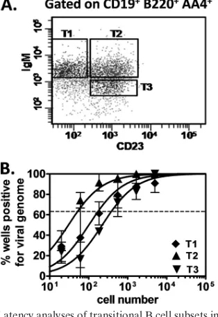
[image:5.585.82.503.68.293.2]To determine whether MHV68 infection was restricted to a specific subset of transitional B cells, we used antibodies di-rected toward an accepted combination of cell surface markers (CD19, B220, AA4, IgM, and CD23) to isolate T1, T2, and T3 transitional B cells (3). Splenocytes were harvested from B6
FIG. 3. Latency analyses of developing B cells in the spleen. For these experiments, B6 mice were infected i.p. or i.n. with 104PFU of MHV68, and spleens were harvested at 15 or 42 to 48 days postinoculation. For each sample group in each experiment (n⫽3 experiments), splenocytes from 5 mice were pooled. Following blocking, cells were stained with anti-CD19-allophycocyanin/Cy7 and anti-AA4-allophycocyanin. (A) Flow cytometric cell sorting was performed to isolate purified transitional B cells (CD19⫹AA4⫹) and mature B cells (CD19⫹AA4⫺). Mean purities for each population were as follows: transitional B cells, 97.5%⫾0.7%; mature B cells, 98.4%⫾0.6%. (B) To determine the frequency of cells that harbored latent virus, sorted populations were subjected to limiting dilution nested PCR for viral genome, exactly as described for Fig. 1. The frequency of cells positive for viral genome was calculated by Poisson distribution analysis of mean data from 3 experiments. On graphs, the dashed line at 63.2% indicates the point at which one viral genome-positive cell per reaction is predicted to occur. Thexaxis shows the numbers of cells per reaction; theyaxis shows the percentages of 12 reactions positive for viral genome. (C) To determine the frequencies of cells that reactivate from latency and carry preformed infectious virus, limiting dilutionex vivoreactivation assays and preformed virus assays were performed, exactly as described for Fig. 1. Thexaxis shows the numbers of cells per reaction; theyaxis shows the percentages of 12 reactions positive for CPE.
on November 8, 2019 by guest
http://jvi.asm.org/
mice 15 days postinoculation, stained with antibodies, and T1 (CD19⫹ B220⫹ AA4⫹ IgMhi CD23⫺), T2 (CD19⫹ B220⫹
AA4⫹ IgMhi CD23⫹), and T3 (CD19⫹B220⫹ AA4⫹ IgMlo
CD23⫹) populations were isolated by flow cytometry (Fig. 5A). Each subset was then subjected to limiting dilution PCR anal-ysis for viral genome (Fig. 5B). Interestingly, all three cell types were infected at high frequencies: 1 in 180 T1 B cells, 1 in 50 T2 B cells, and 1 in 390 T3 B cells. It is unclear why individual subsets of transitional B cells exhibited higher frequencies of infection than did the bulk population of transitional B cells, but this observation may reflect a very low level of infection in the excluded IgM⫺ CD23⫺ population of CD19⫹ B220⫹ AA4⫹ cells. Regardless, these results demonstrate that all three transitional B cell subsets are infected by MHV68.
Summary. It is widely accepted that mature B cells are a primary reservoir for both human and murine gammaherpes-viruses. However, the means by which these viruses access the B cell compartment to establish and maintain lifelong latency are poorly understood. Although it is likely that gammaher-pesviruses initially infect circulating B cells at or near their primary anatomical entry site, these mature cells have finite life spans that, on their own, would not be sufficient to perpetuate long-term infection. One intriguing possibility is that gamma-herpesviruses utilize the self-renewing reservoir of developing B cells as an access point to maintain lifelong infection in the circulating mature B cell compartment. Because the bone mar-row functions as both a primary lymphoid organ for hemato-poiesis and a secondary lymphoid organ for activation of naïve or memory immune cells, it stands to reason that this dynamic microenvironment could provide a unique setting for the con-fluence of the initially infected circulating mature B cells with their progenitor counterparts. In work described here, we dem-onstrate that immature B cells in the bone marrow and tran-sitional B cells in the spleen are reservoirs for MHV68 latency. Because these developing B cell subsets all have a high rate of turnover and a short life span, our finding that they carry viral genome throughout chronic infection strongly suggests that the virus recurrently infects these cells or that it somehow indefi-nitely prolongs their life spans. These findings support the possibility that infection of developing B cells plays a critical
FIG. 5. Latency analyses of transitional B cell subsets in the spleen. For these experiments, B6 mice were infected i.p. or i.n. with 104PFU of MHV68, and spleens were harvested at 15 days postinoculation. For each sample group in each experiment (n⫽2 experiments), spleno-cytes from 5 mice were pooled. Following blocking, cells were stained with rat anti-mouse antibodies: CD19-allophycocyanin/Cy7, B220-peri-dinin chlorophyll protein-Cy/5.5 (clone RA3-6B2; eBioscience), AA4-allophycocyanin, IgM-phycoerythrin/Cy7, and CD23-fluorescein iso-thiocyanate (clone B3B4; BD Biosciences). (A) Flow cytometric cell sorting was performed to isolate purified transitional B cell subsets T1 (CD19⫹B220⫹AA4⫹IgMhiCD23⫺), T2 (CD19⫹B220⫹AA4⫹IgMhi CD23⫹), and T3 (CD19⫹B220⫹AA4⫹IgMloCD23⫹). Mean purity of
all sorted subsets for transitional B cell markers (CD19⫹AA4⫹) was
[image:6.585.83.240.288.517.2]⬎95%. Purities within subset gates were as follows: T1, 89.3%; T2, 88.5%; T3, 86.5%. (B) To determine the frequency of cells that har-bored latent virus, sorted populations were subjected to limiting dilu-tion nested PCR for viral genome, exactly as described for Fig. 1. The frequency of cells positive for viral genome was calculated by Poisson distribution analysis of data from 2 experiments. On graphs, the dashed line at 63.2% indicates the point at which one viral genome-positive cell per reaction is predicted to occur. Thexaxis shows the numbers of cells per reaction; theyaxis shows the percentages of 12 reactions positive for viral genome.
FIG. 4. Detection of transitional B cells expressing mLANA⫹ during chronic infection by using a recombinant marker virus. For these experiments, B6 mice were infected i.n. with 104PFU of MHV68.ORF73la, and spleens were harvested at various times postinoculation. For each sample group in each experiment (n⫽ 3 experiments), splenocytes from 3 mice were pooled. Following blocking, cells were stained with anti-CD19-allophycocyanin/Cy7 and anti-AA4-allophycocyanin. Subsequently, cells were loaded with the-lactamase substrate CCF2/AM (In-vitrogen). Flow cytometry was used to identify transitional B cells (CD19⫹AA4⫹) that expressed mLANA/-lactamase, as previously described (25). (A) Representative flow cytometry plots from samples at 16 days postinoculation. Plots have been pregated on CD19⫹AA4⫹cells. The inner box indicates the gate for mLANA⫹cells. (B) Reciprocal frequency of mLANA⫹transitional B cells at 7, 16, 28, 42, and 90 days postinoculation calculated from flow cytometric analyses.
13050 NOTES J. VIROL.
on November 8, 2019 by guest
http://jvi.asm.org/
role in the normal pathogenesis of gammaherpesvirus infec-tions.
The most widely held paradigm for EBV entry into the resting memory B cell compartment is via virus-driven differ-entiation ofde novo-infected naïve B cells (35). However, it is conceivable that infection of the self-renewing reservoir of developing B cells is actually the key access point for entry into the B cell lineage. In such a model, passive or virus-driven differentiation coupled with LANA expression could result in segregation of the viral genome into mature B cells. In support of this hypothesis, KSHV is detected in mature human B cells and monocytes following reconstitution of NOD/SCID mice with KSHV-infected CD34⫹stem cells (42). Whether or not this occurs following natural infection of a fully immunocom-petent host remains to be tested. While we have found that all developing B cell subsets carry viral genome during chronic infection, further experiments will be necessary to determine whetherde novoinfection of each subset occurs simultaneously or whether infection occurs at an early stage of development (e.g., pro-pre-B cells), followed by differentiation of the in-fected cells. One other important implication of our studies is the possibility that infection at developmental stages preceding selection could influence the outcome of normal hematopoie-sis, resulting in the generation of B cells that would not nor-mally have survived this process. This concept is supported by studies of the EBV latency protein LMP2A, whose expression in transgenic mice is associated with the bypass of normal B cell developmental checkpoints in the bone marrow (6). Future work will be necessary to determine whether such a finding is applicable in the context of natural infection. In summary, our results demonstrate that developing B cells are a major reser-voir for MHV68 latency, providing the possibility that infection of this unique cellular compartment plays a key role in gam-maherpesvirus pathogenesis and the maintenance of lifelong latency.
This work was supported by NIH grant CA139984 and NIH COBRE Center for Molecular and Tumor Virology grant P20-RR018724. M.S.N. was supported by an American Heart Association Predoctoral Fellowship (0815151E).
We thank Robert Chervenak, Deborah Chervenak, and Shannon Mumphrey for expert assistance with flow cytometry experiments. We thank David Allman for helpful discussions.
REFERENCES
1.Allman, D. M., S. E. Ferguson, V. M. Lentz, and M. P. Cancro.1993. Peripheral B cell maturation. II. Heat-stable antigen(hi) splenic B cells are an immature developmental intermediate in the production of long-lived marrow-derived B cells. J. Immunol.151:4431–4444.
2.Allman, D., R. C. Lindsley, W. DeMuth, K. Rudd, S. A. Shinton, and R. R. Hardy.2001. Resolution of three nonproliferative immature splenic B cell subsets reveals multiple selection points during peripheral B cell maturation. J. Immunol.167:6834–6840.
3.Allman, D., and S. Pillai.2008. Peripheral B cell subsets. Curr. Opin. Im-munol.20:149–157.
4.Bertolini, L., F. Falzetti, G. Lanzilli, O. Moscone, M. Longo, C. Bangrazi, and G. Gorini.1991. Lymphocyte receptors as differentiation markers in a cell line autonomously arisen “in vitro” from a human normal bone marrow, p. 77–89. InR. Wegmann and M. Wegmann (ed.), Recent advances in cellular and molecular biology. Peeter Press, Leuven, Belgium.
5.Bertolini, L., M. L. Aebischer, F. Ameglio, A. Angeloni, I. Delaroche, A. Faggioni, A. Fruscalzo, G. Gorini, A. Serafino, G. Starace, and A. Tabilio.
2005. Phenotypic and genotypic characteristics of new euploid-diploid lym-phoblastoid B cell lines EBV⫹, normal human bone marrow derived, spon-taneously overgrown in vitro. J. Virol. Methods126:91–100.
6.Caldwell, R. G., R. C. Brown, and R. Longnecker.2000. Epstein-Barr virus
LMP2A-induced B-cell survival in two unique classes of ELMP2A trans-genic mice. J. Virol.74:1101–1113.
7.Cardin, R. D., J. W. Brooks, S. R. Sarawar, and P. C. Doherty.1996. Progressive loss of CD8⫹T cell-mediated control of a gamma-herpesvirus in the absence of CD4⫹T cells. J. Exp. Med.184:863–871.
8.Chen, T., and S. D. Hudnall.2006. Anatomical mapping of human herpes-virus reservoirs of infection. Mod. Pathol.19:726–737.
9.Collins, C. M., J. M. Boss, and S. H. Speck.2009. Identification of infected B-cell populations by using a recombinant murine gammaherpesvirus 68 expressing a fluorescent protein. J. Virol.83:6484–6493.
10.Corbellino, M., L. Poirel, G. Bestetti, M. Pizzuto, J. Aubin, M. Capra, C. Bifulco, E. Berti, H. Agut, G. Rizzardini, M. Galli, and C. Parravicini.1996. Restricted tissue distribution of extralesional Kaposi’s sarcoma-associated herpesvirus-like DNA sequences in AIDS patients with Kaposi’s sarcoma. AIDS Res. Hum. Retroviruses12:651–657.
11.Deeg, H. J., and G. Socie.1998. Malignancies after hematopoietic stem cell transplantation: many questions, some answers. Blood91:1833–1844. 12.Dutia, B. M., S. J. Reid, D. D. Drummond, Y. Ligertwood, I. Bennet, W.
Rietberg, O. Silvia, M. A. Jarvis, and A. A. Nash.2009. A novel Cre recom-binase imaging system for tracking lymphotropic virus infection in vivo. PLoS One4:e6492.
13.Flano, E., I. Kim, J. Moore, D. L. Woodland, and M. A. Blackman.2003. Differential gamma-herpesvirus distribution in distinct anatomical locations and cell subsets during persistent infection in mice. J. Immunol.170:3828– 3834.
14.Frizzera, G., D. W. Hanto, K. J. Gajl-Peczalska, J. Rosai, R. W. McKenna, R. K. Sibley, K. P. Holahan, and L. L. Lindquist.1981. Polymorphic diffuse B-cell hyperplasias and lymphomas in renal transplant recipients. Cancer Res.41:4262–4279.
15.Hanto, D. W., G. Frizzera, K. J. Gajl-Peczalska, and R. L. Simmons.1985. Epstein-Barr virus, immunodeficiency, and B cell lymphoproliferation. Transplantation39:461–472.
16.Henry, M., A. Uthman, A. Geusau, A. Rieger, L. Furci, A. Lazzarin, P. Lusso, and E. Tschachler.1999. Infection of circulating CD34⫹cells by HHV-8 in patients with Kaposi’s sarcoma. J. Invest. Dermatol.113:613–616. 17.Hoang, M. P., D. B. Dawson, Z. R. Rogers, R. H. Scheuermann, and B. B.
Rogers.1998. Polymerase chain reaction amplification of archival material for Epstein-Barr virus, cytomegalovirus, human herpesvirus 6, and parvovi-rus B19 in children with bone marrow hemophagocytosis. Hum. Pathol.
29:1074–1077.
18.Kikuta, H., Y. Sakiyama, S. Matsumoto, T. Oh-Ishi, T. Nakano, T. Na-gashima, T. Oka, T. Hironaka, and K. Hirai. 1993. Fatal Epstein-Barr virus-associated hemophagocytic syndrome. Blood82:3259–3264. 19.Li, H., K. Ikuta, J. W. Sixbey, and S. A. Tibbetts.2008. A
replication-defective gammaherpesvirus efficiently establishes long-term latency in mac-rophages but not in B cells in vivo. J. Virol.82:8500–8508.
20.Li, Y., R. Wasserman, K. Hayakawa, and R. R. Hardy.1996. Identification of the earliest B lineage stage in mouse bone marrow. Immunity5:527–535. 21.Loder, F., B. Mutschler, R. J. Ray, C. J. Paige, P. Sideras, R. Torres, M. C.
Lamers, and R. Carsetti.1999. B cell development in the spleen takes place in discrete steps and is determined by the quality of B cell receptor-derived signals. J. Exp. Med.190:75–89.
22.Luppi, M., P. Barozzi, T. F. Schulz, G. Setti, K. Staskus, R. Trovato, F. Narni, A. Donelli, A. Maiorana, R. Marasca, S. Sandrini, G. Torelli, and J. Sheldon.2000. Bone marrow failure associated with human herpesvirus 8 infection after transplantation. N. Engl. J. Med.343:1378–1385.
23.Marques, S., S. Efstathiou, K. G. Smith, M. Haury, and J. P. Simas.2003. Selective gene expression of latent murine gammaherpesvirus 68 in B lym-phocytes. J. Virol.77:7308–7318.
24.McClain, K., R. Gehrz, H. Grierson, D. Purtilo, and A. Filipovich.1988. Virus-associated histiocytic proliferations in children. Frequent association with Epstein-Barr virus and congenital or acquired immunodeficiencies. Am. J. Pediatr. Hematol. Oncol.10:196–205.
25.Nealy, M. S., C. B. Coleman, H. Li, and S. A. Tibbetts.2010. Use of a virus-encoded enzymatic marker reveals that a stable fraction of memory B cells expresses latency-associated nuclear antigen throughout chronic gam-maherpesvirus infection. J. Virol.84:7523–7534.
26.Pavlova, B. G., H. H. Mu¨hlberger, H. Strobl, R. Grill, A. Haslberger, F. Varga, H. Auer, R. Heinz, J. Salamon, and A. Stacher.1995. B lymphocytes with latent EBV infection appearing in long-term bone marrow cultures (HLTBMCs) from haematological patients induce lysis of stromal microen-vironment. Br. J. Haematol.89:704–711.
27.Purtilo, D. (ed.).1984. Immune deficiency and cancer: Epstein-Barr virus and lymphoproliferative malignancies. Plenum Medical Book Co., New York, NY.
28.Ragona, G., A. Tabilio, A. Fruscalzo, L. Annino, A. Angeloni, and L. Berto-lini.1988. Presence of EBV infected cells in the bone marrow from trans-plant donors, p. 231–235.InD. Ablashi, A. Ariad, G. Krueger, J. Pagano, and G. Pearson (ed.), Epstein-Barr virus and human disease. Humana Press, Clifton, NJ.
29.Reisman, R. P., and M. A. Greco.1984. Virus-associated hemophagocytic syndrome due to Epstein-Barr virus. Hum. Pathol.15:290–293.
on November 8, 2019 by guest
http://jvi.asm.org/
30.Revoltella, R. P., E. Vigneti, A. Fruscalzo, M. Park, G. Ragona, G. Rocchi, and E. Calef.1989. Epstein-Barr virus DNA sequences in precursor mono-cyte-macrophage cell lines established from the bone marrow of children with maturation defects of haematopoiesis. J. Gen. Virol.70:1203–1215. 31.Risdall, R. J., R. W. McKenna, M. E. Nesbit, W. Krivit, H. H. Balfour, R. L.
Simmons, and R. D. Brunning.1979. Virus-associated hemophagocytic syn-drome: a benign histiocytic proliferation distinct from malignant histiocyto-sis. Cancer44:993–1002.
32.Shapiro, R. S., K. McClain, G. Frizzera, K. J. Gajl-Peczalska, J. H. Kersey, B. R. Blazar, D. C. Arthur, D. F. Patton, J. S. Greenberg, and B. Burke.1988. Epstein-Barr virus associated B cell lymphoproliferative disorders following bone marrow transplantation. Blood71:1234–1243.
33.Sullivan, J. L., B. A. Woda, H. G. Herrod, G. Koh, F. P. Rivara, and C. Mulder.1985. Epstein-Barr virus-associated hemophagocytic syndrome: vi-rological and immunopathological studies. Blood65:1097–1104.
34.Sunil-Chandra, N. P., S. Efstathiou, J. Arno, and A. A. Nash.1992. Viro-logical and pathoViro-logical features of mice infected with murine gamma-her-pesvirus 68. J. Gen. Virol.73:2347–2356.
35.Thorley-Lawson, D. A.2001. Epstein-Barr virus: exploiting the immune sys-tem. Nat. Rev. Immunol.1:75–82.
36.Tibbetts, S. A., L. F. van Dyk, S. H. Speck, and H. W. Virgin.2002. Immune control of the number and reactivation phenotype of cells latently infected with a gammaherpesvirus. J. Virol.76:7125–7132.
37.Tibbetts, S. A., J. Loh, V. van Berkel, J. S. McClellan, M. A. Jacoby, S. B. Kapadia, S. H. Speck, and H. W. Virgin.2003. Establishment and
mainte-nance of gammaherpesvirus latency are independent of infective dose and route of infection. J. Virol.77:7696–7701.
38.Tibbetts, S. A., J. S. McClellan, S. Gangappa, S. H. Speck, and H. W. Virgin.
2003. Effective vaccination against long-term gammaherpesvirus latency. J. Virol.77:2522–2529.
39.Watanabe, H., M. Koide, K. Fukuchi, Y. Takagi, S. Tomoyasu, N. Tsu-ruoka, and K. Gomi.1997. Presence of Epstein-Barr virus genome in the bone marrow of patients with hematopoietic malignancies. Acta Haema-tol.98:32–36.
40.Weck, K. E., M. L. Barkon, L. I. Yoo, S. H. Speck, and I. V. Virgin, H. W.
1996. Mature B cells are required for acute splenic infection, but not for establishment of latency, by murine gammaherpesvirus 68. J. Virol.70:6775– 6780.
41.Weck, K. E., S. S. Kim, H. W. Virgin, and S. H. Speck.1999. B cells regulate murine gammaherpesvirus 68 latency. J. Virol.73:4651–4661.
42.Wu, W., J. Vieira, N. Fiore, P. Banerjee, M. Sieburg, R. Rochford, W. Harrington, and G. Feuer.2006. KSHV/HHV-8 infection of human hema-topoietic progenitor (CD34⫹) cells: persistence of infection during hema-topoiesis in vitro and in vivo. Blood108:141–151.
43.Zlokarnik, G., P. A. Negulescu, T. E. Knapp, L. Mere, N. Burres, L. Feng, M. Whitney, K. Roemer, and R. Y. Tsien.1998. Quantitation of transcription and clonal selection of single living cells with beta-lactamase as reporter. Science279:84–88.
44.Zutter, M. M., P. J. Martin, G. E. Sale, H. M. Shulman, L. Fisher, E. D. Thomas, and D. M. Durnam.1988. Epstein-Barr virus lymphoproliferation after bone marrow transplantation. Blood72:520–529.
13052 NOTES J. VIROL.