0022-538X/10/$12.00 doi:10.1128/JVI.00851-10
Copyright © 2010, American Society for Microbiology. All Rights Reserved.
Alpha Interferon and Not Gamma Interferon Inhibits Salmonid
Alphavirus Subtype 3 Replication
In Vitro
䌤
Cheng Xu, Tz-Chun Guo, Stephen Mutoloki, Øyvind Haugland,
Inderjit S. Marjara, and Øystein Evensen*
Norwegian School of Veterinary Science, Department of Basic Sciences and Aquatic Medicine, P.O. Box 8146 Dep., N-0033 Oslo, Norway
Received 21 April 2010/Accepted 13 June 2010
Salmonid alphavirus (SAV) is an emerging virus in salmonid aquaculture, with SAV-3 being the only subtype
found in Norway. Until now, there has been little focus on the alpha interferon (IFN-␣)-induced antiviral
responses during virus infectionin vivoorin vitroin fish. The possible involvement of IFN-␥in the response
to SAV-3 is also not known. In this study, the two IFNs were cloned and expressed as recombinant proteins
(recombinant IFN-␣ [rIFN-␣] and rIFN-␥) and used for in vitro studies. SAV-3 infection in a permissive
salmon cell line (TO cells) results in IFN-␣and IFN-stimulated gene (ISG) mRNA upregulation. Preinfection
treatment (4 to 24 h prior to infection) with salmon rIFN-␣ induces an antiviral state that inhibits the
replication of SAV-3 and protects the cells against virus-induced cytopathic effects (CPE). The antiviral state
coincides with a strong expression of Mx and ISG15 mRNA and Mx protein expression. When rIFN-␣ is
administered at the time of infection and up to 24 h postinfection, virus replication is not inhibited, and cells are not protected against virus-induced CPE. By 40 h postinfection, the alpha subunit of eukaryotic initiation
factor 2 (eIF2␣) is phosphorylated concomitant with the expression of the E2 protein as assessed by Western
blotting. Postinfection treatment with rIFN-␣ results in a moderate reduction in E2 expression levels in
accordance with a moderate downregulation of cellular protein synthesis, an approximately 65% reduction by
60 h postinfection. rIFN-␥has only a minor inhibitory effect on SAV-3 replicationin vitro. SAV-3 is sensitive
to the preinfection antiviral state induced by rIFN-␣, while postinfection antiviral responses or postinfection
treatment with rIFN-␣is not able to limit viral replication.
Salmon pancreas disease virus (SPDV) is the causative agent of pancreas disease (PD) in Atlantic salmon and rainbow trout and is an emerging pathogen in Europe and North America (21). SPDV belongs to the genusAlphaviruswithin the family
Togaviridaeand is phylogenetically related to arthropod-borne alphavirus groups such as the Semliki Forest virus (SFV) group, the Sindbis virus (SINV) group, and Venezuelan equine encephalitis virus (VEEV)/Eastern equine encephalitis virus (EEEV) (24). SPDV was later termed salmonid alphavirus (SAV) and has been divided into subtypes (14), and six sub-types have now been recognized, SAV-1 to SAV-6 (9). SAV-1 was first isolated from farmed Atlantic salmon in Ireland and Scotland (37). Subsequently, sleeping disease virus (SDV) (and later SAV-2), which affects mainly rainbow trout, was discov-ered in the United Kingdom and France (35, 38). A third and new subtype, SAV-3, is found exclusively in Norway and affects both Atlantic salmon and rainbow trout (14, 33). Three addi-tional subtypes of SAV from Scotland and Ireland have been described (9). Diseased fish are clinically characterized by in-appetence, fecal casts, and emaciation, with main pathological changes found in pancreas, heart, and skeletal muscle (21, 25). Immunity to alphavirus infections in salmonids is poorly un-derstood and has not been studied in any detail. In a previous study, interferon (IFN) responses were not detected following
SAV-1 infection in salmon parr, possibly due to the low sen-sitivity of the detection method used (7). A more recent study showed Mx mRNA upregulation in response to SAV infection
in vitro by using a macrophage cell line from salmon and suggested that Mx has an antiviral function against SAV (11). However, so far, no studies have been carried out to examine the functional aspects of type I IFN responses related to SAV infections of salmonidsin vivoorin vitro.
Type I interferons (alpha/beta IFN [IFN-␣/]) and interfer-on-stimulated genes (ISGs) are known to play a pivotal role in innate immune responses against viral infections in all living organisms (18) and, therefore, also in fish (42). Mice devoid of type I IFN receptors are extremely susceptible to alphavirus infections, whereas there is no apparent role for IFN-␥(22). Studies of infections in higher vertebrates have shown that the IFN-␣/ pathway represents the primary protective response against alphavirus infection involving the limitation of virus replication (1, 13). The antiviral effect of mammalian type I IFN is exerted through binding to the receptor of IFN-␣/, triggering the JAK-STAT pathway, which results in the expres-sion of ISGs, some of which encode antiviral proteins, includ-ing Mx, ISG15, and double-stranded RNA (dsRNA)-depen-dent protein kinase R (PKR) (29). Numerous ISGs such as 2⬘,5⬘-oligoadenylate synthetase (OAS)/RNase L, zinc finger antiviral protein (ZAP), Mx, and ISG15 likely execute a direct or indirect inhibition of alphavirus replication in higher verte-brates (5, 16, 19), although the detailed mechanisms are not understood. The importance of ISGs in relation to alphavirus infections has not been studied for fish. Type I IFN and several of its stimulated genes have been cloned from Atlantic salmon,
* Corresponding author. Mailing address: Norwegian School of Vet-erinary Science, Department of Basic Sciences and Aquatic Medicine, P.O. Box 8146 Dep., N-0033 Oslo, Norway. Phone: 47 229 64500. Fax: 47 225 97310. E-mail: [email protected].
䌤Published ahead of print on 23 June 2010.
8903
on November 8, 2019 by guest
http://jvi.asm.org/
and their analogous antiviral functions are also partly charac-terized, which allows a detailed characterization of the re-sponses at the transcript level. Furthermore, the antiviral na-ture of Atlantic salmon IFN was recently characterized by neutralizing IFN with specific antibodiesin vitro (3, 23). Mx and ISG15 have been cloned from Atlantic salmon (26, 27), and their functional antiviral activity has been demonstrated against another salmon virus, infectious pancreatic necrosis virus (17). Atlantic salmon PKR protein has been cloned, but no functional studies have been performed. However, PKR from Japanese flounder (Paralichthys olivaceus) was recently characterized and shown to increase the phosphorylation of the alpha subunit of eukaryotic initiation factor 2 (eIF2␣) and inhibit the replication of rhabdovirus in flounder em-bryonic cells (41). Atlantic salmon PKZ has a direct inhib-itory effect on protein synthesis after transient expression in Chinook salmon embryo (CHSE) cells (4).
In this study we were interested in understanding IFN-␣ -and IFN-␥-induced responses to SAV-3 infectionin vitroand the possible role of these cytokines in controlling virus repli-cation during the early stages of infection. The two IFNs were cloned and expressed as recombinant proteins and used forin vitro studies using a salmon macrophage cell line (36) as a model for SAV infection (11). We show that in SAV-3-infected cell cultures, IFN-␣and ISG mRNAs are upregulated and Mx protein expression is strongly induced. By 40 h postinfection, eIF2␣is phosphorylated, which coincides with a strong expres-sion of the E2 protein of SAV-3. There is no clear arrest of macromolecular synthesis at early times postinfection (first 24 h postinfection), while from 36 h, there is a moderate downregulation of protein synthesis. The treatment of cell cultures with recombinant IFN-␣(rIFN-␣) 4 to 24 h prior to infection (preinfection treatment) induces an antiviral re-sponse that markedly inhibits the replication of SAV-3 and protects against cytopathic effects (CPE). Treatment with rIFN-␣at time of infection and up to 24 h postinfection has a time-dependent effect on limiting SAV-3 replication: the later the treatment, the less efficient, and later treatment will not protect against CPE. Preinfection treatment (24 h) with IFN-␥ has only a minor inhibitory effect on the replication of SAV-3
in vitro.
MATERIALS AND METHODS
Virus isolation and cell culture.Chinook salmon embryonic cells (CHSE-214; ATCC CRL-1681) were maintained at 20°C with L-15 medium (Invitrogen) supplemented with 5% fetal bovine serum (FBS),L-glutamine, and gentamicin. TO cells (macrophage cell line), originating from salmon head kidney leukocytes (36), were grown at 20°C in HMEM (Eagle’s minimal essential medium [MEM] with Hanks’ balanced salt solution [BSS]) supplemented withL-glutamine, MEM nonessential amino acids, gentamicin sulfate, and 10% FBS. The SAV-3 isolate (named H10) used in this study was isolated from heart of an Atlantic salmon with clinical symptoms of PD. The isolate was propagated by inoculating 80% confluent CHSE-214 cells maintained with growth medium supplemented with 2% FBS. The isolate was identified as a SAV-3 variant by sequencing (14) and was passaged nine times in cell culture before being used in this study.
Protein synthesis shutdown.TO cells were infected with SAV-3 at a multi-plicity of infection (MOI) of 20. At different time points postinfection, the cells were washed three times with phosphate-buffered saline (PBS) and then incu-bated for 30 min in Dulbecco’s modified Eagle’s medium lacking methionine and supplemented with 0.1% FBS and 20Ci [35S]methionine/ml. The cells were then harvested sequentially at 12, 24, 36, 48, 60, and 72 h and lysed by using CelLytic M reagent (Sigma). Protein was separated by SDS-PAGE and blotted onto a polyvinylidene difluoride (PVDF) membrane. The membrane was
ex-posed in a PhosphorImager cassette and then scanned by using a Typhoon imager (GE Healthcare). The protein amount measured by densitometry was quantified with ImageJ software, and the value was expressed relative to a mock-infected control and corrected for the protein amount loaded into each lane.
RNA isolation and cDNA synthesis.Total RNA was isolated by using the RNeasy Plus minikit (Qiagen), and the concentration of RNA was determined by spectrophotometry (Nanodrop ND1000). For each sample, 500 ng of total RNA was subjected to cDNA synthesis using a Transcriptor first-strand cDNA kit (Roche) in a total volume of 20l. The synthesized cDNA was stored at⫺20°C until further use.
Quantitative real-time PCR.Quantitative PCR was performed by using Light-Cycler 480 SYBR green I master mix and the LightLight-Cycler 480 system (Roche). For each gene, 2l of cDNA was used as a template in a mixture of specific primers (10M) in a final volume of 20l. The mixtures were first incubated at 95°C for 10 min, followed by 40 amplification cycles (10 s at 95°C, 20 s at 60°C, and 8 s at 72°C). The sequences of primers used to assess thein vivoandin vitro
expressions of Mx, ISG15,␥IP10, and viral E2 are given in Table 1. For the viral E2 gene, the reaction mix contained 10l of LightCycler 480 Probes Master, 1 l of primer-probe mix (final concentrations, 0.9M each primer and 0.25M probe), 2l of cDNA template, and 7l water and was incubated for 10 min at 95°C, followed by 45 amplification cycles (10 s at 95°C, 30 s at 60°C, and 1 s at 72°C). The specificity of the PCR products from each primer pair was confirmed by melting-curve analysis and subsequent agarose gel electrophoresis. The 2⫺⌬⌬CTmethod was used to calculate the gene products as described elsewhere
previously (20). 2⫺⌬⌬CTis the relative mRNA expression representing the fold
induction over the control group. All quantifications were normalized to-actin (endogenous gene).
Cloning, expression, and purification of recombinant IFN-␣and IFN-␥.Total RNA from the head kidney of Atlantic salmon was used as a template for cDNA synthesis by using a Transcriptor first-strand cDNA synthesis kit (Roche). For initial cloning, two pairs of primers, IFN␣-F1 and IFN␣-R1, and IFN␥-F1 and IFN␥-R1, were designed according to the Atlantic salmon salmo salar IFN-␣1 (SasaIFN-␣1) mRNA sequence (GenBank accession no. AY216594) and the Atlantic salmon SasaIFN-␥ mRNA sequence (GenBank accession no. AY795563). For IFN-␣, we amplified a region from 42 bp downstream of the start codon of the open reading frame (ORF) to 27 bp downstream of the stop codon. For IFN-␥, a region from 107 bp upstream of the start codon of the ORF to 75 bp downstream of the stop codon was amplified. The PCR products were purified by using the QIAquick gel extraction kit (Qiagen) and cloned into the pGEM-T Easy vector (Promega). The region coding for the predicted full-length forms without the signal peptide was subcloned from pGEM-T into the prokary-otic vector 32c (Novagen) by using primer sets IFN␣-F1 and pET-IFN␣-R1, and pET-IFN␥-F1 and pET-IFN␥-R1, containing NdeI and XhoI restriction sites. Truncated forms of IFN-␣(trIFN-␣) and IFN-␥(trIFN-␥), with deletions of the C-terminal 28 amino acids (aa) and 29 aa, respectively, were also constructed by using different reverse primers (pET-IFN␣-R2 and pET-IFN␥-R2). The vectors were designed to express a C-terminal 6⫻His fusion protein to facilitate the purification of recombinant proteins. Full-length and truncated forms of IFN-␣and IFN-␥were designated rIFN-␣and trIFN-␣and rIFN-␥and trIFN-␥, respectively, which will be used throughout. The recombinant vectors pET-rIFN␣and pET-trIFN␣, and pET-rIFN␥and pET-tr IFN␥, were confirmed by DNA sequencing and were transformed into the bacterial host, BL21(DE3), for expression driven by the T7 polymerase. Induction was carried out at 37°C for 2 h with 1 mM isopropyl thiogalactopyranoside (IPTG). The purification of 6⫻His-tagged rIFN was performed under denaturing conditions by using a His-Bind purification kit (Novagen). Protein concentrations were determined by using the Quick Start Bradford protein assay kit (Bio-Rad) with bovine serum albumin (BSA) as a standard. The expression and the purity of recombinant IFN-␣and IFN-␥were checked on a 12% Bis-Tris precast SDS-PAGE gel (Invitrogen Life Technologies) stained with PageBlue protein staining solution (Fermentas). Western blot analysis was performed to confirm the identity of the recombinant IFN-␣by using a monoclonal anti-polyhistidine antibody (Sigma).
Cloning, expression, and purification of E2 and preparation of E2 antiserum.
Viral RNA was extracted from the cell supernatant by using a QIAamp viral RNA minikit (Qiagen) and used as a template for cDNA synthesis by using a Transcriptor first-strand cDNA synthesis kit (Roche). A pair of primers, pET14b-E2-F and pET14b-E2-R, was designed according to the E2 gene of the Norwegian salmonid alphavirus isolate (GenBank accession no. AY604236). The cloning, expression, and purification of recombinant E2 proteins were performed as described above. The purified recombinant E2 protein was sent to PickCell Laboratories BV (Netherlands) for rabbit immunization and subsequently
on November 8, 2019 by guest
http://jvi.asm.org/
acterized by Western blotting and immunofluorescence antibody test (IFAT) staining of SAV-3-infected cultures.
Induction of ISG expression by recombinant IFN-␣and IFN-␥in vitro.TO cells were seeded in 24-well plates and cultured until confluent. For IFN-␣, the cells were stimulated with 2.5g/ml rIFN-␣and trIFN-␣, nontreated cells were included as negative control, and samples were harvested at 3, 6, 12, and 24 h poststimulation. The cells were also treated with 10-fold serial dilutions of rIFN-␣with an initial starting concentration of 0.47 mg/ml; at 24 h, samples were taken, and RNA was extracted. The induction of ISGs by IFN-␣was documented by studying the gene upregulation of Mx and ISG15 by using real-time PCR. For IFN-␥, purified rIFN-␥was serially diluted from 0.33 mg/ml in cell medium and incubated with TO cells for 24 h. The induction of ISGs by IFN-␥was docu-mented by studying the gene upregulation of␥IP10 (gamma-IP CXCL10-like chemokine) using real-time PCR. The data are expressed as the mean fold changes in gene expression⫾standard errors of different dilutions of the inter-feron-treated group relative to the nontreated control group after normalization to-actin.
Antiviral assays.A CPE reduction assay was used to measure the protective effect of IFN-␣against cytopathic effects in pretreated and infected cells (8). TO cells grown in 96-well plates were treated with a serial dilution of rIFN-␣and trIFN-␣, and rIFN-␥and trIFN-␥, for 24 h and subsequently infected with 1 MOI of SAV-3. Virus was left on the cells for 2 h to adsorb (15°C), after which the cells were washed three times with PBS. Untreated cells, infected and noninfected, were included as controls. The cell viability of the cell cultures subjected to the different treatments was assayed by using the CellTiter 96 AQueous One solution cell proliferation assay kit (Promega) at day 10 postinfection when strong CPE
developed in untreated cells. The cell cultures were incubated at 15°C for 4 h, and the absorbance was measured at 490 nm by using a microplate reader (Tecan). For the virus yield reduction assay, culture supernatant and cell total RNA from infected cells in 24-well plates were collected. The titration of virus was done with CHSE-214 cells by the 50% tissue culture infective dose (TCID50) method as described previously by Ka¨rber (15), and viral RNA was quantified by real-time PCR.
Detection of Mx and E2 expression by IFAT.Cells seeded into 24-well culture plates were fixed with 4% paraformaldehyde for 30 min. After being washed with PBS, the cells were permeabilized with 0.1% Triton X-100 for 5 min on ice. The cells were washed once in PBS and blocked with 5% dry milk in PBS for 2 h before being incubated for 1 h with primary antibody. Anti-salmonid Mx (diluted 1:400; kindly provided by Jo-Ann Leong, Hawaii Institute of Marine Biology) and anti-E2 polyclonal antibody were used to detect Mx and E2 protein expres-sions, respectively. The cells were washed and incubated with Alexa 594 or Alexa 488 Fluor goat anti-rabbit IgG (Molecular Probes, Invitrogen) diluted 1:200 for 1 h. Finally, the cells were washed and examined by using a fluorescence micro-scope (Olympus). When nuclear counterstaining was used, it included Hoechst 33324 dye at 5g/ml.
Protein analysis of E2, Mx, and p-eIF2␣in virus-infected cells.TO cells were infected with SAV-3 (MOI of 1) or left uninfected. At days 1, 2, 3, and 4 postinfection, uninfected and infected cells were treated with a 103
[image:3.585.48.541.80.452.2]dilution of IFN-␣for 16 h or left untreated. The cells were then lysed by using CelLytic M reagent (Sigma) and scraped from the dish. Expressions of the E2 (antiserum prepared as described above), phosphorylated eIF2␣(p-eIF2␣) (Cell Signaling), Mx, and actin (Sigma-Aldrich) proteins were detected by Western blotting. TABLE 1. Primer and probe sequences for cloning and quantitative real-time PCRa
Primer Sequence Use GenBank
accession no.
IFN␣-F1 CAGTATGCAGAGCGTGTGT pGEMT cloning AY216594
IFN␣-R1 CGTAGCTTCTGAAATGAGTCTGG
pET-IFN␣-F1 GCGCATATGTGTGACTGGATCCGACAC pET32 cloning
pET-IFN␣-R1 GCGCTCGAGGTACATCTGTGCTGCAAG
pET-IFN␣-R2 GCGCTCGAGGTTCATTTTTCTCAGAAC
IFN␣-F2 TGGGAGGAGATATCACAAAGC qPCR
IFN␣-R2 TCCCAGGTGACAGATTTCAT
Mx-F TGCAACCACAGAGGCTTTGAA qPCR U66475
Mx-R GGCTTGGTCAGGATGCCTAAT
pET14b-E2-F GCGCATATGGCTGTGTCTGCGTCGCCT pET14b cloning
pET14b-E2-R GCGCTCGAGTTACGCACGAGCCCCAGG
SAV-3 E2-F CAGTGAAATTCGATAAGAAGTGCAA qPCR EF675594
SAV-3 E2-R TGGGAGTCGCTGGTAAAGGT
E2 probe FAM-5⬘-AGCGCTGCCCAAGCGACCG-3⬘-MGB
IFN␥-F1 AGGCGGTCTCGTTAAGTCAA pGEMT cloning AY795563
IFN␥-R1 TAAACTGACCCAAGATCAGC
pET32-IFN␥-F GCGCATATGGCTCAGTACACATCAATT pET32 cloning
pET32-IFN␥-R1 GCGCTCGAGCATGATGCTTGATTTGAG
pET32-IFN␥-R2 GCGCTCGAGTTTCTCGTAGATGGTAAT
IFN␥-F4 CTAAAGAAGGACAACCGCAG qPCR
IFN␥-R4 CACCGTTAGAGGGAGAAATG
␥IP10-F TGCCAGAACATGGAGATCAT qPCR EF619047
␥IP10-R TTTACTGCACACTCCTTTGGTT
ISG15-F AAGTGATGGTGCTGATTACGG qPCR AY926456
ISG15-R CACCGTTAGAGGGAGAAATG
-actin-F CCAGTCCTGCTCACTGAGGC qPCR AF012125
-actin-R GGTCTCAAACATGATCTGGGTCA
aRestriction sites are underlined. qPCR, quantitative PCR; FAM, 6-carboxyfluorescein; MGB, MGB-TaqMan probe.
on November 8, 2019 by guest
http://jvi.asm.org/
Statistical analysis.All statistical analyses of gene expression results were performed with the help of GraphPad Prism 5.0 (GraphPad Software Inc.). Two-way analysis of variance (ANOVA) was used to calculate differences in the CPE protection assay at different concentrations. The significant level for rejec-tion of hypothesis 0 (Ho) was set to aPvalue of⬍0.05.
RESULTS
Responses to SAV-3 infection in TO cells.The initial studies
were designed to characterize the expression of IFN-␣and Mx mRNA in response to SAV-3 infection in TO cells. The kinet-ics of the virus infection were monitored by real-time reverse transcription (RT)-PCR analysis of the expression of the
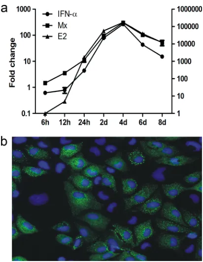
pro-tein E2-encoding gene, which was detected as early as 6 h postinfection and increased 1,600-fold from 6 h to 24 h postin-fection (Fig. 1a). Over the same time period (6 h to 24 h), IFN-␣transcript levels increased 4-fold, while Mx expression levels increased 10-fold (Fig. 1a). From 24 h postinfection to 4 days postinfection (dpi), there was a marked and parallel in-crease in levels of IFN-␣, Mx, and E2 transcripts, all peaking at 4 dpi and declining at later times (Fig. 1a). Staining for the Mx protein at 8 days postinfection showed widespread expression in the infected cell culture (Fig. 1b). These findings provided a strong indication that SAV-3 infection will not result in a shutoff of interferon or interferon-induced responses.
rIFN-␣ induces expression of ISGs in salmon cell lines.
With the purpose to conduct functional studies with recombi-nant IFN-␣ using a macrophage cell line of Atlantic salmon (TO cells) (36), we first cloned and expressed full-length IFN-␣ (rIFN-␣) and tested its functional activity. The obtained se-quence was aligned with SasaIFN-␣1 (GenBank accession no. AY216594) and SasaIFN-␣2 (accession no. AY216595) se-quences, and the amino acid similarities were 96.1% and 99.3%, respectively. As a negative control, we constructed a truncated form of IFN-␣(trIFN-␣), with a deletion of a 28-aa stretch at the C-terminal end (31). Both full-length and trun-cated IFN-␣ were expressed in Escherichia coli BL21(DE3) cells using the pET prokaryotic expression system. The protein yield in the cell soluble fraction was much lower than that of the inclusion bodies (data not shown); thus, the purification of proteins was performed under denaturing conditions. A 6⫻His tag added at the C-terminal end of the protein facilitated purification with a His-Bind column. The expression and pu-rification of recombinant proteins were identified by SDS-PAGE and Western blot analysis using an anti-His monoclonal antibody (Fig. 2a and b). The concentrations of purified full-length and truncated IFN-␣were 0.47 mg/ml and 0.48 mg/ml, respectively. Dilutions of rIFN-␣ and trIFN-␣ refer to these initial concentrations in all experiments except when stated otherwise. A similar characterization was performed for IFN-␥ by SDS-PAGE for the full-length and the truncated forms of purified samples (Fig. 2c). The protein concentration for pu-rified full-length rIFN-␥was 0.33 mg/ml.
The biological function and activity of rIFN-␣were assessed by measuring the expression of the interferon-induced Mx and ISG15 genes in TO cells using real-time RT-PCR after stim-ulation with rIFN-␣. At 24 h poststimulation, Mx mRNA was found to be strongly expressed after rIFN-␣treatment, while
FIG. 1. (a) Expression levels of IFN-␣, Mx, and viral E2 mRNA in TO cells infected with 1 MOI of SAV-3. The leftyaxis shows the fold increase of IFN-␣and Mx mRNA relative to the nontreated control, and the rightyaxis shows the fold increase of viral E2 transcript levels relative to infected cells at 6 h. The results are expressed as means⫾ standard errors of the means (SEM) (n⫽ 2). (b) Cytoplasmic Mx protein expression shown by indirect immunofluorescence in TO cells infected with SAV-3. Cells were fixed at 8 days postinfection. Nuclear staining was done with Hoechst 33324 dye.
FIG. 2. (a) Expression and purification of mature and truncated IFN-␣(SDS-PAGE). Lanes: 1, protein marker; 2, full-length recombinant IFN-␣(rIFN-␣); 3, purified rIFN-␣; 4, truncated IFN-␣(trIFN-␣); 5, purified trIFN-␣. (b) Western blot analysis of rIFN-␣(lane 1) and trIFN-␣ (lane 2) in bacterial lysates. The predicted molecular masses of full-length and truncated IFN-␣are approximately 19.4 and 16.0 kDa, respectively (M, marker lane). (c) Expression and purification of recombinant and truncated IFN-␥. Lanes: 1, protein marker; 2, full-length IFN-␥(rIFN-␥); 3, purified rIFN-␥; 4, trIFN-␥; 5, purified trIFN-␥. The predicted molecular masses of full-length and truncated IFN-␥are approximately 19.7 and 16.3 kDa, respectively.
on November 8, 2019 by guest
http://jvi.asm.org/
[image:4.585.60.266.65.330.2] [image:4.585.111.472.595.668.2]the truncated form was nonfunctional, and no expression of Mx was seen (Fig. 3a). By serial dilution we found that 0.47 ng/ml of rIFN-␣ still induced the expression of Mx (2.5-fold upregulation) (106dilution) (Fig. 3b). The expression of ISG15
mRNA by 12 h post-rIFN-␣treatment was 1,000-fold upregu-lated, increasing to⬎1,500-fold by 24 h (Fig. 3c). The expres-sion of the Mx protein was confirmed by indirect immunoflu-orescence 24 h after rIFN-␣stimulation. TO cells were stained positive with an anti-Mx antibody (Fig. 4a), while cells without treatment showed no staining (Fig. 4b).
rIFN-␣induced a preinfection antiviral state that inhibits
SAV-3 replication. We then went on to study the antiviral
effects after pretreatment (24 h) of TO cells with rIFN-␣. All studies were carried out using cell cultures originating from Atlantic salmon (Salmo salar L.), while we also included a limited study using CHSE cells (of Chinook salmon origin [Oncorhynchus tshawytscha]). We used the CPE reduction as-say and the virus yield reduction asas-say (virus titer estimation) to measure the antiviral activity of rIFN-␣on SAV-3 replica-tion. TO cells were pretreated with a serial dilution of full-length and truncated IFN-␣for 24 h and subsequently infected with 1 MOI of SAV-3. rIFN-␣and trIFN-␣were added to the cell cultures in culture medium, and the cultures were incu-bated for 24 h. At 0 h, medium was aspirated, virus was added, and the cultures were incubated for up to 10 days without changing the medium. Controls included infected cells not
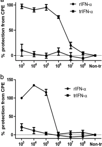
treated with rIFN-␣ or cells left uninfected. At 10 dpi when strong CPE occurred in the infected, non-IFN-␣-treated cells, viability was assayed by using an MTS [3-(4,5-dimethylthiazol- 2-yl)-5-(3-carboxymethoxyphenyl)-2-(4-sulfophenyl)-2H-tetra-zolium, inner salt] assay. Protection from CPE in rIFN-␣ -pretreated cells was dose dependent, with significant differences (P⬍0.05) between nontreated, infected cells and cells pretreated with rIFN-␣ at 103-, 104-, 105-, and 106-fold
dilutions (Fig. 5a), corresponding to rIFN-␣ concentrations from 0.47 g to 0.47 ng ml⫺1. Conversely, trIFN-␣ gave no
protection against CPE (Fig. 5a). Antiviral activity was also demonstrated for CHSE cells, and significant protection (P⬍
0.05) was seen for cells treated with rIFN-␣at 103-, 104-, and
105-fold dilutions (Fig. 5b).
Morphologically, no CPE was observed for TO cells pre-treated with rIFN-␣at 103-, 104-, and 105-fold dilutions, while
at a 106-fold dilution, CPE were observed (Fig. 6). Cultures
pretreated with trIFN-␣were not protected against CPE in-duced by SAV-3 infection even at the highest concentration used (Fig. 6).
[image:5.585.87.502.69.161.2]The purity of the cloned E2 protein (Fig. 7a) and the spec-ificity of the anti-E2 rabbit antiserum were documented by Western blotting (Fig. 7b) and IFAT staining (Fig. 7c) of SAV-3-infected cultures, and the inhibition of viral replication in IFN-␣-treated cells was further demonstrated by an analysis of virus replication and viral protein (E2) synthesis. TO cells
FIG. 3. Induction of Mx and ISG15 expression in TO cells after treatment with full-length recombinant and truncated IFN-␣. (a and b) Time course expression levels of Mx (a) and dose-dependent induction of Mx mRNA by rIFN-␣(b). Purified rIFN-␣was diluted serially 10-fold from 0.47 mg/ml and incubated with TO cells for 24 h. The data are expressed as the mean fold changes in gene expression⫾SEM of different dilutions for the rIFN-␣-treated group relative to the nontreated control group after normalization to-actin (n⫽2). (c) ISG15 mRNA transcripts of TO cells stimulated with 2.5g/ml rIFN-␣and trIFN-␣. The data are expressed as the mean fold changes in gene expression⫾standard errors for the IFN-␣-treated group relative to the nontreated control group after normalization to-actin (n⫽2).
FIG. 4. (a) Detection of Mx protein expression by indirect immunofluorescence in TO cells stimulated with 2.5g/ml rIFN-␣for 24 h and then fixed and stained. (b) Nontreated control cells.
on November 8, 2019 by guest
http://jvi.asm.org/
[image:5.585.82.499.549.703.2]treated with 0.47g/ml rIFN-␣totally inhibited the expression of the E2 protein at 2 days postinfection (Fig. 7e), while non-treated cells showed positive staining for E2 at the same time point (Fig. 7f). The antiviral activity of rIFN-␣was confirmed by the virus yield reduction assay: the virus titer was reduced in TO cells treated with rIFN-␣diluted 103-, 104-, and 105-fold
(103- to 105-fold reduction) compared with nontreated control
cells (Fig. 7d). These results showed that the pretreatment of cells with rIFN-␣ strongly inhibited SAV-3 replication and protected cells against virus-induced cell lysis (CPE).
Timing of rIFN-␣ treatment of SAV-3-infected cells. We
then explored the effect of the timing of rIFN-␣ treatment (0.47g/ml) relative to when the cells were infected, starting IFN-␣treatment 24 h prior to infection and ending 24 h postin-fection. Virus replication was assayed by examining the relative expression of E2 by real-time RT-PCR (Fig. 8), and at the same time, we evaluated the development of CPE in pre- and posttreated infected cell cultures. Treatment 24 h prior to infection inhibited SAV-3 replication 40,000-fold relative to nontreated controls (Fig. 8). Preinfection treatment (⫺4 h) gave close to a 2,000-fold reduction in E2 gene expression and protection against CPE in infected cultures (Fig. 8). Treatment closer to the time of infection (⫺2 h and 0 h) and postinfection did not protect against CPE; however, there was a marked reduction in E2 gene expression levels relative to those of nontreated cell cultures. Treatment 24 h postinfection gave a 3-fold reduction in replication levels and strong CPE. The results clearly show that the timing of the IFN-␣treatment is critical for the induction of the antiviral state, and its magni-tude is dependent on the dose (Fig. 5a) and time (Fig. 8). While an antiviral state can be established through preinfec-tion treatment, concurrent or postinfecpreinfec-tion treatment with rIFN-␣will not protect infected cells against CPE, indicating that the virus has developed a strategy to evade the IFN-induced antiviral response at later stages of the infection cycle.
SAV-3 infection results in phosphorylation of eIF2␣ cells
and moderate protein synthesis downregulation.These
find-ings led us to further characterize the expression of viral pro-tein (E2) and Mx and the phosphorylation of eIF2␣by West-ern blotting over a 96-h period postinfection. TO cells were infected at an MOI of 1 with SAV-3 (0 h), and one parallel culture was also treated with rIFN-␣(0.47g/ml) at 24, 48, 72, and 96 h postinfection. Control treatments included noninfect-ed/rIFN-␣-treated and infected/non-rIFN-␣-treated cells (Fig. 9). After rIFN-␣treatment, the cells were left to grow for an additional 16 h (after each treatment time) to assess responses to treatment. For simplicity, we refer to time points when rIFN-␣ was added. E2 expression was seen at 24 h (faint staining) postinfection in cultures treated with rIFN-␣and left untreated, while Mx was not detected at this time point (Fig. 9). Faint staining for p-eIF2␣ was also seen. From 48 h
on-FIG. 5. Cytopathic effect (CPE) reduction assay expressed as per-cent protection from CPE in TO cells (a) and in CHSE cells (b). Cell cultures were stimulated with 10-fold serial dilutions of rIFN-␣and trIFN-␣ 24 h prior to infection with 1 MOI of SAV-3. The lowest concentration with a biological effect (106dilution) corresponds to 0.47
ng/ml, while the highest concentration used (103dilution) was 0.47 g/ml for IFN-␣. For trIFN-␣, the highest concentration used was 0.48
[image:6.585.78.247.73.315.2]g/ml (n⫽3) (means⫾SEM). Non-tr, nontreated cultures.
FIG. 6. Protection against CPE observed by morphology. The top row shows cells treated with rIFN-␣, which showed dose-dependent protection against CPE at 103and 105dilutions, while initial CPE were seen at a 106dilution. Conversely, trIFN-␣did not show any protection
against CPE (bottom row). Shown are representative findings for two independent experiments. NT, not treated; NI, not infected.
on November 8, 2019 by guest
http://jvi.asm.org/
[image:6.585.81.510.547.695.2]wards, there was marked E2 and Mx expression and staining for p-eIF2␣ in the SAV-3-infected cells not treated with IFN-␣. The combination of infection and rIFN-␣ treatment gave the same results as those for the SAV-3-infected-only
FIG. 7. (a) Purification of recombinant pET14b-E2 protein analyzed by SDS-PAGE. Lanes: 1, protein marker; 2, whole bacteria after induction; 3, soluble fraction after sonication; 4, insoluble fraction after sonication; 5, purified recombinant protein. (b) Characterization of polyclonal antibody against E2 by Western blotting. Lanes: 1, pET14b-E2 after induction; 2, pET14b-E2 without induction; 3, purified E2. (c) Reactivity of rabbit antibody against E2 by IFAT of CHSE cells infected with SAV-3 using a 1:400 dilution of the serum. (d) Virus yield reduction assay. TO cells in 24-well plates were stimulated with 10-fold serial dilutions of rIFN-␣for 24 h and then infected with 1 MOI of SAV-3. Cell culture supernatants were collected when strong CPE were observed for the control infected cells. The virus titer of the supernatants at 10 days postinfection was determined by the TCID50method (n⫽2) (means⫾SEM). (e) Inhibition of E2 protein synthesis in interferon-treated TO cells.
[image:7.585.114.472.67.392.2]TO cells were treated with 0.47g/ml IFN-␣for 24 h and subsequently infected with 1 MOI of SAV-3. At 2 days postinfection, cells were stained with anti-E2 antibody, and no cells were found to express virus protein. (f) TO cells without IFN treatment infected as described in the text and stained by IFAT for E2, showing distinct cytoplasmic staining in infected cells.
FIG. 8. Timing of IFN-␣treatment of SAV-3-infected cells. Treat-ment 4 to 24 h prior to infection results in a marked reduction in virus replication (2,000- to 40,000-fold reduction) as measured by real-time PCR and protection against CPE. rIFN-␣ treatment at the time of infection or up to 24 h postinfection gives a reduction in virus repli-cation but not protection against CPE. TO cells were infected with an MOI of 1, and collection was done at 4 days postinfection. Shown are representative data from two independent experiments, expressed rel-ative to the nontreated control (NT). More details are given in Mate-rials and Methods.
FIG. 9. Protein analysis of E2, Mx, and p-eIF2␣in virus-infected cells. TO cells were infected with SAV-3 (MOI of 1) or left uninfected, and at 24, 48, 72, and 96 h postinfection, uninfected (⫺) and infected (⫹) cells were treated with 0.47g/ml of rIFN-␣for 16 h or left untreated. Cells were lysed by using CelLytic M reagent (Sigma) and scraped from the dish. The expression of the E2, Mx, p-eIF2␣, and actin proteins was detected by Western blotting.
on November 8, 2019 by guest
http://jvi.asm.org/
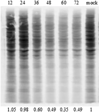
[image:7.585.43.283.521.636.2] [image:7.585.318.524.565.658.2]cells although with a moderate reduction in levels of E2 ex-pression at 48 and 72 h (Fig. 9). In noninfected cells treated with rIFN-␣, there was marked Mx expression, while eIF2␣ was not found to be phosphorylated at any time point (Fig. 9). The finding that SAV-3 infection resulted in the phosphoryla-tion of eIF2␣prompted us to characterize the effect of virus infection on protein synthesis. We found a moderate reduction of protein synthesis in TO cells by 36 h (40% reduction), which was more prominent by 48 and 60 h postinfection, with 50% and 65% reductions, respectively (Fig. 10).
IFN-␥has a limited inhibiting effect on SAV-3 replicationin
vitro.On the basis that it was previously shown that rIFN-␥has a limited effect on alphavirus replication in mice (21), we wanted to understand any antiviral effects induced by rIFN-␥ in fish cells. The functional activity of rIFN-␥was documented by its ability to induce the expression of␥IP10 (Fig. 11a). The CPE reduction assay showed that rIFN-␥ has no ability to rescue pretreated cells from CPE caused by SAV-3 infection (Fig. 11b), while there was an approximately 10-fold reduction of virus titers at the highest rIFN-␥concentration, determined by real-time PCR (1:200 dilution, corresponding to 1.65g/ml) (Fig. 11c).
DISCUSSION
This is the first report showing that IFN-␣ has a strong inhibitory effect on SAV-3 replication in vitro in permissive salmonid cell lines. This finding is in conformity with what was shown previously for other alphavirus species in higher verte-brates, where the stimulation of IFN-␣/pathways represents the primary protective response and plays a key role in pro-tection against infection through the limitation of virus repli-cation (1, 13, 32, 40). IFN-␥limits virus replication of SAV-3 marginally, again in concordance with what was found previ-ously for higher vertebrates (22). These findings have implica-tions for the understanding of host-pathogen interacimplica-tions in SAV infections of salmonid fish and possibly provide new avenues for disease prevention.
The tools available to study thein vivoeffects of type I IFN in infected fish at a functional level are very limited.In vitro
methods using permanent cell lines permissive for virus infec-tion are thus a good opinfec-tion for funcinfec-tional studies when this can be combined with treatment with functionally active cytokines. SAV-3 infection of TO cell was characterized by a rapid in-crease in viral E2 gene copy numbers from 12 to 24 h postin-fection, representing a more-than-2-log10increase over a 12-h
[image:8.585.78.247.69.259.2]period (Fig. 1). Over the same period, there was a strong upregulation of IFN-␣and Mx transcripts, and these findings
FIG. 10. Residual protein synthesis in TO cells infected with SAV-3. Confluent TO cells were grown in a six-well plate and infected as described in Materials and Methods. The membrane was exposed in a PhosphorImager cassette and then scanned by using a Typhoon imager. Numbers above the figure represent hours postpulsing. The protein amount was quantified by densitometry with ImageJ software as described in Materials and Methods. The results show 40, 51, and 65% reduction by 36, 48, and 60 h postinfection, respectively. Mock infection is shown at the far right.
FIG. 11. (a) Induction of ISG mRNA by rIFN-␥in TO cells. Purified rIFN-␥was serially diluted from 0.33 mg/ml in cell medium and incubated with TO cells for 24 h. The data are expressed as the mean fold changes in gene expression⫾standard errors of different dilutions of the IFN-␥-treated group relative to the nontreated control group after normalization to-actin (n⫽2) (⫾SEM). (b) Cytopathic effect reduction assay of IFN-␥-treated TO cells infected with SAV-3. TO cells were stimulated with serial dilutions of rIFN-␥and trIFN-␥for 24 h and subsequently infected with 1 MOI of SAV-3. There is no significant difference in protection against CPE between cell cultures pretreated with rIFN-␥and trIFN-␥(n⫽4) (⫾SEM). Non-tr, nontreated cells; Non-inf, noninfected; OD490, optical density at 490 nm. (c) Real-time PCR quantification of virus in cell supernatants from TO cells stimulated with serial dilutions of mature IFN-␥for 24 h and subsequently infected with 1 MOI of SAV-3. Cell supernatants were collected when a strong CPE was observed in the control infected cells (8 days postinfection). The data are expressed as the mean crossing-point values⫾standard errors using real-time PCR of different dilutions of an IFN-␥-treated group relative to the nontreated infected control group (n⫽2) (⫾SEM).
on November 8, 2019 by guest
http://jvi.asm.org/
[image:8.585.49.542.516.633.2]are in conformity with data from a previous study (11) using a different subtype of SAV (SAV-1). To what extent the cell line used in this study reflects a truein vivosituation is difficult to determine, but TO cells have morphological and functional traits of salmon macrophages (35) and support the replication of a number of salmonid viruses, like salmon anemia virus (36) and infectious pancreatic necrosis virus (3). Furthermore, the treatment of TO cells with low concentrations of rIFN-␣ in-duced strong expression levels of IFN-inin-duced genes, Mx and ISG15, and also a strong expression of Mx at the protein level (shown by IFAT), indicating that a functionally active cell line was used. The induction of an antiviral state in the pretreated cells was dependent on dose and time relative to infection, and protection against CPE was obtained when rIFN-␣was added to the cells earlier than 4 h prior to infection and at a dose higher than 0.47g/ml. Treatment at the time of infection (or 2 h prior to infection) and postinfection gave no protection against CPE, although there was a reduction in viral copy numbers.
In a recent study by Gahlawat et al. (11), it was proposed that the suppression of Mx responses correlates with SAV replication; however, those studies did not include a functional assessment of the responses involved. Those researchers showed a difference in mRNA IFN-␣and Mx expression levels in two cell lines of salmon origin, and they proposed a possible interference with IFN signaling and the suppression of Mx responses (11). Their observations were based solely on the observation that a cell line with low susceptibility had high Mx responses and vice versa. From our studies we have no indi-cation that SAV-3-infected cells have impaired Mx responses (Fig. 1 and 9), and the treatment of TO cells with rIFN-␣ followed by SAV-3 infection did not influence the expression of Mx as assessed by Western blotting. Furthermore, a strong expression of Mx postinfection seems to have little influence on virus replication. On the other hand, the timing of the antiviral responses relative to infection is obviously of major importance. This is concordant with a recent study where it was shown that mRNA of the Old World alphaviruses encoded by subgenomic RNA is resistant to eIF2␣phosphorylation, while genomic RNA replication is highly sensitive to eIF2␣ phos-phorylation (34). It was also found that the replication of genomic RNA ceased with the onset of eIF2␣ phosphoryla-tion, which again correlated with the appearance of structural proteins. We also find that the synthesis of the spike protein (E2) coincided with the phosphorylation of eIF2␣in SAV-3-infected and rIFN-␣-treated/SAV-3-infected cells (Fig. 9), with some reduction of staining intensity of E2 with increasing p-eIF2␣staining, but still, there was a successful production of virus progeny (Fig. 1). The antiviral state induced through preinfection treatment results in strong ISG induction, a marked inhibition of virus replication, and no E2 staining (Fig. 7 and 8). Thus, in line with what was described previously for other alphaviruses (28), the exact antiviral mechanisms con-trolling viral replication are not known; it seems that protein translation from genomic viral RNA (incoming positive strand) is sensitive to IFN-␣-induced responses. At the same time, it cannot be ruled out that the synthesis of spike mRNA driven by the 26S promoter is resistant to IFN-␣-induced cellular responses, but further studies are needed to understand the details of the mechanisms involved.
A known strategy by which alphaviruses evade the IFN re-sponse is through global shutoff of host gene expression (28). The transcriptional shutoff caused by Old World alphaviruses depends on a nonstructural protein (nsP2), and in addition, nsP2 of Sindbis virus has an anti-interferon effect, likely through decreased IFN production, and mutations in nsP2 can attenuate Sindbis virus cytopathogenicity (10, 12). There is no protein shutoff in SAV-3-infected cells, but there is a down-regulation of protein synthesis. This is more in line with New World alphaviruses that result in a more moderate downregu-lation of protein synthesis (2, 10, 12), where the capsid protein possibly plays a role in the arrest of macromolecular synthesis during early stages of infection, which will limit the formation of antiviral proteins in infected cells (2, 12). The New World alphavirus VEEV is also able to antagonize the IFN response using distinct mechanisms independent of host shutoff by in-hibiting the phosphorylation of cytoplasmic signal transducer and activator of transcription 1 (STAT-1), which prevents the nuclear translocation of phosphorylated STAT-1/STAT-2 het-erodimers (30). The result is a reduced formation of antiviral effector molecules like Mx and ISG15. More-recent studies have added complexity to the picture described above and have shown that VEEV nonstructural proteins arrested cellular translation (39). To what extent SAV-3 employs specific mech-anisms to counteract the IFN response by targeting certain signaling proteins involved in the IFN pathway or IFN signal-ing pathway needs to be investigated. A better understandsignal-ing of the induction of IFN-␣and related responses and possible antiviral effects and regulation of host gene expression im-posed by nonstructural and/or structural proteins of SAV-3 is strongly needed, and it is our understanding that this can best be studied by using reverse genetics methods.
The strong upregulation of IFN-␣ mRNA in response to SAV-3 infection is in contrast to what has been seen for SINV infection of permissive cell lines, while it is in conformity with what was observed for VEEV infection (in the same cell lines), where an upregulation of IFN-␣mRNA transcripts postinfec-tion was observed (39). The aquatic alphaviruses are defined as a separate clade within the genus Alphavirus, and they are phylogenetically different from Old World and New World alphaviruses (24). Their identification has raised interesting questions as to the origin of different variants of alphaviruses in general. While the Old World and New World alphaviruses have been classified into arthrogenic and neurotropic variants, the sal-monid alphaviruses do not seem to fall into either of these cate-gories, and they are not transferred by vectors (21). However, in a recent study (6), histopathological changes were found in the central nervous system following infection of Atlantic salmon with the SAV-3 subtype, while no such changes were found for SAV-1 variants. It remains to be decided if this is a general phenomenon of SAV-3 infections, but this observation combined with the sim-ilarities in cell responses to infection raises an interesting possi-bility that SAV-3 is functionally closer to the VEEV variants than the Old World viruses like SINV and Semliki Forest virus.
ACKNOWLEDGMENTS
This study was carried out with financial support from the Norwe-gian Research Council, project no. 182035, Immunization Strategies against Viral Pathogens of Atlantic Salmon.
on November 8, 2019 by guest
http://jvi.asm.org/
We are also indebted to Jo-Ann Leong, School of Ocean, Earth Science Technology, Hawaii Institute of Marine Biology, for providing the antibody to the Mx protein.
REFERENCES
1.Aguilar, P. V., S. Paessler, A. S. Carrara, S. Baron, J. Poast, E. Wang, A. C. Moncayo, M. Anishchenko, D. Watts, R. B. Tesh, and S. C. Weaver.2005. Variation in interferon sensitivity and induction among strains of eastern equine encephalitis virus. J. Virol.79:11300–11310.
2.Aguilar, P. C., S. C. Weaver, and C. F. Basler.2007. Capsid protein of eastern encephalitis virus inhibits host cell gene expression. J. Virol.81:
3866–3876.
3.Berg, T. S. K., B. Sun, and B. Robertsen.2009. An antiserum against Atlantic salmon IFNa1 detects IFN and neutralizes antiviral activity produced by poly I:C stimulated cells. Dev. Comp. Immunol.33:638–645.
4.Bergan, V., R. Jagus, S. Lauksund, O. Kileng, and B. Robertsen.2008. The Atlantic salmon Z-DNA binding protein kinase phosphorylates translation initiation factor 2 alpha and constitutes a unique orthologue to the mam-malian dsRNA-activated protein kinase R. FEBS J.275:184–197. 5.Brehin, A. C., I. Casademont, M. P. Frenkiel, C. Julier, A. Sakuntabhai, and
P. Despres.2009. The large form of human 2⬘,5⬘-oligoadenylate synthetase (OAS3) exerts antiviral effect against Chikungunya virus. Virology384:216– 222.
6.Christie, K. E., D. A. Graham, M. F. McLoughlin, S. Villoing, D. Todd, and D. Knappskog.2007. Experimental infection of Atlantic salmon Salmo salar pre-smolts by i.p. injection with new Irish and Norwegian salmonid alphavi-rus (SAV) isolates: a comparative study. Dis. Aquat. Organ.75:13–22. 7.Desvignes, L., C. Quentel, F. Lamour, and A. Le Ven.2002. Pathogenesis and
immune response in Atlantic salmon (Salmo salar L.) parr experimentally infected with salmon pancreas disease virus (SPDV). Fish Shellfish Immu-nol.12:77–95.
8.Ferreira, P. C., M. L. Peixoto, M. A. Silva, and R. R. Golgher.1979. Assay of human interferon in Vero cells by several methods. J. Clin. Microbiol.
9:471–475.
9.Fringuelli, E., H. M. Rowley, J. C. Wilson, R. Hunter, H. Rodger, D. A. Graham.2008. Phylogenetic analyses and molecular epidemiology of Euro-pean salmonid alphaviruses (SAV) based on partial E2 and nsP3 gene nucleotide sequences. J. Fish Dis.31:811–823.
10.Frovola, E. I., R. R. Fayzulin, S. H. Cook, D. E. Griffin, C. M. Rice, and I. Frolov.2002. Roles of nonstructural protein nsp2 and alpha/beta interferon in determining the outcome of Sindbis virus infection. J. Virol.76:11254– 22264.
11.Gahlawat, S. K., A. E. Ellis, and B. Collet.2009. Expression of interferon and interferon-induced genes in Atlantic salmon Salmo salar cell lines SHK-1 and TO following infection with salmon alphavirus SAV. Fish Shellfish Immunol.26:672–675.
12.Garmashova, N., R. Gorchakov, E. Volkova, S. Paessler, E. Frolova, and I. Frolov.2007. The Old World and New World alphaviruses use different virus-specific proteins for induction of transcriptional shutoff. J. Virol.81:
2472–2484.
13.Grieder, F. B., and S. N. Vogel.1999. Role of interferon and interferon regulatory factors in early protection against Venezuelan equine encephalitis virus infection. Virology257:106–118.
14.Hodneland, K., A. Bratland, K. E. Christie, C. Endresen, and A. Nylund.
2005. New subtype of salmonid alphavirus (SAV), Togaviridae, from Atlan-tic salmon Salmo salar and rainbow trout Oncorhynchus mykiss in Norway. Dis. Aquat. Organ.66:113–120.
15.Ka¨rber, G.1931. Beitrag zur kollektiven Behandlung pharmakologischer Reihenversuche. Arch. Exp. Pathol. Pharmakol.162:480–483.
16.Landis, H., A. Simon-Jodicke, A. Kloti, C. Di, J. J. Schnorr, S. Schneider-Schaulies, H. P. Hefti, and J. Pavlovic.1998. Human MxA protein confers resistance to Semliki Forest virus and inhibits the amplification of a Semliki Forest virus-based replicon in the absence of viral structural proteins. J. Vi-rol.72:1516–1522.
17.Larsen, R., T. P. Røkenes, and B. Robertsen.2004. Inhibition of infectious pancreatic necrosis virus replication by Atlantic salmon Mx1 protein. J. Vi-rol.78:7938–7944.
18.Lenschow, D. J., N. V. Giannakopoulos, L. J. Gunn, C. Johnston, A. K. O’Guin, R. E. Schmidt, B. Levine, and H. W. Virgin IV.2005. Identification of interferon-stimulated gene 15 as an antiviral molecule during Sindbis virus infection in vivo. J. Virol.79:13974–13983.
19.Lenschow, D. J., C. Lai, N. Frias-Staheli, N. V. Giannakopoulos, A. Lutz, T. Wolff, A. Osiak, B. Levine, R. E. Schmidt, A. Garcia-Sastre, D. A. Leib, A. Pekosz, K. P. Knobeloch, I. Horak, and H. W. Virgin IV.2007.
IFN-stimu-lated gene 15 functions as a critical antiviral molecule against influenza, herpes, and Sindbis viruses. Proc. Natl. Acad. Sci. U. S. A.104:1371–1376. 20.Livak, K. J., and T. D. Schmittgen.2001. Analysis of relative gene expression data using real-time quantitative PCR and the 2(⫺delta delta C(T)) method. Methods25:402–408.
21.McLoughlin, M. F., and D. A. Graham.2007. Alphavirus infections in sal-monids—a review. J. Fish Dis.30:511–531.
22.Mu¨ller, U., U. Steinhoff, L. F. Reis, S. Hemmi, J. Pavlovic, R. M. Zinkerna-gel, and M. Aguet.1994. Functional role of type I and type II interferons in antiviral defense. Science264:1918–1921.
23.Ooi, E. L., N. Verjan, I. Haraguchi, T. Oshima, H. Kondo, I. Hirono, T. Aoki, H. Kiyono, and Y. Yuki.2008. Innate immunomodulation with recombinant interferon-alpha enhances resistance of rainbow trout (Oncorhynchus mykiss) to infectious hematopoietic necrosis virus. Dev. Comp. Immunol.
32:1211–1220.
24.Powers, A. M., A. C. Brault, Y. Shirako, E. G. Strauss, W. Kang, J. H. Strauss, and S. C. Weaver.2001. Evolutionary relationships and systematics of the alphaviruses. J. Virol.75:10118–10131.
25.Raynard, R., and G. Houghton.1993. Development towards an experimental protocol for the transmission of pancreas disease of Atlantic salmonSalmo salar. Dis. Aquat. Organ.15:123–128.
26.Robertsen, B., G. Trobridge, and J. A. Leong.1997. Molecular cloning of double-stranded RNA inducible MX genes from Atlantic salmon (Salmo salarL.). Dev. Comp. Immunol.21:397–412.
27.Rokenes, T. P., R. Larsen, and B. Robertsen.2007. Atlantic salmon ISG15: expression and conjugation to cellular proteins in response to interferon, double-stranded RNA and virus infections. Mol. Immunol.44:950–959. 28.Ryman, K., and W. B. Klimstra.2008. Host responses to alphavirus
infec-tion. Immunol. Rev.225:27–45.
29.Samuel, C. E.2001. Antiviral actions of interferons. Clin. Microbiol. Rev.
14:778–809.
30.Simmons, J. D., L. J. White, T. E. Morrison, S. A. Montgomery, A. C. Whitmore, R. E. Johnston, and H. T. Heise.2009. Venezuelan equine en-cephalitis virus disrupts STAT1 signaling by distinct mechanisms indepen-dent of host shutoff. J. Virol.83:10571–10581.
31.Slutzkia, M., D. A. Jaitin, T. B. Yehezkela, and G. Schreiber.2006. Varia-tions in the unstructured C-terminal tail of interferons contribute to differ-ential receptor binding and biological activity. J. Mol. Biol.360:1019–1030. 32.Stanton, G. J., R. E. Lloyd, M. Sarzotti, and J. E. Blalock.1989. Protection of mice from Semliki Forest virus infection by lymphocytes treated with low levels of interferon. Mol. Biother.1:305–310.
33.Taksdal, T., A. B. Olsen, I. Bjerkas, M. J. Hjortaas, B. H. Dannevig, D. A. Graham, and M. F. McLoughlin.2007. Pancreas disease in farmed Atlantic salmon, Salmo salar L., and rainbow trout, Oncorhynchus mykiss (Wal-baum), in Norway. J. Fish. Dis.30:545–558.
34.Ventoso, I., M. A. Sanz, S. Molina, J. J. Berlanga, L. Carrasco, and M. Esteban. 2006. Translational resistance of late alphavirus mRNA to eIF2alpha phosphorylation: a strategy to overcome the antiviral effect of protein kinase PKR. Genes Dev.20:87–100.
35.Villoing, S., M. Bearzotti, S. Chilmonczyk, J. Castric, and M. Bremont.2000. Rainbow trout sleeping disease virus is an atypical alphavirus. J. Virol.
74:173–183.
36.Wergeland, H. I., and R. A. Jakobsen.2001. A salmonid cell line (TO) for production of infectious salmon anaemia virus (ISAV). Dis. Aquat. Organ.
44:183–190.
37.Weston, J. H., M. D. Welsh, M. F. McLoughlin, and D. Todd.1999. Salmon pancreas disease virus, an alphavirus infecting farmed Atlantic salmon, Salmo salar L. Virology256:188–195.
38.Weston, J., S. Villoing, M. Bremont, J. Castric, M. Pfeffer, V. Jewhurst, M. McLoughlin, O. Rodseth, K. E. Christie, J. Koumans, and D. Todd.2002. Comparison of two aquatic alphaviruses, salmon pancreas disease virus and sleeping disease virus, by using genome sequence analysis, monoclonal re-activity, and cross-infection. J. Virol.76:6155–6163.
39.Yin, J., C. L. Gardner, C. W. Burke, K. D. Ryman, and W. B. Klimstra.2009. Similarities and differences in antagonism of neuron alpha/beta interferon responses by Venezuelan equine encephalitis and Sindbis alphaviruses. J. Vi-rol.83:10036–10047.
40.Zhang, Y., C. W. Burke, K. D. Ryman, and W. B. Klimstra.2007. Identifi-cation and characterization of interferon-induced proteins that inhibit al-phavirus replication. J. Virol.81:11246–11255.
41.Zhu, R., Y. B. Zhang, Q. Y. Zhang, and J. F. Gui.2008. Functional domains and the antiviral effect of the double-stranded RNA-dependent protein kinase PKR from Paralichthys olivaceus. J. Virol.82:6889–6901. 42.Zou, J., C. Tafalla, J. Truckle, and C. J. Secombes.2007. Identification of a
second group of type I IFNs in fish sheds light on IFN evolution in verte-brates. J. Immunol.179:3859–3871.