0022-538X/11/$12.00 doi:10.1128/JVI.02319-10
Copyright © 2011, American Society for Microbiology. All Rights Reserved.
VEGFR2 and Src Kinase Inhibitors Suppress Andes Virus-Induced
Endothelial Cell Permeability
䌤
Elena E. Gorbunova,
1Irina N. Gavrilovskaya,
1Timothy Pepini,
1,2and Erich R. Mackow
1,2*
Department of Molecular Genetics and Microbiology1and Molecular and Cellular Biology Program,2
Stony Brook University, Stony Brook, New York 11794-5122
Received 5 November 2010/Accepted 14 December 2010
Hantaviruses predominantly infect human endothelial cells and, in the absence of cell lysis, cause two diseases resulting from increased vascular permeability. Andes virus (ANDV) causes a highly lethal acute pulmonary edema termed hantavirus pulmonary syndrome (HPS). ANDV infection enhances the permeability of endothelial cells in response to vascular endothelial growth factor (VEGF) by increasing signaling responses directed by the VEGFR2-Src-VE-cadherin pathway, which directs adherens junction (AJ) disassembly. Here we demonstrate that inhibiting pathway-specific VEGFR2 and Src family kinases (SFKs) blocks ANDV-induced endothelial cell permeability. Small interfering RNA (siRNA) knockdown of Src within ANDV-infected endo-thelial cells resulted in an⬃70% decrease in endothelial cell permeability compared to that for siRNA controls. This finding suggested that existing FDA-approved small-molecule kinase inhibitors might similarly block ANDV-induced permeability. The VEGFR2 kinase inhibitor pazopanib as well as SFK inhibitors dasatinib, PP1, bosutinib, and Src inhibitor 1 dramatically inhibited ANDV-induced endothelial cell permeability. Consistent with their kinase-inhibitory concentrations, dasatinib, PP1, and pazopanib inhibited ANDV-in-duced permeability at 1, 10, and 100 nanomolar 50% inhibitory concentrations (IC50s), respectively. We further
demonstrated that dasatinib and pazopanib blocked VE-cadherin dissociation from the AJs of ANDV-infected endothelial cells by >90%. These findings indicate that VEGFR2 and Src kinases are potential targets for therapeutically reducing ANDV-induced endothelial cell permeability and, as a result, capillary permeability during HPS. Since the functions of VEGFR2 and SFK inhibitors are already well defined and FDA approved for clinical use, these findings rationalize their therapeutic evaluation for efficacy in reducing HPS disease. Endothelial cell barrier functions are disrupted by a number of viruses that cause hemorrhagic, edematous, or neurologic disease, and as a result, our findings suggest that VEGFR2 and SFK inhibitors should be consid-ered for regulating endothelial cell barrier functions altconsid-ered by additional viral pathogens.
Hantaviruses predominantly infect endothelial cells (ECs) and nonlytically cause diseases associated with dramatic in-creases in vascular permeability (12, 51, 54, 66, 82, 83, 98). Andes virus (ANDV) infection results in acute pulmonary edema and respiratory insufficiency termed hantavirus pulmo-nary syndrome (HPS) or hantavirus cardiopulmopulmo-nary syn-drome (HCPS) (7, 8, 12, 17, 19, 32, 47, 55, 57, 66, 68, 98). Endothelial cells within vast pulmonary capillary beds provide a primary means for ANDV infection to increase capillary permeability and cause pulmonary edema (7, 8, 32). Interen-dothelial cell adherens junctions (AJs) form a fluid barrier within capillaries that regulates permeability of the vascular endothelium (11, 53). However, endothelial cell AJs must dis-sociate in order to permit immune cell extravasation and repair of capillary damage, and thus, opposing signals regulate endo-thelial cell responses that control AJ disassembly (9, 11, 56). Maintaining vascular integrity is of fundamental importance for preventing edema, and as a result, vascular permeability is tightly regulated by redundant systems that act on a unique set of endothelial cell-specific receptors, AJ proteins, and signal-ing pathway effectors (11, 13, 20, 24, 90).
Acute pulmonary edema and hypoxia are hallmarks of HPS disease, and hypoxic conditions alone are capable of inducing acute pulmonary edema (5, 8, 12, 18, 32, 42, 47, 64, 66, 89). Hypoxia induces the expression of vascular endothelial growth factor (VEGF) within pulmonary endothelial cells, and VEGF was originally named vascular permeability factor for its ability to induce tissue edema (5, 10, 13, 14, 48, 59, 64, 70, 89). Secreted VEGF acts locally in an autocrine or paracrine man-ner to activate VEGFR2 receptors on endothelial cells, and VEGFR2 activation induces the internalization of VE-cad-herin from AJs and paracellular permeability (11, 13, 15, 22, 23, 53). In fact, even small changes in vascular permeability result in large changes in fluid efflux within pressurized vessels (79). Intracellularly, VEGFR2-induced permeability is di-rected by Src/Rac/PAK signaling responses (23, 24, 64). Src family kinases (SFKs) are recruited to the cytoplasmic tails of VEGFR2 receptors and link VEGFR2-directed signaling re-sponses to downstream pathway targets that induce changes in VE-cadherin and regulate interendothelial cell adherence. VEGFR2-Src pathway activation directs the disassembly of VE-cadherin from AJs and increases paracellular permeability of the endothelium, which results in edema (23, 34).
Hypoxia causes high-altitude pulmonary edema through the induction of permeabilizing VEGF responses (5, 42). HPS patients are acutely hypoxic, and hyperoxygenation of patients reduces HPS mortality (7, 8, 12, 32, 47, 66, 98).In vitro, endo-thelial cells are not permeabilized by hantavirus infection
* Corresponding author. Mailing address: Department of Molecular Genetics and Microbiology, Stony Brook University, Life Sciences, Rm. 126, Stony Brook, NY 11794-5222. Phone: (631) 632-7014. Fax: (631) 632-9797. E-mail: Erich.Mackow@stonybrook.edu.
䌤Published ahead of print on 22 December 2010.
2296
on November 7, 2019 by guest
http://jvi.asm.org/
alone or by the addition of tumor necrosis factor alpha (TNF-␣) to monolayers (27, 31, 44, 87). However, the perme-ability of endothelial cells infected by Hantaan virus (HTNV), ANDV, and New York 1 virus (NY-1V) is dramatically en-hanced in response to VEGF days after infection and this is not observed following infection by nonpathogenic hantavi-ruses Tula virus (TULV) and Prospect Hill virus (PHV) (26, 27, 31).
VEGFR2-induced endothelial cell permeability is positively or negatively regulated by3 integrins; Robo1/4, Edg-1, and Tie-2 receptors; and surface proteoglycans, syndecans, and galectin-3, which interact with VEGFR2 or downstream sig-naling pathways (6, 24, 40, 60, 63, 75–77, 79, 90). Pathogenic hantaviruses uniquely bind inactive␣v3integrin conformers, and this suggests a means for cell-associated hantaviruses to alter VEGFR2 signaling responses at late times postinfection (p.i.) (25, 26, 29, 31, 61, 74). VEGFR2 receptors form a com-plex with ␣v3 integrins that is required for endothelial cell migration (6), and Src is critical for this receptor synergy (75). Pathogenic hantaviruses bind ␣v3 and block ␣v3-directed endothelial cell migration in response to VEGF (27–29, 31, 61, 74). Paradoxically, knocking out3or inhibiting␣v3also
pro-motes VEGFR2-directed signaling responses and enhances en-dothelial cell permeability (75). Consistent with this, ANDV-in-fected endothelial cells are hyperpermeabilized by VEGF (27). This results from hyperphosphorylation of VEGFR2 and the dis-sociation of VE-cadherin from the AJs of ANDV-infected endo-thelial cells (26, 31, 58). Thus, ANDV enhances the permeability of infected endothelial cells by augmenting responses of the VEGFR2-Src-VE-cadherin signaling pathway (26, 27, 31).
These findings suggest that ANDV-induced edema may be blocked by inhibiting VEGFR2- and SFK-directed pathway activation and that this edemagenic pathway provides several potential therapeutic targets for regulating ANDV-induced endothelial cell permeability. Although there are no effective therapeutics for treating HPS patients or hantavirus disease (41), VEGFR2 signaling pathways have been prominent tar-gets of anticancer therapies (1–3, 37, 65, 71, 78, 86). These clinically available small-molecule kinase inhibitors may simi-larly regulate ANDV-induced permeability and have therapeu-tic utility against HPS.
In this study, we investigate whether FDA-approved small-molecule inhibitors which target VEGFR2 and SFK signaling responses (33, 43, 71, 85) are able to inhibit ANDV-induced endothelial cell permeability. Our findings demonstrate that small interfering RNA (siRNA) knockdown of Src inhibits ANDV-induced endothelial cell permeability, and this finding rationalized testing of small-molecule kinase inhibitors that target the VEGFR2-Src signaling pathway as inhibitors of ANDV-induced vascular permeability. Drugs which inhibit VEGFR2 or SFK responses (1–3, 37, 78, 86) were found to block ANDV-induced endothelial cell permeability. The SFK inhibitor dasatinib (2) blocked ANDV-induced permeability
⬎70% at nanomolar concentrations, and these findings ratio-nalize testing VEGFR2-Src inhibitors as potential therapeutics against hantavirus-induced disease. Since these molecules are already FDA approved for clinical use, they provide the po-tential for rapid evaluation and implementation in HPS pa-tients.
MATERIALS AND METHODS
Cells and virus.VeroE6 cells (ATCC CRL-1586) were grown in Dulbecco’s modified Eagle’s medium (DMEM) containing 10% fetal calf serum (FCS;
Sigma), penicillin (100g/ml), streptomycin sulfate (100g/ml), and
amphoter-icin B (50g/ml) (Gibco). Human umbilical vein endothelial cells (HUVECs)
were purchased from Cambrex Inc. and grown in endothelial cell basal medium
2 (EBM-2; Clonetics) supplemented with gentamicin (50g/ml), amphotericin B
(50g/ml), and 10% FCS (Sigma). Andes virus (CHI-7913; ANDV) (61) was
kindly provided by B. Hjelle (University of New Mexico), cultivated in a biosafety level 3 (BSL-3) facility as previously described (27), and determined to be mycoplasma free (Roche). ANDV titers were determined as previously de-scribed (25, 29). HUVEC monolayers were ANDV or mock infected at a
mul-tiplicity of infection (MOI) of 0.5, and cells were⬎90% infected with ANDV as
determined by immunoperoxidase staining.
Immunoperoxidase staining of hantavirus-infected cells.Rabbit polyclonal antinucleocapsid serum directed against the NY-1V nucleocapsid protein was used to detect ANDV-infected cells as previously described (29). Briefly, infected endothelial cell monolayers were fixed with 100% methanol and incubated with antinucleocapsid serum (1:5,000) followed by goat anti-rabbit–horseradish per-oxidase (HRP) secondary antibody (1:5,000; Amersham Biosciences). Nucleo-capsid protein-expressing cells were identified by staining with
3-amino-9-ethyl-carbazole (0.026% in 0.1 M sodium acetate, pH 5.2, and 0.03% H2O2) and
quantitated by microscopy (29).
VEGFR2 and Src inhibitors.The VEGF receptor kinase inhibitor pazopanib
{GSK-VEG10003; 5-[[4-[(2,3-dimethyl-2H
-indazol-6-yl)methylamino]-2-pyrimidi-nyl]amino]-2-methylbenzolsulfonamide} (37, 50, 71, 78) and the Src family kinase
inhibitors dasatinib {Sprycel; N
-(2-chloro-6-methylphenyl)-2-[[6-[4-(2-hydroxy-ethyl)-1-piperazinyl]-2-methyl-4-pyrimidinyl]amino]-5-thiazolecarboxamide mono-hydrate} (2, 39, 65), PP1 {pyrazolopyrimidine 1; pyrazolopyrimidine,
4-amino-5-(4-methylphenyl)-7-(t-butyl)pyrazolo[3,4-d]pyrimidine} (2, 3), Src inhibitor 1 {SKI-1;
4-(4⬘-phenoxyanilino)-6,7-dimethoxyquinazoline, 6,7-dimethoxy-N
-(4-phenoxyphe-nyl)-4-quinazolinamine} (2), and bosutinib {SKI-606; 4-[(2,4-dichloro-5-methoxy-phenyl) amino]-6-methoxy-7-[3-(4-methylpiperazin-1-yl) propoxy] quinoline-3-car-bonitrile} (2, 93) were purchased from Selleck Chemicals and evaluated for endothelial cell cytotoxicity by trypan blue exclusion. Cytotoxicity for these FDA-approved drugs has been previously reported (33, 43, 71, 85), and inhibitor concen-trations used were based on prior studies of drug cytotoxicity and Src inhibition studies. Briefly, serially diluted inhibitors were added to endothelial cells and after 24 h 0.4% trypan blue was added to cells. Cell viability was determined by quanti-fying the percentage of cells excluding trypan blue, and results were consistent with prior studies of these FDA-approved clinically available drugs (2, 3, 37, 39, 50, 65, 71,
78, 93). Concentrations of inhibitor resulting in⬍5% change in cell viability were
used as maximum inhibitor concentrations in subsequent experiments.
Quantitative RT-PCR (qRT-PCR) and siRNA analysis.Total cellular RNA was extracted from mock-infected or ANDV-infected HUVECs using the
RNeasy minikit (Qiagen) (30, 69). Total RNA (1g) was reverse transcribed
using the Transcriptor first-strand cDNA synthesis kit (Roche) using oligo(dT)18
primer (25°C for 10 min, 55°C for 30 min, and 85°C for 5 min). TaqMan primers (Applied Biosystems) were used to amplify human Src and glyceraldehyde-3-phosphate dehydrogenase (GAPDH) in duplicate using an Applied Biosystems 7300 PCR machine under the following thermocycling parameters: 50°C for 2 min, 95°C for 10 min, 95°C for 15 s, and 60°C for 1 min for 40 cycles (69). Src mRNA levels were normalized to GAPDH mRNA levels, and the fold changes in mRNA levels were compared between ANDV- and mock-infected cells (30, 69). The fold change in Src mRNA level was determined using the threshold
cycle (2⫺⌬⌬CT) method and plotted (⫾standard error of the mean [SEM]) using
GraphPad Prism 5 software (69).
Src knockdown experiments were performed using SureSilencing small inter-fering RNAs (siRNAs) against Src or negative-control siRNA (si-NEG2) pur-chased from SA Biosciences (69). Briefly, endothelial cells were transfected with siRNAs using SureFECT reagent (SA Biosciences). Total RNA was purified as described above and analyzed by qRT-PCR (30).
Endothelial cell permeability assay.A previously described endothelial cell permeability assay was used to assess the effect of kinase inhibitors on ANDV-induced permeability (26, 27, 31). Briefly, human endothelial cells were plated on
Costar Transwell plates (3-m pores; Corning) and confluent monolayers were
infected with pathogenic ANDV at an MOI of 0.5 or mock infected. Prior cytotoxicity of these FDA-approved agents has been previously reported (33, 43, 71, 85), and minimal cytotoxic concentrations of drugs which caused no increase in endothelial cell permeability by themselves were used in assays. Three days postinfection (p.i.), cells were pretreated with kinase inhibitors at noncytotoxic concentrations (33, 43, 71, 85) or mock treated (15 min) prior to the addition of
on November 7, 2019 by guest
http://jvi.asm.org/
0.5 mg/ml of fluorescein isothiocyanate (FITC)-dextran and VEGF (100 ng/ml) to the upper chamber. Monolayer permeability was assayed by analyzing
FITC-dextran present in the lower chamber (100l) at indicated times using a BioTek
FLx800 fluorimeter (26, 27, 31). The fold change in FITC-dextran fluorescence intensity over that of mock-treated controls was used as a measure of the paracellular permeability of endothelial cell monolayers. Hantavirus infections were monitored by immunoperoxidase staining as described above (29, 61).
VE-cadherin internalization.In order to monitor internalization of VE-cad-herin, human endothelial cells were grown on gelatin-coated coverslips and were infected with pathogenic ANDV at an MOI of 0.5 or mock infected (31). Three days postinfection, endothelial cells were starved and pretreated for 15 min with the VEGFR2 inhibitor pazopanib (100 nM) or the SFK inhibitor dasatinib (1 nM) or untreated prior to VEGF addition (100 ng/ml, 30 min). Cells were incubated with an antibody to the extracellular domain of VE-cadherin (BV9; Clonetics) at 4°C for 1 h (23, 31). After antibody removal and washing, cells were incubated at 37°C (1 h) to permit intracellular trafficking of antibody-bound VE-cadherin. Cells were subsequently washed with a mild acid solution (2 mM phosphate-buffered saline [PBS]–glycine, pH 2.0; three times for 5 min each) (23, 31) in order to remove VE-cadherin antibody that was not internalized or were left untreated. Cells were paraformaldehyde fixed and Triton X-100 permeabil-ized prior to incubation with an FITC-tagged anti-mouse secondary antibody and examined on an Olympus IX51 microscope (31).
RESULTS
ANDV infects the endothelial cell lining of capillaries and results in patient hypoxia and acute pulmonary edema leading to respiratory distress (7, 12, 17, 32, 57, 66, 68, 98). Hypoxia transcriptionally induces VEGF, a permeabilizing growth fac-tor that acts on endothelial cells and is 50,000 times more potent than histamine in causing edema (5, 10, 13–15, 18, 48, 89). In vitro, ANDV-infected endothelial cells are hyperre-sponsive to the permeabilizing effects of VEGF (26, 27, 31). Our recent findings indicate that ANDV infection of endothe-lial cells results in increased VEGFR2 phosphorylation, en-hanced VE-cadherin internalization, and increased paracellu-lar permeability (26, 31, 58). These findings suggest both a mechanism for ANDV-induced edema and a means for ther-apeutically targeting the VEGFR2-Src-VE-cadherin signaling pathway that directs these responses.
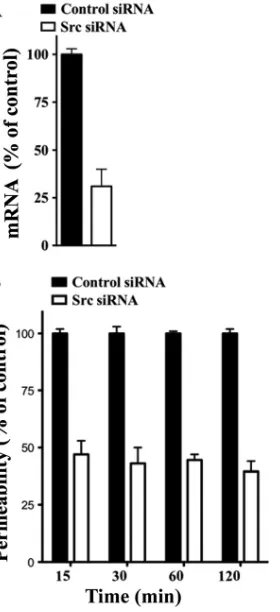
Src knockdown decreases ANDV-induced endothelial cell permeability.The VEGFR2 cytoplasmic tail recruits Src ki-nases which activate a specific signaling pathway that results in VE-cadherin internalization and increased paracellular perme-ability of endothelial cells (23, 52). ANDV-infected endothelial cells are hyperpermeabilized by VEGF (27), suggesting that VEGFR2-Src signaling responses direct ANDV-induced per-meability. Here we determine if knocking down Src blocks endothelial cell permeability induced by ANDV infection. We transfected endothelial cells with control or Src-specific siRNAs and assayed ANDV-induced endothelial cell perme-ability 3 days postinfection. Quantitative RT-PCR analysis in-dicates that endothelial cells transfected with Src siRNAs spe-cifically reduced Src mRNA levels by ⬃70% (Fig. 1A). Endothelial cells similarly transfected with Src or control siRNAs were subsequently analyzed for their effect on ANDV-induced permeability (27, 69). We found that siRNAs to Src reduced the hyperpermeability of ANDV-infected endothelial cells 55 to 65% compared to control siRNA at all time points after VEGF addition (Fig. 1B). These findings suggest that ANDV-induced hyperpermeability occurs via a VEGFR2-Src pathway (23, 24) and that inhibiting Src is a viable mechanism for reducing ANDV-induced endothelial cell permeability. These findings further suggested that chemical inhibitors of
VEGFR2 and Src kinases may similarly inhibit ANDV-in-duced endothelial cell permeability.
VEGFR2-Src pathway inhibitors reduce ANDV-induced en-dothelial cell permeability.Since VEGF-induced vasculariza-tion is required for tumor growth, small molecules which in-hibit VEGFR2 signaling responses are already in various stages of human clinical trials as anticancer therapies (2, 37, 65, 71, 78, 86). VEGFR2 signaling responses are inhibited by pazopanib (1, 37, 71, 78, 86), while Src activation is blocked by dasatinib, bosutinib, PP1, and Src inhibitor 1 (2, 65). Interest-ingly, compounds that target the VEGFR2-Src signaling path-way also have the potential to inhibit ANDV-induced perme-ability in vitro and reduce edema in HPS patients. Here we address the ability of commercially available drugs which in-hibit VEGFR2-Src signaling responses to block ANDV-in-duced endothelial cell permeability.
[image:3.585.351.491.69.375.2]Human endothelial cells were grown on Transwell plates and infected with ANDV for 3 days prior to assessment of
FIG. 1. Src knockdown inhibits ANDV-induced EC permeability. (A) Endothelial cells were transfected with Src or scrambled siRNA, and Src mRNAs were analyzed by qRT-PCR (69). mRNA levels were quantitated and standardized to GAPDH mRNA levels and presented as a percentage of controls. (B) Endothelial cells were plated on Transwell inserts and infected at an MOI of 0.5 in triplicate with ANDV. One day postinfection (p.i.), cells were transfected with con-trol or Src-specific siRNAs using SureFECT siRNA transfection re-agent. Three days p.i., VEGF (100 ng/ml) and FITC-dextran were added to medium in the upper chamber, and the presence of FITC-dextran in the lower chamber was quantitated at indicated times (26, 27, 31). The percent change in FITC-dextran over controls is presented as a measure of EC monolayer permeability (27).
on November 7, 2019 by guest
http://jvi.asm.org/
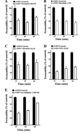
monolayer permeability in response to VEGF (27). Endothe-lial cells were treated with increasing concentrations of poten-tially inhibitory compounds (2, 65, 78, 86), and the permeabil-ity of ANDV-infected endothelial cells was determined and compared to that of untreated controls (Fig. 2A to D). Figure 2 defines concentrations of kinase inhibitors which block ANDV-induced EC permeability approximately 50% (IC50s). Dasatinib and PP1 had the lowest IC50s (1 and 10 nM, respec-tively), with IC50s of pazopanib and bosutinib being 100 nM and 10M, respectively (Fig. 2A to D).
In Fig. 3, we assessed the abilities of VEGFR2 and SFK inhibitors to block ANDV-induced permeability at their IC50s from 15 to 60 min after VEGF addition. The Src family inhib-itors dasatinib and PP1, which had the lowest IC50s of the kinase inhibitors tested, inhibited ANDV-induced endothelial cell permeability by 50 to 60% and 40 to 70%, respectively (Fig. 3B and D). The VEGFR2 kinase inhibitor pazopanib also inhibited ANDV-induced permeability by 40 to 60% at nano-molar IC50levels (Fig. 3A). Src inhibitor 1 and bosutinib in-hibited ANDV-induced permeability up to 40 to 50% but only at IC50s of 360 nM and 10M, respectively (Fig. 3C and E).
Inhibitors blocked ANDV-induced endothelial cell perme-ability at IC50s consistent with their reported VEGFR2 or SFK inhibitory concentrations (2, 65, 71, 78, 86). These findings demonstrate that commercially available small-mol-ecule VEGFR2 and SFK inhibitors block ANDV-induced permeability at nanomolar concentrations.
[image:4.585.43.302.69.346.2]Dasatinib and pazopanib prevent VE-cadherin internaliza-tion in ANDV-infected HUVECs.The internalization of VE-cadherin from AJs is a downstream effect of VEGFR2-Src
FIG. 2. IC50s of compounds that block ANDV-induced EC perme-ability. The concentrations of VEGFR2 and SFK inhibitors required to inhibit ANDV-induced endothelial cell permeability by 50% (IC50s) were determined. Endothelial cells were ANDV infected, and 3 days postinfection the permeability of cells in response to VEGF addition was determined (27) in the presence or absence of increasing amounts of kinase inhibitor. Endothelial cell permeability was determined as described for Fig. 1, and the permeability of ANDV- versus mock-infected controls was determined to be 100%. The effect of inhibitors is presented as the percentage of ANDV-induced permeability of inhibitor-treated monolayers 3 days postinfection and 30 min post-VEGF and FITC-dextran addition (27). Data are derived from at least three different experiments for each condition.
FIG. 3. VEGFR2-Src inhibitors block ANDV-induced permeabil-ity. Endothelial cells were plated on vitronectcoated Transwell in-serts and infected at an MOI of 0.5 in triplicate with ANDV. Three days postinfection, the permeability of ANDV- and mock-infected endothelial cell monolayers was determined as described for Fig. 1 (27) at indicated times in the presence or absence of the kinase inhib-itors pazopanib (A), dasatinib (B), bosutinib (C), PP1 (D), and Src inhibitor 1 (E). The percent change in FITC-dextran over controls is presented as a measure of EC monolayer permeability (27). Data are derived from two independent experiments performed in triplicate with comparable results.
on November 7, 2019 by guest
http://jvi.asm.org/
[image:4.585.281.537.70.495.2]activation, and VE-cadherin internalization is increased by ANDV infection of endothelial cells (26, 31). Here we deter-mined whether VEGFR2 and SFK inhibitors are capable of blocking VE-cadherin disassembly following ANDV infection of endothelial cells. We monitored VE-cadherin internaliza-tion using a previously described acid wash approach which removes antibody bound to extracellular VE-cadherin and per-mits quantitation of only intracellularly immunostained VE-cadherin (23, 31). We found that dasatinib and pazopanib inhibited ANDV-induced VE-cadherin internalization 65 to 70% and resulted in VE-cadherin localization nearly identical to that of mock-infected cells (Fig. 4). These data suggest that dasatinib and pazopanib inhibit the permeability of hantavirus-infected endothelial cells by blocking signaling responses that induce VE-cadherin internalization. These results further sug-gest that VEGFR2-Src signaling responses which direct AJ disassembly may be therapeutically targeted in order to inhibit hantavirus-induced endothelial cell permeability. Our findings rationalize testing of these existing, FDA-approved com-pounds for therapeutic efficacy in HPS animal models and clinical HPS cases.
DISCUSSION
Pulmonary edema and hypoxia are hallmarks of HPS disease and linked to VEGFR2-directed responses of endothelial cells, which are the primary targets of hantavirus infection. Hypoxia induces the secretion of VEGF, a factor which is known for its
potent ability to permeabilize capillaries and cause edema. In fact, patient hyperoxygenation has contributed to reducing HPS mortality to ⬃40% (7, 8, 12, 32, 47, 66, 98). In vitro, endothelial cells infected by HFRS- and HPS-causing hanta-viruses, but not nonpathogenic TULV, result in a dramatic enhancement of permeability in response to VEGF (26, 27, 31). ANDV-enhanced endothelial cell permeability results from the hyperactivation of VEGFR2 signaling pathways, VE-cadherin internalization, and the dissociation of AJs which normally maintain vascular integrity (26, 31, 58).
Here we demonstrate that clinically relevant FDA-approved small-molecule inhibitors of the VEGFR2 signaling pathway are able to block ANDV-induced endothelial cell permeability. Our results indicate that drugs which specifically target the VEGFR2 or Src kinases inhibit ANDV-induced endothelial cell permeability and the internalization of VE-cadherin from AJs. The observed decreases in endothelial cell permeability which we observed are likely to be clinically significant, since even a 2-fold change in permeability results in a dramatic change in fluid flux across pressurized vessels (20, 79, 91). Although there are currently no therapeutics for hantavirus-induced disease (41), these findings directly suggest therapeu-tic targets and approaches for reducing endothelial cell per-meability caused by hantavirus infection.
Pazopanib (GSK-VEG10003) is a VEGF receptor-specific inhibitor which blocks VEGFR2 kinase activity at an IC50of 30 nM (71, 78). Our findings indicate that pazopanib similarly inhibits ANDV-induced endothelial cell permeability at a nanomolar level and suggest that the VEGFR2 kinase may also be a therapeutic target for regulating hantavirus-induced capillary permeability. However, there are additional ap-proaches for blocking VEGFR2 responses that should simi-larly be entertained (78). The use of the VEGF antibody be-vacizumab (Avastatin), which prevents VEGFR2 activation, has been used clinically to inhibit tumor growth by blocking angiogenesis (78). Suntinib, sorafenib, and asitinib are addi-tional drugs which specifically target the VEGFR2 kinase and have been used clinically to inhibit metastatic renal cell carci-noma (78). Each of these inhibitory drugs is currently used clinically for cancer therapy or is being evaluated in human clinical studies (33, 43, 71, 78, 85). Our findings rationalize testing of these kinase inhibitors for additional indications and clinical application in reducing hantavirus-induced edema.
[image:5.585.125.202.69.214.2]These studies also demonstrate that FDA-approved drugs that are clinically used as SFK inhibitors are also capable of blocking ANDV-induced permeability. Dasatinib sensitively inhibits all SFK members, including c-Src, Lck, Fyn, and Yes, with an IC50 of 1.1 nM (39). Dasatinib is FDA approved (Sprycel) for clinical treatment of patients with chronic my-eloid leukemia to reduce cancer cell growth (33, 43, 65, 72). SFK inhibitors also affect cancer progression by inhibiting an-giogenesis and endothelial cell permeability (39, 45). Pharma-cologic blockade of SFKs or knocking out Src suppresses tumor cell extravasation and stabilizes endothelial barrier function (39, 45). Ourin vitrofindings suggest that dasatinib may also have short-term therapeutic utility against hanta-virus-induced endothelial cell permeability. Additional SFK inhibitors PP1, Src inhibitor 1, and bosutinib also inhibited ANDV-induced permeability at nanomolar to micromolar concentrations, respectively. As a result, dasatinib and other
FIG. 4. Dasatinib and pazopanib block VE-cadherin internaliza-tion in ANDV-infected HUVECs. Human endothelial cells were mock infected or infected with pathogenic ANDV at an MOI of 0.5. Three days postinfection, endothelial cells were pretreated with kinase inhib-itors dasatinib (1 nM/liter) and pazopanib (100 nM/liter) or left un-treated for 15 min prior to VEGF addition (100 ng/ml, 2 h). Cells were incubated with an antibody to the extracellular domain of VE-cadherin (BV9; Clonetics) at 4°C for 1 h (23, 26, 31). After antibody removal and washing, cells were incubated at 37°C in 5% CO2(31) (1 h) to permit intracellular trafficking of antibody-bound VE-cadherin (23). Cells were subsequently washed with a mild acid solution (2 mM PBS-glycine, pH 2.0; three times for 5 min each) in order to remove VE-cadherin antibody that was not internalized or were left untreated (23, 26, 31). Cells were paraformaldehyde fixed and Triton X-100 permeabilized prior to incubation with an FITC-tagged anti-mouse secondary antibody and examined on an Olympus IX51 microscope. Data are presented as a percentage of cells with internalized VE-cadherin in ANDV-infected ECs pretreated only with VEGF (control) versus mock-treated infected cells (n⫽500;P ⬍0.001) (31). Data
represent the results from two independent experiments.
on November 7, 2019 by guest
http://jvi.asm.org/
SFK inhibitors may be rationalized as potential inhibitors of HPS disease and tested in the Syrian hamster HPS disease model (35).
The SFK inhibitor PP1 has been shown to reduce vascular permeability and cerebral edema in response to stroke and to have protective effects on cardiovascular disease (45). PP1 reduced edema and myocardial injury following myocardial infarction via a mechanism that inhibits endothelial cell per-meability (45). This is important for hantaviruses since HPS is also referred to as hantavirus cardiopulmonary syndrome (HCPS) for cardiogenic symptoms observed in hantavirus pa-tients (8, 32, 47). The ability of SFK inhibitors to block endo-thelial cell permeability under ischemic conditions (95, 96) may further suggest the utility of this therapeutic approach in HPS/ HCPS patients.
Interestingly, targeting SFKs may also reduce the induction of VEGF in response to hypoxia and thus downregulate secre-tion of the VEGFR2 permeability activator (5, 18, 48, 64, 70, 89). VEGF expression requires Src activation, and antisense knockdown of Src decreases VEGF expression induced by hypoxia (16, 22, 64). These findings further suggest that drugs which target SFKs may similarly downregulate VEGF induc-tion of hypoxia-inducible factor 1␣ (HIF1␣), which forms an autocrine loop that serves to further induce VEGF under hy-poxic conditions (22, 23, 64, 89). One paper suggests that ANDV induces VEGF at early times postinfection, although the low-level early VEGF response suggested was transient and not demonstrated to contribute to permeability or to co-incide with VEGF transcription (84). However, VEGF may be secreted from immune, endothelial, or epithelial cells in re-sponse to hypoxia and still act on endothelial cells (13, 14, 70, 89). Since HPS patients present with hallmark hypoxia, target-ing HIF1␣itself may be entertained as a means for reducing capillary permeability in HPS patients alone or in combination with VEGFR2 or SFK inhibitors.
Additional inhibitors of VEGFR2-directed permeability block ancillary signaling pathways that increase AJ stability directly (21, 24, 62, 90). Angiopoietin 1 (Ang-1) and sphin-gosine-1-phosphate (S1P) inhibit VEGFR2-directed perme-ability by binding to Tie-2 and Edg-1 receptors, respectively (21). In fact, like VEGF, Ang-1 is an endothelial cell-specific growth factor that enhances vascular stability and dominantly blocks VEGF-directed permeability (24, 27, 31, 38, 56, 90). We have previously shown that both Ang-1 and S1P inhibit ANDV- and HTNV-induced endothelial cell permeability (27, 31), and an S1P analog, FTY720, is already in human clinical studies (81). Collectively, our findings suggest several means for enhancing the capillary integrity of hantavirus-infected en-dothelial cells that may be considered therapeutically through the use of single or combined therapeutic approaches. Since VEGFR2 and SFK inhibitors are already FDA approved for use in humans, they could be immediately rationalized for use in clinical HPS cases.
Interestingly, the use of SFK inhibitors may be warranted for use against a number of infectious agents that impact vascular permeability, induce edema, or cause cerebral hemorrhage or edematous disease (4, 67, 92, 97). Dengue virus infects human endothelial cells and increases capillary permeability, resulting in hemorrhagic or edematous disease (4, 97). Several addi-tional viruses alter endothelial cell barrier functions or
blood-brain barrier (BBB) integrity, an effect that contributes to neurological symptoms at late times postinfection (46, 67, 73, 92, 97). Although with some viruses permeability may result from immune responses that impact the endothelium or BBB, VEGFR2 and SFK inhibitors which enhance endothelial cell integrity may still be of therapeutic utility in stabilizing the vasculature during these and other viral infections.
Studies performed here were not aimed at inhibiting ANDV entry or replication and instead analyzed the effect of inhibi-tors 3 days post-ANDV infection, when infected endothelial cells are hyperresponsive to VEGF (26, 27, 31). Although antiviral compounds that block virus entry or reduce viral rep-lication may be prophylactic, these approaches may not be efficacious against HPS or hemorrhagic fever with renal syn-drome (HFRS) (41, 94). Ribavirin and interferon, which pro-phylactically inhibit hantavirus replication, have little effect on hantavirus disease once patients are symptomatic (36, 41, 49, 80, 88). This is probably because hantavirus patients already have respiratory (HPS) or hemorrhagic (HFRS) symptoms when they seek medical attention and are at a point when continued hantavirus replication may no longer be required to cause disease (41). As a result, small-molecule inhibitors tar-geting cellular responses that contribute to disease may have a substantial advantage over antiviral approaches for these and other viruses with long disease onsets or which occur in the midst of high-level neutralizing antibodies. Further, the inves-tigation of potential therapeutics which are already FDA ap-proved for clinical use may provide for the rapid implementa-tion of inhibitors with known funcimplementa-tions in regulating specific aspects of viral disease or which may be combined with com-pounds which reduce disease by restricting inflammatory re-sponses that also impact capillary integrity (20, 79, 91).
ACKNOWLEDGMENTS
We thank Valery Matthys and Nadine Dalrymple for helpful discus-sions and critical review of the manuscript.
This work was supported by National Institutes of Health grants R01AI47873, PO1AI055621, R21AI1080984, and U54AI57158 (Northeast Biodefense Center [director, W. I. Lipkin]).
REFERENCES
1.Adjei, A. A.2007. Novel small-molecule inhibitors of the vascular endothelial
growth factor receptor. Clin. Lung Cancer8(Suppl. 2):S74–S78.
2.Aleshin, A., and R. S. Finn.2010. SRC: a century of science brought to the
clinic. Neoplasia12:599–607.
3.Bain, J., et al.2007. The selectivity of protein kinase inhibitors: a further
update. Biochem. J.408:297–315.
4.Basu, A., and U. C. Chaturvedi.2008. Vascular endothelium: the battlefield
of dengue viruses. FEMS Immunol. Med. Microbiol.53:287–299.
5.Berger, M. M., et al.2005. Hypoxia impairs systemic endothelial function in individuals prone to high-altitude pulmonary edema. Am. J. Respir. Crit.
Care Med.172:763–767.
6.Borges, E., Y. Jan, and E. Ruoslahti.2000. Platelet-derived growth factor receptor beta and vascular endothelial growth factor receptor 2 bind to the
beta 3 integrin through its extracellular domain. J. Biol. Chem.275:39867–
39873.
7.Bustamante, E. A., H. Levy, and S. Q. Simpson.1997. Pleural fluid
charac-teristics in hantavirus pulmonary syndrome. Chest112:1133–1136.
8.Chang, B., M. Crowley, M. Campen, and F. Koster.2007. Hantavirus
car-diopulmonary syndrome. Semin. Respir. Crit. Care Med.28:193–200.
9.Corada, M., et al.1999. Vascular endothelial-cadherin is an important de-terminant of microvascular integrity in vivo. Proc. Natl. Acad. Sci. U. S. A.
96:9815–9820.
10.Dehler, M., E. Zessin, P. Bartsch, and H. Mairbaurl.2006. Hypoxia causes permeability oedema in the constant-pressure perfused rat lung. Eur. Respir.
J.27:600–606.
11.Dejana, E., F. Orsenigo, and M. G. Lampugnani.2008. The role of adherens junctions and VE-cadherin in the control of vascular permeability. J. Cell Sci.
121:2115–2122.
on November 7, 2019 by guest
http://jvi.asm.org/
12.Duchin, J. S., et al.1994. Hantavirus pulmonary syndrome: a clinical de-scription of 17 patients with a newly recognized disease. The Hantavirus
Study Group. N. Engl. J. Med.330:949–955.
13.Dvorak, H. F.2006. Discovery of vascular permeability factor (VPF). Exp.
Cell Res.312:522–526.
14.Dvorak, H. F., L. F. Brown, M. Detmar, and A. M. Dvorak.1995. Vascular permeability factor/vascular endothelial growth factor, microvascular
hyper-permeability, and angiogenesis. Am. J. Pathol.146:1029–1039.
15.Dvorak, H. F., et al.1991. Distribution of vascular permeability factor (vas-cular endothelial growth factor) in tumors: concentration in tumor blood
vessels. J. Exp. Med.174:1275–1278.
16.Eliceiri, B. P., et al.1999. Selective requirement for Src kinases during
VEGF-induced angiogenesis and vascular permeability. Mol. Cell4:915–924.
17.Enria, D., et al.1996. Hantavirus pulmonary syndrome in Argentina.
Possi-bility of person to person transmission. Medicina56:709–711.
18.Forsythe, J. A., et al.1996. Activation of vascular endothelial growth factor
gene transcription by hypoxia-inducible factor 1. Mol. Cell. Biol.16:4604–
4613.
19.Galeno, H., et al.2002. First human isolate of Hantavirus (Andes virus) in
the Americas. Emerg. Infect. Dis.8:657–661.
20.Gamble, J. R., et al.2000. Angiopoietin-1 is an antipermeability and
anti-inflammatory agent in vitro and targets cell junctions. Circ. Res.87:603–607.
21.Garcia, J. G., et al.2001. Sphingosine 1-phosphate promotes endothelial cell barrier integrity by Edg-dependent cytoskeletal rearrangement. J. Clin.
In-vest.108:689–701.
22.Gavard, J.2009. Breaking the VE-cadherin bonds. FEBS Lett.583:1–6. 23.Gavard, J., and J. S. Gutkind.2006. VEGF controls endothelial-cell
perme-ability by promoting the beta-arrestin-dependent endocytosis of
VE-cad-herin. Nat. Cell Biol.8:1223–1234.
24.Gavard, J., V. Patel, and J. S. Gutkind. 2008. Angiopoietin-1 prevents VEGF-induced endothelial permeability by sequestering Src through mDia.
Dev. Cell14:25–36.
25.Gavrilovskaya, I. N., E. J. Brown, M. H. Ginsberg, and E. R. Mackow.1999. Cellular entry of hantaviruses which cause hemorrhagic fever with renal
syndrome is mediated by beta3 integrins. J. Virol.73:3951–3959.
26.Gavrilovskaya, I. N., E. E. Gorbunova, and E. R. Mackow.2010. Pathogenic hantaviruses direct the adherence of quiescent platelets to infected
endo-thelial cells. J. Virol.84:4832–4839.
27.Gavrilovskaya, I. N., E. E. Gorbunova, N. A. Mackow, and E. R. Mackow.
2008. Hantaviruses direct endothelial cell permeability by sensitizing cells to the vascular permeability factor VEGF, while angiopoietin 1 and
sphin-gosine 1-phosphate inhibit hantavirus-directed permeability. J. Virol. 82:
5797–5806.
28.Gavrilovskaya, I. N., T. Peresleni, E. Geimonen, and E. R. Mackow.2002. Pathogenic hantaviruses selectively inhibit beta3 integrin directed
endothe-lial cell migration. Arch. Virol.147:1913–1931.
29.Gavrilovskaya, I. N., M. Shepley, R. Shaw, M. H. Ginsberg, and E. R. Mackow.1998. Beta3 integrins mediate the cellular entry of hantaviruses
that cause respiratory failure. Proc. Natl. Acad. Sci. U. S. A.95:7074–7079.
30.Geimonen, E., et al.2002. Pathogenic and nonpathogenic hantaviruses dif-ferentially regulate endothelial cell responses. Proc. Natl. Acad. Sci. U. S. A.
99:13837–13842.
31.Gorbunova, E., I. N. Gavrilovskaya, and E. R. Mackow.2010. Pathogenic hantaviruses Andes virus and Hantaan virus induce adherens junction dis-assembly by directing vascular endothelial cadherin internalization in human
endothelial cells. J. Virol.84:7405–7411.
32.Hallin, G. W., et al.1996. Cardiopulmonary manifestations of hantavirus
pulmonary syndrome. Crit. Care Med.24:252–258.
33.Hegedus, C., et al.2009. Interaction of nilotinib, dasatinib and bosutinib with ABCB1 and ABCG2: implications for altered anti-cancer effects and
phar-macological properties. Br. J. Pharmacol.158:1153–1164.
34.Holmes, K., O. L. Roberts, A. M. Thomas, and M. J. Cross.2007. Vascular endothelial growth factor receptor-2: structure, function, intracellular
sig-nalling and therapeutic inhibition. Cell. Signal.19:2003–2012.
35.Hooper, J. W., T. Larsen, D. M. Custer, and C. S. Schmaljohn.2001. A lethal
disease model for hantavirus pulmonary syndrome. Virology289:6–14.
36.Huggins, J. W., et al.1991. Prospective, double-blind, concurrent, placebo-controlled clinical trial of intravenous ribavirin therapy of hemorrhagic fever
with renal syndrome. J. Infect. Dis.164:1119–1127.
37.Iwamoto, F. M., et al.2010. Phase II trial of pazopanib (GW786034), an oral multi-targeted angiogenesis inhibitor, for adults with recurrent glioblastoma (North American Brain Tumor Consortium Study 06-02). Neuro Oncol.
12:855–861.
38.Jain, R. K., and L. L. Munn.2000. Leaky vessels? Call Ang1! Nat. Med.
6:131–132.
39.Johnson, F. M., et al.2010. Phase 1 pharmacokinetic and drug-interaction
study of dasatinib in patients with advanced solid tumors. Cancer116:1582–
1591.
40.Jones, C. A., et al.2008. Robo4 stabilizes the vascular network by inhibiting
pathologic angiogenesis and endothelial hyperpermeability. Nat. Med.14:
448–453.
41.Jonsson, C. B., J. Hooper, and G. Mertz.2008. Treatment of hantavirus
pulmonary syndrome. Antiviral Res.78:162–169.
42.Kaner, R. J., et al.2000. Lung overexpression of the vascular endothelial growth factor gene induces pulmonary edema. Am. J. Respir. Cell Mol. Biol.
22:657–664.
43.Kantarjian, H., E. Jabbour, J. Grimley, and P. Kirkpatrick.2006. Dasatinib.
Nat. Rev. Drug Discov.5:717–718.
44.Khaiboullina, S. F., D. M. Netski, P. Krumpe, and S. C. St. Jeor.2000. Effects of tumor necrosis factor alpha on Sin Nombre virus infection in vitro.
J. Virol.74:11966–11971.
45.Kim, M. P., S. I. Park, S. Kopetz, and G. E. Gallick.2009. Src family kinases as mediators of endothelial permeability: effects on inflammation and
me-tastasis. Cell Tissue Res.335:249–259.
46.Kortekaas, J., O. Ergonul, and R. J. Moormann.2010. Interventions against West Nile virus, Rift Valley fever virus, and Crimean-Congo hemorrhagic
fever virus: where are we? Vector Borne Zoonotic Dis.10:709–718.
47.Koster, F., et al.2001. Rapid presumptive diagnosis of hantavirus cardiopul-monary syndrome by peripheral blood smear review. Am. J. Clin. Pathol.
116:665–672.
48.Kourembanas, S., et al.1998. Hypoxic responses of vascular cells. Chest
114:25S–28S.
49.Krakauer, T., J. W. Leduc, J. C. Morrill, A. O. Anderson, and H. Krakauer.
1994. Serum levels of alpha and gamma interferons in hemorrhagic fever
with renal syndrome. Viral Immunol.7:97–101.
50.Kumar, R., et al.2007. Pharmacokinetic-pharmacodynamic correlation from mouse to human with pazopanib, a multikinase angiogenesis inhibitor with
potent antitumor and antiangiogenic activity. Mol. Cancer Ther.6:2012–
2021.
51.Lahdevirta, J.1982. Clinical features of HFRS in Scandinavia as compared
with East Asia. Scand. J. Infect. Dis. Suppl.36:93–95.
52.Lamalice, L., F. Le Boeuf, and J. Huot.2007. Endothelial cell migration
during angiogenesis. Circ. Res.100:782–794.
53.Lampugnani, M. G., and E. Dejana.2007. Adherens junctions in endothelial
cells regulate vessel maintenance and angiogenesis. Thromb. Res.120(Suppl.
2):S1–S6.
54.Lee, H. W.1982. Hemorrhagic fever with renal syndrome (HFRS). Scand. J.
Infect. Dis. Suppl.36:82–85.
55.Levis, S., J. Rowe, S. Morzunov, D. Enria, and S. St. Jeor. 1997. New hantaviruses causing hantavirus pulmonary syndrome in central America.
Lancet349:998–999.
56.London, N. R., K. J. Whitehead, and D. Y. Li.2009. Endogenous endothelial
cell signaling systems maintain vascular stability. Angiogenesis12:149–158.
57.Lopez, N., P. Padula, C. Rossi, M. E. Lazaro, and M. T. Franze-Fernandez.
1996. Genetic identification of a new hantavirus causing severe pulmonary
syndrome in Argentina. Virology220:223–226.
58.Mackow, E. R., and I. N. Gavrilovskaya.2009. Hantavirus regulation of
endothelial cell functions. Thromb. Haemost.102:1030–1041.
59.Manalo, D. J., et al.2005. Transcriptional regulation of vascular endothelial
cell responses to hypoxia by HIF-1. Blood105:659–669.
60.Markowska, A. I., F. T. Liu, and N. Panjwani.2010. Galectin-3 is an impor-tant mediator of VEGF- and bFGF-mediated angiogenic response. J. Exp.
Med.207:1981–1993.
61.Matthys, V. S., E. E. Gorbunova, I. N. Gavrilovskaya, and E. R. Mackow.
2010. Andes virus recognition of human and Syrian hamster beta3 integrins
is determined by an L33P substitution in the PSI domain. J. Virol.84:352–
360.
62.McVerry, B. J., and J. G. Garcia.2004. Endothelial cell barrier regulation by
sphingosine 1-phosphate. J. Cell. Biochem.92:1075–1085.
63.Morgan, M. R., M. J. Humphries, and M. D. Bass.2007. Synergistic control
of cell adhesion by integrins and syndecans. Nat. Rev. Mol. Cell Biol.8:957–
969.
64.Mukhopadhyay, D., et al.1995. Hypoxic induction of human vascular
endo-thelial growth factor expression through c-Src activation. Nature375:577–
581.
65.Nam, S., et al.2005. Action of the Src family kinase inhibitor, dasatinib
(BMS-354825), on human prostate cancer cells. Cancer Res.65:9185–9189.
66.Nolte, K. B., et al.1995. Hantavirus pulmonary syndrome in the United States: a pathological description of a disease caused by a new agent. Hum.
Pathol.26:110–120.
67.Paddock, C. D., et al.2006. Fatal hemorrhagic fever caused by West Nile
virus in the United States. Clin. Infect. Dis.42:1527–1535.
68.Padula, P. J., et al. 1998. Hantavirus pulmonary syndrome outbreak in Argentina: molecular evidence for person-to-person transmission of Andes
virus. Virology241:323–330.
69.Pepini, T., E. E. Gorbunova, I. Gavrilovskaya, J. E. Mackow, and E. R. Mackow.2010. Andes virus regulation of cellular microRNAs contributes to
hantavirus-induced endothelial cell permeability. J. Virol.84:11929–11936.
70.Pham, I., et al.2002. Hypoxia upregulates VEGF expression in alveolar epithelial cells in vitro and in vivo. Am. J. Physiol. Lung Cell. Mol. Physiol.
283:L1133–L1142.
71.Podar, K., et al.2006. The small-molecule VEGF receptor inhibitor
on November 7, 2019 by guest
http://jvi.asm.org/
panib (GW786034B) targets both tumor and endothelial cells in multiple
myeloma. Proc. Natl. Acad. Sci. U. S. A.103:19478–19483.
72.Porkka, K., et al.2010. Dasatinib 100 mg once daily minimizes the occur-rence of pleural effusion in patients with chronic myeloid leukemia in chronic phase and efficacy is unaffected in patients who develop pleural effusion.
Cancer116:377–386.
73.Pulzova, L., M. R. Bhide, and K. Andrej.2009. Pathogen translocation across
the blood-brain barrier. FEMS Immunol. Med. Microbiol.57:203–213.
74.Raymond, T., E. Gorbunova, I. N. Gavrilovskaya, and E. R. Mackow.2005. Pathogenic hantaviruses bind plexin-semaphorin-integrin domains present at the apex of inactive, bent alphavbeta3 integrin conformers. Proc. Natl. Acad.
Sci. U. S. A.102:1163–1168.
75.Reynolds, A. R., et al.2009. Stimulation of tumor growth and angiogenesis by
low concentrations of RGD-mimetic integrin inhibitors. Nat. Med.15:392–
400.
76.Reynolds, A. R., et al.2004. Elevated Flk1 (vascular endothelial growth factor receptor 2) signaling mediates enhanced angiogenesis in
beta3-inte-grin-deficient mice. Cancer Res.64:8643–8650.
77.Reynolds, L. E., et al.2002. Enhanced pathological angiogenesis in mice
lacking beta3 integrin or beta3 and beta5 integrins. Nat. Med.8:27–34.
78.Rini, B. I. 2009. Vascular endothelial growth factor-targeted therapy in
metastatic renal cell carcinoma. Cancer115:2306–2312.
79.Robinson, S. D., L. E. Reynolds, L. Wyder, D. J. Hicklin, and K. M. Hodi-vala-Dilke.2004. Beta3-integrin regulates vascular endothelial growth
fac-tor-A-dependent permeability. Arterioscler. Thromb. Vasc. Biol.24:2108–
2114.
80.Rusnak, J. M., et al.2009. Experience with intravenous ribavirin in the treatment of hemorrhagic fever with renal syndrome in Korea. Antiviral Res.
81:68–76.
81.Sanchez, T., et al.2003. Phosphorylation and action of the immunomodu-lator FTY720 inhibits vascular endothelial cell growth factor-induced
vascu-lar permeability. J. Biol. Chem.278:47281–47290.
82.Schmaljohn, C.2001. Bunyaviridae and their replication, p. 1581–1602.In
D. M. Knipe et al. (ed.), Fields virology, 4th ed., vol. 1. Lipppincott Williams & Wilkins, Philadelphia, PA.
83.Schmaljohn, C., and B. Hjelle.1997. Hantaviruses: a global disease problem.
Emerg. Infect. Dis.3:95–104.
84.Shrivastava-Ranjan, P., P. E. Rollin, and C. F. Spiropoulou.2010. Andes virus disrupts the endothelial cell barrier by induction of vascular endothelial
growth factor and downregulation of VE-cadherin. J. Virol.84:11227–11234.
85.Sleijfer, S., et al.2009. Pazopanib, a multikinase angiogenesis inhibitor, in patients with relapsed or refractory advanced soft tissue sarcoma: a phase II study from the European organisation for research and treatment of cancer-soft tissue and bone sarcoma group (EORTC study 62043). J. Clin. Oncol.
27:3126–3132.
86.Sonpavde, G., T. E. Hutson, and C. N. Sternberg.2008. Pazopanib, a potent orally administered small-molecule multitargeted tyrosine kinase inhibitor
for renal cell carcinoma. Expert Opin. Invest. Drugs17:253–261.
87.Sundstrom, J. B., et al.2001. Hantavirus infection induces the expression of RANTES and IP-10 without causing increased permeability in human lung
microvascular endothelial cells. J. Virol.75:6070–6085.
88.Tamura, M., H. Asada, K. Kondo, M. Takahashi, and K. Yamanishi.1987. Effects of human and murine interferons against hemorrhagic fever with
renal syndrome (HFRS) virus (Hantaan virus). Antiviral Res.8:171–178.
89.Tang, N., et al.2004. Loss of HIF-1alpha in endothelial cells disrupts a hypoxia-driven VEGF autocrine loop necessary for tumorigenesis. Cancer
Cell6:485–495.
90.Thurston, G., et al.2000. Angiopoietin-1 protects the adult vasculature
against plasma leakage. Nat. Med.6:460–463.
91.Thurston, G., et al.1999. Leakage-resistant blood vessels in mice
transgeni-cally overexpressing angiopoietin-1. Science286:2511–2514.
92.Verma, S., M. Kumar, U. Gurjav, S. Lum, and V. R. Nerurkar.2010. Re-versal of West Nile virus-induced blood-brain barrier disruption and tight junction proteins degradation by matrix metalloproteinases inhibitor.
Virol-ogy397:130–138.
93.Vultur, A., et al.2008. SKI-606 (bosutinib), a novel Src kinase inhibitor, suppresses migration and invasion of human breast cancer cells. Mol. Cancer
Ther.7:1185–1194.
94.Wahl-Jensen, V., et al.2007. Temporal analysis of Andes virus and Sin
Nombre virus infections of Syrian hamsters. J. Virol.81:7449–7462.
95.Weis, S., et al.2004. Src blockade stabilizes a Flk/cadherin complex, reducing edema and tissue injury following myocardial infarction. J. Clin. Invest.
113:885–894.
96.Weis, S. M., and D. A. Cheresh.2005. Pathophysiological consequences of
VEGF-induced vascular permeability. Nature437:497–504.
97.Wu-Hsieh, B. A., Y. T. Yen, and H. C. Chen.2009. Dengue hemorrhage in a
mouse model. Ann. N. Y. Acad. Sci.1171(Suppl. 1):E42–E47.
98.Zaki, S., et al.1995. Hantavirus pulmonary syndrome: pathogenesis of an
emerging infectious disease. Am. J. Pathol.146:552–579.