isolation of a limited number of virulent phages so far. These phages are all members of theTwortlikevirus, displaying little vari-ance. We present two novel closely related (95.9% DNA homology) lytic myoviruses, Romulus and Remus, with double-stranded DNA (dsDNA) genomes of 131,333 bp and 134,643 bp, respectively. Despite their relatedness toStaphylococcusphages K, G1, ISP, and Twort andListeriaphages A511 and P100, Romulus and Remus can be proposed as isolates of a new species within the Twortlikevirusgenus. A distinguishing feature for these phage genomes is the unique distribution of group I introns compared to that in other staphylococcal myoviruses. In addition, a hedgehog/intein domain was found within their DNA polymerase genes, and an insertion sequence-encoded transposase exhibits splicing behavior and produces a functional portal protein. From a phage therapy application perspective, Romulus and Remus infected approximately 70% of the testedS. aureusisolates and displayed promising lytic activity against these isolates. Furthermore, both phages showed a rapid initial adsorption and demon-strated biofilm-degrading capacity in a proof-of-concept experiment.
I
n addition to asymptomatic colonization of the skin andmuco-sal surfaces,Staphylococcus aureusis responsible for various
in-fections of the skin, systemic inin-fections, and sepsis (1–3). Like
many other bacteria,S. aureusproduces biofilms which are
diffi-cult to eradicate (4). In combatting this versatile opportunistic
pathogen, bacteriophages have become an interesting alternative
to antibiotics.Staphylococcusphages belong to the viral families
Myoviridae, Podoviridae, and Siphoviridae, with the last being temperate so far and therefore not suitable for phage therapy.
The myoviruses infectingS. aureusappear to be genotypically
and proteomically related and have been classified into the
sub-familySpounavirinae, genusTwortlikevirus(5,6). This genus
in-cludes staphylococcal phages K (7), G1, Twort (8), Sb-1 (9), ISP
(10,11), A5-W, Staph1N, Fi200W, P4W, 676Z, A3R, MSA6 (12,
13), SA11 (14), and GH15 (15). Their double-stranded genomes
contain 127,188 bp to 140,194 bp, display a G⫹C content of 30.04
to 30.60%, and contain 183 to 217 open reading frames (ORFs).
Staphylococcusphage 812 likely is a twortlikevirus as well, but only fragments of its genome sequence were deposited in the National
Center for Biotechnology Information database (16).
Phages ISP, Sb-1, 812, and K were able to infect
antibiotic-resistant S. aureus isolates (9, 11, 17, 18) and therefore show
promising therapeutic potential. For phage K, its ability to remove
and preventS. aureusbiofilms was reported as well (19), while the
administration of phage Sb-1 was proved to be effective in a cystic
fibrosis patient (9).
A characteristic feature of twortlikeviruses infectingS. aureusis
the interruption of genes with self-splicing mobile elements. This is illustrated by introns encoding endonucleases within the
endo-lysin genes and the DNA polymerase genes of K and G1 (7,8). For
phage Twort, multiple self-splicing group I introns were experi-mentally demonstrated within a gene with unknown function and
within the ribonucleotide reductase large-subunit gene (20,21).
Further, two introns were predicted within the large terminase
gene as well as an intein-encoding region in the helicase gene (8).
Phages Sb-1, ISP, Staph1N, Fi200W, P4W, 676Z, A3R, MSA6, and A5-W display an intron distribution similar to those of K and G1
(13), whereas phage GH15 lacks all previously described introns
(15). Although splicing in phages was first believed to be a
regula-tory mechanism of DNA metabolism (22,23), the discovery of
introns in late genes (8,21,24) challenges the relevance of this
hypothesis. So far, no other solid prediction of the meaning of splicing in phages can be made. Moreover, the appearance of split genes is more plentiful in phages infecting Gram-positive bacteria
(25).
In this report, we present a microbiological and molecular ex-amination of two newly isolated phages, Romulus and Remus, as well as a proof-of-concept experiment which illustrates their bio-film-degrading potential. Furthermore, both phages carry in-trons, a hedgehog/intein domain, and an insertion sequence in their genome-interrupting multiple open reading frames. Nota-bly, Romulus and Remus represent a new species within the genus
Twortlikevirus, which calls for expanding the existing taxonomy within this genus.
Received4 October 2012Accepted7 December 2012
Published ahead of print9 January 2013
Address correspondence to Rob Lavigne, rob.lavigne@biw.kuleuven.be.
Supplemental material for this article may be found athttp://dx.doi.org/10.1128
/JVI.02763-12.
Copyright © 2013, American Society for Microbiology. All Rights Reserved.
doi:10.1128/JVI.02763-12
on November 7, 2019 by guest
MATERIALS AND METHODS
Bacterial isolates and culture conditions.For phage enrichment pur-poses, a collection ofS. aureusisolates was obtained from J. Verhaegen (Laboratory of Clinical Bacteriology and Mycology, Campus Gasthuis-berg, UZ Leuven, Belgium) and the Institut Pasteur of Brussels (Belgium). AdditionalStaphylococcusisolates were provided by M. Vaneechoutte (Department of Clinical Chemistry, Microbiology and Immunology, Gh-ent University, Belgium), T. Ito (DepartmGh-ent of Bacteriology, Juntendo University, Tokyo, Japan), the Centre for Food and Microbial Technology (University of Leuven, Belgium), the Department of Pathology, Bacteri-ology and Poultry Diseases (Ghent University, Belgium), A. Toledo-Arana (Instituto de Agrobiotecnología and Departamento de Producción Agraria, Universidad Pública de Navarra, Spain), and C. Loc-Carrillo (VA SLC Health Care System, Department of Orthopaedics, Salt Lake City, UT) for host range screening. Isolates were grown in liquid Mueller-Hin-ton (MH) medium at 37°C, on solid MH medium (1.5% agar), or in MH soft agar overlays (0.35% agar).
Sampling and bacteriophage enrichment.Raw inlet sewage water was collected in sterile Falcon tubes and homogenized by shaking at 4°C, after which bacteria and large waste particles were removed by centrifu-gation (30 min at 3,000⫻gand 4°C, Sorvall Legend RT⫹; Thermo Fisher Scientific Inc., Waltham, MA) and filtration (Durapore polyvinylidene difluoride [PVDF] membrane, 0.45m; Millipore Corporation, Billerica, MA). After enrichment (26) in liquid MH medium (overnight at 37°C) against a selectedS. aureushost strain, chloroform was added and the bacterial debris was removed by centrifugation (30 min at 3,000⫻gand 4°C). Finally, potential phages were collected by centrifugation (90 min at 28,000⫻gand 4°C), and the resulting pellet was dissolved in phage buffer (10 mM Tris-HCl, 10 mM MgSO4, 150 mM NaCl; pH 7.5) and spotted on a soft agar lawn containing the selected host. The presence of phages was evaluated after incubation (overnight at 37°C). In case of lysis spots, mul-tiple single-plaque isolations were performed to obtain a pure phage iso-late.
Bacteriophage amplification and purification. High-titer phage stocks were obtained through amplification in liquid MH medium. Fol-lowing visible lysis of the liquid culture, the lysate was incubated with chloroform for 10 min to kill any residual bacteria. Phages were collected through centrifugation (30 min at 3,000⫻gand 4°C) Phage particles were purified and concentrated by polyethylene glycol (PEG) 8000 precipita-tion (27) and separated from contaminants by sedimentation in a CsCl (MP Biomedicals, Solon, OH) gradient {1.33, 1.45, 1.50, and 1.70 g/cm3in SM buffer (50 mM Tris-HCl, 100 mM NaCl, 8 mM MgSO4, 0.01% gelatin [Sigma-Aldrich]; pH 7.5)} in Ultra-Clear tubes (Beckman Coulter, Inc., Fullerton, CA). Finally, the phage preparation was dialyzed against phage buffer using Slide-A-Lyzer G2 dialysis cassettes (2K MWCO; Pierce, Rockford, IL) and stored at 4°C.
Electron microscopic imaging.Phage particles were collected by cen-trifugation at 25,000⫻gfor 1 h and washed twice in 0.1 M ammonium acetate (pH 7.0) using a Beckman high-speed centrifuge and a JA-18.1 fixed-angle rotor. Following deposition on carbon-coated copper grids and staining with 2% (wt/vol) potassium phosphotungstate (pH 7.0), phages were visualized in a Philips EM 300 transmission electron micro-scope (28).
Host range screening.The host range was analyzed by spotting serial dilutions of phage on a soft agar lawn ofS. aureus,Staphylococcus epider-midis, and Staphylococcus haemolyticusisolates. The results were con-firmed with a plaque assay which permitted assessment of the efficiency of plating, i.e., the relative phage titer on a bacterial strain compared to the maximum titer observed.
Killing curve and adsorption curve.For adsorption experiments, 100-l phage samples were taken at fixed intervals (20 s and 1, 2, 5, 7, 10, 15, 20, and 25 min) and transferred to 850l of liquid MH medium supplemented with 50l of chloroform to lyse the remaining bacteria (26,
29). The number of unadsorbed or reversibly adsorbed phages was deter-mined, and for each time point during the adsorption experiment, the
adsorption rate constant was calculated. This value,k, is calculated by the
equationk⫽2.3 Btlog
冉
P0
P
冊
wherePrepresents the number of unadsorbed or reversibly adsorbed phages,P0the initial number of phages,B the bacterial titer, andtthe time after infection.To generate a killing curve at different multiplicities of infection (MOI; the ratio of phage particles to host cells), a bacterial culture was infected at an optical density at 600 nm (OD600) of 0.3. The OD600of the infected cultures was monitored every 20 min for 6 h and compared with that of an uninfected bacterial culture.
Biofilm degradation.Biofilm degradation was examined using the MBEC assay (30,31) in which 96 polystyrene pegs (Nunc-Immuno TSP; Thermo Fischer Scientific, Tournai, Belgium) are used to grow 96 identi-cal biofilms. Overnight cultures grown in liquid MH medium were di-luted (1:200) in 200l of MH medium supplemented with 1% (wt/vol) glucose. The peg lid was placed in the microtiter plate, sealed with Para-film, and incubated at 37°C without shaking. Phage treatment of the bio-films occurred by placing the peg lid in 200l of phage buffer containing a specific amount of phages. To quantify the attached biofilms, the pegs were first washed briefly with phage buffer. Next, the biofilm mass was stained with 200l of a 0.1% (wt/vol) crystal violet (Merck) solution in 5% (vol/vol) isopropanol, 5% (vol/vol) methanol, and 90% (vol/vol) phosphate-buffered saline (PBS; 137 mM NaCl, 10 mM phosphate, 2.7 mM KCl; pH 7.4) for 30 min, washed again, and air dried for 30 min. The bound crystal violet was removed with 200l of 33% glacial acetic acid. The absorbance of crystal violet at 600 nm was measured using a Multi-skan RC (Thermo Labsystems, Vantaa, Finland).
Results of treatment with different phages and titers were compared to treatment with phage buffer using a two-tailed Studentttest (P⬍0.05). Biophysical stability.The pH stability of the phage particles was tested by incubation in pH buffer (150 mM potassium chloride, 10 mM potassium dihydrogen phosphate, 10 mM sodium citrate, 10 mM boric acid; adjusted to pH 1 to pH 13) for 24 h at room temperature. Titration of the phage samples and comparison with a control sample incubated in phage buffer permitted the calculation of the relative survival of the phages. Similarly, the temperature stability was examined by incubation in phage buffer at different temperatures (⫺20°C, 4°C, 16°C, 37°C, 42°C, and 50°C), with 4°C as a control.
DNA isolation and genome sequencing.Isolation of phage DNA was performed as described earlier (27). Sequencing of phage Remus was per-formed by Sanger shotgun sequencing and 454 sequencing (Plate-forme d’Analyses Génomiques at Laval University, Quebec, Quebec, Canada). Raw sequences arising from Sanger sequencing were filtered for vector contamination, and poor-quality reads were eliminated using the Pregap4 program (32). These were combined with the raw 454 sequence reads and assembled using the MIRA sequence assembly package, version 3.2.1.15 (33). The resulting assembly was imported into the Gap5 program (32), in which it was edited and proofread. Additional shotgun sequencing was done to elucidate ambiguities in A and T stretches. The phage Romulus genome was sequenced by 454 sequencing and upon comparison with that of phage Remus was verified with shotgun sequencing.
In silicoanalysis.Phage genomes were autoannotated using MyRAST (http://blog.theseed.org/servers/presentations/t1/running-a-job-with-the -desktop-rast.html) and then proofread in Kodon (Applied Maths, Austin, TX). Shine-Dalgarno sequences of these predicted open reading frames and potential alternative start codons were checked manually. Using BLASTP (34) and HHpred (35) with databases available in August 2011 and in June 2012, putative protein functions were assigned for Remus and Romulus, re-spectively. Promoters were predicted using MEME (36), followed by manual verification. TransTerm (37) and ARNold (38,39) were used to detect poten-tial terminators, and the free energy of their secondary structures was calcu-lated using Mfold (40). tRNAs were predicted using tRNAscan-SE (41) and ARAGORN (42). A comparative genome figure was generated using CGView (43), and DNA homology between phages was examined with EMBOSS stretcher (44,45).
on November 7, 2019 by guest
http://jvi.asm.org/
Proteome analysis.Phage proteins were dried by extraction with methanol-chloroform (1:0.75, vol/vol). The protein pellet was resus-pended in loading buffer (1% [wt/vol] sodium dodecyl sulfate [SDS], 6% [wt/vol] sucrose, 100 mM 1,4-dithiothreitol, 10 mM Tris-HCl [pH 6.8], 0.0625% [wt/vol] bromophenol blue) (46) and loaded onto a 12% poly-acrylamide gel. Following electrophoresis (SDS-PAGE) (1 h at 200 V), the gel was stained with Simply Blue SafeStain (Invitrogen) and fragmented and trypsinized according to the protocol of Shevchenko et al. (47). The eluted peptides were subjected to electrospray ionization tandem mass spectrometry (ESI-MS-MS) on a LCQ Classic (ThermoFinnigan, San Jose, CA) equipped with a nano-LC column switching system as described earlier (48). The resulting MS-MS data were analyzed using Mascot, ver-sion 2.3.01, and Sequest, verver-sion 1.2.0.208, against a local database of all possible phage proteins.
Nucleotide sequence accession numbers.The nucleotide sequences of Romulus and Remus have been deposited in the NCBI database under accession numberJX846613.
RESULTS
Isolation and morphology. Three phage propagation strains (Staphylococcus aureusPS47, PS92, and PSPB) and two clinical
isolates (S. aureusKS8 and KS13) were selected for phage
enrich-ment. Both clinical isolates carry genes conferring resistance to
methicillin, penicillin, tetracycline, and aminoglycosides (11).
Analysis of 23 sewage samples of the UZ Leuven Campus Gas-thuisberg (Belgium), collected in January 2009, resulted in suc-cessful phage enrichment against PS47 and PSPB. In each case, plaques were about 1 mm in diameter and clear. Restriction anal-ysis of both phage isolates (data not shown) revealed two similar but slightly different patterns, implying the isolation of two closely related lytic phages. The phages were named after Romulus and Remus, twin brothers and founders of Rome.
Based on their morphology examined by transmission electron
microscopy (Fig. 1), Romulus and Remus were classified as
mem-bers of the familyMyoviridae. Both phages possess an isometric
head with a diameter of 90 nm and a contractile tail with a length of 204 nm and a width of 17 nm. These dimensions correspond to
those of other twortlikeviruses infecting S. aureus(5).
Conse-quently, phage SA11, closely related to Romulus and Remus, was
presumably erroneously described as a siphovirus (14).
Host range screening.A collection of 90S. aureusisolates, 9S. haemolyticusisolates, and 1S. epidermidisisolate was used to assess the host range and corresponding efficiency of plating (see Table S1 in the supplemental material). For completeness, the host range of phage ISP was added as additional isolates were examined
compared to earlier results (11). Romulus and Remus were able to
infect 69% and 68% of all testedS. aureusisolates, respectively,
which is considerably lower than the 87% infected by ISP. Further,
while all humanS. aureusisolates were sensitive to ISP infection,
Romulus and Remus infected 28 and 30 out of 36S. aureusisolates
from patients, respectively. In contrast to the 21 phage propaga-tion strains infected by ISP, only 19 and 18 of the 31 phage prop-agation strains were sensitive to Romulus and Remus, respec-tively. However, all the pig isolates which were insensitive to ISP infection were killed by Romulus and Remus. Similar to the case
with ISP (11), none of theS. haemolyticusorS. epidermidisisolates
showed sensitivity to Romulus and Remus infection. In general, Romulus and Remus displayed a moderate to low efficiency of plating, while a notable number of human isolates was infected
with a high efficiency of plating by Remus.S. aureusisolates from
rabbits are infected with a high efficiency of plating by all three phages.
Infection parameters.To examine the infection parameters,
the adsorption to and subsequent infection ofStaphylococcus
au-reussubsp.aureusRosenbach ATCC 6538 at different MOIs in
liquid culture were studied. Adsorption experiments were
per-formed in the presence of approximately 3⫻108bacterial cells/ml.
The adsorption assay (data not shown) indicated that approxi-mately 60% of the Romulus and Remus particles were irreversibly
adsorbed within 1 min, with adsorption rate constants of 2.1⫻
10⫺9ml/min and 2.2⫻10⫺9ml/min, respectively. Furthermore,
for both phages 80% of the viral particles were bound after 25 min,
with adsorption rate constants of 2.6⫻10⫺11ml/min and 3.7⫻
10⫺11ml/min, respectively. These adsorption rate constants
illus-trate an initial rapid adsorption followed by a reduced rate leading
to maximum adsorption, as was already observed forEscherichia
phage T2 in 1961 (49).
Romulus and Remus showed a similar infection pattern onS.
aureussubsp.aureusRosenbach ATCC 6538 (data not shown) and were both able to kill the culture at MOIs of 1 and 10. In case of both phages, new phage particles were liberated after a lytic cycle of approximately 40 min. At an MOI of 0.1 the optical density at 600 nm of the bacterial culture decreased, but not to a steady
minimum within the time period of the experiment. In contrast,S.
aureussubsp.aureusRosenbach ATCC 6538 was only attenuated
by ISP (11). Considering therapeutic implementation, killing
curves were generated as well forS. aureusKS23 isolated from a
patient at the UZ Leuven Campus Gasthuisberg (11). Again, the
optical densities upon infection with Romulus and Remus pro-gressed in parallel (data not shown). Further, the bacterial culture
FIG 1Morphology of Romulus and Remus. Shown are transmission electron microscopic images of myoviruses Romulus (A) and Remus (B) negatively stained with 2% (wt/vol) potassium phosphotungstate (pH 7.0). Scale bars represent 100 nm.
on November 7, 2019 by guest
http://jvi.asm.org/
[image:3.585.120.472.67.191.2]was killed for all three MOIs tested, while ISP could only cause a decrease in the optical density of which the minimum was not reached within the time period of the experiment (data not shown).
Biofilm degradation: proof-of-concept experiment.Before a proof-of-concept experiment was carried out to illustrate the
bio-film-degrading potential of phages ISP, Romulus, and Remus,S.
aureusPS47—a phage propagation strain characterized by a high relative biofilm-forming capacity (data not shown) and sensitivity to all three phages (see Table S1 in the supplemental material)—
was selected. Using the MBEC assay (30,31), we evaluated the
effects of ISP, Romulus, and Remus on a 24-h pregrown biofilm of
S. aureusPS47 (Fig. 2). Phage treatment for 2 to 6 h did not result in any substantial biofilm degradation, in contrast to when phages were applied for 24 h. The effects of ISP and Romulus treatments
were significant only when 109phages per peg were applied, while
107to 109Remus particles per peg resulted in a significant decrease
of biofilm mass. When applied at 109phages per peg, ISP,
Romu-lus, and Remus were able to degrade theS. aureusPS47 biofilm by
approximately 37.8%, 34.4%, and 60.4%, respectively.S. aureus
Xen29, used to examine the biofilm-degrading activity of phage K
(19), is considered to display an intermediate biofilm-degrading
capacity using the MBEC assay in this study (data not shown). Hence, the biofilm-degrading potentials of phages ISP, Romulus, Remus, and K cannot be compared due to the variation in
biofilm-forming capacities betweenS. aureusPS47 andS. aureusXen29.
Biophysical stability.To determine optimal storage and appli-cation conditions, the survival of Romulus and Remus at different temperatures and pH values was tested. Both phages appeared to be stable at 4°C and 16°C. At 37°C, the phage titers dropped by approximately 1 logarithmic unit, and at 42°C and 50°C, the log-arithmic decrease was considerable. This should be taken into account in case of medical use. The pH range 5 to 9 caused no loss of phage infectivity, while a pH of 10 led to a logarithmic reduc-tion of 1.5 and 4.5 units for Romulus and Remus, respectively.
Like phage ISP (11), Romulus and Remus are therefore not
suit-able for oral administration without manipulation.
Genome analysis. (i) General features.The linear double-stranded DNA of Remus with terminally redundant ends
com-prises 134,643 bp (JX846612) encoding 189 putative ORFs (Fig. 3;
see alsoTable S2 in the supplemental material). The 131,333-bp genome of Romulus (JX846613), on the other hand, is highly similar to that of Remus, except for a 4.4-kb deletion resulting in the absence of 12 putative ORFs (ORF123 to ORF134), a 1.1-kb insertion leading to three additional putative genes (ORF140A to ORF140C), and two point mutations (see Table S3 in the supplemental material). Forty-three of these pre-dicted genes are transcribed from the minus strand, including the lysis module. A combination of prediction programs fol-lowed by manual verification predicted 61 and 60 promoters and 26 and 25 terminators for Remus and Romulus,
respec-tively. Furthermore, one tRNA encoding tRNASerwas detected
in an intergenic region. In addition, Romulus and Remus do not encode virulence-associated or toxic proteins and are therefore potentially safe to use in phage therapy.
(ii) Comparative genome analysis.Bioinformatic analysis of the Romulus and Remus genomes revealed 95.9% identity at the nu-cleotide level. A DNA comparison of both phage genomes with the genomes of K (AY176327), G1 (AY954969), Twort (NC_007021), Sb-1 (HQ163896), and ISP (FR852584) and the recently published phages Staph1N (JX080300), Fi200W (JX080303), P4W (JX080305), 676Z (JX080302), A3R (JX080301), MSA6 (JX080304), A5-W (EU418428), SA11 (JX194239), and GH15 (JQ686190) is represented inTable 1. The homology with the closely related phages (⬎90% nucleotide homology) K, G1, Sb-1, and ISP is as low as the homology between K, G1, Sb-1, and ISP on one hand and Twort on the other hand (approximately 55%). Furthermore, SA11 and the eight phages
described by Łobocka et al. (13) and Gu and coworkers (15)
(nucle-otide identity between 88.6% and 99.9%) display a nucle(nucle-otide iden-tity between 53.2% and 58.7% to Romulus and Remus. In addition,
FIG 2Phage-mediated biofilm degradation.S. aureusPS47 biofilms grown on pegs for 24 h were treated with 109, 108, or 107PFU of ISP, Romulus, or Remus for 24 h. Remaining biofilm mass was represented relative to the negative control, treatment with phage buffer. Equality of variances was analyzed using an F test; an asterisk marks significant values examined with a two-tailed Studentttest (P⬍0.05). Mean values and corresponding standard deviations were based on six independent experiments.
on November 7, 2019 by guest
http://jvi.asm.org/
[image:4.585.135.451.63.269.2]Romulus, Remus, and SA11 share only 53.2%, 53.7%, and 55.8%
DNA identity, respectively, with phage Twort. Figure 3shows a
BLASTN comparison of the genomes of phages Remus, K, and Twort.
Despite this relatively low DNA similarity, Romulus and Re-mus display the same modular genome organization as K, G1, Twort, and ISP. Three major modules encoding virion proteins, DNA metabolism functions, and proteins necessary for cell lysis can be distinguished. Predicted structural proteins of Romulus, Remus, and SA11 show approximately 70 to 80% sequence
iden-tity to proteins of the other twortlikeviruses infectingS. aureusand
approximately 60% identity to proteins of the Listeria phages
A511 and P100 (5,50,51). In general, other proteins encoded by
the Romulus and Remus genomes displayed an average 50% ho-mology with proteins of K, G1, ISP, and Twort. For 60 and 58% of the proteins predicted for Remus and Romulus, no function could be assigned using BLASTP or HHpred, and for 109 and 118 pro-teins, no database counterpart was found at all. The lysis module, for example, appeared to be significantly different from the mod-ule observed in phages K, G1, ISP, and Twort. While the holins of
Romulus and Remus are homologous to the Twort holin, the en-dolysin, interrupted by a group I intron in K, G1, and ISP, is replaced by two presumed endolysins homologous to siphoviral endolysins in Romulus and Remus.
(iii) Genes interrupted by self-splicing elements.The pres-ence of mobile elements in essential genes of Romulus and Remus is a characteristic feature among most known staphylococcal
myoviruses (7,8,11,13). In the case of Romulus and Remus, six
protein-encoding genes were found to be interrupted by six cod-ing and two noncodcod-ing introns, an insertion sequence, and a hedghoge/intein domain. The distribution of the coding introns,
however, is dissimilar from those observed before inS. aureus
phages (7,8,11,13).
First, the terminase large-subunit gene is fragmented by three group I introns of which the first and the third encode a homing
endonuclease (Fig. 4A). The three introns were shown to be
un-related, whereas the embedded homing endonucleases, named
I-RoReI and I-RoReII, appeared to be mutually homologous,
imply-ing a common evolutionary ancestor. In addition, HHpred analysis revealed relatedness of these homing endonucleases with
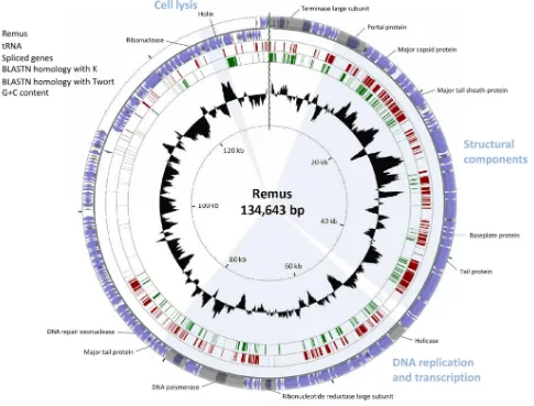
FIG 3BLASTN genome comparison of the phages representing the three species identified withinTwortlikevirus. The outer ring presents the ORFs of the circularized phage Remus genome (blue) and the numbering of every 10 ORFs. The two other rings display the BLASTN homology between Remus on one hand and K (red) and Twort (green) on the other hand. The inner ring shows the G⫹C content of the Remus genome (black). The three functional modules in the genome and some major predicted gene functions are indicated.
on November 7, 2019 by guest
http://jvi.asm.org/
[image:5.585.54.539.67.436.2]Bacillus thuringiensisphage 03058-36 I-Bth0305I, the first and un-til now only member of the EDxHD family of homing
endonu-cleases discovered in a phage genome (52) (Table 2).
Further-more, I-RoReI and I-RoReII share homology with the homing
endonucleases residing within the group I intron in the terminase
large-subunit genes ofStaphylococcusphages MSA6 (I-MsaI) and
Twort (8,13).
An insertion sequence comprising a transposase gene (ORF14) and an additional gene (ORF13) was detected between ORF12 and ORF15, both homologous to parts of the gene encoding the portal
protein in K, G1, ISP, and Twort (Fig. 4B). The transposase gene is
preceded by the predicted promoter P5. According to BLAST analysis, the transposase in Romulus and Remus is homologous to
the IS605family transposase OrfB. The insertion sequences of this
family typically comprise two genes encoding OrfA and OrfB,
serving together as the functional transposase (53). ORF13 and
ORF14 are transcribed from the same strand, whereas the IS605
family gene encoding OrfA is transcribed from the reverse strand
(54). This raises the question of whether ORF13 is part of the
insertion sequence. This question is strengthened by the imperfect
palindromic sequences (29 to 30 nucleotides [nt]), which replace
the inverted terminal repeats in the IS605family of insertion
se-quences (55), and the direct target repeats (7 nt) flanking ORF14.
Third, the helicase gene of Romulus and Remus is split into two
open reading frames, ORF49 and ORF50 (Fig. 4C). The insert is
approximately 245 nucleotides in length and probably constitutes a group I intron lacking an open reading frame. The interruption occurs at the same location as where a hedgehog/intein (Hint)
domain is found in the helicase gene of phage Twort (8).
Next, the ribonucleotide (or ribonucleoside disphosphate) re-ductase large-subunit gene is interrupted by two group I introns (Fig. 4D). Again, the introns are not related. The embedded
hom-ing endonucleases, I-RoReIII and I-RoReIV, are homologous to
I-TevI and I-HmuI, members of the GIY-YIG and HNH families,
respectively (56, 57) (Table 2). The ribonucleotide reductase
large-subunit gene itself is related to thePolaribacter irgensii23-P
[image:6.585.39.549.79.270.2]ribonucleotide reductase large-subunit gene and does not show any protein homology with the ribonucleotide reductase large-subunit gene of phages K, G1, Twort, ISP, Sb-1, A5-W, Staph1N, Fi200W, P4W, 676Z, A3R, or MSA6.
TABLE 1Comparative nucleotide analysis between genomes ofS. aureusphages present in the NCBI databasea
Phage
% Nucleotide identity
K G1 Twort ISP A5-W Staph1N Fi200W P4W 676Z A3R MSA6 Sb-1 GH15 SA11 Romulus Remus
K 100 90.3 56.1 90.6 91.4 91.3 89.2 89.8 89.4 93.5 88.6 96.6 80.0 58.4 57.6 57.9
G1 90.3 100 55.2 99.5 98.4 98.3 97.5 97.0 97.4 92.0 97.0 91.0 84.5 57.8 55.1 55.8
Twort 56.1 55.2 100 55.3 55.4 55.4 55.8 55.9 55.8 56.6 55.4 55.9 55.2 55.8 54.5 54.9
ISP 90.6 99.5 55.3 100 98.7 98.8 97.9 97.4 97.8 92.4 97.3 90.6 84.9 57.8 55.2 55.9
A5-W 91.4 98.4 55.4 98.7 100 99.9 97.3 98.1 97.3 93.2 96.1 90.6 84.5 58.0 55.5 56.2
Staph1N 91.3 98.3 55.4 98.8 99.9 100 97.4 98.2 97.4 93.3 96.2 90.5 84.6 58.0 55.5 56.1
Fi200W 89.2 97.5 55.8 97.9 97.3 97.4 100 99.1 99.6 94.1 95.4 88.9 83.9 57.5 54.9 55.6
P4W 89.8 97.0 55.9 97.4 98.1 98.2 99.1 100 99.0 94.7 94.9 89.2 83.6 57.6 55.1 55.8
676Z 89.4 97.4 55.8 97.8 97.3 97.4 99.6 99.0 100 94.3 95.3 88.8 83.9 57.5 54.9 55.6
A3R 93.5 92.0 56.6 92.4 93.2 93.3 94.1 94.7 94.3 100 90.0 92.5 81.5 58.0 56.4 57.1
MSA6 88.6 97.0 55.4 97.3 96.1 96.2 95.4 94.9 95.3 90.0 100 88.8 82.7 58.7 56.3 56.9
Sb-1 96.6 91.0 55.9 90.6 90.6 90.5 88.9 89.2 88.8 92.5 88.8 100 79.7 58.4 57.7 57.9
GH15 80.0 84.5 55.2 84.9 84.5 84.6 83.9 83.6 83.9 81.5 82.7 79.7 100 57.3 55.4 56.0
SA11 58.4 57.8 55.8 57.8 58.0 58.0 57.5 57.6 57.5 58.0 58.7 58.4 57.3 100 82.1 84.6
Romulus 57.6 55.1 53.2 55.2 55.5 55.5 54.9 55.1 54.9 56.4 56.3 57.7 55.4 82.1 100 95.9
Remus 57.9 55.8 54.9 55.9 56.2 56.1 55.6 55.8 55.6 57.1 56.9 57.9 56.0 84.6 95.9 100
aThe nucleotide identity between the genomes of each set of two phages was calculated using EMBOSS stretcher. Homologies lower than 60% are shaded. Complete (100%)
homology is indicated by bold type.
TABLE 2Overview of the predicted homing endonucleases embedded in group I introns in the Romulus and Remus genomesa
Homing
endonuclease ORF Target gene Homolog
Homing endonuclease family
Prediction
program E value
I-RoReI ORF2 Terminase large subunit I-Bth0305I (B. thuringiensisphage 0305⌽8–36)
EDxHD HHpred 6.00E⫺17
I-RoReII ORF5 Terminase large subunit I-Bth0305I (B. thuringiensisphage 0305⌽8–36)
EDxHD HHpred 6.00E⫺17
I-RoReIII ORF67 Ribonucleotide reductase large subunit
I-TevI (E. coliphage T4) GIY-YIG BLASTP/HHpred 6.00E⫺10/1.10E⫺24
I-RoReIV ORF69 Ribonucleotide reductase large subunit
I-HmuI (B. subtilisphage SPO1) HNH HHpred 7.20E⫺40
I-RoReV ORF76 DNA polymerase I-HmuI (B. subtilisphage SPO1) HNH HHpred 1.70E⫺21
I-RoReVI ORF81 DNA repair protein I-DmoI (Desulfurococcus mobilis) LAGLIDADG HHpred 2.50E⫺24
a
For each predicted homing endonuclease, the encoding ORF and the target gene are given. Further, the homologous homing endonuclease, its family, the prediction program, and corresponding E value are indicated.
on November 7, 2019 by guest
http://jvi.asm.org/
[image:6.585.40.545.569.706.2]The DNA polymerase gene not only is interrupted by a group I intron but also contains a Hint domain, an autopro-cessing domain which undergoes a protein splicing reaction (58) (Fig. 4E). This Hint domain is located at the end of ORF75,
with predicted N- and C-terminal boundaries at amino acid positions 732 and 1140, and typically contains a histidine and an asparagine residue at the C terminus, while a threonine residue follows the downstream splice site. The group I intron
FIG 4Split genes in Romulus and Remus. Shown is a graphical representation of the splicing events in the terminase large-subunit gene (A), the gene encoding the portal protein (B), the helicase gene (C), the ribonucleotide reductase large-subunit gene (D), the DNA polymerase gene (E), and the gene encoding a DNA repair protein (F) in phages Romulus and Remus compared to those in phages K, G1, ISP, and Twort orPolaribacter irgensii23-P. The open reading frames are presented as boxes, and the size of the corresponding amino acid sequence is indicated within each box. Group I intron-encoded homing endonucleases are depicted in blue. The insertion sequence-encoded transposase is depicted in dark blue. P5 represents the predicted promoter preceding ORF14. In the cases of K, G1, and ISP, the ORF numbering corresponds to that of ISP. H, histidine; N, asparagine; T, threonine.
on November 7, 2019 by guest
http://jvi.asm.org/
[image:7.585.51.528.67.606.2]codes for a homing endonuclease, I-RoReV, which is also
re-lated to I-HmuI (Table 2).
The sixth group I intron-invaded gene is the gene coding for a
DNA repair protein (Fig. 4F). This intron codes for I-RoReVI, an
endonuclease similar to I-DmoI, an archaeal intron-encoded homing
endonuclease belonging to the LAGLIDADG family (Table 2);
BLASTP analysis showed homology with other members of this fam-ily as well (data not shown). Remarkably, an LAGLIDADG homing endonuclease has never been reported for a phage genome before.
The homolog of this DNA repair protein inStaphylococcusphages
MSA6 and 812 is interrupted by a group I intron as well (13,16). In
addition, the embedded homing endonuclease in Romulus and
Re-mus, I-RoReVI, resembles the corresponding homing endonuclease
in MSA6 and 812 (13,16).
It should be mentioned that the above-mentioned group I in-trons were predicted with bioinformatic tools and therefore re-quire experimental evidence.
Structural proteome analysis of Remus.Mass spectrometric analysis of 20 gel slices of the Remus structural proteome resulted
in the identification of 19 proteins (Table 3). Homologs of 18 of
these proteins were already identified in earlier proteome studies
(11,59), while Gp42 of Remus can now be considered a
compo-nent of the virus particle as well. Various Remus proteins were detected throughout the SDS-PAGE gel, illustrated by the major capsid (Gp18) and the major tail sheath (Gp25) protein, suggest-ing their abundant presence in the Remus virion. Similar
obser-vations were made for phage ISP (11).
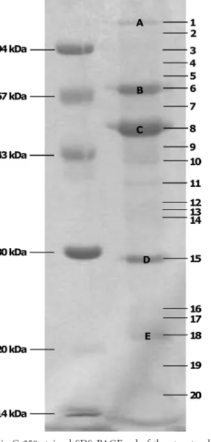
The five major protein bands (Fig. 5) of the Remus structural
proteome are very similar to the bands visualized for phage ISP
(11). However, band D of Remus is localized several kilodaltons
lower. Because for ISP no corresponding molecular mass was found, this band was thought to be a proteolytically processed
[image:8.585.40.553.79.315.2]form of the tail sheath protein (59,60). If this hypothesis is correct,
TABLE 3Characteristics of the ESI-MS-MS-identified proteins of bacteriophage Remusa
Gp Band no.
Molecular mass (kDa)
No. of identified peptides
Sequence
coverage (%) HHpred search E value ISP homolog
Molecular mass (kDa)
Gp12 7 33.8 4 15.0 Portal protein (HK97) 5.8E⫺32 Gp8 64.1
Gp15 7 26.1 4 19.7 Portal protein (HK97) 2.2E⫺30 Gp8 64.1
Gp16 19 28.8 2 9.8 Prohead protease (HK97) 9.5E⫺34 Gp9 28.6
Gp18 1, 2, 4–12 51.0 16 52.9 Major capsid protein (HK97) 5.2E⫺05 Gp11 51.2
Gp21 11 33.6 2 9.3 Gp14 33.7
Gp25 5–9, 11, 14–16, 18–20
64.6 24 51.9 Tail sheath protein (Mu) 3.1E⫺61 Gp18 64.5
Gp26 18, 19 12.4 4 55.4 Tail tube protein (T4) 0.74 Gp19 15.9
Gp36 14 34.9 1 5.7 Cell wall peptidase (NlpC) 2.7E⫺24 Gp28 34.6
Gp38 13 30.9 9 36.0 Baseplate structural protein (T4) 96 Gp30 29.3
Gp39 16 20.1 2 13.3 Gp31 20.0
Gp41 9 39.2 3 9.2 Baseplate J-like protein (P2) 8.5E⫺43 Gp33 39.2
Gp42 3, 5 102.7 6 9.0 Tail protein (P2) 1.6E⫺4 Gp34 116.3
Gp43 17 19.2 3 20.8 Baseplate structural protein (T4) 3.7E⫺35 Gp35 19.2
Gp44 1, 2, 4–6 129.4 20 24.9 Gp36 129.1
Gp46 6, 14 72.8 5 9.7 Gp38 72.6
Gp85 15, 16, 18, 19 23.4 8 61.4 Gp70 23.2
Gp86 16, 18–20 18.2 6 54.7 Major tail protein (lambda) 9.5E⫺21 Gp71 17.9
Gp87 20 7.6 2 50.0 Gp72 7.8
Gp97 19 17.7 3 19.7 Gp83 17.8
aFor each gene product (Gp), the band number(s) (according toFig. 5), the molecular mass, the number of identified peptides (⬎99% protein identification probability with
manual validation), and the protein sequence coverage are listed. Furthermore, the putative protein function according to HHpred and the corresponding E value are shown, as well as homologous ISP proteins and their molecular masses.
FIG 5Coomassie G-250-stained SDS-PAGE gel of the structural proteome of bacteriophage Remus. A low-molecular-mass marker is indicated on the left. The numbers on the right correspond to the analyzed gel pieces of which the identified proteins are listed inTable 2. Prominent protein bands are indicated with capital letters.
on November 7, 2019 by guest
http://jvi.asm.org/
[image:8.585.344.496.355.672.2]HK97, and P2 was detected. Moreover, the tail protein Gp26 is homologous to the tail tube protein of T4, and Gp38, Gp41, and Gp43 are proposed to be components of the Remus baseplate. The molecular masses of Gp26 (12.4 kDa) and Gp42 (102.7 kDa) are markedly lower than the 15.9 kDa and 116.3 kDa of the putative tail tube protein and corresponding tail protein of phage ISP, probably resulting in different dimensions of the contractile myo-viral tail.
DISCUSSION
The presence of the pathogenic bacteriumS. aureusis often
asso-ciated with hospital environments (61,62), while bacteriophages
are expected to be widespread in locations populated by their bac-terial host. Therefore, this research focused on hospital sewage for phage isolation, resulting in the isolation of the novel phages Ro-mulus and Remus. Phage isolation attempts in this research,
how-ever, have revealed that the isolation of new virulent
Staphylococ-cusphages is not a straightforward approach, illustrated by the
limited number of virulentStaphylococcusphages described in the
scientific literature (7–9,11–15,63,64).
Like the majority of the known virulentStaphylococcusphages,
Romulus and Remus were assigned to the genusTwortlikevirus
based on their morphological features, including a characteristic
baseplate at the end of the tail (5,13). Furthermore, both phages
were shown to infect a considerable number ofS. aureusisolates,
which was also observed for twortlikeviruses K, ISP, Sb-1, and
MSA6 (9,11,12,17). From a genome perspective, Romulus and
Remus are very closely related to each other and to phage SA11 but are substantially different from the other twortlikeviruses infect-ingS. aureus. At the DNA level, Romulus, Remus, and SA11 dis-play no more than 60% homology with these staphylococcal myo-viruses, suggesting that they represent a new species within
Twortlikevirus(Fig. 3). This places Romulus, Remus, and SA11 in
a new clade withinTwortlikevirus(representative phage, Remus),
next to and clearly divergent from phages K, G1, ISP, A5-W, Staph1N, Fi200W, P4W, 676Z, A3R, MSA6, and Sb-1 on one hand (representative phage, K) and phage Twort on the other hand (representative phage, Twort).
AlthoughStaphylococcus phages MSA6, Twort, and 812 are
quite distantly related to Romulus and Remus at the DNA level, these phages contain similar group I intron-embedded homing endonucleases. Interestingly, the related homing endonucleases target functional homologs. In addition to the ribonucleotide
re-ductase large-subunit gene ofBacillus subtilisbacteriophage SP
(24), the DNA polymerase gene of Romulus and Remus provides
a second example of a group I intron and an intein coding se-quence within the same gene. Furthermore, an insertion sese-quence in Romulus and Remus is unusually located between ORF12 and ORF15, genes encoding parts of the portal protein. For Gp12 and Gp15, peptides were detected in the same band. This implies that
infection patterns of Romulus and Remus appeared more prom-ising than those of ISP, implying their suitability for efficient
con-trol of sensitiveS. aureusisolates. Furthermore, the
biofilm-de-grading potential of Romulus and Remus makes their therapeutic application even more attractive. In consideration of their thera-peutic implementation, the temperature and pH range in which Romulus and Remus are stable were determined.
To conclude, Romulus and Remus are considered to be
mem-bers of a new species withinTwortlikevirus, containing a unique
scale of group I introns, an intein, and an insertion sequence in their genomes. In addition, the characterization of both phage isolates, comprising microbiological as well as molecular aspects, demonstrated their appropriateness for therapeutic application.
ACKNOWLEDGMENTS
This work was supported by the Federal Public Service of Health, Food Chain Safety and Environment, Belgium (contract RT 08/6 PHAGE).
We thank H.-W. Ackermann for performing the electron microscopic imaging.
REFERENCES
1.Jarraud S, Mougel C, Thioulouse J, Lina G, Meugnier H, Forey F, Nesme X, Etienne J, Vandenesch F.2002. Relationships between Staph-ylococcus aureusgenetic background, virulence factors,agrgroups (al-leles), and human disease. Infect. Immun.70:631– 641.
2.Lowy FD.1998.Staphylococcus aureusinfections. N. Engl. J. Med.339: 520 –532.
3.Veien NK.1998. The clinician’s choice of antibiotics in the treatment of bacterial skin infection. Br. J. Dermatol.139:30 –36.
4.Patel R.2005. Biofilms and antimicrobial resistance. Clin. Orthop. Relat. Res.437:41– 47.
5.Klumpp J, Lavigne R, Loessner MJ, Ackermann HW.2010. The SPO1-related bacteriophages. Arch. Virol.155:1547–1561.
6.Lavigne R, Darius P, Summer E, Seto D, Mahadevan P, Nilsson A, Ackermann H, Kropinski A.2009. Classification ofMyoviridae bacterio-phages using protein sequence similarity. BMC Microbiol.9:224. 7.O’Flaherty S, Coffey A, Edwards R, Meaney W, Fitzgerald GF, Ross RP.
2004. Genome of staphylococcal phage K: a new lineage of Myoviridae infecting gram-positive bacteria with a low G⫹C content. J. Bacteriol. 186:2862–2871.
8.Kwan T, Liu J, DuBow M, Gros P, Pelletier J. 2005. The complete genomes and proteomes of 27Staphylococcus aureusbacteriophages. Proc. Natl. Acad. Sci. U. S. A.102:5174 –5179.
9.Kvachadze L, Balarjishvili N, Meskhi T, Tevdoradze E, Skhirtladze N, Pataridze T, Adamia R, Topuria T, Kutter E, Rohde C, Kutateladze M. 2011. Evaluation of lytic activity of staphylococcal bacteriophage Sb-1 against freshly isolated clinical pathogens. Microb. Biotechnol.4:643– 650.
10. Merabishvili M, Pirnay J-P, Verbeken G, Chanishvili N, Tediashvili M, Lashkhi N, Glonti T, Krylov V, Mast J, Van Parys L, Lavigne R, Volckaert G, Mattheus W, Verween G, De Corte P, Rose T, Jennes S, Zizi M, De Vos D, Vaneechoutte M.2009. Quality-controlled small-scale production of a well-defined bacteriophage cocktail for use in human clinical trials. PLoS One4:e4944. doi:10.1371/journal.pone.0004944. 11. Vandersteegen K, Mattheus W, Ceyssens P-J, Bilocq F, De Vos D,
Pirnay J-P, Noben J-P, Merabishvili M, Lipinska U, Hermans K,
on November 7, 2019 by guest
http://jvi.asm.org/
gne R.2011. Microbiological and molecular assessment of bacteriophage ISP for the control of Staphylococcus aureus. PLoS One6:e24418. doi:10 .1371/journal.pone.0024418.
12. Kwiatek M, Parasion S, Mizak L, Gryko R, Bartoszcze M, Kocik J.2012. Characterization of a bacteriophage, isolated from a cow with mastitis, that is lytic againstStaphylococcus aureusstrains. Arch. Virol.157:225– 234.
13. Łobocka M, Hejnowicz MS, Da˛browski K, Gozdek A, Kosakowski J, Witkowska M, Ulatowska MI, Weber-Da˛browska B, Kwiatek M, Para-sion S, Gawor J, Kosowska H, Głowacka A.2012. Genomics of staphy-lococcal Twort-like phages—potential therapeutics of the post-antibiotic era. Adv. Virus Res.83:143–216.
14. Kim MS, Myung H.2012. Complete genome ofStaphylococcus aureus phage SA11. J. Virol.86:10232.
15. Gu J, Liu X, Lu R, Li Y, Song J, Lei L, Sun C, Feng X, Du C, Yu H, Yang Y, Han W.2012. Complete genome sequence of Staphylococcus aureus bacteriophage GH15. J. Virol.86:8914 – 8915.
16. Kaspárek P, Pantùcek R, Kahánková J, Rùzicková V, Doskar J.2007. Genome rearrangements in host-range mutants of the polyvalent staphy-lococcal bacteriophage 812. Folia Microbiol. (Praha)52:331–338. 17. O’Flaherty S, Ross RP, Meaney W, Fitzgerald GF, Elbreki MF, Coffey A.
2005. Potential of the polyvalent anti-Staphylococcus bacteriophage K for control of antibiotic-resistant staphylococci from hospitals. Appl. Envi-ron. Microbiol.71:1836 –1842.
18. Pantùcˇek R, Rosypalová A, Doškaø J, Kailerová J, Rùžicˇková V, Borecká P, Š. Snopková Horváth R, Götz F, Rosypal S.1998. The polyvalent staphylococcal phage812: its host-range mutants and related phages. Virology246:241–252.
19. Kelly D, McAuliffe O, Ross RP, Coffey A.2012. Prevention of Staphy-lococcus aureusbiofilm formation and reduction in established biofilm density using a combination of phage K and modified derivatives. Lett. Appl. Microbiol.54:286 –291.
20. Landthaler M, Begley U, Lau NC, Shub DA.2002. Two self-splicing group I introns in the ribonucleotide reductase large subunit gene of Staphylococcus aureusphage Twort. Nucleic Acids Res.30:1935–1943. 21. Landthaler M, Shub DA.1999. Unexpected abundance of self-splicing
introns in the genome of bacteriophage Twort: introns in multiple genes, a single gene with three introns, and exon skipping by group I ribozymes. Proc. Natl. Acad. Sci. U. S. A.96:7005–7010.
22. Goodrich-Blair H, Scarlato V, Gott JM, Xu M-Q, Shub DA.1990. A self-splicing group I intron in the DNA polymerase gene ofBacillus subtilis bacteriophage SPO1. Cell63:417– 424.
23. Gott JM, Shub DA, Belfort M.1986. Multiple self-splicing introns in bacteriophage T4: evidence from autocatalytic GTP labeling of RNA in vitro. Cell47:81– 87.
24. Lazarevic V, Soldo B, Düsterhöft A, Hilbert H, Mauël C, Karamata D. 1998. Introns and intein coding sequence in the ribonucleotide reductase genes ofBacillus subtilistemperate bacteriophage SP. Proc. Natl. Acad. Sci. U. S. A.95:1692–1697.
25. Edgell DR, Belfort M, Shub DA.2000. Barriers to intron promiscuity in bacteria. J. Bacteriol.182:5281–5289.
26. Adams MH. 1959. Bacteriophages. Interscience Publishers Inc., New York, NY.
27. Sambrook J, Russell DW.2001. Molecular cloning: a laboratory manual, 3rd ed. Cold Spring Harbor Laboratory Press, Cold Spring Harbor, NY. 28. Ackermann HW.2009. Basic phage electron microscopy, p 113–126.In
Clokie MRJ, Kropinski AM (ed), Bacteriophages: methods and protocols, vol 1. Isolation, characterization, and interactions. Humana Press, Clif-ton, NJ.
29. Ceyssens PJ, Lavigne R, Mattheus W, Chibeu A, Hertveldt K, Mast J, Robben J, Volckaert G.2006. Genomic analysis ofPseudomonas aerugi-nosaphages LKD16 and LKA1: establishment of the phiKMV subgroup within the T7 supergroup. J. Bacteriol.188:6924 – 6931.
30. Ceri H, Olson M, Morck D, Storey D, Read R, Buret A, Olson B.2001. The MBEC assay system: multiple equivalent biofilms for antibiotic and biocide susceptibility testing. Methods Enzymol.337:377–385.
31. Ceri H, Olson ME, Stremick C, Read RR, Morck D, Buret A.1999. The Calgary Biofilm Device: new technology for rapid determination of anti-biotic susceptibilities of bacterial biofilms. J. Clin. Microbiol.37:1771– 1776.
32. Bonfield JK, Whitwham A.2010. Gap5— editing the billion fragment sequence assembly. Bioinformatics26:1699 –1703.
33. Chevreux B, Wetter T, Suhai S.1999. Genome sequence assembly using
trace signals and additional sequence information. Comput. Sci. Biol. Proc. German Conf. Bioinformatics99:45–56.
34. Altschul SF, Gish W, Miller W, Myers EW, Lipman DJ.1990. Basic local alignment search tool. J. Mol. Biol.215:403– 410.
35. Söding J, Biegert A, Lupas AN.2005. The HHpred interactive server for protein homology detection and structure prediction. Nucleic Acids Res. 33:W244 –W248. doi:10.1093/nar/gki408.
36. Bailey TL, Elkan C.1994. Fitting a mixture model by expectation maxi-mization to discover motifs in biopolymers, p 28 –36.InProceedings of the Second International Conference on Intelligent Systems for Molecular Biology. AAAI Press, Menlo Park, CA.
37. Ermolaeva MD, Khalak HG, White O, Smith HO, Salzberg SL.2000. Prediction of transcription terminators in bacterial genomes. J. Mol. Biol. 301:27–33.
38. Gautheret D, Lambert A.2001. Direct RNA motif definition and identi-fication from multiple sequence alignments using secondary structure profiles. J. Mol. Biol.313:1003–1011.
39. Macke T, Ecker D, Gutell R, Gautheret D, Case DA, Sampath R.2001. RNAMotif—a new RNA secondary structure definition and discovery al-gorithm. Nucleic Acids Res.29:4724 – 4735.
40. Zuker M.2003. Mfold web server for nucleic acid folding and hybridiza-tion predichybridiza-tion. Nucleic Acids Res.31:3406 –3415.
41. Lowe TM, Eddy SR.1997. tRNAscan-SE: a program for improved detec-tion of transfer RNA genes in genomic sequence. Nucleic Acids Res.25: 955–964.
42. Laslett D, Canback B.2004. ARAGORN, a program to detect tRNA genes and tmRNA genes in nucleotide sequences. Nucleic Acids Res.32:11–16. 43. Stothard P, Wishart DS.2005. Circular genome visualization and
explo-ration using CGView. Bioinformatics21:537–539.
44. Myers EW, Miller W.1988. Optimal alignments in linear space. Comput. Appl. Biosci.4:11–17.
45. Rice P, Longden I, Bleasby A.2000. EMBOSS: the European Molecular Biology Open Software Suite. Trends Genet.16:276 –277.
46. Moak M, Molineux IJ.2004. Peptidoglycan hydrolytic activities associ-ated with bacteriophage virions. Mol. Microbiol.51:1169 –1183. 47. Shevchenko A, Wilm M, Vorm O, Mann M.1996. Mass spectrometric
sequencing of proteins from silver-stained polyacrylamide gels. Anal. Chem.68:850 – 858.
48. Lavigne R, Noben JP, Hertveldt K, Ceyssens PJ, Briers Y, Dumont D, Roucourt B, Krylov VN, Mesyanzhinov VV, Robben J, Volckaert G. 2006. The structural proteome ofPseudomonas aeruginosabacteriophage phiKMV. Microbiology152:529 –534.
49. Baylor MB, Silver SD.1961. Studies on the additivity of action of genes affecting host range in coliphage T2. Virology14:167–176.
50. Carlton RM, Noordman WH, Biswas B, de Meester ED, Loessner MJ. 2005. Bacteriophage P100 for control ofListeria monocytogenesin foods: genome sequence, bioinformatic analyses, oral toxicity study, and appli-cation. Regul. Toxicol. Pharmacol.43:301–312.
51. Klumpp J, Dorscht J, Lurz R, Bielmann R, Wieland M, Zimmer M, Calendar R, Loessner MJ.2008. The terminally redundant, nonpermuted genome ofListeriabacteriophage A511: a model for the SPO1-like myo-viruses of gram-positive bacteria. J. Bacteriol.190:5753–5765.
52. Taylor GK, Heiter DF, Pietrokovski S, Stoddard BL.2011. Activity, specificity and structure of I-Bth0305I: a representative of a new homing endonuclease family. Nucleic Acids Res.39:9705–9719.
53. Kersulyte D, Akopyants NS, Clifton SW, Roe BA, Berg DE.1998. Novel sequence organization and insertion specificity of IS605and IS606: chi-maeric transposable elements ofHelicobacter pylori. Gene223:175–186. 54. Kersulyte D, Mukhopadhyay AK, Shirai M, Nakazawa T, Berg DE.
2000. Functional organization and insertion specificity of IS607, a chime-ric element ofHelicobacter pylori. J. Bacteriol.182:5300 –5308.
55. Barabas O, Ronning DR, Guynet C, Hickman AB, Ton-Hoang B, Chandler M, Dyda F. 2008. Mechanism of IS200/IS605 family DNA transposases: activation and transposon-directed target site selection. Cell 132:208 –220.
56. Goodrich-Blair H, Shub DA.1996. Beyond homing: competition be-tween intron endonucleases confers a selective advantage on flanking ge-netic markers. Cell84:211–221.
57. Quirk SM, Bell-Pedersen D, Belfort M.1989. Intron mobility in the T-even phages: high frequency inheritance of group I introns promoted by intron open reading frames. Cell56:455– 465.
58. Hall TMT, Porter JA, Young KE, Koonin EV, Beachy PA, Leahy DJ.