0022-538X/09/$12.00 doi:10.1128/JVI.00394-09
Copyright © 2009, American Society for Microbiology. All Rights Reserved.
X4 Human Immunodeficiency Virus Type 1 gp120 Down-Modulates
Expression and Immunogenicity of Codelivered Antigens
䌤
Avi-Hai Hovav,
1* Michael Santosuosso,
2Maytal Bivas-Benita,
3Andre Plair,
2Alex Cheng,
2Mazal Elnekave,
1Elda Righi,
2Tao Chen,
2Satoshi Kashiwagi,
2Michael W. Panas,
3Shi-Hua Xiang,
4Karina Furmanov,
1Norman L. Letvin,
3and Mark C. Poznansky
2Institute of Dental Sciences, Hebrew University-Hadassah School of Dental Medicine, Jerusalem, Israel1; Partners AIDS Research Center,
Department of Infectious Diseases, Massachusetts General Hospital, Harvard Medical School, Charlestown, Massachusetts 021292;
Division of Viral Pathogenesis, Beth Israel Deaconess Medical Center, Harvard Medical School, Boston, Massachusetts 021153;
and Cancer Immunology and AIDS, Dana Farber Cancer Institute, Binney Street, Boston, Massachusetts 021154
Received 23 February 2009/Accepted 28 July 2009
In order to increase the immune breadth of human immunodeficiency virus (HIV) vaccines, strategies such as immunization with several HIV antigens or centralized immunogens have been examined. HIV-1 gp120 protein is a major immunogen of HIV and has been routinely considered for inclusion in both present and future AIDS vaccines. However, recent studies proposed that gp120 interferes with the generation of immune response to codelivered antigens. Here, we investigate whether coimmunization with plasmid-encoded gp120 alters the immune response to other coadministered plasmid encoded antigens such as luciferase or ovalbumin in a mouse model. We found that the presence of gp120 leads to a significant reduction in the expression level of the codelivered antigen in vivo. Antigen presentation by antigen-presenting cells was also reduced and resulted in the induction of weak antigen-specific cellular and humoral immune responses. Importantly, gp120-mediated immune interference was observed after administration of the plasmids at the same or at distinct locations. To characterize the region in gp120 mediating these effects, we used plasmid constructs
encoding gp120 that lacks the V1V2 loops (⌬V1V2) or the V3 loop (⌬V3). After immunization, the⌬V1V2, but
not the⌬V3 construct, was able to reduce antigen expression, antigen presentation, and subsequently the
immunogenicity of the codelivered antigen. The V3 loop dependence of this phenomenon seems to be limited to V3 loops known to interact with the CXCR4 molecule but not with CCR5. Our study presents a novel mechanism by which HIV-1 gp120 interferes with the immune response against coadministered antigen in a polyvalent vaccine preparation.
Genetic immunization is a powerful approach for eliciting robust cellular immune response, which is known to play an important role in controlling human immunodeficiency virus (HIV) infection (12). Vaccination studies using nonviral “naked” plasmid DNA or recombinant virus-based vectors have been reported to generate protective T-cell immunity against various bacterial and viral pathogens (7, 19). These studies usually involve a prime-boost immunization regi-men, where plasmid DNA is often used as a priming vector. Due to the large genetic diversity among HIV-1 strains, a desired vaccine should induce broadly reactive immune re-sponses (6). To achieve this goal, different strategies were developed to increase the spectrum of T- and B-cell epitopes in a vaccine construct. Such strategies include coimmunization with several HIV antigens (polyvalent vaccine), administration of a synthetic antigen consisting of various epitopes, and im-munization with centralized HIV-1 antigens (2, 10, 13). The last approach uses computational algorithms based on consen-sus, recent common ancestor, or center of the tree analyses. The use of such vaccines was shown to increase immune
breadth in comparison to immunization with conventional vac-cination approaches (22).
The HIV-1 envelope is the only component present on the virus surface and thus represents a critical target for vaccine-elicited neutralizing antibodies. HIV-1 envelope plays a central role in viral entry and the mechanisms by which this is achieved have been well defined. In addition, the envelope protein can elicit CD8⫹and CD4⫹T-cell responses in humans, nonhuman primates and mice, providing the rationale that the envelope is a major HIV immunogen (1, 9, 17). More recently, secondary functions of the envelope protein have been identified, includ-ing its ability to dysregulate immune responses. For example, HIV gp120, a component of the HIV envelope, induces apop-tosis in CD4⫹ T cells that allow the virus to escape virus-specific immunity (18, 29). We have shown recently that a high level of gp120 can reduce the efficacy of T-cell-mediated im-munity during SHIV infection (26). HIV-1 gp120 can also act as a B-cell mitogen, stimulating antibody production and acti-vation of complement, which leads to a formation of complexes that protect the virus from effector cells and facilitate its in-fection (16). HIV-1 gp120 was also found to inhibit major histocompatibility complex (MHC) class I presentation of T-cell epitopes of other antigen in vitro (14). More importantly, there is evidence that vaccination with gp120 can enhance viral replication and even decrease protection provided by other antigens (4, 25). In a recent study the HIV envelope was
* Corresponding author. Mailing address: Institute of Dental Sci-ences, Hebrew University-Hadassah Faculty of Dental Medicine, P.O. Box 122722, Jerusalem 91120, Israel. Phone: 2-6758802. Fax: 972-2-6758561. E-mail: [email protected].
䌤Published ahead of print on 19 August 2009.
10941
on November 8, 2019 by guest
http://jvi.asm.org/
MATERIALS AND METHODS
Vectors.The codon-optimized HIV-1 X4 HXBIIenvor firefly luciferase genes were cloned into the VRC vector. The empty VRC vector was kindly provided by Gary Nabel, Vaccine Research Center, National Institute of Allergy and Infec-tious Disease, National Institutes of Health. The pACB-OVA plasmid was a gift from Maripat Corr (University of California at San Diego). The V3 loop- or V1V2 loop-deleted gp120 HXBII plasmids were generously provided by Joseph Sodroski (Dana Farber Cancer Institute, Boston, MA). The X4KB-9, R5YU-2, and R5ADA plasmid constructs were provided by J. Sodroski and X4 NL4-3 gp120 was derived from an expressor plasmid from M. Martin. The Con-S vector was a gift from Barton F. Haynes (Duke University, Durham, NC). All plasmids used in the present study were prepared by using the EndoFree plasmid Giga kit (Qiagen), and the levels of endotoxin in the DNA preparations were less than 0.1 EU perg of DNA. Equivalent expression of all constructs used in the present study was demonstrated by Northern analysis of RNA expression in transfected HEK 293T cells as previously described (26).
Mice and immunizations.Six- to eight-week-old female BALB/c and B6 mice were purchased from Jackson Laboratories and maintained under specific-pathogenic-free conditions. Research on mice was approved by the Beth Israel Deaconess Institutional Animal Care and Use Committee. Mice were injected intramuscularly with two plasmids in various combinations as indicated in the text (50g of DNA/per plasmid in 100l of total injection volume, 50l was delivered into each quadricep muscle).
Antibodies and reagents.The following monoclonal antibodies were used in the study: PerCP-Cy5.5-conjugated anti-CD8␣(53-6.7; BD Biosciences), phy-coerythrin (PE)- or Alexa Fluor 700-conjugated anti-gamma interferon (anti-IFN-␥; XMG1.2; BD Biosciences), allophycocyanin-conjugated anti-inter-leukin-2 (anti-IL-2; JHS6-5H4; BD Biosciences), and allophycocyanin-Cy7-con-jugated anti-CD4 (GK1.5; BD Biosciences). The PE-conallophycocyanin-Cy7-con-jugated SIINFEKL-specific MHC class I tetramers were purchased from Beckman Coulter.
MHC class I tetramer analysis.Blood was collected from individual mice in RPMI 1640 medium containing 40 U of heparin per ml, and peripheral blood mononuclear cells were isolated by using Lympholyte-M (Cedarlane). Cells were washed with phosphate-buffered saline (PBS) containing 2% fetal bovine serum (FBS) and stained for 15 min at room temperature with PE-conjugated MHC class I tetramer folded with the following peptide: H-2Kb/SIINFEKL tetramer
(specific for the OVA immunodominant epitope SIINFEKL) or with H-2Dd
/R GPGRAFVTI tetramers (specific for the gp120 p18 immunodominant epitope). The cells were then stained with anti-CD8␣antibody for an additional 15 min at room temperature, washed once, and fixed with PBS containing 2% paraformal-dehyde. Samples were collected on an LSR II instrument (BD Biosciences) and analyzed by using the FlowJo software (Tree Star).
Splenocyte stimulation and intracellular cytokine staining.Splenocytes were harvested from individual mice and red blood cells were lysed by using ACK buffer. The cells were then washed with PBS plus 2% FBS, counted and resus-pended (4⫻106
cells/tube) in RPMI 1640 medium (Cellgro, Herndon, VA) supplemented with 10% FBS, 25 mM HEPES, 2 mML-glutamine, 20 U of penicillin/ml, 20g of streptomycin/ml, 1 mM sodium pyruvate, and 0.1 mM nonessential amino acids. For T-cell stimulation, cells were incubated with Golgi Plug (2l/ml), anti-CD28 (2g/ml), anti-CD49d (2g/ml), and 2g of lucif-erase peptide pool per ml. The pool consisted of overlapping 15-mer peptides spanning the firefly luciferase protein and was used such that each peptide was present at a concentration of 2g/ml. To measure Con-S-specific T-cell re-sponses, splenocytes were exposed to two sets of pooled peptides consisting of overlapping 15-mer peptides spanning the entire Con-S protein sequence. Un-stimulated cells were incubated with all of the above reagents except for the peptides. As a positive control, splenocytes were incubated with phorbol myris-tate acemyris-tate (2g/ml), ionomycin (10g/ml), and Golgi Plug. The cells were
luciferin injection, photonic emissions were measured by using an IVIS-100 charge-coupled device camera. Luciferase quantification was done by using Liv-ing Image software to identify and measure regions of interest.
Antigen presentation assay.The draining lymph nodes (LNs) were collected from immunized mice and treated with collagenase type II (1 mg/ml; Worthing-ton Biochemicals) and DNase I (1 mg/ml; Roche) solution in PBS plus 2% FBS for 20 min at 37°C in a shaker bath. Then, 20l of EDTA 0.5 M was added to the digested LN, and the incubation proceeded for an additional 10 min. The cells were then washed, filtered, irradiated (3,000 rads) and plated at twofold dilutions in a 96-well plate starting at 106cells/well. The RF33.70 hybridoma (a
gift from Kenneth Rock, University of Massachusetts), which produces IL-2 upon specific recognition of SIINFEKL in the context of H-2Kb
presented by antigen-presenting cells (APCs), was added to the plate (105
cells/well), the plate was incubated for 30 h, and the supernatants were then collected. IL-2 levels in the supernatant were determined by using the OptEIA IL-2 ELISA kit (BD Biosciences) according to the manufacturer’s instructions.
Enzyme-linked immunosorbent assay (ELISA).Blood was drawn from mice and sera were stored at⫺70°C prior to use. Ninety-six-well plates (Maxisorp; Nunc) were coated overnight at 4°C with 1g of the recombinant OVA antigen/ well in 0.1 M sodium carbonate (pH 9.5) solution. Plates were washed twice with PBS–0.05% Tween 20 and blocked with PBS–10% FBS (2 h at room tempera-ture). Subsequently, mouse serum samples diluted serially in PBS were added to the wells for 2 h of incubation at room temperature. Plates were then washed five times with PBS–0.05% Tween 20 and either horseradish peroxidase (HRP)-conjugated goat anti-mouse immunoglobulin G (IgG; KPL) or HRP-(HRP)-conjugated goat anti-rabbit IgG (Bio-Rad) as appropriate. After incubation for 1 h at room temperature, plates were washed seven times, and 100l of Sure Blue reagent (KPL) was added. Reactions were stopped after 30 min by the addition of TMB stop solution (KPL). The absorption was read at 405 nm using an ELISA reader (SPECTRA max PLUS).
TUNEL staining.Staining for apoptotic cells was performed by using a com-mercial TUNEL (terminal deoxynucleotidyltransferase-mediated dUTP-biotin nick end labeling) histochemical assay (DeadEnd Colorimetric TUNEL System; Promega). In brief, sections were placed in 0.85% NaCl solution for 3 min before 100l of a 20-g/ml proteinase K mixture was added to the slides, followed by incubation for 10 min. After the slides were washed in PBS for 5 min, 100l of equilibration buffer was placed on the slides for 10 min, followed by a 1-h incubation of rTdT reaction mix (composed of 98l of equilibration buffer, 1l of rTdT enzyme, and 1l of biotinylated nucleotide mix) at 37°C. Reactions were terminated in 2⫻SSC (1⫻SSC is 0.15 M NaCl plus 0.015 M sodium citrate) for 15 min, and the slides were then washed three times for 5 min each time in PBS. Endogenous peroxidases were then blocked with 0.3% hydrogen peroxide for 5 min. After the slides were washed in PBS again (as mentioned before), the tissues were incubated in 100l of streptavidin-HRP (1:500 dilution in PBS) for 30 min. The slides were washed in PBS again, and then 100l of 3,3⬘ -diamino-benzidine (DAB) solution (composed of 50l of 20⫻DAB substrate buffer, 950 l of distilled H2O, 50l of 20⫻DAB chromogen, and 50l of 20⫻hydrogen
peroxide) was added to the sections, followed by incubation for 10 min. The slides were finally washed in distilled H2O several times before Permount
mount-ing medium (Fisher) was added, coverslips were applied, and the edges were sealed with nail polish. Slides were then viewed under light and fluorescence microscopes. TUNEL-positive cells were counted, and the number of positive cells for 10 high-power fields was determined.
Statistical analysis.Data were expressed as means⫾the standard error of the means (SEM). Statistical tests were performed by using one-way analysis of variance and the Studentttest, and aPvalue of⬍0.05 was considered significant.
on November 8, 2019 by guest
http://jvi.asm.org/
RESULTS
HIV-1 gp120 reduces T-cell responses elicited against
coad-ministered antigen. We explored the influence of HIV-1
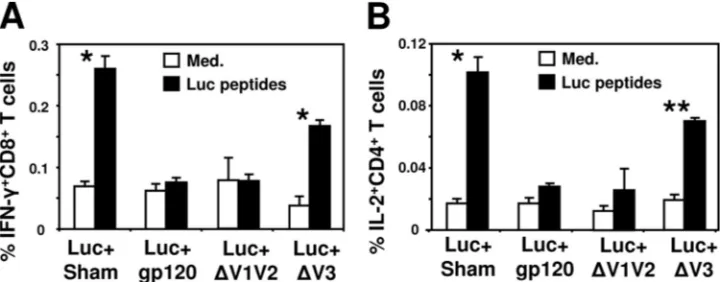
gp120 protein on the immune responses generated against another antigen in a polyvalent vaccine preparation. Mice were coimmunized with plasmids encoding the gp120 gene and the firefly luciferase gene (Luc⫹gp120). Control mice were coinjected with sham plasmid (Luc⫹Sham) or plasmid DNA encoding the ovalbumin (OVA) gene as a control foreign an-tigen (Luc⫹Ctrl). Two weeks after the immunization, spleno-cytes were harvested and evaluated for their capability to pro-duce IFN-␥after exposure to a pool of peptides spanning the luciferase protein (Fig. 1). The percentages of luciferase-spe-cific CD8⫹T cells that produced IFN-␥in Luc⫹ gp120-immu-nized mice were lower than that observed in mice immugp120-immu-nized with Luc⫹Sham or Luc⫹Ctrl (P⬍0.001) (Fig. 1A). A similar reduction in IFN-␥-producing CD4⫹T cells was also observed in Luc⫹gp120-immunized mice in comparison to mice immu-nized with Luc⫹Sham or Luc⫹Ctrl (P⬍0.001) (Fig. 1B). The production of IL-2 by luciferase-specific CD8⫹ or CD4⫹ T cells was also reduced due to the codelivery of gp120 and
luciferase plasmids (data not shown). These findings indicate that HIV-1 gp120 interferes with the elicitation of T-cell im-mune responses against coadministered vaccine antigens. This effect is specific to gp120, since expression of a control protein (OVA) did not interfere with luciferase-specific T-cell re-sponses.
The presence of gp120 down-modulates expression of
coad-ministered antigen.The amount of antigen expressed in the
host is known to play a major role in the magnitude of the immune response that is generated to it. Using luciferase as an antigen allows us to examine its expression level in the mice by using IVIS. Mice were immunized as described above and the kinetics of luciferase expression were measured at day 6 and 14 postimmunization. Our analysis indicates that at day 6 postimmunization expression of luciferase in mice immu-nized with Luc⫹gp120 was 1.36 log lower than that mea-sured in Luc⫹Sham-immunized mice (P⬍0.00001) (Fig. 2). At 2 weeks postimmunization the levels of luciferase were almost undetectable in Luc⫹gp120-immunized mice, whereas expression of this antigen was still detectable in mice injected with Luc⫹Sham. In contrast, an insignificant reduction in
lu-FIG. 1. The presence of HIV-1 gp120 decreases antigen-specific T-cell responses. BALB/c mice were coimmunized with plasmid DNA encoding the luciferase protein (50 g) and plasmid (50 g) encoding either gp120 (Luc⫹gp120), OVA (Luc⫹Ctrl), or sham plasmid (Luc⫹Sham). At 2 weeks postimmunization splenocytes were harvested and exposed for 6 h to medium alone (Med.) or luciferase pooled peptides (2g/ml). The data are presented as the percentages of CD8⫹T cells (A) or CD4⫹T cells (B) staining positively for IFN-␥, and represent the means of five mice per group⫾the SEM.*,P⬍0.001 IFN-␥production by Luc⫹gp120 compared to the other groups.
FIG. 2. HIV-1 gp120 reduces expression of codelivered antigen. BALB/c mice were coimmunized with plasmid DNA encoding the luciferase protein (50g) and plasmid (50g) encoding either gp120 (Luc⫹gp120), OVA (Luc⫹Ctrl), or sham plasmid (Luc⫹Sham). The levels of luciferase expression were measured on days 6 and 14 postimmunization using IVIS and are quantitated as relative light units (RLU). (A) Mean values of the amount of luciferase expressed by groups of four to five mice⫾the SEM following the immunization. (B) Representative images of luciferase expression on day 6 postimmunization are shown.*,P⬍0.00001 (luciferase expression in Luc⫹gp120-immunized mice compared to Luc⫹ Sham-immunized mice).
on November 8, 2019 by guest
http://jvi.asm.org/
ciferase expression (0.12 log) was seen in mice coimmunized with the luciferase- and control antigen-encoding plasmids ei-ther 1 or 2 weeks postimmunization (P⫽0.056 andP⫽0.16, respectively) (Fig. 2). These results demonstrate that expres-sion of gp120 down-modulates adjacent coadministered anti-gen expression. The small decrease in luciferase expression in the presence of a control protein suggests that gp120 may suppress luciferase expression.
The HIV-1 gp120 V3 loop domain contributes to the
reduc-tion in coadministered luciferase expression.We next sought
to identify the domain in the HIV-1 gp120 envelope protein that is involved in modulating the expression of luciferase. Since the variable loops of gp120 are known to modulate many of its biological functions, we immunized mice with the plasmid DNA expressing luciferase and the plasmid DNA encoding the gp120 protein lacking the V1 and V2 loops (Luc⫹⌬V1V2) or the V3 loop (Luc⫹⌬V3). Immunization with Luc⫹⌬V1V2 re-sulted in reduced expression of luciferase similar to the level observed after immunization with Luc⫹gp120 (P ⫽ 0.499) (Fig. 3A and B). In contrast, the absence of the V3 loop in mice immunized with Luc⫹⌬V3 did not influence significantly the expression of luciferase, and the level of expression was comparable to that seen in Luc⫹Sham-immunized mice (P⫽
0.420). The V3 loop region is known to determine coreceptor usage (CXCR4 [X4] versus CCR5 [R5]) during viral infection. Since the HXBII gp120 envelope protein binds the CXCR4 molecule (X4), we tested whether other X4 gp120 proteins could reduce the luciferase expression following immunization. As shown in Fig. 3C, coimmunization of mice with luciferase plasmid and the X4 KB-9 or X4 NL4-3 gp120 plasmid con-structs resulted in decreases of luciferase expression levels comparable to those observed for the HXBII gp120 construct. In contrast, immunization of mice with luciferase plasmid, to-gether with the R5 gp120 constructs, R5 YU-2 and R5 ADA, did not significantly reduce the expression level of luciferase. These results suggest that the V3 loop of the HIV-1 gp120 protein contributes to the down-modulation of expression of coinjected antigen. In addition, these results suggest that in a murine model the X4 gp120 clones, but not R5 gp120 clones, have the ability to downregulate antigen expression after co-immunization.
V3 loop of X4gp120 contributes to the reduction of
lucif-erase-specific T-cell responses.To examine whether the
dele-tion of the V3 loop region from the gp120 sequence also affects the immune response elicited against the luciferase protein, we assessed the magnitude of luciferase-specific T-cell responses
FIG. 3. The reduction in antigen expression level is V3 loop dependent. Groups of BALB/c mice were immunized with plasmid DNA encoding the luciferase protein together with sham plasmid (Luc⫹Sham), plasmid encoding the gp120 protein (Luc⫹gp120), or the gp120 mutants lacking the V1V2 loops (Luc⫹⌬V1V2) or V3 loop (Luc⫹⌬V3). The levels of luciferase expression were measured on day 5 postimmunization using IVIS. (A) Mean values of the amount of luciferase expressed by groups of four to five mice⫾the SEM following the immunization. (B) Representative images of luciferase expression in the immunized mice 6 days after the immunization are shown. (C) BALB/c mice were immunized with Luc⫹Sham, Luc⫹Ctrl, or with various plasmids expressing different X4 and R5 gp120 envelope proteins. Six days after the immunization the mean values⫾the SEM of the amount of luciferase expressed by group of four mice were examined.*,P⬍0.005 (luciferase expression in Luc⫹ gp120-or Luc⫹⌬V1V2-immunized mice compared to Luc⫹Sham-immunized mice); **, P ⬍ 0.05 (luciferase expression compared to Luc⫹ Ctrl-immunized mice).
on November 8, 2019 by guest
http://jvi.asm.org/
generated in the immunized mice. Splenocytes from the im-munized mice were prepared 2 weeks after the immunization, and their capability to produce cytokines was measured by ICS after stimulation with luciferase pooled peptides. The number of IFN-␥-producing luciferase-specific CD8⫹T cells was con-siderably reduced in mice immunized with Luc⫹gp120 or Luc⫹⌬V1V2. Immunization of mice with Luc⫹⌬V3 resulted in a higher level of IFN-␥⫹CD8⫹T cells than that observed in Luc⫹gp120- or Luc⫹⌬V1V2-immunized mice; however, the magnitude of the response was slightly lower than that seen after immunization with Luc⫹Sham (P⫽0.003). Analysis of IL-2 production by CD4⫹T cells showed a pattern similar to that found for IFN-␥⫹CD8⫹T cells (Fig. 4B). These findings indicate that the V3 loop is involved in gp120-mediated im-mune interference against coimmunized antigen in a polyva-lent vaccination.
Intact gp120-specific immunity and increased apoptosis
levels at the injection site.We evaluated whether the
gp120-specific immune responses generated after polyvalent immu-nization are influenced by the presence of the other immuno-gen. Mice were immunized as described above, and the magnitude of the CD8⫹T cells was examined using H-2Dd/p18
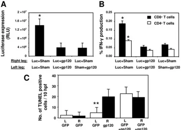
MHC class I tetramer. As demonstrated in Fig. 5A, the kinetics of p18-specific CD8⫹T cells were not affected by the presence of luciferase and were similar to those mea-sured in gp120⫹Sham-immunized mice. We next measured the level of apoptosis in the injection site. To allow precise identification of the injection site in the muscle, green fluores-cent protein (GFP) instead of luciferase expressing plasmid was coinjected with gp120 or its mutants. Muscles were col-lected 5 and 15 days postimmunization, and the apoptosis levels were assessed by a TUNEL assay. Injection of GFP or luciferase alone resulted in low apoptosis levels at the injection site either on day 5 or 15 (Fig. 5B). Coadministration of GFP and gp120 (GFP⫹gp120) led to a dramatic increase in the level of apoptosis at the injection site (P⬍0.0001). High numbers of
[image:5.585.112.473.69.210.2]FIG. 4. Deletion of the V3 loop region eliminates the negative effect of HIV-1 gp120 on antigen-specific T-cell responses. BALB/c mice were immunized with plasmid DNA encoding the luciferase protein together with sham plasmid (Luc⫹Sham), plasmid encoding the gp120 protein (Luc⫹gp120), or the gp120 mutants lacking the V1V2 loops (Luc⫹⌬V1V2) or V3 loop (Luc⫹⌬V3). Two weeks postimmunization splenocytes were harvested and exposed for 6 h to medium alone (Med.) or luciferase pooled peptides (2g/ml). The data are presented as the percentages of CD8⫹T cells (A) or CD4⫹T cells (B) staining positively for IFN-␥or IL-2, respectively, and represent the means of five mice per group⫾the SEM.*,P⬍0.001;**,P⬍0.01 (cytokine production by Luc⫹Sham- or Luc⫹⌬V3-immunized mice compared to the other groups).
FIG. 5. gp120 induces a CD8⫹T-cell response and apoptosis. (A) gp120-specific CD8⫹ T-cell responses were measured in BALB/c mice coimmunized with gp120-encoding plasmid and sham plasmid (Sham⫹gp120) or luciferase-encoding plasmid (Luc⫹gp120) and in naive mice. The data are presented as the percentages of H-2Dd/p18 tetramer⫹CD8⫹T cells and represent the mean of four mice per group⫾the SEM. (B) Mice received intramuscular injections of plasmids encoding luciferase or GFP with sham plasmids (Luc⫹Sham, GFP⫹Sham, respectively), GFP and gp120-encoding plasmid (GFP⫹gp120) or the gp120 mutants lacking the V1V2 loops (GFP⫹⌬V1V2) or V3 loop (GFP⫹⌬V3). Mice were sacrificed at day 5 or 15 and muscles from these animals harvested and fixed in paraformaldehyde. Sections (5m) were then cut; alternate sections were stained with hematoxylin and eosin to identify the site of injection according to GFP expression and then stained for TUNEL. The data are presented as TUNEL-positive cells in 10 high-power fields and represent the mean of four mice per group⫾the SEM.P⬍0.0001 (apoptosis level in GFP⫹gp120-immunized mice compared to GFP⫹Sham-immunized mice).
on November 8, 2019 by guest
http://jvi.asm.org/
apoptotic cells were also observed in GFP⫹ ⌬ V1V2-immu-nized mice. In contrast, when GFP was injected with the gp120
⌬V3 mutant no increase in the numbers of apoptotic cells was found in comparison to GFP⫹Sham (P⫽0.21) or Luc⫹Sham (P⫽0.39) (Fig. 5B). Our analysis indicates that the immune response against gp120 is not influenced by the presence of other antigens in a polyvalent vaccination. In addition, immu-nization with gp120 generates considerable V3 loop-mediated apoptosis at the site of injection.
Antigen-presenting activity is inhibited due to the presence
of gp120 in a polyvalent vaccine.We postulated that the
re-duction in the level of luciferase expression due to the coex-pression of gp120 might limit the amount of luciferase taken up by APCs. As a result, less antigenic stimulation is applied to T and B lymphocytes and that may explain the lower immu-nogenicity found in the mice (21). To address this, we assessed the level of antigen presenting activity by APCs in the different immunization groups of mice. In order to do so we moved from a luciferase construct to an OVA-expressing plasmid, due to the immunological tools available for measuring OVA-specific antigen presentation levels in B6 (H-2Kb) mice. B6 mice were
immunized with plasmid DNA encoding the OVA antigen, together with one of following plasmids: (i) sham plasmid (OVA⫹Sham), (ii) plasmids encoding the gp120 gene (OVA⫹gp120), (iii) gp120 constructs lacking the V1 and V2 loops (OVA⫹⌬V1V2), or (iv) gp120 constructs lacking the V3
loop (OVA⫹⌬V3). We first examined whether gp120 was ca-pable of suppressing the immune response in the different mouse strain (B6 versus BALB/c) and in a context of OVA protein antigen. Using H-2Kbtetramer specific for the
immu-nodominant SIINFEKL epitope we found low percentages of SIINFEKL-specific CD8⫹ T cells responses in the blood of mice immunized with OVA⫹gp120 or OVA⫹⌬V1V2 plasmids (Fig. 6A). In contrast, immunization with OVA⫹⌬V3 plasmids resulted in high SIINFEKL-specific CD8⫹ T-cell responses similar to that measured in the blood of OVA⫹ Sham-immu-nized mice. We next measured the production of OVA-specific antibodies in the sera of the B6 immunized mice 3 weeks postimmunization. As in the case of the T-cell responses, the production of OVA-specific IgG antibodies was significantly reduced when the gp120 or its⌬V1V2-lacking construct was coexpressed with the OVA antigen (P⬍0.005), whereas the presence of the⌬V3-lacking construct did not affect the anti-bodies levels (Fig. 6B). To test whether the presence of gp120 influenced antigen-presenting activity in this experimental set-ting, we collected cells from the local draining LNs 5 days after the immunization and tested the stimulatory capability of the APCs. The collected LN cells were cocultured with a T-cell hybridoma that has a T-cell receptor specific for the SIINFEKL epitope in the context of the H-2Kbclass I
mol-ecule. Since this T-cell hybridoma secretes IL-2 upon acti-vation as a response to antigen presented by APCs, the
FIG. 6. Antigen-presenting activity is down-modulated due to the presence of HIV-1 gp120. Groups of B6 mice were immunized with plasmid DNA encoding the OVA protein together with sham plasmid (OVA⫹Sham), plasmid encoding the gp120 gene (OVA⫹gp120), or the gp120 mutants lacking the V1V2 loops (OVA⫹⌬V1V2) or V3 loop (OVA⫹⌬V3). (A) Magnitude of SIINFEKL-specific CD8⫹T cells in the peripheral blood of the mice 14 days postimmunization as detected with an H-2Kb/SIINFEKL tetramer and represent means of five mice per group⫾the
SEM (B) Three weeks after the immunization, OVA-specific antibody responses generated in the mice were measured by ELISA. The presented data are the titers of OVA-specific IgG antibodies and represent the means of five mice per group⫾ the SEM. (C) Five days after the immunization, the local draining LN cells were collected, irradiated, and cocultured with the SIINFEKL-specific T-cell hybridoma for 24 h, and the IL-2 concentrations in the supernatants were quantitated by ELISA. The presented data are the IL-2 concentrations in the supernatants and represent the mean values from four mice per group ⫾ the SEM. *,P ⬍ 0.005; **,P ⬍ 0.05 (cytokine production by OVA⫹Sham- or OVA⫹⌬V3-immunized mice compared to mice immunized with OVA⫹gp120).
on November 8, 2019 by guest
http://jvi.asm.org/
hybridoma can be used as an indicator of APC activity by monitoring its production of IL-2 (Fig. 6C). Higher levels of IL-2 were detected after the addition of LN cells from OVA⫹Sham- or OVA⫹⌬V3-immunized mice than that measured by APCs derived from mice immunized with OVA-gp120 or OVA⫹⌬V1V2 plasmids. Therefore, these findings suggest that the reduced immunogenicity of anti-gens coexpressed with gp120 is due to a lower level of presentation of the coadministered antigen.
Immune interference due to gp120 occurs after
immuniza-tion at a separate locaimmuniza-tion.Our results suggest that the
reduc-tion in antigen expression is not due to simple competireduc-tion between two antigens that were coimmunized but to specific immune suppressive activities of gp120. Therefore, we exam-ined whether gp120 is capable of reducing expression of other antigens injected at a distal site. BALB/c mice were coimmu-nized in both legs with Luc⫹Sham or Luc⫹gp120, and a third group was injected in the right leg with Luc⫹Sham and in the left leg with Sham⫹gp120. Analysis of luciferase expression in the right leg revealed that luciferase levels were reduced in mice receiving gp120 and luciferase in different legs in com-parison to Luc⫹Sham-immunized mice (Fig. 7A). The reduc-tion was similar to that found in mice injected with gp120 and
[image:7.585.110.474.68.331.2]luciferase at the same site (Luc⫹gp120). We then assessed the induction of T-cell immunity against luciferase in the different groups of mice. Mice that were injected with gp120 and lucif-erase either in the same or distant locations generated consid-erably lowered CD8⫹ and CD4⫹ T-cell responses than that observed in Luc⫹Sham-immunized mice (Fig. 7B). We hy-pothesized that a soluble factor such as gp120 would be able to mediate this reduction of T-cell immunity; however, we were unable to measure gp120 in the serum (data not shown). Fur-thermore, we examined the potential indirect effect of gp120 via the production of type I IFNs as demonstrated previously (28) and again we were unable to measure differences between the gp120⫹Luc or the Sham⫹Luc (Table 1). Next, we tested the level of apoptotic cells in the immunization site when the gp120 and the coimmunized antigen (GFP) were injected in separate legs. Muscles were collected 5 days after the immu-nization and the apoptosis levels were assessed by a TUNEL assay. Immunization with the GFP plasmid resulted in a low number of apoptotic cells in the injected muscles (Fig. 7C). Interestingly, expression of gp120 in the opposite leg does not increase the apoptosis level seen in the GFP-injected muscle. Taken together, these data suggest that gp120 can reduce the expression and immunogenicity of codelivered antigen even at
FIG. 7. gp120-mediated immune interference also occurs after administration of the plasmids at separate anatomic sites. BALB/c mice (n⫽
4) were immunized in both legs with plasmid DNA encoding the luciferase protein (50g) and plasmid (50g) encoding the gp120 (Luc⫹gp120) or sham plasmid (Luc⫹Sham). A third group (n⫽8) was immunized in the right leg with Luc⫹Sham and in the left leg with Sham⫹gp120. The levels of luciferase expression in the right leg were measured on day 5 postimmunization using IVIS. (A) Mean values of the amount of luciferase expressed by the mice⫾the SEM after the immunization. (B) At 2 weeks postimmunization, splenocytes were harvested and exposed for 6 h to luciferase pooled peptides (2g/ml). The data are presented as the percentages of CD8⫹or CD4⫹T cells staining positively for IFN-␥and represent the means of four mice per group⫾the SEM. (C) Mice were immunized as described in panel A but instead of the luciferase plasmid, a GFP-expressing plasmid was used. The mice were sacrificed 5 days after the immunization, and muscles from these animals were harvested and fixed in paraformaldehyde. Sections (5m) were then cut, and alternate sections were stained with hematoxylin and eosin to identify the site of injection according to GFP expression and then stained for TUNEL. The data are presented as TUNEL-positive cells in 10 high-power fields and represent the means of four mice per group⫾the SEM.*,P⬍0.01 (luciferase expression or IFN-␥secretion compared to the other groups);**,
P⬍0.05 (difference between the number of apoptotic cells in GFP⫹Sham- versus GFP⫹gp120-injected legs).
on November 8, 2019 by guest
http://jvi.asm.org/
a distant location but not as a result of induction of apoptosis at that distal site.
Coimmunization with luciferase and the consensus gp120 (Con-S) did not interfere with antigen expression and immune
induction. In an attempt to increase cross-protection and
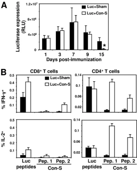
epitope diversity of gp120, after vaccination, recent studies used a novel gp120 antigen that represent a centralized or consensus sequence of various HIV-1 envelope proteins (13, 22). We examined whether such an antigen will retain the immunosuppressive effect as the HIV-1 gp120 protein. To test this question, we immunized BALB/c mice with plasmid DNA encoding the luciferase and Con-S, a consensus sequence of group M HIV-1 envelope protein. The Con-S protein shares high similarity with the HXIIB gp120 protein (⬎80%), and the differences are limited to the variable loops. In addition, Con-S gp120 is known to interact with CCR5. In contrast to HIV-1 HXIIB gp120, coexpression of luciferase and Con-S did not decrease luciferase expression in the immunized mice (Fig. 8A). However, we found significant reduction in luciferase expression in Luc⫹Con-S-immunized mice at 15 days postim-munization. We next assessed the immunogenicity of luciferase in the immunized mice (Fig. 8B). No reduction in luciferase-specific T-cell response was observed due to the coexpression of Con-S. Moreover, the CD8⫹ T-cell responses developed against luciferase were considerably higher in the presence of Con-S. Cellular immune responses were also detected against the Con-S antigen (Fig. 8B). These data suggest that, in con-trast to X4 gp120 envelope proteins, Con-S does not interfere with the induction of immune response against coimmunized antigen.
DISCUSSION
In this study we present evidence to support a novel mech-anism by which HIV-1 gp120 interferes with the induction of the immune response against other antigens in a polyvalent vaccine preparation. We have shown that gp120 downregulates the expression of the coimmunized antigen, resulting in limited antigen presentation by APCs and weak immunogenicity. Al-though a small reduction in protein expression might occur due to the expression of two plasmids in the same cell, our data clearly indicate that this is not the case with gp120. We also excluded the possibility that downregulation of antigen expres-sion might be due to apoptosis of APC, since the ability of gp120 to induce apoptosis in the muscle did not correlate with
[image:8.585.43.284.82.169.2]express the various antigens (8, 24). HIV-1 gp120 was previ-ously reported to mediate robust production of type I IFNs by plasmacytoid DC (pDC) (23). However, we could not detect an increase in the level of IFN-␣ in the serum of the gp120-immunized mice, suggesting that antigen expression is not af-fected by type I IFNs in our system. Although not exhaustive, our data support the concept that an accessory molecule or cell mediates a portion of this phenotype.
FIG. 8. The R5 gp120 Con-S antigen does not interfere with ex-pression and immune induction against coadministered antigen. BALB/c mice were coimmunized with plasmid DNA encoding the luciferase protein (50g) and plasmid (50g) encoding gp120 Con-S (Luc⫹Con-S) or sham plasmid (Luc⫹Sham). (A) Kinetics of lucifer-ase expression are presented as measured by IVIS during the 2 weeks after the immunization and represent the mean of four mice per group⫾the SEM. (B) At 2 weeks postimmunization, splenocytes were harvested and exposed for 6 h to luciferase pooled peptides (2g/ml) or to Con-S peptides divided into two pools (“Pep. 1” and “Pep. 2”) (2
g/ml). The data are presented as the percentages of CD8⫹or CD4⫹ T cells staining positively for IFN-␥or IL-2 and represent the means of four mice per group⫾the SEM.*,P⬍0.01 (expression of luciferase in Luc⫹Sham- compared to Luc⫹Con-S-immunized mice).
IFN-␣concentrations in serum were determined by ELISA.
on November 8, 2019 by guest
http://jvi.asm.org/
[image:8.585.300.540.299.598.2]A potential explanation for the reduced antigen load and reduced immune response could be either APC or T-cell lo-calization. HIV-1 gp120 is able to directly interact with che-mokine receptors; for example, X4-gp120 has been shown to mediate signaling in murine and rat cells (3, 11, 28). Although there are no specific reports that indicate that R5 HIV-1 gp120 can trigger murine CCR5, this protein has been shown to bind this chemokine receptor on rat cells (20). It is possible that the reason that X4 gp120 rather than R5 gp120 down-modulates the expression and immunogenicity of codelivered antigens is because the former protein can trigger its cognate murine coreceptor while the latter cannot. Furthermore, these gp120 chemokine receptor interactions could impair the ability of APCs to pick up antigen, migrate to the local LNs, or provide the correct chemotactic signals to recruit naive T cells. Al-though our data support this hypothesis, they do not exclude a potential effect on T-cell migration into the LN and interaction with APC either directly through gp120 or indirectly via a downstream mediator.
One could also speculate that in the absence of sufficient antigen load, DCs might not take up, process, and present antigen efficiently; therefore, the effect of gp120 on protein expression could result in immune interference. Nevertheless, the weak immunogenicity elicited against the coimmunized antigen can be explained by the ability of gp120 to interfere directly with antigen presentation in APCs. HIV-1 gp120 can prevent pDC activation and maturation by inhibiting IFN-␣ production and, as a result, downregulate immunogenicity (15). A recent study by Toapanta et al. reported that HIV-1 gp120 interferes with HIV-1 Gag immunogenicity in plasmid-immunized mice. However, this effect of gp120 was observed only with HIV-1 Gag and not with other antigens, and no correlation was found with the level of antigen expression (27). We believe that this inconsistency could be related to the different analysis tools and methods used to track antigen expression and quantify immune responses in the respective studies. Nevertheless, both studies found that gp120-specific immunogenicity was not affected in the coimmunized mice and that gp120 can interfere with the immune response to antigen expressed at a distant location. Interestingly, Lopez et al. re-ported that the HIV envelope inhibits MHC class I presenta-tion of cytomegalovirus protective epitope (14). Accordingly, after antigen uptake at the injection site, gp120 epitopes might compete with other epitopes for presentation on APCs. This hypothesis fits our observation that the gp120-specific immune response is not reduced in Luc⫹gp120-immunized mice. How-ever, the high antigen-presenting activity observed in the LNs when gp120⌬V3 mutant were coinjected supports the view that gp120 mediates immune interference by reducing antigen expression. We suggest that expression of gp120 in a murine model leads to the generation of a soluble factor(s) that could regulate antigen expression in the host cells. The induction of such a factor would be specific to X4 gp120 envelope and may involve the V3 loop region of gp120 and potentially mediate its interaction with CXCR4. This would most appropriately be tested in the context of human CXCR4 in a humanized mouse model.
DNA vaccines are regularly used as a priming immunogen in prime-boost vaccinations, since this immunization strategy is efficient in priming antigen-specific T cells in various
physio-logical compartments (5). The immune interference induced by gp120 in HIV vaccine trials might be overlooked because of several reasons such as inefficient expression of gp120 after immunization, the use of strong boosting immunogens, and repetitive immunizations with plasmid DNA. Removal of the V3 loop from the gp120 protein in order to avoid its immune interference is not a recommended strategy, since the V3 loop is the most immunogenic domain of gp120. Modification of the V3 loop seems to be a more successful way to avoid the neg-ative influence of gp120 on coadministered antigens while pre-serving its immunogenicity. Indeed, we showed that the R5 Con-S, a computer-based synthetic gp120 protein, is an excel-lent substitute for the HIV-1 gp120 protein. In fact, the Con-S even enhanced the CD8⫹T-cell response elicited against the codelivered antigen. Based on our observation that the V3 loop is involved in gp120-induced immune interference, it may be speculated that the different tropism of HIV-1IIIB gp120 and Con-S gp120 (CXCR4 versus CCR5, respectively) is the cause of this phenomenon (13). In summary, the present study describes a novel mechanism in a murine model by which HIV-1 gp120 interferes with immune induction against coim-munized antigen. The potential of gp120 to interfere with immune induction during vaccination should be addressed in more clinically relevant models, since these findings are likely to impact future HIV vaccine design.
ACKNOWLEDGMENTS
We thank Feng Gao and Beatrice Hahn for providing the Con-S construct and for Joseph Sodroski for assistance in interpreting data-sets and provision of the X4gp120 and R5gp120 expressor constructs. This study received financial support from the Israeli Ministry of Health (300000-4790 [A.-H.H.]). M.C.P. was supported by National Institutes of Health grant RO1 AI49757, N.L.L. was supported by the National Institute of Allergy and Infection Diseases Center for HIV/ AIDS Vaccine Immunology (AI067854), and M.S. was funded by the Canadian Institutes of Heath Research.
REFERENCES
1.Amara, R. R., J. M. Smith, S. I. Staprans, D. C. Montefiori, F. Villinger, J. D. Altman, S. P. O’Neil, N. L. Kozyr, Y. Xu, L. S. Wyatt, P. L. Earl, J. G. Herndon, J. M. McNicholl, H. M. McClure, B. Moss, and H. L. Robinson. 2002. Critical role for Env as well as Gag-Pol in control of a simian-human immunodeficiency virus 89.6P challenge by a DNA prime/recombinant mod-ified vaccinia virus Ankara vaccine. J. Virol.76:6138–6146.
2.Bansal, A., B. Jackson, K. West, S. Wang, S. Lu, J. S. Kennedy, and P. A. Goepfert.2008. Multifunctional T-cell characteristics induced by a polyvalent DNA prime/protein boost human immunodeficiency virus type 1 vaccine regimen given to healthy adults are dependent on the route and dose of administration. J. Virol.82:6458–6469.
3.Brainard, D. M., W. G. Tharp, E. Granado, N. Miller, A. K. Trocha, X. H. Ren, B. Conrad, E. F. Terwilliger, R. Wyatt, B. D. Walker, and M. C. Poznansky.2004. Migration of antigen-specific T cells away from CXCR4-binding human immunodeficiency virus type 1 gp120. J. Virol.78:5184–5193. 4.Buge, S. L., H. L. Ma, R. R. Amara, L. S. Wyatt, P. L. Earl, F. Villinger, D. C. Montefiori, S. I. Staprans, Y. Xu, E. Carter, S. P. O’Neil, J. G. Herndon, E. Hill, B. Moss, H. L. Robinson, and J. M. McNicholl. 2003. gp120-alum boosting of a Gag-Pol-Env DNA/MVA AIDS vaccine: poorer control of a pathogenic viral challenge. AIDS Res. Hum. Retrovir.19:891–900. 5.Donnelly, J. J., B. Wahren, and M. A. Liu.2005. DNA vaccines: progress and
challenges. J. Immunol.175:633–639.
6.Gao, F., H. X. Liao, B. H. Hahn, N. L. Letvin, B. T. Korber, and B. F. Haynes. 2007. Centralized HIV-1 envelope immunogens and neutralizing antibodies. Curr. HIV Res.5:572–577.
7.Gilbert, S. C., V. S. Moorthy, L. Andrews, A. A. Pathan, S. J. McConkey, J. M. Vuola, S. M. Keating, T. Berthoud, D. Webster, H. McShane, and A. V. Hill.2006. Synergistic DNA-MVA prime-boost vaccination regimes for ma-laria and tuberculosis. Vaccine24:4554–4561.
8.Gribaudo, G., S. Ravaglia, A. Caliendo, R. Cavallo, M. Gariglio, M. G. Martinotti, and S. Landolfo.1993. Interferons inhibit onset of murine cyto-megalovirus immediate-early gene transcription. Virology197:303–311.
on November 8, 2019 by guest
http://jvi.asm.org/
F. Gao, and B. F. Haynes.2006. A group M consensus envelope glycoprotein induces antibodies that neutralize subsets of subtype B and C HIV-1 primary viruses. Virology353:268–282.
14.Lopez, D., Y. Samino, U. H. Koszinowski, and M. Del Val.2001. HIV envelope protein inhibits MHC class I presentation of a cytomegalovirus protective epitope. J. Immunol.167:4238–4244.
15.Martinelli, E., C. Cicala, D. Van Ryk, D. J. Goode, K. Macleod, J. Arthos, and A. S. Fauci.2007. HIV-1 gp120 inhibits TLR9-mediated activation and IFN-␣secretion in plasmacytoid dendritic cells. Proc. Natl. Acad. Sci. USA 104:3396–3401.
16.Muller, S., and H. Kohler.1997. B-cell superantigens in HIV-1 infection. Int. Rev. Immunol.14:339–349.
17.Musey, L., J. Hughes, T. Schacker, T. Shea, L. Corey, and M. J. McElrath. 1997. Cytotoxic-T-cell responses, viral load, and disease progression in early human immunodeficiency virus type 1 infection. N. Engl. J. Med.337:1267– 1274.
18.Perfettini, J. L., M. Castedo, T. Roumier, K. Andreau, R. Nardacci, M. Piacentini, and G. Kroemer.2005. Mechanisms of apoptosis induction by the HIV-1 envelope. Cell Death Differ.12(Suppl. 1):916–923.
19.Prud’homme, G. J.2005. DNA vaccination against tumors. J. Gene Med. 7:3–17.
20.Ronaldson, P. T., and R. Bendayan.2006. HIV-1 viral envelope glycoprotein
potently suppress gene expression following gene delivery using liposome(⫺)DNA complexes. Mol. Ther.12:451–459.
25.Staprans, S. I., A. P. Barry, G. Silvestri, J. T. Safrit, N. Kozyr, B. Sumpter, H. Nguyen, H. McClure, D. Montefiori, J. I. Cohen, and M. B. Feinberg. 2004. Enhanced SIV replication and accelerated progression to AIDS in macaques primed to mount a CD4 T-cell response to the SIV envelope protein. Proc. Natl. Acad. Sci. USA101:13026–13031.
26.Stevceva, L., V. Yoon, A. Carville, B. Pacheco, M. Santosuosso, B. Korioth-Schmitz, K. Mansfield, and M. C. Poznansky.2008. The efficacy of T cell-mediated immune responses is reduced by the envelope protein of the chimeric HIV-1/SIV-KB9 virus in vivo. J. Immunol.181:5510–5521. 27.Toapanta, F. R., J. K. Craigo, R. C. Montelaro, and T. M. Ross.2007.
Reduction of anti-HIV-1 Gag immune responses during coimmunization: immune interference by the HIV-1 envelope. Curr. HIV Res.5:199–209. 28.Tran, P. B., D. Ren, and R. J. Miller.2005. The HIV-1 coat protein gp120
regulates CXCR4-mediated signaling in neural progenitor cells. J. Neuro-immunol.160:68–76.
29.Trushin, S. A., A. Algeciras-Schimnich, S. R. Vlahakis, G. D. Bren, S. Warren, D. J. Schnepple, and A. D. Badley.2007. Glycoprotein 120 binding to CXCR4 causes p38-dependent primary T-cell death that is facilitated by, but does not require cell-associated CD4. J. Immunol.178:4846–4853.