Open Access
R E S E A R C H
© 2010 Hartigan et al; licensee BioMed Central Ltd. This is an Open Access article distributed under the terms of the Creative Commons Attribution License (http://creativecommons.org/licenses/by/2.0), which permits unrestricted use, distribution, and reproduction in any medium, provided the original work is properly cited.
Research
Museum material reveals a frog parasite
emergence after the invasion of the cane toad in
Australia
Ashlie Hartigan, David N Phalen and Jan Šlapeta*
Abstract
Background: A parasite morphologically indistinguishable from Myxidium immersum (Myxozoa: Myxosporea) found in gallbladders of the invasive cane toad (Bufo marinus) was identified in Australian frogs. Because no written record exists for such a parasite in Australian endemic frogs in 19th and early 20th century, it was assumed that the cane toad
introduced this parasite. While we cannot go back in time ourselves, we investigated whether material at the museum of natural history could be used to retrieve parasites, and whether they were infected at the time of their collection (specifically prior to and after the cane toad translocation to Australia in 1935).
Results: Using the herpetological collection at the Australian Museum we showed that no myxospores were found in any animals (n = 115) prior to the cane toad invasion (1879-1935). The green and golden bell frog (Litoria aurea), the Peron's tree frog (Litoria peronii), the green tree frog (Litoria caerulea) and the striped marsh frog (Limnodynastes peronii) were all negative for the presence of the parasite using microscopy of the gallbladder content and its histology. These results were sufficient to conclude that the population was free from this disease (at the expected minimum
prevalence of 5%) at 99.7% confidence level using the 115 voucher specimens in the Australian Museum. Similarly, museum specimens (n = 29) of the green and golden bell frog from New Caledonia, where it was introduced in 19th
century, did not show the presence of myxospores. The earliest specimen positive for myxospores in a gallbladder was a green tree frog from 1966. Myxospores were found in eight (7.1%, n = 112) frogs in the post cane toad introduction period.
Conclusion: Australian wildlife is increasingly under threat, and amphibian decline is one of the most dramatic examples. The museum material proved essential to directly support the evidence of parasite emergence in Australian native frogs. This parasite can be considered one of the luckiest parasites, because it has found an empty niche in Australia. It now flourishes in > 20 endemic and exotic frog species, but its consequences are yet to be fully understood.
Background
Museum material is important in comparisons between historical and contemporary animal ranges in relation to environmental modifications [1]. Investigation of patho-gens in museum material has been scarce, despite their offering a unique insight into parasite emergence throughout the museum's sampling period, often span-ning several centuries [2].
Museum collections of amphibians have been used effectively in the global investigation into amphibian decline and historical emergence of external frog malfor-mations [1,3]. Examination of museum material sup-ported the 'out of Africa' origin of the amphibian chytrid (Batrachochytrium dendrobatidis) [4]. International trade of amphibians, that began in the mid-1930's, spread the chytrid which decimated amphibian populations world-wide [4]. Exotic introductions of myxosporeans are well known to the aquaculture industry. The myxosporean,
Myxobolus cerebralis (Plehn 1905), which causes whirling disease in salmonid fish was introduced from Europe to 26 other countries [5] and has significantly impacted wild
* Correspondence: jan.slapeta@sydney.edu.au
1 Faculty of Veterinary Science, University of Sydney, New South Wales 2006,
Australia
and farmed fish populations in North America signifi-cantly [6].
A myxosporean parasite Myxidium immersum (Lutz 1889) (Myxozoa; Myxosporea) was suggested to have been introduced to a wide spectrum of Australian native frogs during the 20th century with the exotic cane toad
(Bufo marinus) [7,8]. This assumption is based on the absence of any written record for myxosporean parasite in Australian endemic frogs in 19th and early 20th century
[7]. Myxidium immersum was found in gallbladders of at least 12 species of Litoria, 4 species of Limnodynastes, and one each of Mixophyes, Ranidella, and Uperoleia in eastern Australia (Queensland, New South Wales) [7]. Moreover, a Myxidium sp. (myxospores closely resem-bling Myxidium immersum) infection was found in a green tree frog (Litoria caerulea) with hepatitis [9], and circumstantial evidence suggests that myxosporeans are important pathogens of frogs and despite being recogn-ised as potential disease agents there has been little inves-tigation into their threat status [10,11]. Currently, no life cycle for any frog myxosporean parasite has been eluci-dated [12]. It is speculated, that myxosporean parasites in frogs will require an invertebrate host, because the major-ity of myxosporean parasites in fish alternate between vertebrate and invertebrate hosts [13].
The historical emergence of Myxidium immersum in Australian amphibians can be effectively investigated by directly showing its absence in frogs prior to the well doc-umented cane toad introduction into Queensland, Aus-tralia in 1935. To test the hypothesis that the emergence of myxosporeans in Australian frogs occurred after the introduction of the cane toad, we dissected frog voucher specimens from the Australian Museum to microscopi-cally examine the presence of myxosporean stages before and after the cane toad translocation.
Results
No gallbladder myxospores found in frogs prior to 1935
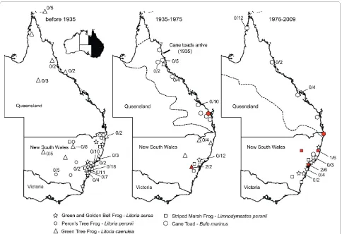
A catalogue of the Australian Museum, Sydney was que-ried for five frog species; the green and golden bell frog (Litoria aurea), the green tree frog (Litoria caerulea), the Peron's tree frog (Litoria peronii), the striped marsh frog (Limnodynastes peronii), and the introduced cane toad (Bufo marinus, syn. Rhinella marina). All specimens prior to 1935, for the green and golden bell frog (n = 27), the green tree frog (n = 48), the Peron's tree frog (n = 16) and the striped marsh frog (n = 24) representing Austra-lian endemic species were retrieved (Additional file 1). No myxospores were found in the total of 115 voucher specimens collected before 1935. The museum speci-mens were from eastern Australia, mostly from New South Wales (Figure 1).
Assuming the test sensitivity and specificity to be per-fect and the expected minimum prevalence to be 5%, the
probability of observing zero positives in a sample of 115 frogs from before 1935 is 0.003. These results are suffi-cient to conclude that the population was free from this disease (at the expected minimum prevalence of 5%) at 99.7% confidence level. Assuming only 90% test sensitiv-ity we have exceeded the 66 animals as the minimum number (p = 0.05) to be taken from a population with a negative result to consider the population as free of the disease (expecting minimum prevalence of 5%). Using 115 animals and test sensitivity of 90%, it is adequate to conclude, that the population is free from disease at the 99.5% confidence level.
No myxospores were found in a selection of museum specimens of the green and golden bell frog from New Caledonia (n = 29). The green and golden bell frog was introduced to New Caledonia in 19th century [14].
The earliest specimen positive for gallbladder myxospores is from 1966
In total, 112 voucher specimens and their catalogue records were recovered for frogs after 1935 (Figure 1, Additional file 1); the green and golden bell frog (n = 34), the green tree frog (n = 12), the striped marsh frog (n = 18) - representing Australian endemic species, and the introduced cane toad (n = 48). The earliest specimen pos-itive for myxospores spores was from 1966 (green tree frog, New South Wales, AM#99574). Myxospores spores were found in eight (7.1%, 8/112) Australian frog vouch-ers in the post cane toad introduction period (Figure 1, 2; Additional file 1). The myxospore positive native frogs, the green and golden bell frog (AM#148744, AM#153967), the green tree frog (AM#99754, AM#99575) and the striped marsh frog (AM#158418, AM#162439), were from New South Wales. Two cane toads from New South Wales and Queensland were posi-tive for myxospores (AM#158500, AM#59922), the earli-est is from 1967.
Ultrastructurally, myxospores resembled Myxidium
spores for which the number of ridges on the surface of
and Sprague (1940) [15], who have provided the following measurements ranges for myxospores found in toad gall-bladders: length and width, 11.8-14.2 × 7.5-10.0 μm, polar capsule length and width 3.5-4.5 × 3.3-4.2 μm, and recorded 7-9 transverse striations. Myxospores found in Australian frogs including the cane toad by Delvinquier (1986) [7] had the following characteristics: length and width 12.3-13.3 × 7.3-7.8 μm and 5-10 transverse stria-tions (polar capsule measurements were not reported).
Discussion
All vertebrates harbour a large diversity of parasites with often intricate life histories. For parasites whose life cycle comprises more than one specific host, the survival of such a parasite is dependent on sympatry and interaction with its host [16]. Removing a host from its natural envi-ronment where it has coevolved with its surroundings over a long historical trajectory may have major deleteri-ous consequences for its parasites. However, parasites that do not need an additional host and can be sustained easily in the environment will be more likely to travel
[image:3.595.57.541.94.426.2]with their hosts especially if successively translocated multiple times across a great geographical distance, as experienced in the case of the cane toad. The cane toad was introduced around the world from its native range of Central and South America through the Caribbean into Queensland, Australia in 1935 [8]. The 101 translocated cane toads were bred and their progeny released as a bio-logical control agent for a sugar cane industry suffering from beetle damage [8]. A single host lungworm parasite with a direct life-cycle (Rhabdias pseudosphaerocephala) is endemic to the cane toad in Central and South Ameri-cas and it has been translocated with the cane toad into the Australia but has not infected Australian endemic frog species [17]. Translocation of a multi-host parasite with the cane toad has yet to be unambiguously demon-strated. Host specificity is a fundamental property of all parasites, and parasite faunas of invasive species consist of a mixture of authentic parasites translocated from its original range and those newly encountered parasites that had broad enough specificity to take the advantage of the invasive host [18].
Almost all we know about myxosporean parasites comes from their investigation in fish. Out of more than 2200 species, about 2000 are fish Myxosporea, of which we know the life-cycle of only 33. The parasite alternates between vertebrate and invertebrate hosts; asexual devel-opment occurs in vertebrate hosts leading to the produc-tion of resistant myxospores, and sexually in the invertebrate host leading to the production of fragile aquatic actinospores [6,13]. The introduction of a myxo-sporean parasite into Australian frogs from the cane toad has been assumed based on morphological evidence from extant frogs and the absence of written records of such gallbladder parasite in scientific records [7]. However, the two-host life cycle of Myxosporea and the unknown spec-ificity for either vertebrate or invertebrate hosts has prompted us to re-examine this hypothesis using museum material. Indeed, the absence of Myxosporea in the gallbladders in frogs collected and catalogued prior to
the introduction of the cane toad supports a recent emer-gence of the parasite. It is further supported by the absence of Myxosporea in the green and golden bell frog gallbladders from New Caledonia, where it is the only frog introduced in late 19th century. However, does this
ultimately victimise the cane toad? First, we do not know the life cycle of the parasite, or if it requires an inverte-brate host. The majority (possibly all) of myxosporean species, including the pathogenic Myxobolus cerebralis
requires its aquatic oligochaete host being infected by myxospores produced in salmonid fish. The success behind the global distribution of Myxobolus cerebralis is in the cosmopolitan distribution of its dominant inverte-brate host Tubifex tubifex and its transcontinental trade [6]. However, direct transmission, fish-to-fish, was docu-mented for several Enteromyxum species [19]. We know nothing about the life cycle in frogs, but it is likely that
Myxidium immersum uses an invertebrate host in its life-cycle, and therefore international trade may also be impli-cated in Myxidium immersum distribution across continents similar to the spread of the chytrid fungus [4].
Myxidium immersum is described as a species with an extremely broad host specificity [7,15]; however, what might seem like a single morphospecies may mask a cryp-tic species complex for which molecular methods may resolve this difficulty [20]. We are currently collecting material to genetically characterise extant isolates from Australian frogs (AH, DNP, JŠ unpublished data).
Conclusion
While the cane toad may appear as a prime suspect, the deleterious effects of successive cane toad translocations seems unfavourable to a parasite with a two host life cycle such as Myxidium immersum due to its complexity. The low prevalence (7%) in the museum material suggests that only 7 animals would be Myxidium-positive when first translocated into Australia, because the founding cane toad population was only 101 individuals [8]. If, indeed these were the founding animals for the dispersal of the parasite, than Myxidium immersum is one of the luckiest parasites in the world because it is found flourishing in > 20 endemic and exotic frog species and had even reached a distribution beyond the current cane toad range. The origin of the parasite needs to be considered further with regards to its status in the source population, an intro-duction with other frogs brought to Australia for labora-tory purposes and accidental introduction via infected invertebrate hosts.
Methods
Voucher museum material
[image:4.595.57.290.95.425.2]A catalogue of the Australian Museum, Sydney was que-ried for the green and golden bell frog (Litoria aurea), the green tree frog (Litoria caerulea), the Peron's tree frog
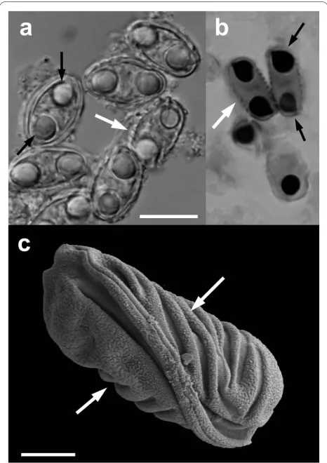
Figure 2 Myxidium cf. immersum spores recovered from the Aus-tralian Museum. Light microscopy (A), histological section through the gallbladder stained with Giemsa (B) and scanning electron micros-copy on black background. White arrows show the surface ridges and black arrows show two polar capsules per myxospore. Scale bar 10 μm (A, B), 2.5 μm (C); AM#158500 (A, C; Bufo marinus) and AM#162439 (B;
(Litoria peronii), the striped marsh frog (Limnodynastes peronii), and the introduced cane toad (Bufo marinus, syn. Rhinella marina). Vouchers were originally fixed in ethanol or formalin for an unknown period of time prior to transfer into 70% ethanol. In total, 256 voucher speci-mens and their catalogue records (denoted by AM#) were recovered, including all specimens prior to 1935 (Addi-tional file 1). A selection of museum specimens of the green and golden bell frog from New Caledonia was included (n = 29).
Extraction of frog gallbladders
A small incision (0.5-1 cm) on the belly of the frog voucher was used to reach the gallbladder and preserve the integrity of the specimen. For sufficiently large speci-mens the content of the gallbladder was aspirated with a disposable G22-needle. For smaller specimens the entire gallbladder was removed. The individual gallbladder con-tents and the gallbladder were stored in 70% ethanol until examination or processing.
Examination of gallbladders for myxospores
For a wet mount examination the entire gallbladder con-tents were examined using 20 × and 40 × objectives. Myxospores were measured/photographed using a 100 × objective BX51 microscope equipped with a DP70 cam-era (Olympus Australia); a minimum of 10 myxospores from each individual was measured and morphological details evaluated. Myxospores were prepared for scan-ning electron microscopy; fixed in 2% OsO4 in 0.2 M sodium cacodylate buffer (pH 7.0), rinsed in distilled water, dehydrated with a graded acetone series, critical point dried, coated with gold and examined using a Zeiss ULTRA plus FE. Individual gallbladders were selected for paraffin embedding, sectioning, Giemsa staining and examined for the presence of myxospores.
Statistical analysis
The probability of observing no positive samples in the total number of samples collected was calculated using FreeCalc v2, an epidemiological probability calculator assisting with the planning and analysis of surveys to demonstrate freedom from disease [21]; http://www.aus-vet.com.au/content.php?page=software - distributed by AusVet Animal Health Services.
We assumed 100% specificity of our detection of myxo-spores, because it relied on microscopic of the whole gall-bladder content coupled with histology and Giemsa staining. We calculated that 57, 29 and 14 animals (p = 0.05) is the minimum number of animals to be taken from a population with negative result to consider the popula-tion is free from disease using 5%, 10% and 20% minimum expected prevalence, respectively. We also evaluated a conservative 90% test sensitivity yielding 66, 32 and 15
animals (p = 0.05) as the minimum number be taken from a population with negative result to consider the popula-tion is free of the disease expecting minimum prevalence of 5%, 10% and 20%, respectively.
The minimum expected prevalence is the lowest level of prevalence that we aimed to detect. Delvinquier (1986) detected the parasite in 50% (6/12) specimens of the striped marsh frog (Limnodynastes peronii), 46% (5/11) specimens of the green tree frog (Litoria caerulea), 42% (8/19) specimens of the Peron's tree frog (Litoria peronii), and 13/34 (38%) specimens of the cane toad (Bufo mari-nus); the green an golden bell frog (Litoria aurea) was not surveyed [7]. Despite these numbers we restricted our-selves to a conservative 5% minimum prevalence. In other words, the minimum expected prevalence means that if parasite were to be present below 5% level, our survey would not be able to detect it. This minimum expected prevalence was considered sufficient, because (i) Delvin-quier (1986) [7] has detected much higher prevalence in Australian frogs and (ii), we calculated 13% (7/60) preva-lence using our survey of museum frogs from 1975 onwards.
Additional material
Competing interests
The authors declare that they have no competing interests.
Authors' contributions
All authors contributed to this study. JŠ and AH designed the study. AH per-formed the experiments and collected data. AH and JŠ analysed the data. JŠ, AH and DNP drafted the manuscript. All authors read and approved the final manuscript.
Acknowledgements
This project was financially supported by the Australian Academy of Science, ARC/NH&MRC Network for Parasitology and Dr William Richards Award in Vet-erinary Pathology (University of Sydney). We thank Iva Dyková, Ivan Fiala, Karrie Rose, Navneet Dhand, Michael Tyler and Glenn Shea for their insight, Ross Sad-lier and Cecilie Beatson for access into the Australian Museum, technical sup-port from the Electron Microscopy Unit (Australian Key Centre for Microscopy and Microanalysis).
Author Details
Faculty of Veterinary Science, University of Sydney, New South Wales 2006, Australia
References
1. Tingley MW, Beissinger SR: Detecting range shifts from historical species occurrences: new perspectives on old data. Trends Ecol Evol 2009, 24:625-633.
2. Suárez AV, Tsutsui ND: The value of museum collections for research and society. BioScience 2004, 54:66-74.
3. Johnson PTJ, Lunde KB, Zelmer DA, Werner JK: Limb deformities as an emerging parasitic disease in amphibians: Evidence from museum specimens and resurvey data. Conserv Biol 2003, 17:1724-1737. Additional file 1 Summary of the Australian Museum frog specimens and presence of Myxidium cf. immersum.
Received: 25 May 2010 Accepted: 10 June 2010 Published: 10 June 2010
This article is available from: http://www.parasitesandvectors.com/content/3/1/50 © 2010 Hartigan et al; licensee BioMed Central Ltd.
4. Weldon C, du Preez LH, Hyatt AD, Muller R, Speare R: Origin of the amphibian chytrid fungus. Emerg Infect Dis 2004, 10:2100-2105. 5. Whipps CM, El-Matbouli M, Hedrick RP, Blazer V, Kent ML: Myxobolus
cerebralis internal transcribed spacer 1 (ITS-1) sequences support recent spread of the parasite to North America and within Europe. Dis
Aq Org 2004, 60:105-108.
6. Hedrick RP, Adkison MA, El-Matbouli M, MacConnell E: Whirling disease: re-emergence among wild trout. Immunol Rev 1998, 166:365-376. 7. Delvinquier BLJ: Myxidium immersum (Protozoa: Myxosporea) of the
cane toad, Bufo marinus, in Australian Anura, with a synopsis of the genus in amphibians. Aust J Zoo 1986, 34:843-853.
8. Easteal S: The history of introductions of Bufo marinus (Amphibia: Anura) - a natural experiment in evolution. Biol J Linn Soc 1981, 16:93-113.
9. Hill BD, Green PE, Lucke HA: Hepatitis in the green tree frog (Litoria caerulea) associated with infection by a species of Myxidium. Aust Vet J
1997, 75:910-911.
10. Green DE, Gray MJ, Miller DL: Disease monitoring and biosecurity. In
Amphibian Ecology and Conservation: A Handbook of Techniques Edited by:
Dodd CK. Oxford: Oxford Press; 2009:481-505.
11. Sitjà-Bobadilla A: Can Myxosporean parasites compromise fish and amphibian reproduction? Proc R Soc B 2009, 276:2861-2870.
12. Eiras JC: An overview on the myxosporean parasites in amphibians and reptiles. Acta Parasitol 2005, 50:267-275.
13. Lom J, Dyková I: Myxozoan genera: definition and notes on taxonomy, life cycle terminology and pathogenic species. Folia Parasitol (Praha)
2006, 53:1-32.
14. Bauer AM: The Herpetofauna of New Caledonia Ithaca, NY: Society for the Study of Amphibians and Reptiles; 2000.
15. Kudo R, Sprague V: On Myxidium immersum (Lutz) and M. serotinum n. sp., two myxosporidian parasites of Salientia of South and North America. Rev Med Trop Parasitol Bacteriol Clin Lab 1940, 6:65-73. 16. Bush AO, Fernandez JC, Esch GW, Seed JR: Parasitism: The diversity and
ecology of animal parasites Cambridge: Cambridge University Press; 2001.
17. Dubey S, Shine R: Origin of the parasite of an invading species, the Australian cane toad (Bufo marinus): are lungworms Australian or American? Mol Ecol 2008, 17:4418-4424.
18. Torchin ME, Laffrety KD, Dobson AP, McKenzie VJ, Kurtis AM: Introduced species and their missing parasites. Nature 2003, 421:628-630. 19. Diamant A: Fish-to-fish transmission of a marine myxosporean. Dis Aq
Org 1997, 30:99-105.
20. Poulin R, Keeney DB: Host specificity under molecular and experimental scrutiny. Trends Parasitol 2007, 24:24-28.
21. Cameron AR, Baldock FC: A new probability formula for surveys to substantiate freedom from disease. Prev Vet Med 1998, 34:1-17.
doi: 10.1186/1756-3305-3-50
Cite this article as: Hartigan et al., Museum material reveals a frog parasite
emergence after the invasion of the cane toad in Australia Parasites & Vectors