REVIEW
Beyond the replication-competent
HIV reservoir: transcription
and translation-competent reservoirs
Amy E. Baxter
1,2, Una O’Doherty
3*and Daniel E. Kaufmann
1,2*Abstract
Recent years have seen a substantial increase in the number of tools available to monitor and study HIV reservoirs. Here, we discuss recent technological advances that enable an understanding of reservoir dynamics beyond classical assays to measure the frequency of cells containing provirus able to propagate a spreading infection (replication-competent reservoir). Specifically, we focus on the characterization of cellular reservoirs containing proviruses able to transcribe viral mRNAs (so called transcription-competent) and translate viral proteins (translation-competent). We suggest that the study of these alternative reservoirs provides complementary information to classical approaches, crucially at a single-cell level. This enables an in-depth characterization of the cellular reservoir, both following reactivation from latency and, importantly, directly ex vivo at baseline. Furthermore, we propose that the study of cellular reservoirs that may not contain fully replication-competent virus, but are able to produce HIV mRNAs and proteins, is of biological importance. Lastly, we detail some of the key contributions that the study of these transcrip-tion and translatranscrip-tion-competent reservoirs has made thus far to investigatranscrip-tions into HIV persistence, and outline where these approaches may take the field next.
Keywords: HIV reservoirs, CD4 T cells, Flow cytometry, RNA flow cytometry, Fluorescence in situ hybridization
© The Author(s) 2018. This article is distributed under the terms of the Creative Commons Attribution 4.0 International License (http://creativecommons.org/licenses/by/4.0/), which permits unrestricted use, distribution, and reproduction in any medium, provided you give appropriate credit to the original author(s) and the source, provide a link to the Creative Commons license, and indicate if changes were made. The Creative Commons Public Domain Dedication waiver (http://creativecommons.org/ publicdomain/zero/1.0/) applies to the data made available in this article, unless otherwise stated.
Background
Despite over 30 years of research and the tremendous successes of combined anti-retroviral therapy (ART), HIV remains a chronic disease for which there is no cure. In individuals receiving ART, the amount of cir-culating virus in the plasma is brought down to unde-tectable levels, as measured by current standard clinical assays. However, the virus is able to persist in the form of integrated proviruses in a predominantly CD4 T cell reservoir and will rebound from this cellular reservoir if therapy is discontinued [1–5]. Therefore, a key challenge for the field is how to identify cellular reservoirs of HIV [6], and crucially, how to measure the impact of potential
cure strategies on the replication-competent reservoir [7] as well as defective proviruses capable of expressing HIV proteins [8, 9].
Multiple techniques have been proposed, developed, and successfully utilized to identify the reservoir. Many of these techniques will be discussed in detail elsewhere in this series. Broadly, the majority of approaches focus on either the very early (DNA), or the very late (infec-tious virus) products of the viral life cycle. This focus has many advantages, but there are key limitations to be considered. For example, common PCR based tech-niques including the measure of total and integrated HIV DNA [2, 10] vastly overestimate the size of the reservoir due to the high prevalence of integrated, but “defective” proviruses [9, 11, 12]. On the other end of the scale, the Quantitative Viral Outgrowth Assay (Q-VOA), [4, 5, 13] and variants [14–16] may underestimate the size of the reservoir, as not all replication-competent proviruses are inducible with one round of stimulation [11] or able to
Open Access
*Correspondence: unao@pennmedicine.upenn.edu; daniel.kaufmann@ umontreal.ca
1 CR-CHUM, Université de Montréal, Montréal, QC, Canada 3 Department of Pathology and Laboratory Medicine, Division
of Transfusion Medicine and Therapeutic Pathology, University of Pennsylvania, Philadelphia, PA, USA
propagate in the in vitro conditions required for detec-tion. Crucially, such approaches provide population-level, rather than single-cell population-level, information allowing only a quantification of the relative size of the reservoir, rather than in-depth reservoir characterization.
With these challenges in mind, we and others have sought a different way of characterizing and understand-ing HIV persistence (see Fig. 1). For example, while the maintenance of intact, replication-competent viruses is clearly a major barrier to HIV eradication, can transcrip-tion or translatranscrip-tion-competent proviruses contribute to HIV pathogenesis on ART, and provide key insights into HIV persistence? We suggest that proviruses that may not be fully replication-competent, but that are capable of transcribing viral mRNAs and translating viral proteins, provide an additional dimension to persistence studies; and that the elimination of such proviruses should be considered in the context of a cure. Furthermore, we pro-pose that the in-depth analysis of the cellular HIV reser-voir at baseline, i.e. those cells containing proviruses that spontaneously produce viral products in ART-treated
individuals in the absence of stimulation or reactivation, enables a deeper understanding and informative quantifi-cation of the response to latency reversing agents (LRAs) in the context of “shock/kick and kill” [17] and alternative cure strategies [18–20]. Here, we detail the initial stud-ies of the transcription and translation-competent reser-voirs, which have recently overcome issues of specificity and sensitivity, to begin to address these questions.
The approaches we describe uniquely investigate HIV reservoirs at the single-cell level; termed here cellular HIV reservoirs. The use of the word “cellular” distin-guishes these measures from the more prevalent popu-lation-level analyses utilized in the field. Popupopu-lation-level analysis provide crucial insight into the size and nature of the reservoir; however we and others have demonstrated that studying the reservoir at a single-cell level can pro-vide an additional critical understanding of the heteroge-neity of the reservoir.
Lastly, we have avoided the term “latent” when describ-ing these cellular HIV reservoirs since this phrase is com-monly used to describe cells containing a provirus that
Silent reservoir
Transcription-competent
reservoir
Translation-competent
reservoir
Replication-competent
reservoir
Proviral DNA
viral mRNA
viral proteins
infectious viral particles
HIV protein
HIV mRNA
Translaon competent cellular reservoirs
Transcripon competent cellular reservoirs
Single cell analysis based on viral product detecon
HIV protein
CD4
Translaon competent cellular reservoirs
Single cell analysis based on viral products and host markers
a b
c
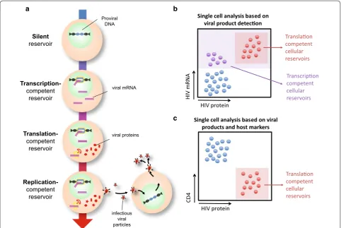
Fig. 1 Defining and identifying HIV reservoirs. a Schematic detailing the naming conventions used to identify different aspects of the HIV reservoir.
[image:2.595.57.540.363.686.2]is transcriptionally silent. However, we and others have shown that a rare subset of HIV-infected cells in indi-viduals on long-term ART can express HIV mRNA and proteins in the absence of a spreading infection. By this definition, these cells are not latent at the time of detec-tion, but, as has been suggested, might cycle back to a latent state and thus contribute to the latent HIV reser-voir [21, 22].
Summary of HIV transcription and translation
The transcription and translation of the HIV genome has been studied in detail in vitro (reviewed in [23]). Briefly, the first fully spliced transcripts encode the HIV acces-sory proteins Tat and Rev [23, 24]. Tat is an essential reg-ulatory protein for viral replication, which binds the HIV TAR (Trans-Acting Response element) RNA, inducing transcription [23]. In concert, Rev promotes HIV RNA nuclear export by binding the Rev Responsive Element (RRE) present in partially spliced and unspliced RNA [23]. Thus, as Tat and Rev protein levels increase, par-tially spliced RNAs are exported. In this manner, other accessory proteins, in addition to HIV Envelope (Env), are made. Lastly, unspliced mRNA forms are exported to the cytoplasm such that Gag and Pol are also translated, and viral particles are produced.
In addition, there are multiple levels of post-tran-scriptional regulation that can impact expression of viral mRNAs and proteins. These include mRNA splic-ing, RNA processing by microRNAs and nuclear export, as well as control at the translation level [23, 25]. In the context of HIV latency, these points of regulation remain underexplored [21, 26]. However, such post-transcrip-tional regulation should be taken into consideration when measuring HIV reservoirs based on detection of transcription or translation products. For example, a cell that is able to transcribe HIV mRNAs may not be able to translate HIV proteins, due to control at the post-tran-scriptional level [27].
While many studies have probed the control of HIV expression in T cell lines and activated T cells, little is known about the control of HIV expression in more qui-escent or resting primary T cells. It is clear that activated T cells are much more effective at producing infectious virus than quiescent cells, producing 100-fold more HIV Gag RNA per provirus [28]. Whether HIV gene regula-tion has unique differences between resting and activated cells requires more investigation both in vitro and in vivo; primary models suggest that while splice products form in resting cells, the levels of fully and partially spliced mRNAs are ~ 100-fold lower than in activated cells [28]. Thus, further work building on the lessons learned from the study of in vitro latency models is required to deter-mine how HIV expression is controlled in vivo [29].
Measuring transcription‑competent cellular reservoirs
Relatively early in the epidemic, prior to the discovery and widespread implementation of potent ART regi-mens, multiple groups reported the detection of HIV RNA species within CD4 T cells from chronically HIV-infected individuals using PCR-based approaches [30, 31]. The advent of potent ART-induced viral suppres-sion saw the detection of such cell-associated (CA)-RNA applied to the latent HIV reservoir. In the late 2000s, Fis-cher and colleagues provided a key insight into the signif-icance of this transcription-competent reservoir (Fig. 1a) by monitoring multiple forms of RNA within cells, and measuring the frequency of RNA-expressing cells at lim-iting dilution in HIV-infected individuals as they began therapy. They observed that HIV CA-RNA measures decayed drastically when compared to HIV DNA meas-ures within the same individual [32], and suggested that ~ 5% of cells containing HIV DNA also expressed HIV RNA in individuals on ART [33]. Importantly, more recent work using a nested PCR approach confirmed that the HIV mRNAs detected predominantly resulted from genuine HIV mRNA transcription, rather than chimeric read-through products transcribed from host promot-ers [34]. This work clearly demonstrated the relevance of cellular RNA-based measures for investigations in cure strategies, and is discussed in depth elsewhere in this series [35]. As with measures of HIV DNA, most classical CA-RNA measures are based upon modified versions of real-time PCR for various HIV mRNA species [36]. Cru-cially, therefore, this approach provides population-level information, allowing a quantification of the relative size of the reservoir in HIV-infected individuals, but does not enable an in-depth analysis of the cellular nature of the reservoir. With this in mind, we and others have applied various approaches to detect single cells containing a provirus able to produce HIV RNA species; termed the transcription-competent cellular reservoir.
HIV [39, 40]. While powerful, microscopy-based ISH is limited by its relatively low throughput. In the context of chronic, untreated HIV infection, the prevalence of HIV-infected cells is sufficient to enable detection, but still requires laborious analysis of many sections to obtain robust quantitation. However the frequency of such cells is dramatically reduced in ART-treated individuals. Thus, additional, complementary high-throughput techniques were required to investigate very high number of cells to identify these rare events and characterize the cellular reservoir that persisted in individuals on ART.
The late 1990s saw the advent of a new era in immunol-ogy; that of multiparametric flow cytometry. This high-throughput approach was soon applied to the study of cellular HIV sanctuaries in HIV-infected, untreated indi-viduals. Patterson and colleagues pioneered an approach based on reverse-transcriptase (RT)-PCR-based amplifi-cation and Fluoresence ISH (FISH) detection of intracel-lular HIV RNA [41], and later a probe-based approach termed SUSHI (simultaneous ultrasensitive subpopula-tion staining/hybridizasubpopula-tion in situ, [42–44]). While these approaches provided a key proof of concept for the field, as the authors note, the frequencies of HIV mRNA+ cells detected with these assays are generally higher than would be predicted based on measurements of integrated HIV DNA [41]. This indicates a potential issue with false positive detection that may hamper interpretation of this data.
Building on this pioneering initial work, in recent years a new version of these ISH technologies sought to over-come the issues of high background/nonspecific stain-ing and low signal-to-noise ratios, which limited earlier iterations. In 2012 Wang et al. [45] detailed a microscopy technique known as RNAscope. This approach builds on a branched DNA (bDNA) technique described previously [46], but added additional levels of stringency to reduce off-target binding. Briefly, a series of DNA probes are designed whereby each probe has two sections; the first recognizes the target mRNA and the second forms part of a conserved “tail” sequence. The probes are designed such that pairs of probes which recognize adjacent regions of the target mRNA each contain one half of this conserved tail. Only this combined “tail” sequence can be recognized by a DNA pre-amplifier, which in turn is recognized by a secondary amplifier. This amplified structure is then labeled with a fluorescent probe, or an alkaline phosphatase or horseradish peroxidase (HRP) molecule. The requirement for the two probes (known as a “Z”) to bind adjacent to one another in order for the pre-amplifier to bind substantially reduces off-target binding.
Those in the HIV cure field quickly recognized the significance of this approach. The application of this
technique to microscopy has been advanced in particu-lar by the Estes laboratory, who have demonstrated the increased sensitivity and high specificity of this assay when compared to alternative ISH approaches (see Table 1 [47, 48]). The low background is particularly strik-ing; the team imaged nearly 70 mm2 of uninfected tissue from rhesus macaques and identified only two false-posi-tive RNA+ cells [47]. Recently, this group has successfully applied this technology to quantify transcription-compe-tent cellular SIV reservoirs across a broad range of tissues in both untreated and ART-treated animals, confirming the predominance of lymphoid tissues as a key reservoir [49]. While HIV RNA+ cells were identified in untreated subjects, further work is required to determine if such cells can be readily identified in ART-treated individuals.
In parallel, this approach was applied to flow cytom-etry, and developed by our group and others in col-laboration with the company Affymetrix (now part of ThermoFisher) into a commercial RNAflow assay known as PrimeFlowTM. It was quickly utilized for the high-throughput, high-sensitivity detection of cellu-lar mRNAs [50]. Thus far, three groups have reportedly applied this RNAflow technology to the flow-cytometric study of transcription-competent HIV reservoirs (Fig. 1b, Table 1), with variations in terms of the specificity of the assay and therefore applicability of the approach to stud-ying samples directly from HIV-infected, and particularly ART-treated, individuals [51]. While Altfeld and col-leagues successfully applied the technique to the detec-tion of in vitro HIV-infected cells and cell lines, they reported that the sensitivity of this iteration was unlikely to be sufficient to detect HIV mRNA-expressing cells directly in HIV-infected subjects [52]. Similarly, we noted that the GagPol probes used in this study showed rela-tively high background (in the range of ~ 1000 GagPol mRNA false-positive events per million CD4 T cells in HIV-uninfected donors) precluding the detection of the transcription-competent reservoir in our hands [53, 54].
More recently, however, Grau-Expósito et al. [55] reported a high-sensitivity version of the RNAflow assay which used 50 probes sets designed against the
GagPol region of the conserved HXB2 genome. While
Table 1 C omparison of single ‑c ell appr oaches t o measur
e the tr
anscription ‑ and tr ansla tion ‑c ompet en t r eser voirs C ellular r eser voir meas ‑ ur ed A ssa y A ssa y o ver view A dv an tages Limita tions Pot en tial applica tions K ey r ef er enc es Transcr iption-compet ent cellular r eser voir RNA flo w c yt ometr y D et ec
tion of cells expr
ess
-ing HIV RNA in suspension by fluor
escence in
situ
hybr
idisation (FISH) using
branched DNA (bDNA) technology
H
igh thr
oughput
In depth phenot
yping of
single cells
H
ighly flexible and adapt
-able Back gr ound obser ved in HIV -uninf ec ted individuals Labour int ensiv e (2–3 da ys pr ot ocol) H igh star
ting cell number
requir ed LRA scr eening Reser voir quantification
In depth phenot
yping of the r eser voir Biomar ker disco ver y [ 52 , 55 ] Simultaneous ultrasensitiv e subpopulation stain -ing/h ybr idization in situ (SUSHI) D et ec
tion of cells expr
ess
-ing HIV RNA in suspension by fluor
escence in situ hybr idisation (FISH) H igh thr oughput H
igher than pr
edic
ted fr
e-quencies of
mRNA
+ cells
obser
ved
Reser
voir quantification
Phenot
yping of single cells
, including m yeloid cells [ 41 – 44 ] Con ventional in situ h ybr idi -zation D et ec
tion of cells expr
ess
-ing HIV RNA in
situ using
radiolabelled or enz
ymatic det ec tion Tissue le vel inf or mation Limit
ed cell phenot
yping
Labour int
ensiv
e
Reser
voir quantification in
tissues [ 37 , 38 , 47 , 48 ] RNAS cope D et ec
tion of cells expr
ess
-ing HIV RNA in
situ using
branched DNA amplifica
-tion and det
ec tion Tissue le vel inf or mation H ighly sensitiv
e and specific
Shor
t assa
y duration
Limit
ed cell phenot
yping
Reser
voir quantification in
tissues/whole body [ 47 – 49 ] Translation-compet ent cel -lular r eser voir
Fiber optic ar
ra y scanning technology (F AST ) Antibody-based det ec tion
of cells expr
essing HIV pr
o-tein and do
wn-r
egulating
CD4, in suspension
Relativ
ely high thr
oughput Shor t assa y duration Specializ ed micr oscop y
tools and sof
twar
e
requir
ed
Limit
ed cell phenot
yping LRA scr eening Reser voir quantification [ 62 ] RNA flo w c yt ometr y Concur rent det ec tion of cells expr
essing HIV RNA
by FISH using branched DNA (bDNA) t
echnology
,
and HIV pr
ot
ein in suspen
-sion
H
igh linear
ity and specificit
y
H
igh thr
oughput
In depth phenot
yping of
single cells
H
ighly flexible and adapt
-able Labour int ensiv e (2–3 da ys pr ot ocol) H igh star
ting cell number
requir ed LRA scr eening Reser voir quantification
In depth phenot
[image:5.595.149.454.91.722.2]this “false-positive” population will still effectively con-taminate the true positive HIV-infected population. This contamination therefore precludes an in-depth pheno-typing analysis of these rare HIV mRNA+ cells, particu-larly in samples from ART-treated individuals where the frequencies of mRNA+ cells is close to the limit of detection.
Thus, while this assay shows great promise, the appli-cability for the detection of transcription-competent cel-lular reservoirs in samples from treated patients remains unclear. Previous studies using highly-sensitive, limiting dilution RT-PCR demonstrated that low levels of HIV gag mRNA could be detected in a subset, only ~ 5%, of HIV DNA-containing cells in subjects on ART [33]. Using a dilution assay, Grau-Expósito et al. demonstrated that the detection of mRNA+ cells was linear down to the lowest dilution tested (50 events per million cells). Accordingly, in samples from untreated HIV-infected individuals, the median frequency of mRNA+ events detected was above this threshold at ~ 165 per million CD4 T cells. However, unsurprisingly, these events were much rarer in samples from ART-treated individuals (~ 6–20 per million CD4 T cells in the absence of stimulation [55]). Therefore further validation may be required to ensure that this approach is linear down to the ranges required for the robust evalua-tion of cure therapies.
A further key consideration of such flow-cytometric mRNA-based detection assays is the sensitivity of these approaches in terms of the number of mRNA copies that a cell must express to be detected. To address this question, Baxter et al. performed a confocal microscopy analysis of CD4 T cells from a HIV-negative individual, processed with the HIVRNA/Gag assay. They observed a mean of ~ 7 false-positive GagPol mRNA spots per cell; providing a conservative detection limit of ~ 20 GagPol mRNA copies per cell (+3 standard deviations, [53]). This limit enabled identification of ~ 94% of GagPol mRNA+ cells from a infected individual. Therefore, an HIV-infected cell containing at least 20 copies of HIV mRNA is highly likely to be truly infected (0.15% false positive discovery rate for a Gaussian distribution); however an infected cell with fewer copies of HIV RNA is more likely to be missed. Crucially, the number of spots per cell was closely associated with the total fluorescence intensity of the cell, suggesting this approach enables a relative quan-tification of mRNA copy number [53].
Importantly, however, this analysis makes the assump-tion that each “spot” represents one mRNA copy, which may not be accurate. Furthermore, the number of copies required for detection varies according to the number of probe set pairs that bind to each mRNA; thus the selec-tion of probe sets and the heterogeneity of the target mRNA are key variables [54]. In a hypothetical example,
consider two samples. In the first sample, the probe sets and the viral mRNA sequence match perfectly, therefore if 50 probe sets are available, 50 probe sets will bind. In the second sample, there is a high degree of sequence mismatches with the original sequence used to design the probes; although 50 probe sets are available, only ten are able to bind the target mRNA. Therefore, for a cell in the second sample to reach the same total fluorescence intensity as a cell in the first sample, five-times as many mRNA copies may be required. While this is an oversim-plification, it demonstrates a key point that these assays may “miss” true HIV-infected cells due to sequence het-erogeneity. One potential solution is to design individual probes for each patient after sequencing the patient’s virus, but this may be prohibitively costly. Given this point, and those raised above, work in our laboratory and others is ongoing to increase both the specificity and the sensitivity, and therefore the applicability, of these RNA-flow assays to the detection of the transcription-compe-tent cellular reservoirs.
Measuring translation‑competent cellular reservoirs
expressed at very high levels in HIV-infected cells and each virion incorporates ~ 5000 Gag particles [60]. HIV Gag was detected in a small fraction of resting T cells after direct infection in vitro (Gag+, [28]), however this represented only a minority of the cells containing inte-grated HIV DNA. Whether these Gag+ cells were an in vitro artifact or had a counterpart in vivo was unclear until recently [53, 55, 61]. The first evidence that HIV Gag could be expressed in resting CD4+ T cells in vivo came from the sorting of resting HIV Gag+ non CD4-lin-eage negative PBMCs from HIV-infected subjects. In the order of ~ 1 Gag+ cell per million PBMCs were detected from ART-treated individuals [61]. However, this tech-nique was labor intensive and fraught with false positives. While Gag+ cells were enriched for HIV DNA, only 10% of the sorted Gag+ cells contained HIV DNA. Thus, this approach provided key evidence that HIV protein expres-sion likely occurred in T cells in ART-treated subjects, but indicated that more sensitive methods were required.
Detection of HIV cellular reservoirs was further advanced by exploiting HIV’s ability to downregulate CD4 as a surrogate marker for cellular reservoirs (Fig. 1c) [53, 55, 62]. A well-known function of Nef, Env and Vpu is the downmodulation of CD4 in activated T cell infec-tion [63–68]. In vitro experiments showed that after direct infection of resting CD4+ T cells a subset of cells with integrated HIV DNA were Gag+ and negative for surface CD4, suggesting internalization and downregu-lation of CD4 [62]. Sorted Gag+CD4− cells contained HIV proviruses by Alu-gag PCR, proving the presence of Gag was not due to bound virions. Moreover, extensive phenotyping confirmed that these were genuine TCRαβ CD4 T cells with internalized CD4. Mutational analysis showed that Nef and Env, but not Vpu, were required for CD4 internalization, suggesting that if an HIV-infected cell downregulates CD4 it is likely that additional HIV open reading frames (including env, nef, tat, and rev) are intact and expressed. Thus, to express Gag and to down-regulate CD4, a large fraction of the 3′ and 5′ regions of the HIV genome must be intact.
These in vitro experiments suggested that an approach combining detection of Gag protein expression with CD4 downregulation could be used to identify trans-lation-competent cellular reservoirs. However, sorting strategies, while useful for proof of principle, proved impractical. Thus, the O’Doherty lab introduced a dif-ferent approach (Table 1, [62]). They exploited a rare cell detection technique used in cancer detection, FAST (Fiber-optic Array Scanning Technology [69–71]), to scan up to 20 million cells adhered to a slide, followed with Automated Digital Microscopy to confirm the cel-lular phenotype. Applying this rationale and technol-ogy enabled imaging of high numbers of PBMCs from
ART-treated patients, stained for intracellular CD4 and Gag protein. Indeed, they identified Gag+ cells at low frequencies (0.33–2.7 events per million PBMCs), many of which were CD4−, or showed punctate internalized CD4 staining. The absence of surface CD4 suggests that indeed, the majority of these cells contain a transla-tion-competent HIV provirus and are distinct from the false-positive Gag+ events observed in HIV-uninfected individuals [62]. The key strength of FAST combined with Automated Digital Microscopy is the lower false positive rate compared to classical Gag staining by flow cytometry. While FAST has the potential to be high throughput, the technique is still in early development, the confirmation of positive results by Automated Digi-tal Microscopy is time intensive and this technology is not widely available. Therefore, alternative methods to detect the translation-competent cellular reservoir were required.
Combining measures of transcription
and translation‑competent cellular reservoirs
bringing these two advances together enabled the detec-tion of 0.5–1 HIVRNA+/Gag+ events per million CD4 T cells.
Importantly, the high specificity and flow-cytometric basis of this approach enabled multi-parameter, in-depth phenotyping of the translation-competent cellular HIV reservoir that were not previously possible. For exam-ple, consistent with observations made by the O’Doherty laboratory [62], cells identified as HIVRNA+/Gag+ strongly downregulated CD4. Moreover, HIVRNA+/Gag+ cells were enriched in the circulating T follicular helper cell popu-lation [53] and cells expressing inhibitory receptors, consistent with previous reports [72–75]. These exam-ples demonstrate the importance of a low false-positive detection in measuring HIV cellular reservoirs.
Lastly, while Grau-Expósito et al. [55] focused on the transcription-competent cellular reservoir, they also identified a subset of mRNA-expressing cells which expressed viral Gag protein, and thus were also able to identify the translation-competent reservoir as a subpop-ulation of the transcription-competent cellular reservoir. An area of key further interest is to determine what fea-tures (viral or host) may distinguish these two different reservoirs.
Taken together, this work demonstrates that the detec-tion of multiple HIV viral products, or the downstream consequences of these products such as loss of CD4 expression, can overcome the issue of false positive events. Furthermore, we suggest that this multi-faceted approach increases the likelihood that a translation-com-petent cellular reservoir contains a replication-comtranslation-com-petent provirus. Nonetheless, careful controls for false positive signals are imperative and additional work is required to determine which fraction of the translation-competent cellular reservoir is truly replication-competent.
Why measure transcription
and translation‑competent cellular reservoirs?
Closing the gap between DNA quantitation and measures of replication‑competent virus
A crucial caveat in the measurement of transcription/ translation-competent cellular reservoirs is that not all cells detected by these assays may contain a virus able to initiate a spreading infection in vivo: a replication-competent provirus. However, we suggest that the detec-tion of cells containing proviruses able to produce viral mRNA and proteins is biologically and scientifically rel-evant. Secondly, we propose that the populations of HIV-infected cells detected by these approaches are likely to be highly enriched for replication-competent virus. Thus, measuring the translation-competent cellular reservoir after latency reversal may be an appropriate and informa-tive surrogate for detection of replication-competent
proviruses. Optimistically, such approaches may over-come the gap between the overestimation of the reser-voir size measured by DNA-centric techniques and the reported underestimation of the reservoir size by the Q-VOA.
To address this second point, both the Buzon and Kaufmann laboratories observed associations with their measures of the cellular reservoir and DNA-based measures, which commonly overestimate the size of the translation-competent reservoir [76]. Baxter et al. also observed a correlation between levels of integrated HIV DNA and the frequency of the translation-competent cellular reservoir in samples from ART-treated individu-als following in vitro stimulation with PMA/ionomycin. Interestingly though, DNA measures and the frequency of HIVRNA+/Gag+ cells were not associated at baseline. Importantly, the frequency of the cells detected as tran-scription/translation-competent cellular reservoirs is substantially lower than the number of copies of HIV DNA detected (~ 160-fold lower [55] and ~ 200-fold lower [53]). This difference suggests that measurement of the transcription/translation-competent cellular res-ervoirs identify a population that is substantially closer to the replication-competent reservoir than DNA measures.
Uncovering a unique aspect of the reservoir
A key rationale behind measuring the transcription- and translation-competent reservoirs is the additional level of detailed, complementary information that can be gained from the study of this form of the reservoir. As discussed above, many of the techniques used to identify the tran-scription- and/or translation-competent reservoirs pro-vide information at a single-cell level, as they are often flow cytometry or microscopy-based. This means that an individual cell can be probed for multiple parameters of interest in addition to HIV RNA/protein, such as cellular activation, exhaustion or memory markers [52, 53, 55, 62, 77]. In contrast, PCR-based techniques and the Q-VOA provide only population-level comparative information (i.e. population A contains a higher proportion of HIV DNA than population B). This is particularly important to consider in the context of the wide heterogeneity of the cellular reservoir; when assessing cure strategies it is of paramount importance to understand how all sub-populations of the cellular reservoir respond, rather than treating the reservoir as a homogenous entity. For exam-ple, while it has previously been reported that both the central, transitional and effector memory T cell popula-tions contain HIV DNA, there are conflicting reports regarding whether replication-competent virus is pre-dominantly localized in the central memory compart-ment [78], or the effector memory compartment [79]. CD4 T cells expressing exhaustion markers including PD-1, LAG-3 and TIGIT have been shown to be enriched for HIV DNA, but this enrichment is further dependent on the state of CD4 T cell differentiation [75]. Further-more, expression of multiple inhibitory receptors on CD4 T cells prior to ART has been identified as a predictive biomarker of viral rebound following treatment inter-ruption; this suggests that the expression of such mark-ers may also identify a subpopulation of latently infected cells with a higher proclivity to viral transcription [80]. From only these limited examples, it is apparent that analysis of bulk CD4 memory populations would pre-vent an understanding of these subtleties. While sorting individual CD4 T cell populations for downstream analy-sis is possible, this becomes less feasible when analyzing exceedingly rare CD4 T cell subpopulations, and quickly limited in terms of the number of populations that can be concurrently analyzed. As the approaches we have described for the analysis of the transcription- and trans-lation-competent cellular reservoirs, particularly those which are flow cytometry-based, overcome these limita-tions, these techniques will become increasingly useful for in-depth characterization of the HIV reservoir.
An additional strength of these techniques is the ability to compare in vitro models and validation experiments with in vivo-infected T cells. Spina et al. [81] previously
indicated the limitations of latency models to fully reca-pitulate latency reversal, however we suggest that the lessons learnt from in vitro models can advance in vivo research. For example, the in vitro observations of a rare Gag+ populations in resting CD4 T cells have been sup-ported by the in vivo detection of this population directly in samples from ART-treated individuals [61, 62]. Using the HIVRNA/Gag assay, the in vitro observation of a down-regulation of HLA-Class I on HIVRNA+/Gag+ cells was confirmed. In contrast, however, HLA-Class II-express-ing CD4 T cells were enriched for both HIV mRNAs and protein only in ex vivo samples [53]. Therefore, such approaches can be used to both investigate HIV biology in vivo, but also to build upon key observations made in in vitro models.
Quantifying the HIV reservoir at the single‑cell level in ART‑treated subjects
We further suggest that a highly useful aspect of this type of measurement is the ability to quantify the HIV reservoir in ART-treated individuals at the single-cell level (i.e. directly ex vivo in ART-treated subject sam-ples). Such measurements capture a distinct view of the reservoir; this represents the cells from ART-treated individuals that spontaneously reactivate the provirus to produce HIV mRNA, protein, and perhaps viral particles, in the absence of a spreading infection and/or exogenous stimulation [15, 49, 53]. We speculate that the cells con-taining transcription/translation-competent virus that are producing HIV mRNA and/or protein might revert to a latent state before dying from viral cytotoxicity or immune clearance [22]. Therefore, investigating these cells could provide insight into the single-cell phenotype of the latent reservoir. In addition, plasma sequences identified during viral rebound following treatment interruption match proviruses in cells that were already expressing HIV mRNA before ART was stopped. This indicates that clones of these proviruses likely contrib-uted to the rebound viremia [56]. Thus, defining those single cells that contain transcription/translation-com-petent viruses and produce viral products during ART may help identify the cell population from which viral rebound may occur.
suppressive ART [36, 82, 83]. Using these approaches, the reservoir is readily quantifiable. In contrast, the detection of alternatively spliced mRNAs by TILDA did not observe spliced mRNA production without in vitro stimulation in all samples studied [84]. Given these differences, we sug-gest that the quantification of this persistent reservoir at the single cell level could provide key insights. However, such studies have only been conducted in detail recently. Using single-cell RNAflow based approaches, HIV mRNA-expressing CD4 T cells were robustly identified in samples from 2 of 6 virally suppressed ART-treated individuals [55], while HIVRNA+/Gag+ CD4 T cells were detected in 8 samples, from a total of 14 [53]. Using the FAST approach, Gag protein+ cells were identified in all five of the subjects studied [61], including one individual who was repeatedly sampled over several years. In those samples where translation/transcription competent cel-lular reservoirs were detected, the frequencies ranged from ~ 10 mRNA+ to ~ 1.0 HIVRNA+/Gag+ events per million CD4 T cells. Given these frequencies, we postu-late that one of the major issues when monitoring this baseline cellular reservoir is the number of cells stud-ied. The lower the total number of cells analyzed in the assay, the lower the probability of detecting the very rare cells infected with HIV [54]. In studies performed by our laboratories we routinely assess two-four million CD4 T cells [53], or six-eighteen million PBMCs [62] to enable detection of these rare cells. The analysis of such a high number of cells is made possible only by the use of the high-throughput approaches, but nonetheless, detection of these rare cells remains challenging requiring signifi-cant expertise and is constrained by the size of available clinical samples. While such limitations must be consid-ered, studying the transcription/translation-competent reservoirs can provide additional information regarding the nature of the HIV reservoir at baseline as well as after stimulation.
Detailing a biologically relevant population
We suggest here that the transcription/translation-competent cellular reservoir may contribute to both the persistent reservoir and importantly the pathogenesis of HIV on ART, and are thus biologically relevant. If this is the case, these cells, not only cells containing replication-competent viruses, need to be considered in the context of HIV cure.
T cell exhaustion and ongoing immune activation are characteristic features of chronic infections [85], includ-ing HIV [86–89], and are driven in part by exposure to persistent antigen [90]. In the presence of suppressive ART, HIV antigen levels should be low, however, p24 and Env protein products can still be detected in the plasma of HIV-infected individuals under long-term (~ 10 years)
of suppressive therapy [9]. Furthermore, ultrasensitive techniques have detected very low level viremia in ART-treated individuals [91, 92]. Additionally, the ongoing production of HIV proteins from “defective” proviruses has been demonstrated [8, 59, 62]. Such observations have led to the term “zombie” proviruses, as while “defec-tive” proviruses may not be “alive”, they still may con-tribute to HIV pathogenesis on ART [59]. These points indicate that the translation-competent cellular reservoir may contribute to continued antigen presence, either through the production of replication-competent virus in the absence of spreading infection at baseline, or through viral protein production only. Crucially though, the pre-cise role of HIV antigen in the persistence of immune activation remains unclear, particularly as HIV anti-gens are very unlikely to be the only drivers of ongoing immune dysfunction; products from microbial transloca-tion [93, 94] and concurrent viral infections such as CMV and EBV are likely to contribute [95]. While further work is required to determine the significance of the transla-tion-competent reservoir in regards to T cell dysfunction, we suggest that the clearance of such translation-com-petent cellular reservoirs may need to be considered in addition to the removal of replication-competent virus in the context of a HIV cure.
In addition to contributing to immune activation, viral protein production, possibly from “defective” proviruses may explain the continued presence of antibodies against HIV [9] and indeed may shape the antibody repertoire. Furthermore, a recent study from the Ho/Siliciano lab suggested that cells expressing viral proteins, even from “defective” proviruses, can be recognized and killed by cytotoxic T lymphocytes (CTL) [58]. In support of this finding, other groups have also reported immune-based clearance of HIV-infected cells measured by loss of HIV/ SIV DNA, implying that some expression of defective proviruses must be occurring [96–99]. Accordingly, anti-HIV CTL activity in vitro is strongly correlated with viral DNA levels in vivo [61]. As with the antibody repertoire, it is likely that such interactions may also shape the CTL landscape.
Lessons from the study of transcription/
translation‑competent reservoirs: an evolving field
with both CD4/CD8 ratio and plasma viral load [53, 55]. In ART-treated individuals, the CD4 T cell count and CD4/CD8 ratio are important indicators of the immu-nological response to therapy. A poor reconstitution of the CD4 T cell compartment is associated with increased morbidity and mortality among ART-treated subjects, and is correlated with a larger latent HIV reservoir [100– 102]. In line with this suggestion, the level of integrated HIV DNA has been inversely associated with CD4 T cell count [103] and CD4/CD8 ratio [103–106]. Correspond-ingly an inverse correlation was also observed between the size of the PMA/ionomycin-inducible translation-competent reservoir and the CD4/CD8 ratio [53]. This suggests that a smaller translation-competent reservoir is also associated with increased immunological recovery in response to ART, indicating the potential clinical signifi-cance of this reservoir measure.
The approaches pioneered to investigate the HIV cellu-lar reservoir have raised the possibility that there may be distinctions between the subsets of cells captured as the translation-competent versus the transcription compe-tent reservoir. For example, T cell memory populations, in particular the central memory population, contains the majority of HIV DNA in subject on ART [103]. While Baxter et al. [53] observed a comparable distribu-tion between the central and effector memory subsets of HIVRNA+/Gag+ cells, Grau-Expósito et al. [55] observed that the effector memory population contained a signif-icantly higher frequency of mRNA+ cells than all other memory subsets. Furthermore they identified the same enrichment in the baseline transcription-competent res-ervoir in ART-treated individuals. While further work is required to determine if the discrepancies between the Buzon and Kaufmann lab studies represent a bio-logically significant difference between the transcription and translation-competent reservoirs, or if this variation is due to experimental/technical or cohort differences, these data demonstrate the variety and detail of informa-tion that such techniques can provide.
The power of this single-cell approach is evident in latency reversal studies, where the RNAflow approach allows simultaneous monitoring of HIV mRNA+ cells, and co-expression of HIV Gag protein, in response to stimulation with PMA/ionomycin and clinical LRAs. For example, while romidepsin stimulation resulted in a ~ fourfold increase in the frequencies of mRNA+ cells, the majority of this population did not express Gag pro-tein, in contrast to stimulation with PMA/ionomycin which led to a substantial increase in the frequency of dual expressing CD4 T cells [55]. This difference may be explained simply by the time point studied, as the kinet-ics of latency reversal are likely to differ between LRAs so the mRNA+ cells may become positive for Gag protein
at a later time point. In support of the former explana-tion, when the kinetics of latency reversal was monitored in vitro, an mRNA+ population rapidly appeared that became Gag protein+ over 48 h [52]. Alternatively, the authors suggest that romidepsin may be able to stimulate HIV transcription, but not translation [27], as has previ-ously been observed in vitro using alternative approaches for inducible reservoir measurement [15, 107]. While in a small clinical trial, romidepsin infusions increased plasma HIV-1 RNA levels in 5 of 6 participants, it has not been determined whether this increase in plasma RNA represents true de novo production of virus from reacti-vated latent proviruses [108], as 3 of these subjects were receiving protease inhibitors as part of their ART. There-fore, further work is required to determine the effective-ness of romidepsin as an LRA.
In complementary experiments, Baxter et al. took a dif-ferent approach and used this technique to address the question: which subsets of CD4 T cells respond to LRAs in vitro by producing HIV mRNA and protein? Cells were stimulated in vitro with the PKC agonists bryosta-tin or ingenol [109, 110], and the LRA-responsive cells were phenotyped using the memory markers CD27 and CD45RA. Surprisingly, reactivation of HIV RNA and pro-tein expression in response to bryostatin predominantly occurred in the effector memory compartment, despite the central memory population containing high levels of integrated HIV DNA. Curiously, the same polarization was not seen with ingenol, which induced reactivation in all memory compartments [53]. This initial data suggests, critically, that not all populations of HIV-infected CD4 T cells will respond to all LRAs equally. Although further work is required to validate and expand on this results, this supports the requirement for combination therapies to target the entire latent reservoir and again highlights the importance of considering the single-cell heterogene-ity of the reservoir in cure strategies.
Future perspectives
the ability to analyze expression and co-expression of multiple markers at a single cell level means that the techniques used for the identification of the transcrip-tion/translation-competent reservoirs can be utilized for screening approaches. This type of analysis has clear potential for use in the identification of biomarkers for latent HIV-infected cells, which could then be preferen-tially targeted by cure strategies.
The application of such single-cell measurements to clinical cure research is a key next step in the develop-ment of these approaches. For example, this approach has the power to determine whether a particular treatment is effective at clearing latent virus from a specific cellu-lar compartment. It remains to be determined, however, how the size of the transcription/translation-competent reservoir may be associated with positive treatment out-come; specifically, if a reduction in the size of the tran-scription/translation-competent reservoir is associated with a longer time to rebound, or post-treatment control, following analytic treatment interruption. In line with this, it will be important to determine if the detection of the transcription/translation-competent reservoirs can provide useful information, when compared to classical measures of HIV DNA or RNA at a population level in this context.
While most of the work shown here has focused on CD4 T cell as the predominant reservoir, alternative cell populations, such as macrophages, have been shown to be infected with HIV. The contribution of this popula-tion to HIV persistence however remains controversial [112–114]. Interestingly, Jambo et al. [115] were able to use a flow-based FISH approach to identify HIV-infected alveolar macrophages in bronchial lavages from chroni-cally infected individuals. While additional studies are required to confirm these results, this initial study indi-cates the power of such approaches to study cell popu-lations other than CD4 and opening up the number of questions that can be addressed.
Lastly, in this review we have focused on assays using flow cytometry and microscopy as a readout. However, the field is now moving beyond viral mRNA/protein detection by flow cytometry, for example by combin-ing scombin-ingle cell sortcombin-ing by FACS with detection of multi-ple SIV mRNAs (including tat/rev, env, gag and LTR) by ultrasensitive PCR. While this initial study enabled detailed in-depth profiling of HIV infected cells in SIV infected macaques during chronic untreated infection [116], it demonstrated a large amount of variation both between infected, mRNA+ cells and also between tis-sues. Furthermore, a recent report has demonstrated the concurrent detection of spliced and unspliced RNA, nuclear DNA and Gag protein by microscopy, using an approach known as multiplex immunofluorescent
cell-based detection of DNA, RNA and Protein (MICD-DRP, [117]). While the latter study focused on in vitro infection, future work will determine how both of these approaches can be applied to detect HIV-infected cells in ART-treated individuals.
Conclusions
We propose that the detection of the transcription and translation-competent cellular reservoirs provides a unique, complementary, approach to identify and probe the cells contributing to HIV persistence at a single cell level. While not all cells identified as transcription and translation-competent cellular reservoirs will harbor replication-competent virus, we propose that such cells, particularly those which express multiple HIV mRNAs, express HIV protein and downregulate CD4, are likely to be enriched for replication-competent virus. We base this speculation on the requirement for the function-ality of multiple genes to bring about this phenotype, including gag, tat, rev, env, and nef. Thus, we suggest that these approaches close the gap between alternative reservoir measurements and provide a closer estimate of HIV reservoir size. Finally, we summarize recent evi-dence supporting the concept that even if such tran-scription/translation-competent proviruses are not replication-competent, understanding and/or removing this cellular reservoir will be important for the develop-ment of cure strategies.
Abbreviations
ART: anti-retroviral therapy; bDNA: branched DNA; CA-RNA: cell associated-RNA; CTL: cytotoxic T lymphocytes; FAST: fiber-optic array scanning technol-ogy; (F)ISH: (fluorescence) in situ hybridization; HRP: horse radish peroxidase; IUPM: infectious units per million; LRA: latency-reversing agent; TAR: trans-acting response element; Q-VOA: quantitative viral outgrowth assay; RT-PCR: reverse transcriptase-PCR; RRE: rev responsive element; SUSHI: simultaneous ultrasensitive subpopulation staining/hybridization in situ.
Authors’ contributions
AEB performed the literature review and wrote the manuscript, with contribu-tions from UO and DEK, who edited the manuscript. All authors read and approved the final manuscript.
Author details
1 CR-CHUM, Université de Montréal, Montréal, QC, Canada. 2 Scripps CHAVI-ID,
La Jolla, CA, USA. 3 Department of Pathology and Laboratory Medicine,
Divi-sion of TransfuDivi-sion Medicine and Therapeutic Pathology, University of Pennsyl-vania, Philadelphia, PA, USA.
Acknowledgements
We thank Mathieu Dubé for the critical reading of this manuscript and assis-tance with figure preparation.
Competing interests
The authors declare that they have no competing interests.
Availability of data and materials
Consent for publication
Not applicable.
Ethics approval and consent to participate
Not applicable.
Funding
AEB is the recipient of a CIHR Postdoctoral Fellowship (Award No. 152536); DEK is supported by a FRQS Research Scholar Award (# 31035), CIHR #377124, NHLBI RO1-HL-092565 and UM1-AI-100663. UO receives support from NIAID R01-AI-120011 and UM1-AI-126617.
Publisher’s Note
Springer Nature remains neutral with regard to jurisdictional claims in pub-lished maps and institutional affiliations.
Received: 23 October 2017 Accepted: 8 January 2018
References
1. Chun TW, Finzi D, Margolick J, Chadwick K, Schwartz D, Siliciano RF. In vivo fate of HIV-1-infected T cells: quantitative analysis of the transi-tion to stable latency. Nat Med. 1995;1:1284–90.
2. Chun TW, Carruth L, Finzi D, Shen X, DiGiuseppe JA, Taylor H, et al. Quantification of latent tissue reservoirs and total body viral load in HIV-1 infection. Nature. 1997;387:183–8.
3. Chun TW, Stuyver L, Mizell SB, Ehler LA, Mican JA, Baseler M, et al. Presence of an inducible HIV-1 latent reservoir during highly active antiretroviral therapy. Proc Natl Acad Sci USA. 1997;94:13193–7. 4. Finzi D, Hermankova M, Pierson T, Carruth LM, Buck C, Chaisson RE,
et al. Identification of a reservoir for HIV-1 in patients on highly active antiretroviral therapy. Science. 1997;278:1295–300.
5. Wong JK, Hezareh M, Günthard HF, Havlir DV, Ignacio CC, Spina CA, et al. Recovery of replication-competent HIV despite prolonged suppression of plasma viremia. Science. 1997;278:1291–5.
6. Bruner KM, Hosmane NN, Siliciano RF. Towards an HIV-1 cure: measuring the latent reservoir. Trends Microbiol. 2015;23:192–203.
7. Rasmussen TA, Lewin SR. Shocking HIV out of hiding: where are we with clinical trials of latency reversing agents? Curr Opin HIV AIDS. 2016;11:394–401.
8. Imamichi H, Natarajan V, Adelsberger JW, Rehm CA, Lempicki RA, Das B, et al. Lifespan of effector memory CD4+ T cells determined by replica-tion-incompetent integrated HIV-1 provirus. AIDS. 2014;28:1091–9. 9. Imamichi H, Dewar RL, Adelsberger JW, Rehm CA, O’Doherty U, Paxinos
EE, et al. Defective HIV-1 proviruses produce novel protein-coding RNA species in HIV-infected patients on combination antiretroviral therapy. Proc Natl Acad Sci USA. 2016;113:8783–8.
10. Vandergeeten C, Fromentin R, Merlini E, Lawani MB, DaFonseca S, Bake-man W, et al. Cross-clade ultrasensitive PCR-based assays to measure HIV persistence in large-cohort studies. J Virol. 2014;88:12385–96. 11. Ho Y-C, Shan L, Hosmane NN, Wang J, Laskey SB, Rosenbloom DIS, et al.
Replication-competent noninduced proviruses in the latent reservoir increase barrier to HIV-1 cure. Cell. 2013;155:540–51.
12. Bruner KM, Murray AJ, Pollack RA, Soliman MG, Laskey SB, Capoferri AA, et al. Defective proviruses rapidly accumulate during acute HIV-1 infec-tion. Nat Publ Group. 2016;22:1043–9.
13. Siliciano JD, Kajdas J, Finzi D, Quinn TC, Chadwick K, Margolick JB, et al. Long-term follow-up studies confirm the stability of the latent reservoir for HIV-1 in resting CD4+ T cells. Nat Med. 2003;9:727–8.
14. Laird GM, Eisele EE, Rabi SA, Lai J, Chioma S, Blankson JN, et al. Rapid quantification of the latent reservoir for HIV-1 using a viral outgrowth assay. PLoS Pathog. 2013;9:e1003398.
15. Bullen CK, Laird GM, Durand CM, Siliciano JD, Siliciano RF. New ex vivo approaches distinguish effective and ineffective single agents for reversing HIV-1 latency in vivo. Nat Med. 2014;20:425–9.
16. Sanyal A, Mailliard RB, Rinaldo CR, Ratner D, Ding M, Chen Y, et al. Novel assay reveals a large, inducible, replication-competent HIV-1 reservoir in resting CD4+ T cells. Nat Med. 2017;73:5858–6099.
17. Deeks SGHIV. Shock and kill. Nature. 2012;487:439–40.
18. Margolis DM, Garcia JV, Hazuda DJ, Haynes BF. Latency reversal and viral clearance to cure HIV-1. Science. 2016;353:aaf6517–7.
19. Lederman MM, Cannon PM, Currier JS, June CH, Kiem HP, Kuritzkes DR, et al. A cure for HIV infection: “not in my lifetime” or “just around the corner”? PAI. 2016;1:154–64.
20. Churchill MJ, Deeks SG, Margolis DM, Siliciano RF, Swanstrom R. HIV reservoirs: what, where and how to target them. Nat Rev Microbiol. 2016;14:55–60.
21. Siliciano RF, Greene WC. HIV latency. Cold Spring Harbor Perspect Med. 2011;1:a007096–6.
22. Murray JM, Zaunders JJ, McBride KL, Xu Y, Bailey M, Suzuki K, et al. HIV DNA subspecies persist in both activated and resting memory CD4+ T cells during antiretroviral therapy. J Virol. 2014;88:3516–26.
23. Karn J, Stoltzfus CM. Transcriptional and posttranscriptional regula-tion of HIV-1 gene expression. Cold Spring Harbor Perspect Med. 2012;2:a006916.
24. Kim S-Y, Byrn R, Groopman J, Baltimore D. Temporal aspects of DNA and RNA synthesis during human immunodeficiency virus infection: evidence for differential gene expression. J Virol. 1989;63:3708–13. 25. Rojas-Araya B, Ohlmann T, Soto-Rifo R. Translational control of the HIV
unspliced genomic RNA. Viruses. 2015;7:4326–51.
26. Van Lint C, Bouchat S, Marcello A. HIV-1 transcription and latency: an update. Retrovirology. 2013;10:67.
27. Mohammadi P, di Iulio J, Muñoz M, Martinez R, Bartha I, Cavassini M, et al. Dynamics of HIV latency and reactivation in a primary CD4+ T cell model. PLoS Pathog. 2014;10:e1004156.
28. Pace MJ, Graf EH, Agosto LM, Mexas AM, Male F, Brady T, et al. Directly infected resting CD4+ T cells can produce HIV Gag without spreading infection in a model of HIV latency. PLoS Pathog. 2012;8:e1002818. 29. Pace MJ, Agosto L, Graf EH, O’Doherty U. HIV reservoirs and latency
models. Virology. 2011;411:344–54.
30. Schnittman SM, Greenhouse JJ, Lane HC, Pierce PF, Fauci AS. Frequent detection of HIV-1-specific mRNAs in infected individuals suggests ongoing active viral expression in all stages of disease. AIDS Res Hum Retrovir. 1991;7:361–7.
31. Graziosi C, Pantaleo G, Butini L, Demarest JF, Saag MS, Shaw GM, et al. Kinetics of human immunodeficiency virus type 1 (HIV-1) DNA and RNA synthesis during primary HIV-1 infection. Proc Natl Acad Sci USA. 1993;90:6405–9.
32. Fischer M, Joos B, Niederöst B, Kaiser P, Hafner R, von Wyl V, et al. Biphasic decay kinetics suggest progressive slowing in turnover of latently HIV-1 infected cells during antiretroviral therapy. Retrovirology. 2008;5:107.
33. Kaiser P, Joos B, Niederöst B, Weber R, Günthard HF, Fischer M. Produc-tive human immunodeficiency virus type 1 infection in peripheral blood predominantly takes place in CD4/CD8 double-negative T lymphocytes. J Virol. 2007;81:9693–706.
34. Pasternak AO, DeMaster LK, Kootstra NA, Reiss P, O’Doherty U, Berkhout B. Minor contribution of chimeric host-HIV readthrough transcripts to the level of HIV cell-associated gag RNA. J Virol. 2015;90:1148–51.
35. Pasternak AO, Berkhout B. What do we measure when we measure cell-associated HIV RNA. Retrovirology. 2018;15(1). https://doi.org/10.1186/
s12977-018-0397-2.
36. Pasternak AO, Lukashov VV. Ben Berkhout. Cell-associated HIV RNA: a dynamic biomarker of viral persistence. Retrovirology. 2013;10:41. 37. Pantaleo G, Graziosi C, Demarest JF, Butini L, Montroni M, Fox CH, et al.
HIV infection is active and progressive in lymphoid tissue during the clinically latent stage of disease. Nature. 1993;362:355–8.
38. Haase AT, Henry K, Zupancic M, Sedgewick G, Faust RA, Melroe H, et al. Quantitative image analysis of HIV-1 infection in lymphoid tissue. Sci-ence. 1996;274:985–9.
40. Li Q, Skinner PJ, Ha S-J, Duan L, Mattila TL, Hage A, et al. Visualizing antigen-specific and infected cells in situ predicts outcomes in early viral infection. Science. 2009;323:1726–9.
41. Patterson BK, Till M, Otto P, Goolsby C, Furtado MR, McBride LJ, et al. Detection of HIV-1 DNA and messenger RNA in individual cells by PCR-driven in situ hybridization and flow cytometry. Science. 1993;260:976–9.
42. Patterson BK, Mosiman VL, Cantarero L, Furtado M, Bhattacharya M, Goolsby C. Detection of HIV-RNA-positive monocytes in peripheral blood of HIV-positive patients by simultaneous flow cytometric analysis of intracellular HIV RNA and cellular immunophenotype. Cytometry. 1998;31:265–74.
43. Patterson BK, Czerniewski MA, Pottage J, Agnoli M, Kessler H, Landay A. Monitoring HIV-1 treatment in immune-cell subsets with ultrasensitive fluorescence-in situ hybridisation. Lancet. 1999;353:211–2.
44. Chargin A, Yin F, Song M, Subramaniam S, Knutson G, Patterson BK. Identification and characterization of HIV-1 latent viral reservoirs in peripheral blood. J Clin Microbiol. 2015;53:60–6.
45. Wang F, Flanagan J, Su N, Wang L-C, Bui S, Nielson A, et al. RNAscope: a novel in situ RNA analysis platform for formalin-fixed, paraffin-embed-ded tissues. J Mol Diagn. 2012;14:22–9.
46. Player AN, Shen LP, Kenny D, Antao VP, Kolberg JA. Single-copy gene detection using branched DNA (bDNA) in situ hybridization. J Histo-chem CytoHisto-chem. 2001;49:603–12.
47. Deleage C, Wietgrefe SW, Del Prete G, Morcock DR, Hao XP, Ander-son JL, et al. Defining HIV and SIV reservoirs in lymphoid tissues. PAI. 2016;1:39–68.
48. Deleage C, Turkbey B, Estes JD. Imaging lymphoid tissues in nonhuman primates to understand SIV pathogenesis and persistence. Curr Opin Virol. 2016;19:77–84.
49. Estes JD, Kityo C, Ssali F, Swainson L, Makamdop KN, Del Prete GQ, et al. Defining total-body AIDS-virus burden with implications for curative strategies. Nat Med. 2017;23:1271–6.
50. Porichis F, Hart MG, Griesbeck M, Everett HL, Hassan M, Baxter AE, et al. High-throughput detection of miRNAs and gene-specific mRNA at the single-cell level by flow cytometry. Nat Commun. 2014;5:5641. 51. Prasad VR, Kalpana GV. FISHing out the hidden enemy: advances
in detecting and measuring latent HIV-infected cells. MBio. 2017;8:e01433–17.
52. Martrus G, Niehrs A, Cornelis R, Rechtien A, García-Beltran W, Lütgehetmann M, et al. Kinetics of HIV-1 latency reversal quantified on the single-cell level using a novel flow-based technique. J Virol. 2016;90:9018–28.
53. Baxter AE, Niessl J, Fromentin R, Richard J, Porichis F, Charlebois R, et al. Single-cell characterization of viral translation-competent reservoirs in HIV-infected individuals. Cell Host Microbe. 2016;20:368–80.
54. Baxter AE, Niessl J, Fromentin R, Richard J, Porichis F, Massanella M, et al. Multiparametric characterization of rare HIV-infected cells using an RNA-flow FISH technique. Nat Protoc. 2017;12:2029–49.
55. Grau-Expósito J, Serra-Peinado C, Miguel L, Navarro J, Curran A, Burgos J, et al. A novel single-cell FISH-flow assay identifies effector memory CD4(+) T cells as a major niche for HIV-1 transcription in HIV-infected patients. MBio. 2017;8:e00876–17.
56. Kearney MF, Wiegand A, Shao W, Coffin JM, Mellors JW, Lederman MM, et al. Origin of rebound plasma HIV includes cells with identical provi-ruses that are transcriptionally active before stopping of antiretroviral therapy. J Virol. 2015;90:1369–76.
57. Barton K, Hiener B, Winckelmann A, Rasmussen TA, Shao W, Byth K, et al. Broad activation of latent HIV-1 in vivo. Nat Commun. 2016;7:12731. 58. Pollack RA, Jones RB, Pertea M, Bruner KM, Martin AR, Thomas AS, et al.
Defective HIV-1 proviruses are expressed and can be recognized by cytotoxic T lymphocytes, which shape the proviral landscape. Cell Host Microbe. 2017;21:494–4.
59. Imamichi H, Smith M, Rehm CA, Catalfamo M, Lane HC. Evidence of production of HIV-1 proteins from “defective” HIV-1 proviruses in vivo: implication for persistent immune activation and HIV-1 pathogenesis. IAS Conference Abstract, Paris; 2017.
60. Briggs JAG, Simon MN, Gross I, Kräusslich H-G, Fuller SD, Vogt VM, et al. The stoichiometry of gag protein in HIV-1. Nat Struct Mol Biol. 2004;11:672–5.
61. Graf EH, Pace MJ, Peterson BA, Lynch LJ, Chukwulebe SB, Mexas AM, et al. Gag-positive reservoir cells are susceptible to HIV-specific cyto-toxic T lymphocyte mediated clearance in vitro and can be detected in vivo. PLoS ONE. 2013;8:e71879.
62. DeMaster LK, Liu X, VanBelzen DJ, Trinité B, Zheng L, Agosto LM, et al. A subset of CD4/CD8 double negative T cells expresses HIV proteins in patients on ART. J Virol. 2015;90:2165–79.
63. Garcia JV, Miller AD. Serine phosphorylation-independent downregula-tion of cell-surface CD4 by nef. Nature. 1991;350:508–11.
64. Willey RL, Maldarelli F, Martin MA, Strebel K. Human immunodeficiency virus type 1 Vpu protein induces rapid degradation of CD4. J Virol. 1992;66:7193–200.
65. Aiken C, Konner J, Landau NR, Lenburg ME, Trono D. Nef induces CD4 endocytosis: requirement for a critical dileucine motif in the membrane-proximal CD4 cytoplasmic domain. Cell. 1994;76:853–64. 66. Geleziunas R, Bour S, Wainberg MA. Correlation between high level
gp160 expression and reduced CD4 biosynthesis in clonal derivatives of human immunodeficiency virus type 1-infected U-937 cells. J Gen Virol. 1994;75(Pt 4):857–65.
67. Rhee SS, Marsh JW. Human immunodeficiency virus type 1 Nef-induced down-modulation of CD4 is due to rapid internalization and degrada-tion of surface CD4. J Virol. 1994;68:5156–63.
68. Chen BK, Gandhi RT, Baltimore D. CD4 down-modulation during infection of human T cells with human immunodeficiency virus type 1 involves independent activities of vpu, env, and nef. J Virol. 1996;70:6044–53.
69. Krivacic RT, Ladanyi A, Curry DN, Hsieh HB, Kuhn P, Bergsrud DE, et al. A rare-cell detector for cancer. Proc Natl Acad Sci USA. 2004;101:10501–4. 70. Hsieh HB, Marrinucci D, Bethel K, Curry DN, Humphrey M, Krivacic RT,
et al. High speed detection of circulating tumor cells. Biosens Bioelec-tron. 2006;21:1893–9.
71. Liu X, Hsieh HB, Campana D, Bruce RH. A new method for high speed, sensitive detection of minimal residual disease. Cytom Part A. 2012;81:169–75.
72. Perreau M, Savoye A-L, De Crignis E, Corpataux J-M, Cubas R, Haddad EK, et al. Follicular helper T cells serve as the major CD4 T cell com-partment for HIV-1 infection, replication, and production. J Exp Med. 2013;210:143–56. http://jem.rupress.org/content/210/1/143.full. 73. Banga R, Procopio FA, Noto A, Pollakis G, Cavassini M, Ohmiti K, et al.
PD-1+ and follicular helper T cells are responsible for persistent HIV-1 transcription in treated aviremic individuals. Nat Med. 2016;22:754–61. 74. Pallikkuth S, Sharkey M, Babic DZ, Gupta S, Stone GW, Fischl MA, et al.
Peripheral T follicular helper cells are the major HIV reservoir within cen-tral memory CD4 T cells in peripheral blood from chronic HIV infected individuals on cART. J Virol. 2015;90:JVI.02883–15–45.
75. Fromentin R, Bakeman W, Lawani MB, Khoury G, Hartogensis W, DaFon-seca S, et al. CD4+ T cells expressing PD-1, TIGIT and LAG-3 contribute to HIV persistence during ART. PLoS Pathog. 2016;12:e1005761. 76. Eriksson S, Graf EH, Dahl V, Strain MC, Yukl SA, Lysenko ES, et al.
Com-parative analysis of measures of viral reservoirs in HIV-1 eradication studies. PLoS Pathog. 2013;9:e1003174.
77. Patterson BK, McCallister S, Schutz M, Siegel JN, Shults K, Flener Z, et al. Persistence of intracellular HIV-1 mRNA correlates with HIV-1-specific immune responses in infected subjects on stable HAART. AIDS. 2001;15:1635–41.
78. Soriano-Sarabia N, Bateson RE, Dahl NP, Crooks AM, Kuruc JD, Margolis DM, et al. Quantitation of replication-competent HIV-1 in populations of resting CD4+ T cells. J Virol. 2014;88:14070–7.
79. Hiener B, Horsburgh BA, Eden J-S, Barton K, Schlub TE, Lee E, et al. Identification of genetically intact HIV-1 proviruses in specific CD4(+) T cells from effectively treated participants. Cell Rep. 2017;21:813–22. 80. Hurst J, Hoffmann M, Pace M, Williams JP, Thornhill J, Hamlyn E, et al.
Immunological biomarkers predict HIV-1 viral rebound after treatment interruption. Nat Commun. 2015;6:8495.
81. Spina CA, Anderson J, Archin NM, Bosque A, Chan J, Famiglietti M, et al. An In-depth comparison of latent HIV-1 reactivation in multiple cell model systems and resting CD4+ T cells from aviremic patients. PLoS Pathog. 2013;9:e1003834.