RESEARCH
In-vitro influence of mycophenolate
mofetil (MMF) and Ciclosporin A (CsA)
on cytokine induced killer (CIK) cell
immunotherapy
Melanie Bremm
1*†, Sabine Huenecke
1†, Olga Zimmermann
1, Verena Pfirrmann
1, Andrea Quaiser
1,
Halvard Bonig
2,3, Jan Soerensen
1, Thomas Klingebiel
1, Eva Rettinger
1, Peter Bader
1and Claudia Cappel
1Abstract
Background: Cytokine-induced-killer (CIK) cells are a promising immunotherapeutic approach for impending relapse following hematopoietic stem cell transplantation (HSCT). However, there is a high risk for treatment failure associated with severe graft versus host disease (GvHD) necessitating pharmaceutical intervention post-transplant. Whether immunosuppression with mycophenolate mofetil (MMF) or Ciclosporin A (CsA) influences the cytotoxic effect of CIK cell immunotherapy is still an open issue.
Methods: CIK cells were generated from PBMC as previously described followed by co-incubation with mycophe-nolic acid (MPA) or CsA. Proliferation, cytotoxicity and receptor expression were investigated following short- (24 h), intermediate- (3 days) and long-term (7 days) MPA incubation with the intention to simulate the in vivo situation when CIK cells were given to a patient with relevant MPA/CsA plasma levels.
Results: Short-term MPA treatment led to unchanged proliferation capacity and barely had any effect on viability and cytotoxic capability in vitro. The composition of CIK cells with respect to T-, NK-like T- and NK cells remained stable. Intermediate MPA treatment lacked effects on NKG2D, FasL and TRAIL receptor expression, while an influence on proliferation and viability was detectable. Furthermore, long-term treatment significantly impaired proliferation, restricted viability and drastically reduced migration-relevant receptors accompanied by an alteration in the CD4/CD8 ratio. CD3+CD56+ cells upregulated receptors relevant for CIK cell killing and migration, whereas T cells showed the
most interference through significant reductions in receptor expression. Interestingly, CsA treatment had no signifi-cant influence on CIK cell viability and the cytotoxic potential against K562.
Conclusions: Our data indicate that if immunosuppressant therapy is indispensable, efficacy of CIK cells is main-tained at least short-term, although more frequent dosing might be necessary.
Keywords: MMF, MPA, CIK cells, Immunosuppressive therapy, Immunotherapy, Allogeneic stem cell transplantation
© 2016 The Author(s). This article is distributed under the terms of the Creative Commons Attribution 4.0 International License (http://creativecommons.org/licenses/by/4.0/), which permits unrestricted use, distribution, and reproduction in any medium, provided you give appropriate credit to the original author(s) and the source, provide a link to the Creative Commons license, and indicate if changes were made. The Creative Commons Public Domain Dedication waiver (http://creativecommons.org/ publicdomain/zero/1.0/) applies to the data made available in this article, unless otherwise stated.
Background
The transplantation of allogeneic hematopoietic stem cells (HSCT) is an established therapy option for the
treatment of relapsed leukemia and other hematological disorders [1, 2].
For prevention and treatment of severe GvHD follow-ing HSCT, the immunosuppressive drug mycophenolate mofetil (MMF; Cellcept) and Ciclosporin A (CsA) may be administered [3]. MMF is a prodrug which is systemi-cally metabolized to the active metabolite mycophenolic acid (MPA). MPA non-competitively inhibits inosine monophosphate dehydrogenase (IMPDH) which plays
Open Access
*Correspondence: melanie.bremm@kgu.de
†Melanie Bremm and Sabine Huenecke contributed equally to this work 1 Clinic for Pediatric and Adolescent Medicine, University Hospital, Theodor-Stern-Kai 7, 60596 Frankfurt/Main, Germany
an important role in the de novo nucleotide synthesis. Thereby, MPA effectively inhibits the cell proliferation depending on de novo nucleotide synthesis [4–7]. CsA is a calcineurin inhibitor, which suppresses the activation of IL-2 transcription leading to a reduced immune response especially of T cells [8]. Patients with aGVHD >grade I and/or immunosuppression are not eligible for CIK cell therapy. Anyhow, relevant MPA plasma levels might still be present at the time of CIK cell treatment due to intra- and inter-patient variability. In addition, CIK cells may cause GvHD necessitating pharmaceutical intervention, which among others may include the administration of MMF. We previously investigated the influence of MMF on NK cells within the scope of a clinical phase I/II study where patients received IL-2 stimulated NK cell immu-notherapy to target high-risk leukemia or tumors. In this evaluation we observed that short-term (24 h) MPA incubation had no or marginal effects on the phenotype and only moderately reduced cytotoxic capability of IL2-stimulated NK cells in contrast to unIL2-stimulated NK cells [9].
In an ongoing study we currently investigate the immunotherapy with cytokine induced killer (CIK) cells derived from peripheral blood mononuclear cells (PBMC) of the stem cell donor via stimulation with interferon (IFN)-γ, OKT-3, IL-2 and IL-15 over a period of 10–12 days [10–13]. CIK cells are a heterogeneous population primarily consisting of a minor contribution of CD3−CD56+ NK cells and a majority of CD3+CD56−
T cells and CD3+CD56+ NK-like T cells [14, 15]. The
cytotoxic activity of CIK cells against several tumor cell lines including leukemia, lymphoma and solid tumors was shown [16–19]. Among CIK cells, CD3+CD56+
NK-like T cells, which are derived from CD3+CD56− T cells
acquiring the CD56 molecule during expansion, showed the strongest proliferation and cytotoxic potential [14, 20, 21]. In first clinical applications we and others showed the safety and feasibility of CIK cell immuno-therapy, including their relatively low propensity for causing GvHD even in only partially MHC-matched recipients [14, 22, 23]. By now, IL-15 activated CIK cells have been licensed as an advanced medicinal product for patients with high-risk leukemia and myelodysplas-tic syndrome (ATMP § 4b Abs. 3 AMG, license number: PEI.A.11630.01.1) [24]. Whether immunosuppressive therapy influences the survival and cytotoxic effect of CIK cell immunotherapy remains an open issue. There-fore, we investigated the in vitro effect of short, interme-diate and long-term MPA incubation in therapeutically relevant concentrations on CIK cells which were manu-factured over a period of 10–12 days according to our study protocol.
Methods
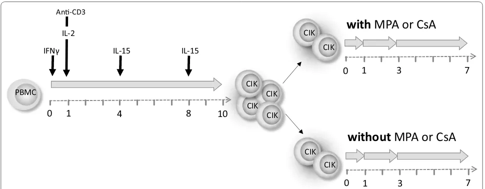
CIK cell generation and cultivation
CIK cells were generated from underweight banked blood of healthy donors (IRB approval 329/10) by stand-ard ficoll separation (Biochrom AG, Berlin Germany). PBMC were adjusted to 3 × 10⁶ cells/ml and cultured
in X-VIVO 10 media (Lonza, Verviers, Belgium) sup-plemented with 10 % fresh frozen plasma (German Red Cross Blood Donor Service, Frankfurt, Germany) in cell culture flasks (Greiner, Nürtingen, Germany) at 37 °C and 5 % CO2. At day 0 of CIK generation 1000 U/
ml IFN-γ (Imukin®, Boeringer Ingelheim Pharma, Ger-many) were added, followed by 100 ng/ml anti-CD3 mAB (OKT3, MACS GMP CD3 pure, Miltenyi Bio-tec, Bergisch Gladbach, Germany) and 500 U/ml IL-2 (Proleukin®S, Novartis Pharma, Nuremberg, Germany) 24 h later (day 1). On days 4 and 8 of culture, cell den-sity was adjusted to 1 × 10⁶ cells/ml and cells were
re-stimulated with 50 ng/ml IL-15 (PeproTech, Rocky Hill, USA) until they were harvested on day 10. CIK cells were split; control flasks were further supplemented only with 50 ng/ml IL-15 every 3 days, test flasks were additionally spiked with a therapeutically relevant MPA concentra-tion of 10 µM (Sigma-Aldrich, Taufkirchen, Germany) or of 5 µg/ml Ciclosporin A (CsA, Sandimmun®, Novartis Pharma GmbH, Nuremberg) after harvesting. The long term series received a further MPA/CsA treatment 3 days following harvesting.
Preparation for ex vivo investigations
For the experiments regarding cytotoxicity, surface receptor expression and cytokine/chemokine secre-tion, bench scale CIK cells of healthy donors were used (n = 6, independent experiments). Co-incubation
experi-ments with/without MPA/CsA started after 10 days of cultivation. Proliferation, cytokine secretion and receptor expression was investigated following short- (24 h), inter-mediate- (3 days) and long-term (7 days) MPA treatment, whereas cytotoxicity was investigated following short- and intermediate-term MPA incubation, only (Fig. 1).
Flow cytometric analyses
following antigens (clones): FITC: TCRγδ (IMMU510), CD62L (DREG56), CD226/DNAM-1 (DX11), CD11a/ LFA-1 (HI111)2, PE: TCRαβ (BW242/412)1, CD314/
NKG2D (ON72)/(149810)4, CD173/CD95L/FAS-Ligand
(NOK-1)2, CD183/CXCR3 (2D7)2, CD195/CCR5 (1C6)2,
CD262/DR5/TRAIL (DJR2-4)3; ECD: CD19 (J3-119),
CD45RO (UCHL1); PC-5.5: CD45 (J.33), CD45RO (UCHL1); PC-7: CD56 (N901/NKH-1); APC: CD3 (UCHT1), CD4 (13B8.2); APC-A700: CD25 (B1.49.9); APC-A750: CD16 (3GB), CD3 (UCHT1); PB: CD14 (RMO52), CD45RA (2H4); KO: CD45 (J.33), CD8 (B9.11) (all mouse IgG1, other than #IgG2a, *IgG2b, all
antibod-ies Beckman Coulter, except 1Miltenyi Biotec, 2BD
Bio-sciences; 3Biolegend, 4R&D Systems). For the assessment
of cell viability 7-AAD was used. Absolute counts were calculated via single-platform using Flow-Count™ fluoro-spheres (Beckman Coulter, Krefeld, Germany). All data were analyzed using CXP system II (Vs 2.2), Navios (Vs. 1.2) or Kaluza (Vs. 1.2) software (Beckman Coulter, Kre-feld, Germany).
Cytotoxicity assay
CIK cell cytotoxicity was tested against THP-1, K562 and MOLT-4 using the non-radioactive europium release cytotoxicity assay as described previously [16,
20]. To avoid the known discharge of the labeling rea-gent BATDA, K562 were previously incubated with Probenecid (Sigma-Aldrich Chemie GmbH, Steinheim, Germany). Target cells and CIK effector cells were co-cultured in triplicates at effector to target (E:T) ratios 40:1, 20:1, 10:1 and 5:1 on U-bottomed 96-well culture
plates (Nunclon™, Thermo Fisher Scientific, Roskilde, Denmark). After 3 h co-incubation at 37 °C, 20 µl super-natant was collected from each well and incubated for 15 min (shaking, 250 rpm) with 200 µl Europium solu-tion (PerkinElmer, Boston, USA). The fluorescence signal, correlating with the amount of destroyed cells, was then measured by a multilabel plate reader (VIC-TOR3™ 1420 multilabel counter, PerkinElmer, Boston USA). Target cells without effector cells were used as negative control. Maximum release (positive control) was obtained by target cell incubation with 16 % Tri-ton™ X-100 solution (Sigma-Aldrich Chemie, Stein-heim, Germany). Percentage of specific cytotoxicity was defined as the loss of target cells in relation to the mono-cultured control.
Cytokine/chemokine analysis
Supernatants of expanded CIK cells after 10 days of cul-tivation and then following 24 h, 3 and 7 days MPA incu-bation (10 µM) with respective controls were collected and assayed using BioLegend LEGENDplex™ (BioLeg-end, San Diego, USA). Data acquisition was performed on a Navios Flow Cytometer and analyzed with the LEGENDplex™ Data Analysis Software (BioLegend, San Diego, USA). The cell density was adjusted to 3 × 10⁶/ml.
The human inflammation 13-plex panel was designed for quantification of the cytokines/chemokines IL-1β, IFN-α, IFN-γ, TNF-α, MCP-1/CCL2, IL-6, IL-8, IL-10, IL-12p70, IL-17A, IL-18, IL-23 and IL-33. The minimum detect-able concentration for the cytokines ranged from 0.6 to 2.1 pg/ml.
An-CD3
IFNγ IL-15
CIK
0 PBMC
IL-2
IL-15
1 4 8
0 1 3 7
with
MPA or CsA
CIK
CIK CIK
0 1 3 7
without
MPA or CsA
CIK CIK CIK
CIK 10
[image:3.595.56.541.87.276.2]Statistical analysis
Statistical analysis was performed using GraphPad Prism 6 for Windows (GraphPad Software, San Diego, USA). Data were compared by paired, non-parametric Fried-mann test and differences were considered as significant for p < 0.05 (*), p < 0.01 (**) and p < 0.001 (***).
Results
Influence of MPA/CsA treatment on CIK cell expansion and viability
CIK cells that were cultivated in the presence of MPA for at least 3 days showed significantly impaired prolif-eration capacity compared to CIK cells cultivated with-out MPA over an equal period of time (p < 0.01; Fig. 2a). These differences were also demonstrated for all three CIK cell subgroups following intermediate- and for T- and NK cells following long-term MPA treatment (p < 0.05). Contrary, no effect of CsA treatment on CIK cell proliferation was observed (Additional file 1: Figure S1A). Interestingly, short-term MPA treatment had no or only marginal effect on CIK cell viability. In contrast, intermediate and long-term MPA treatment reduced CIK cell viability by more than 30 % whereas CsA treatment had no significant influence on CIK cell viability (p < 0.01; Fig. 2b).
Cytotoxic capacity of CIK cells following MPA/CsA treatment
Cytotoxicity of CIK cells with and without MPA treat-ment was investigated against the ALL cell line MOLT-4 and the AML cell lines K562 and THP-1 whereas CsA treatment was investigated against the cell lines K562 and MOLT-4 (Fig. 3). The cytotoxic effect of MPA treated CIK cells against K562 cells was influenced by MPA exposure, though only in the 40:1 ratio a sig-nificant reduction of the killing capacity was obtained
(p < 0.05, Fig. 3a). In contrast, no differences in the lytic activity of CsA incubated and wildtype CIK cells were observed against K562 (Fig. 3b). Regarding MOLT-4 killing, short-term MPA treatment did not affect tar-get cell lysis compared to untreated control CIK cells (Fig. 3c). However we observed a reduced lysis of MOLT-4 cells by CIK cells treated with MPA for 3 days (intermediate term) in the E:T ratios 10:1, 20:1 and 40:1 (p < 0.05). CsA-treated and wildtype CIK cells showed comparable results in MOLT-4 killing (data not shown). Interestingly, no difference in the lysis of THP-1 target cells of CIK cells with and without MPA incubation were determined (Fig. 3d). Long-term MPA treatment resulted in strong reduction of absolute CIK cell number. Therefore, cytotoxicity testing could not be investigated.
Changes in receptor expression and cytokine secretion as consequence of MPA treatment
Although absolute numbers of T cells declined, we observed an alteration in the CD4/CD8 ratio following MPA treatment of CIK cells. Interestingly, the percent-age of CD8+ cells decreased, while the percentage of
CD4+ cells increased (Fig. 4a). Comparing the
propor-tion of CD8+ cells following long-term and without MPA
treatment revealed a significant decrease in CD8+ cells
(p < 0.05). In accordance, intermediate and long-term MPA incubation were associated with a statistically sig-nificant higher percentage of CD4+ cells (p < 0.05 and
p < 0.01).
The characterization of NKG2D receptor on CD4+ and
CD8+ cells showed stable expression upon short- and
intermediate-term MPA treatment. Only following long-term MPA incubation, downregulation of NKG2D was observed (p < 0.05, Fig. 4b). Further analysis also revealed stable expression of NKG2D on CIK cell subpopulations (T, NK and NK-like T cells) during short- and interme-diate-term MPA incubation, whereas reduced NKG2D receptor expression was observed following long-term MPA exposure (T and NK cells: p < 0.05; NK-like T cells: p < 0.01, Fig. 4c).
Regarding CD25, we observed loss of expression when CIK cells were cultivated for longer than the usual cul-ture period of 10 days. However, this reduction in CD25 expression was enhanced following MPA incubation, showing the most pronounced differences for NK-like T cells after intermediate- and long-term MPA treat-ment (p < 0.01) (Fig. 4d). Interestingly, we also observed a reduction in CD25 expression implicated by CsA treat-ment. T and NK-like T cells showed a significant reduc-tion of CD25 expression following three (p < 0.01) and 7 days (p < 0.05) of CsA treatment whereas no significant alteration of CD25 expression on NK cells was observed (Additional file 1: Figure S1B). Furthermore, no changes in NKG2D expression in CsA treated CIK cells were examined (data not shown).
CIK cells T cells NK-like T cells NK cells
0 10 20 30 40 50 60 80 100
Absolute cell count [x10
6 ]
24h 3d 7d
**
** * * * ** * *
24h 3d 7d 24h 3d 7d 24h 3d 7d
4h ated
0 10 20 30 40 50 60 70 80 90 100
Viable cells [%
]
24h
** **
3d 7d
h 0
10 20 30 40 50 60 70 80 90 100
Viable cells [%
]
24h 3d 7d
+CsA 24h +CsA 3d +CsA 7d +MPA 24h
+MPA 3d +MPA 7d untreated 24h
untreated 3d untreated 7d
a
b
[image:5.595.58.539.85.624.2]up-regulated CCR5 upon intermediate- and long-term treatment with MPA (p < 0.05).
We also analyzed a panel of cytokines, i.e. IL-8, IFN-γ, MCP-1/CCL2, IL-6, IL-1β and TNFα, following short-,
intermediate- and long-term MPA exposure or control incubation. Similar to CD25 expression, cytokine release was attenuated by extended cultivation. In other words, impaired functionality in terms of cytokine release was
MOLT- 4
5:1 10:1 20:1 40:1
0 25 50 75 100
E:T ratio
E:T ratio E:T ratio
E:T ratio
* * *
K562
5:1 10:1 20:1 40:1
0 25 50 75 100
lysed targed cells [%
]
lysed targed cells [%
]
lysed targed cells [%
]
lysed targed cells [%
]
*
untreated 24h +MPA 24h
untreated 3d +MPA 3d
THP-1
5:1 10:1 20:1 40:1
0 25 50 75
+CsA 24h +CsA 3d K562
5:1 10:1 20:1 40:1
0 25 50 75 100
a b
c d
[image:6.595.58.540.86.546.2]CD8+ CD4+ 0
20 40 60 80 100
Cells [%
]
Cells [%
]
* *
24h 3d 7d 24h 3d 7d
24h 3d 7d
24h 3d 7d
24h 3d 7d
24h 3d 7d
24h 3d 7d
24h 3d 7d
24h 3d 7d 24h 3d 7d
CD8+NKG2D+ CD4+NKG2D+
0 20 40 60 80 100
* *
0 20 40 60 80 100
NKG2D
+ Cells [%
]
* * * *
T cells NK-like T cells NK cells
0 20 40 60 80 100
CD25
+ Cells [%
]
untreated 24h untreated 7d
+ MPA 24h + MPA 3d + MPA 7d
untreated 3d
* * * * *
T cells NK-like T cells NK cells * * a
b
c
not primarily influenced by MPA, but rather through the prolonged activation period. The only cytokine signifi-cantly reduced after long-term MPA compared to control was TNFα (Additional file 2: Figure S2).
Discussion
Immunotherapeutic strategies are of increasing interest in the therapy of emerging minimal residual disease or incomplete donor chimerism following HSCT. We and others have previously shown that IL-15 stimulated CIK cells are a promising immunotherapeutic approach for the treatment of patients with impending relapse follow-ing allogeneic SCT for acute leukemia or myelodysplastic syndrome [13, 19, 22, 25, 26].
CIK cells consist of a heterogeneous population of CD3+ T cells, CD3+CD56+ NK-like T cells and a minor
part of CD3−CD56+ NK cells [14]. Thereby, CIK cells are
able to kill tumor cells via diverse TCR specificities and in a non-MHC-restricted manner [27]. Different studies indicated an anti-tumor effect of CIK cells against various tumor cells mediated by the receptors or ligands NKG2D, TRAIL, DNAM and FasL [16, 28, 29]. In first clinical applications, CIK cell infusions were well tolerated and showed low incidence of GvHD even in the haplo-identi-cal setting, which was postulated to be mainly due to less trafficking of CIK cells to GvHD sites [30], and due to the lack of corresponding ligands or receptors on normal tis-sues and hematopoietic progenitors.
Beside antileukemic efficacy, CIK cell treatment may be associated with increased risk for developing severe GvHD, especially when being applied in the haploidenti-cal setting and in the early post-transplant period requir-ing immunosuppressive treatment. Furthermore, even if immunosuppression like MMF is stopped in patients at risk for relapse after HSCT, relevant MPA plasma levels might be present due to intra- and inter-patient
variability [31]. Therefore, the influence of immuno-suppressive treatment, on the efficacy of cellular inter-ventions needs to be further investigated. Hence, we analyzed the phenotype, survival and cytotoxic capacity of CIK cells during short-, intermediate- and long-term presence of MMF in vitro.
Within this study, we observed that CIK cell cytotox-icity was maintained during short-term MPA exposure, while intermediate- and long-term MPA treatment atten-uated proliferation capacity, viability and cytotoxicity. Comparable results were obtained by Brehm et al. [9] for NK cells within the scope of a clinical phase I/II study in short- and long-term MPA treatment. In this study, NK cells were co-incubated with MPA just at the beginning of cytokine activation. In our new approach, we first gen-erated the immunotherapeutic cells and then exposed them to MPA. Eissens et al. [32] and Ohata et al. [33] also described that MPA clearly impeded the outgrowth and the cytotoxic effect of NK cells treated with MPA. Unfor-tunately, a short-term MPA treatment was not investi-gated in these two studies. These results are contradicting Shapira et al. [34] who suggested that MMF does not impair GvL-effect or reduce LAK cell activity in mice. However, this might be explained by differing study pro-tocols. Shapira et al. treated mice with MMF for a period of 8 days and afterwards generated autologous LAK cells without further addition of MMF. In contrast, we gener-ated CIK cells according to our study protocol for 10 days and started MPA treatment after harvesting with the intention to simulate in vitro the situation when CIK cells were given to a patient who has therapeutic MPA plasma levels.
Most importantly, none or at most a marginal effect on CIK cell proliferation was seen following short-term MPA incubation. Therefore, our findings are promising, that in case of indispensable immunosuppression post
(See figure on next page.)
Fig. 5 Influence of MPA exposure on receptors involved in CIK cell mediated killing and cell migration. FasL, TRAIL, DNAM, CD11a, CXCR3 and CCR5 were analyzed following short- (24 h), intermediate- (3 days) and long-term (7 days) MPA treatment and all receptors were compared to their indi-vidual controls (only 24 h control is displayed). a–c Analyzing receptors involved in CIK cell mediated killing, we observed a stable expression of FasL, TRAIL and DNAM upon short- and intermediate-term MPA treatment. A reduced expression of FasL and TRAIL on T cells and an increased expres-sion of DNAM following long-term MPA exposure was determined (p < 0.05). d–f With regard to CIK cell migration, no alteration in the expression of CD11a, CXCR3 and CCR5 upon short-term MPA exposure was observed. CD11a was downregulated on T cells and upregulated on NK-like T and NK cells following intermediate- and long-term MPA treatment (p < 0.05 and p < 0.01). CXCR3 was downregulated on T cells upon long-term MPA incubation (p < 0.05), whereas CCR5 was expressed to a higher extend on NK-like T cells. n = 5 independent experiments
(See figure on previous page.)
FasL
T cells NK-T cells NK cells
TRAIL
DNAM
CD11a
CXCR3
CCR5
c 24h 3d 7d
c 24h 3d 7d 0 20 40 60 80 100 120 ns ns * 0 20 40 60 80 100 120 ns ns ns 0 20 40 60 80 100 120 ns ns ns 0 20 40 60 80 100 ns ns ns 0 5 10 15 20 ns ns ns 0 100 200 300 ns * ** 0 20 40 60 ns ns ns 0 20 40 60 ns ns ns 0 5 10 15 20 ns ns ns 0 5 10 15 20 ns * * 0 5 10 15 20 ns ns ns 0 20 40 60 80 100 ns ns * 0 20 40 60 80 100 ns ns ns 0 5 10 15 20 ns ns ns 0 5 10 15 20 ns ns * 0 100 200 300 ns * **
c 24h 3d 7d
c 24h 3d 7d
c 24h 3d 7d
c 24h 3d 7d
c 24h 3d 7d c 24h 3d 7d c 24h 3d 7d
c 24h 3d 7d c 24h 3d 7d c 24h 3d 7d
c 24h 3d 7d c 24h 3d 7d c 24h 3d 7d
c 24h 3d 7d c 24h 3d 7d c 24h 3d 7d
HSCT, the therapy with IL-15 stimulated CIK cells will have at least short-term efficacy. We observed no signifi-cant differences of killing relevant receptor expressions of the NKG2D, TRAIL, DNAM and FasL receptors upon short- and intermediate-term MPA exposure. However, we revealed that T cells and NK-like T cells reacted dif-ferently upon MPA exposure. CD3+CD56+ NK-like T
cells upregulated receptors relevant for CIK cell killing and migration, whereas CD3+ T cells downregulated
these receptors. CsA treatment resulted in a non-signif-icant reduction of proliferation and cytotoxicity, only. These results are comparable to Mehta et al. [35] describ-ing that CsA inhibited anti-CD3-mediated degranula-tion, but did not affect cytotoxicity of CIK cells against tumor targets. Regarding cytokine/chemokine secretion, we observed no significant differences between CIK cells with and without MPA treatment expect for a significant reduction in TNFα secretion. These results are in accord-ance with Liu et al. [36] who published promising results combining CIK cell therapy with CsA treatment also describing the secretion of various cytokines including IL-2 and IL-8. In contrast, Brehm et al. [9] and Nagy et al. [37] reported a significant reduction in the secretion of IFNγ, IL-6 and other cytokines by NK cells upon MPA treatment, but here immune cells were co-incubated with MPA already during cultivation time.
Following infusion, CIK cells are described to accumu-late and persist in tumor sites, resulting in tumor eradica-tion [30]. Wang et al. [38] analyzed CIK cell homing of
18F-FDG labeled CIK cells in leukemia patients by PET/
CT tracking. They figured out, that 1 h following CIK cell infusion, the majority of CIK cells accumulated in the lungs, followed by a migration into brain, heart, liver and spleen at the time points 4 and 8 h post CIK cell ther-apy. Furthermore, tendencies of CIK cell homing into the bone marrow were shown. In the study described herein, we observed that short-term MPA incubation had no pronounced effect on proliferation, viability, cytotoxicity and CIK cell composition. Linking this information to the homing parameters analyzed by Wang et al., we might speculate that during a window of 24 h where hardly any influence of MPA on the CIK cells was seen, CIK cells might migrate to tumor sites and achieve a cytotoxic effect.
Conclusions
In conclusion, already generated IL-15 CIK cells that were co-incubated with MPA for at least 3 days showed signifi-cantly impaired proliferation capacity, restricted viability, alterations in receptor expression and a reduction in their cytotoxic capability compared to CIK cells that were cul-tivated without MPA. Interestingly, CsA treatment had
no significant influence on CIK cell viability and the cyto-toxic potential against K562. However, a short-term MPA incubation had only marginal or reduced effect on CIK cells. The favored strategy is to avoid immunosuppres-sion in patients who received CIK cell immuno-therapy. However, our findings showed, that even in patients with immunosuppression e.g. for treatment of GvHD, CIK cell treatment may have at least short-term efficacy.
Abbreviations
APC: allophycocyanin; APC-A700: APC-Alexa Fluor 700; APC-A750: APC-Alexa Fluor 750; CIK: cytokine-induced killer; CsA: Ciclosporin A; FITC: fluorescein– isothiocyanate; ECD: phycoerythrin-Texas Red®; GvL/T: graft-versus-leukemia/ tumor; DLI: donor lymphocyte infusion; GvHD: graft-versus-host disease; GMP: good manufacturing practice; HSCT: hematopoietic stem cell transplanta-tion; KO: krome orange; MMF: mycophenolate mofetil; MPA: mycophenolic acid; MRD: minimal residual disease; NK: natural killer; PBMC: peripheral blood mononuclear cells; PB: pacific blue™; PC-5: phycoerythrin–cyanine-5; PC-7: phycoerythrin–cyanine-7; PE: phycoerythrin.
Authors’ contributions
Conceived and designed the experiments: MB, SH, CC. Performed the experi-ments: OZ, AQ. Analyzed the data: MB, SH, AQ. Coordinated the research: SH, CC. Contributed reagents/materials/analysis tools: MB, SH, VP, OZ, AQ. Wrote the paper: MB, SH. Revised the manuscript: HB, ER, CC, VP, JS. Supervised the research: PB, TK. All authors read and approved the final manuscript.
Author details
1 Clinic for Pediatric and Adolescent Medicine, University Hospital, Theo-dor-Stern-Kai 7, 60596 Frankfurt/Main, Germany. 2 Division for Translational Development of Cellular Therapeutics, Institute for Transfusion Medicine and Immunohematology, Goethe-University Frankfurt/Main, Frankfurt/Main, Germany. 3 German Red Cross Blood Donor Service Baden-Württemberg-Hessen, Frankfurt/Main, Germany.
Acknowledgements
The authors would like to thank Sibille Betz and Stephanie Erben for the excel-lent technical support.
Competing interests
The authors declare that they have no competing interests.
Availability of data and materials
All data are available in the manuscript or upon request to the authors.
Ethics approval and consent to participate
Donor PBMCs were obtained after written informed consent was given, with approval from the Ethical Review Board of the Medical Faculty of the Univer-sity Hospital Frankfurt (IRB approval 329/10).
Additional files
Additional file 1: Figure S1. Ex vivo CIK cell expansion and CD25
expression upon CsA treatment. (A) CsA treated CIK cells showed compa-rable expansion rates compared to wiltype CIK cells. (B) CD25 expression significantly decreased on T and NK-like T cells following 3 and 7 days of CsA incubation (day 3: p < 0.01 and day 7: p < 0.05).
Additional file 2: Figure S2. Impact of MPA treatment on cytokine
Funding
This project was supported by “Frankfurter Stiftung für krebskranke Kinder” and “Hilfe für krebskranke Kinder e.V.”, as well as “LOEWE Center for Cell and Gene Therapy Frankfurt; funded by: Hessisches Ministerium für Wissenschaft und Kunst (HMWK); funding reference number: III L 5-518/17.004”. The funders had no role in study design, data collection and analysis, decision to publish, or preparation of the manuscript.
Received: 30 April 2016 Accepted: 30 August 2016
References
1. Barrett AJ, Horowitz MM, Pollock BH, Zhang MJ, Bortin MM, Buchanan GR, Camitta BM, Ochs J, Graham-Pole J, Rowlings PA, et al. Bone marrow transplants from HLA-identical siblings as compared with chemotherapy for children with acute lymphoblastic leukemia in a second remission. N Engl J Med. 1994;331:1253–8.
2. Klingebiel T, Handgretinger R, Lang P, Bader P, Niethammer D. Haploi-dentical transplantation for acute lymphoblastic leukemia in childhood. Blood Rev. 2004;18:181–92.
3. Windreich RM, Goyal RK, Joshi R, Kenkre TS, Howrie D, Venkataramanan R. A pilot study of continuous infusion of mycophenolate mofetil for prophylaxis of graft-versus-host-disease in pediatric patients. Biol Blood Marrow Transplant. 2016;22(4):682–9.
4. Allison AC, Eugui EM. Mycophenolate mofetil and its mechanisms of action. Immunopharmacology. 2000;47:85–118.
5. Allison AC, Eugui EM. Mechanisms of action of mycophenolate mofetil in preventing acute and chronic allograft rejection. Transplantation. 2005;80:S181–90.
6. Eugui EM, Allison AC. Immunosuppressive activity of mycophenolate mofetil. Ann N Y Acad Sci. 1993;685:309–29.
7. van Gelder T, Hesselink DA. Mycophenolate revisited. Transpl Int. 2015;28:508–15.
8. Laupacis A, Keown PA, Ulan RA, McKenzie N, Stiller CR. Cyclosporin A: a powerful immunosuppressant. Can Med Assoc J. 1982;126:1041–6. 9. Brehm C, Huenecke S, Esser R, Kloess S, Quaiser A, Betz S, Zimmermann
O, Soerensen J, Passweg JR, Klingebiel T, et al. Interleukin-2-stimulated natural killer cells are less susceptible to mycophenolate mofetil than non-activated NK cells: possible consequences for immunotherapy. Cancer Immunol Immunother. 2014;63:821–33.
10. Rettinger E, Bonig H, Wehner S, Lucchini G, Willasch A, Jarisch A, Soer-ensen J, Esser R, Rossig C, Klingebiel T, Bader P. Feasibility of IL-15-acti-vated cytokine-induced killer cell infusions after haploidentical stem cell transplantation. Bone Marrow Transplant. 2013;48:1141–3.
11. Rettinger E, Kreyenberg H, Merker M, Kuci S, Willasch A, Bug G, Ullrich E, Wels WS, Bonig H, Klingebiel T, Bader P. Immunomagnetic selection or irradiation eliminates alloreactive cells but also reduces anti-tumor potential of cytokine-induced killer cells: implications for unmanipulated cytokine-induced killer cell infusion. Cytotherapy. 2014;16:835–44. 12. Rettinger E, Kuci S, Naumann I, Becker P, Kreyenberg H, Anzaghe M,
Willasch A, Koehl U, Bug G, Ruthardt M, et al. The cytotoxic potential of interleukin-15-stimulated cytokine-induced killer cells against leukemia cells. Cytotherapy. 2012;14:91–103.
13. Rettinger E, Meyer V, Kreyenberg H, Volk A, Kuci S, Willasch A, Koscielniak E, Fulda S, Wels WS, Boenig H, et al. Cytotoxic capacity of IL-15-stimulated cytokine-induced killer cells against human acute myeloid leukemia and rhabdomyosarcoma in humanized preclinical mouse models. Front Oncol. 2012;2:32.
14. Linn YC, Niam M, Chu S, Choong A, Yong HX, Heng KK, Hwang W, Loh Y, Goh YT, Suck G, et al. The anti-tumour activity of allogeneic cytokine-induced killer cells in patients who relapse after allogeneic transplant for haematological malignancies. Bone Marrow Transplant. 2012;47:957–66. 15. Schmidt-Wolf IG, Lefterova P, Mehta BA, Fernandez LP, Huhn D, Blume KG,
Weissman IL, Negrin RS. Phenotypic characterization and identification of effector cells involved in tumor cell recognition of cytokine-induced killer cells. Exp Hematol. 1993;21:1673–9.
16. Kuci S, Rettinger E, Voss B, Weber G, Stais M, Kreyenberg H, Willasch A, Kuci Z, Koscielniak E, Kloss S, et al. Efficient lysis of rhabdomyosarcoma
cells by cytokine-induced killer cells: implications for adoptive immu-notherapy after allogeneic stem cell transplantation. Haematologica. 2010;95:1579–86.
17. Linn YC, Lau LC, Hui KM. Generation of cytokine-induced killer cells from leukaemic samples with in vitro cytotoxicity against autologous and allogeneic leukaemic blasts. Br J Haematol. 2002;116:78–86. 18. Schmidt-Wolf IG, Negrin RS, Kiem HP, Blume KG, Weissman IL. Use
of a SCID mouse/human lymphoma model to evaluate cytokine-induced killer cells with potent antitumor cell activity. J Exp Med. 1991;174:139–49.
19. Schmeel LC, Schmeel FC, Coch C, Schmidt-Wolf IG. Cytokine-induced killer (CIK) cells in cancer immunotherapy: report of the international registry on CIK cells (IRCC). J Cancer Res Clin Oncol. 2015;141:839–49. 20. Cappel C, Huenecke S, Suemmerer A, Erben S, Rettinger E, Pfirrmann V, Klingebiel T, Ullrich E, Bader P, Bremm M. Cytotoxic potential of IL-15-activated cytokine-induced killer cells against human neuroblastoma cells. Pediatr Blood Cancer. 2016;. doi:10.1002/pbc.26147.
21. Lu PH, Negrin RS. A novel population of expanded human CD3+CD56+ cells derived from T cells with potent in vivo antitumor activity in mice with severe combined immunodeficiency. J Immunol. 1994;153:1687–96. 22. Introna M, Borleri G, Conti E, Franceschetti M, Barbui AM, Broady R,
Dander E, Gaipa G, D’Amico G, Biagi E, et al. Repeated infusions of donor-derived cytokine-induced killer cells in patients relapsing after allogeneic stem cell transplantation: a phase I study. Haematologica. 2007;92:952–9. 23. Laport GG, Sheehan K, Baker J, Armstrong R, Wong RM, Lowsky R,
Johnston LJ, Shizuru JA, Miklos D, Arai S, et al. Adoptive immunotherapy with cytokine-induced killer cells for patients with relapsed hematologic malignancies after allogeneic hematopoietic cell transplantation. Biol Blood Marrow Transplant. 2011;17:1679–87.
24. Rettinger E, Huenecke S, Bonig H, Merker M, Jarisch A, Soerensen J, Wil-lasch A, Bug G, Schulz A, Klingebiel T, Bader P. Interleukin-15-activated cytokine-induced killer cells may sustain remission in leukemia patients after allogeneic stem cell transplantation: feasibility, safety and first insights on efficacy. Haematologica. 2016;101:e153–6.
25. Rettinger E, Huenecke S, Bonig H, Merker M, Jarisch A, Soerensen J, Wil-lasch A, Bug G, Schulz A, Klingebiel T, Bader P. IL-15-activated cytokine-induced killer cells may sustain remission in leukemia patients after allogeneic stem cell transplantation: feasibility, safety and first insights on efficacy. Haematologica. 2016;101(4):e153–6.
26. Sangiolo D, Mesiano G, Carnevale-Schianca F, Piacibello W, Aglietta M, Cignetti A. Cytokine induced killer cells as adoptive immunotherapy strat-egy to augment graft versus tumor after hematopoietic cell transplanta-tion. Expert Opin Biol Ther. 2009;9:831–40.
27. Jiang J, Wu C, Lu B. Cytokine-induced killer cells promote antitumor immunity. J Transl Med. 2013;11:83.
28. Pievani A, Borleri G, Pende D, Moretta L, Rambaldi A, Golay J, Introna M. Dual-functional capability of CD3+CD56+CIK cells, a T-cell subset that acquires NK function and retains TCR-mediated specific cytotoxicity. Blood. 2011;118:3301–10.
29. Verneris MR, Kornacker M, Mailander V, Negrin RS. Resistance of ex vivo expanded CD3+CD56+ T cells to Fas-mediated apoptosis. Cancer Immu-nol Immunother. 2000;49:335–45.
30. Nishimura R, Baker J, Beilhack A, Zeiser R, Olson JA, Sega EI, Karimi M, Negrin RS. In vivo trafficking and survival of cytokine-induced killer cells resulting in minimal GVHD with retention of antitumor activity. Blood. 2008;112:2563–74.
31. Bhatia M, Militano O, Jin Z, Figurski M, Shaw L, Moore V, Morris E, Tallamy B, van deVen C, Ayello J, et al. An age-dependent pharmacokinetic study of intravenous and oral mycophenolate mofetil in combination with tacrolimus for GVHD prophylaxis in pediatric allogeneic stem cell trans-plantation recipients. Biol Blood Marrow Transplant. 2010;16:333–43. 32. Eissens DN, Van Der Meer A, Van Cranenbroek B, Preijers FW, Joosten I. Rapamycin and MPA, but not CsA, impair human NK cell cytotoxic-ity due to differential effects on NK cell phenotype. Am J Transplant. 2010;10:1981–90.
33. Ohata K, Espinoza JL, Lu X, Kondo Y, Nakao S. Mycophenolic acid inhibits natural killer cell proliferation and cytotoxic function: a possible disadvan-tage of including mycophenolate mofetil in the graft-versus-host disease prophylaxis regimen. Biol Blood Marrow Transplant. 2011;17:205–13. 34. Shapira MY, Hirshfeld E, Weiss L, Zeira M, Kasir J, Or R, Resnick IB, Slavin
effect or the activity of lymphokine-activated killer (LAK) cells in a murine model. Cancer Immunol Immunother. 2005;54:383–8.
35. Mehta BA, Schmidt-Wolf IG, Weissman IL, Negrin RS. Two pathways of exocytosis of cytoplasmic granule contents and target cell killing by cytokine-induced CD3+CD56+ killer cells. Blood. 1995;86:3493–9. 36. Liu S, Wang X, Lu Y, Xiao J, Liang J, Zhong X, Chen Y. The combined
use of cytokine-induced killer cells and cyclosporine a for the treat-ment of aplastic anemia in a mouse model. J Interferon Cytokine Res. 2015;35:401–10.
37. Nagy SE, Andersson JP, Andersson UG. Effect of mycophenolate mofetil (RS-61443) on cytokine production: inhibition of superantigen-induced cytokines. Immunopharmacology. 1993;26:11–20.