5 (2010) 1367 - 1378
International Journal of
ELECTROCHEMICAL
SCIENCE
www.electrochemsci.org
Density functional theory studies on the geometry and electronic
properties of Mitomycin C, DNA Base Pairs and their complex
S. Eynollahi1, S. Riahi*1,2, M.R. Ganjali2,3 and Parviz Norouzi2,3
1Center of Excellence in Electrochemistry, Faculty of Chemistry, University of Tehran, P. O. Box 14155-6455, Tehran, Iran
2 Institute of Petroleum Engineering, Faculty of Engineering, University of Tehran, P. O. Box 11365- 4563, Tehran, Iran
3 Medical Nanotechnology Research Centre, Medical Sciences ⁄ University of Tehran, Tehran, P.O. Box 14155-6451, Iran
*E-mail: [email protected]
Received: 18 July 2010 / Accepted: 30 July 2010 / Published: 1 September 2010
In this study, we present the work on the physicochemical interaction between the anti-cancer drug molecule mitomycin C (MC) and DNA base pairs. Comprehending the physicochemical properties of this drug, besides the mechanism by which it interacts with DNA base pairs, should eventually permit the rational design of novel anti-cancer or anti-viral drugs. The final purpose is the clarification of this novel class of drugs as potential pharmaceutical agents. The properties of the isolated intercalator mitomycin C (MC) and their stacking interactions with the adenine···thymine (AT) and guanine···cytosine (GC) nucleic acid base pairs were studied by means of the DFTB method. This method was an approximate version of the DFT method and it included the London dispersion energy. The molecular modeling on the complex, formed between MC and DNA base pairs, indicated that this complex was certainly capable of contributing in the formation of a constant intercalation site. The results exhibited that the MC changes affected the DNA base pairs structures with reference to the bond lengths, the bond angles, the torsion angles and the charges.
Keywords: Base pairs, Mitomycin-C, DFTB, Molecular geometry, DNA
1. INTRODUCTION
This chemotherapeutic agent in living cells is activated by flavoenzymes. Furthermore, MC has been used in ophthalmologic procedures and airway surgery to decrease scar formation [1-4].
The investigation of the drug– DNA base pairs interactions is a significant issue, since essential information is recovered regarding the action mechanism of antitumor, antiviral drugs and some carcinogenic compounds. A variety of techniques have been employed for the interactions study of some anticancer drugs with DNA [5-8]. Following the discovery of the nucleic acids electro-activity, many electrochemical approaches have been developed for the nucleic acids analysis and their interactions [9,10]. The interaction between DNA and MC was also studied previously by electrochemical methods [2]. Nevertheless, the types and the mechanism of these interactions did not become fully understood.
The comprehension of these properties is, however, facilitated by the application of computational studies. Among these properties, the most noteworthy one is the nucleobases interaction with other drugs not only in a stacked arrangement but also in other arrangements. For the description of this interaction, the higher correlated theories have proved to be successful. Several experimental and computational reports have been discussing the stacking interaction between the DNA bases and the base pairs [11-19]. Owing to the high cost of the high-level calculations regarding the large complexes, there are only a limited number of papers [20-23] so far on the high-level quantum mechanical calculations (QM) of the stacking energies between the nucleic acid bases and the intercalators.
With reference to the DFT methods, they are a group of precise and reliable QM calculations for computational studies [24-42]. The usage of the density functional tight-binding (DFTB) theory has been very promising [43,44]. The DFTB method also includes the empirical London dispersion energy term, called DFTB-D [45]. It was found that this method was very suitable and accurate for the calculations of the H-bonded and stacked DNA base pairs [46].
With respect to this study, it might contribute to the design of new intercalators [47]. This contribution could take place by investigating the nature of the DNA base pairs-drugs intercalation and the different binding forces, particularly the charge transfer, the dispersion and the electrostatic forces of interaction. For this reason, the geometries, the MC electronic structures and its molecular complexes with the nucleobases were examined with the aid of the DFTB methods. According to the literature survey, the presented work is the first which theoretically studies the interactions between MC and DNA base pairs.
2. COMPUTATIONAL METHODS
stressed that for the QM calculations, the QM-optimized geometries of the base pairs and the intercalators were used. As a result, the interacting molecules were overlaid by their B3LYP/6-31G* optimized geometries, when the idealized geometries were utilized.
The other one-electron properties (dipole moment, polarizability, energies of the frontier molecular orbital) were also determined at the B3LYP/6-31G* level. Concerning the stabilization energies of the selected complexes, they were determined with the DFT calculations and calculated with the DFTB method. In general, the DFT methods are known to be inherently very deficient for the stacking interactions, because they principally ignore the dispersion attraction [49] (Van Duijneveldt et al., 1994). For the improvement of this major deficiency, their extension by an empirical dispersion term. Currently appears to be a very reasonable way. In parallel, it must be considered that the interaction energies were calculated as the difference between the complex energy and the combined energies of the molecules in isolation [50].
3. RESULTS AND DISCUSSION
3.1. MC, GC and AT Characteristics
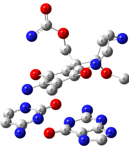
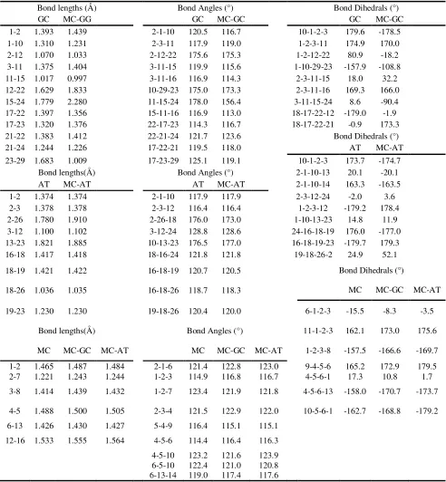
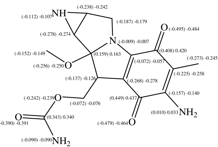
[image:3.612.183.435.459.694.2]The MC structure and geometry as well as the Watson-Crick base pairs (guanine...cytosine (GC) and adenine...thymine (AT)) were optimized through the DFTB calculations. The optimized MC structure is shown in Figure 1. The atom numbering and the atom charges (before and after interaction) of MC are depicted in Figures 2 and 3. The significant computed geometrical parameters are available in Table 1, including the bond lengths, the bond angles and the dihedral angles of MC, before and after the complexes formation (MC···AT and MC···GC).
Table 1. Significant computed geometrical parameters for MC, GC and AT before and after the complex formation (MC···GC and MC···AT)
Bond lengths (Å) Bond Angles (°) Bond Dihedrals (°)
GC MC-GG GC MC-GC GC MC-GC
1-2 1.393 1.439 2-1-10 120.5 116.7 10-1-2-3 179.6 -178.5
1-10 1.310 1.231 2-3-11 117.9 119.0 1-2-3-11 174.9 170.0
2-12 1.070 1.033 2-12-22 175.6 175.3 1-2-12-22 80.9 -18.2
3-11 1.375 1.404 3-11-15 119.9 115.6 1-10-29-23 -157.9 -108.8
11-15 1.017 0.997 3-11-16 116.9 114.3 2-3-11-15 18.0 32.2
12-22 1.629 1.833 10-29-23 175.0 173.3 2-3-11-16 169.3 166.0
15-24 1.779 2.280 11-15-24 178.0 156.4 3-11-15-24 8.6 -90.4
17-22 1.397 1.356 15-11-16 116.9 113.0 18-17-22-12 -179.0 -1.9
17-23 1.320 1.376 22-17-23 114.3 116.7 18-17-22-21 -0.9 173.3
21-22 1.383 1.412 22-21-24 121.7 123.6 Bond Dihedrals (°)
21-24 1.244 1.226 17-22-21 119.5 118.0 AT MC-AT
23-29 1.683 1.009 17-23-29 125.1 119.1 10-1-2-3 173.7 -174.7
Bond lengths(Å) Bond Angles (°) 2-1-10-13 20.1 -20.1
AT MC-AT AT MC-AT 2-1-10-14 163.3 -163.5
1-2 1.374 1.374 2-1-10 117.9 117.9 2-3-12-24 -2.0 3.6
2-3 1.378 1.378 2-3-12 116.4 116.4 1-2-3-12 -179.2 178.4
2-26 1.780 1.910 2-26-18 176.0 173.0 1-10-13-23 14.8 11.9
3-12 1.100 1.102 3-12-24 128.8 128.6 24-16-18-19 176.0 -177.0
13-23 1.821 1.885 10-13-23 176.5 177.0 16-18-19-23 -179.7 179.3
16-18 1.417 1.418 18-16-24 121.8 121.8 19-18-26-2 24.9 52.1
18-19 1.421 1.422 16-18-19 120.7 120.5 Bond Dihedrals (°)
18-26 1.036 1.035 16-18-26 118.7 118.3 MC MC-GC MC-AT
19-23 1.230 1.230 19-18-26 120.4 120.0 6-1-2-3 -15.5 -8.3 -3.5
Bond lengths(Å) Bond Angles (°) 11-1-2-3 162.1 173.0 175.6
MC MC-GC MC-AT MC MC-GC MC-AT 1-2-3-8 -157.5 -166.6 -169.7
1-2 1.465 1.487 1.484 2-1-6 121.4 122.8 123.0 9-4-5-6 165.2 172.9 179.5
2-7 1.221 1.243 1.244 1-2-3 114.9 116.8 116.7 4-5-6-1 17.3 10.8 1.7
3-8 1.414 1.439 1.432 1-2-7 123.4 121.9 121.8 4-5-6-13 -158.0 -170.7 -173.7
4-5 1.488 1.500 1.505 2-3-4 121.5 122.9 122.0 10-5-6-1 -162.7 -168.8 -179.2
6-13 1.426 1.430 1.427 5-4-9 116.4 115.1 115.1
12-16 1.533 1.555 1.564 4-5-6 114.4 116.4 116.3
4-5-10 123.2 121.6 123.9 6-5-10 122.4 121.0 120.8 6-13-14 119.0 117.4 117.6
[image:4.612.67.559.104.636.2][image:5.612.154.483.111.370.2]
highest positive charge belonged to C2,which was due to the transfer of the charge density from C2 to possible hydrogen bonding between O7 and one of the N8 hydrogens.
Figure 2. The atom numbering and the atom charges of MC
Figure 3. The MC atom charges before and after the complex formation with AT and GC (Parentheses include the changes after the complex formation with GC)
1 6 5 4 3 2
O
7O
10 11 12N
13 16 15 14NH
19NH
2
8 9O
17 18 20O
21 22NH
2
24O
23 -0.516 0.096 -0.163 0.291 -0.528 -0.786 -0.436 -0.172 0.674 -0.727 -0.514 -0.436 0.392 -0.502 0.338 0.360 0.448 -0.031 -0.428 -0.062 0.039 -0.086 -0.631 [image:5.612.128.495.407.655.2]
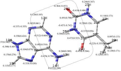
Figure 4. The optimized structure and the charge of guanine/cytosine (GC) base pair & GC...MC, before and after the complex formation (Parentheses include the changes after the complex formation)
Figure 5. The optimized structure and the charge of adenine/thymine (AT) base pair & AT...MC, before and after the complex formation (Parentheses include the changes after the complex formation)
0.472(0.558) -0.491(-0.789) 0.229(0.351) 0.262(0.394) 0.228(0.330) 0.106(0.166) 0.282(0.419) -0.501(-0.800) -0.443(-0.553) -0.393(-0.421) 0.151(0.252) -0.301(-0.385) -0.153(0.041) 0.354(0.409) -0.416(-0.421) 0.564(0.668) -0.428(-0.512) 0.502(0.661) -0.522(-0.660) -0.449(-0.779) 0.228(0.319) 0.129(0.183) -0.406(-0.465) 0.194(0.160) -0.278(-0.149) 0.439(0.590) 0.109(0.152) 0.244(0.349) 0.286(0.410) 0.358(0.424) -0.337(-0.355) 0.162(0.661) -0.512(-0.669) 0.128(0.185) 0.115(0.177) -0.104(0.078) 0.237(0.340) -0.476(-0.786) -0.390(-0.407) 0.170(0.276) -0.335(-0.430) 0.492(0.516) 0.237(0.330) 0.260(0.389) 0.491(0.700)
0.304(0.429) -0.337(0.056)
[image:6.612.100.492.91.348.2] [image:6.612.74.500.410.661.2]
Furthermore, the MC molecule was studied from different angles towards the base pairs and it was seen that the maximum changes of the bond angles and the bond lengths occurred in the quinine ring, indicating that the interactions between the drug and DNA base pairs molecules were stronger in this region. The optimized structures, the atom numbering, the atom charges and the base pairs are depicted in Figures 4 and 5. In Table 1, a selected number of bond angle and length values are listed, demonstrating the most significant changes for AT and GC, respectively.
[image:7.612.88.370.330.430.2]Table 2 visualizes the one-electron properties (dipole moment and polarizability) and the energies of the frontier molecular orbitals (HOMO and LUMO) of MC, using the DFTB computational method. In accordance with the high values of polarizability and dipole moment in this Table for MC, GC and AT, we could conclude that there were 2 kinds of interactions between the intercalator and DNA base pairs; the electrostatic interactions and the dispersion. In addition, the B3LYP/6-31G stabilization energies of the base pairs complexes were obtained to be -12.7 kcal/mol and -23.6 kcal /mol for A···T and G···C, respectively.
Table 2. Dipole moment [D], polarizibility [B3], HOMO and LUMO energies (in eV) of the drug, the bases and the base pairs
Compound HOMO LUMO Dipole moment Polarizability
AT -8.64 3.01 1.28 213.2
GC -7.35 2.74 2.51 223.4
MC -5.55 -2.76 4.14 291.6
A -8.83 3.12 2.49 101.17
T -9.53 2.94 3.88 89.14
G -8.45 3.52 2.76 109.19
C -9.93 3.01 6.12 80.41
Table 2 also displays that all the bases are good electron donors (the base and the base pairs LUMO energies were positive in contrast to those of MC). This ability was further magnified by base pairing. For example, the HOMO energy of guanine (-9.93 eV) increased by 2.85 eV upon pairing by cytosine.
3.2. Complex Formation
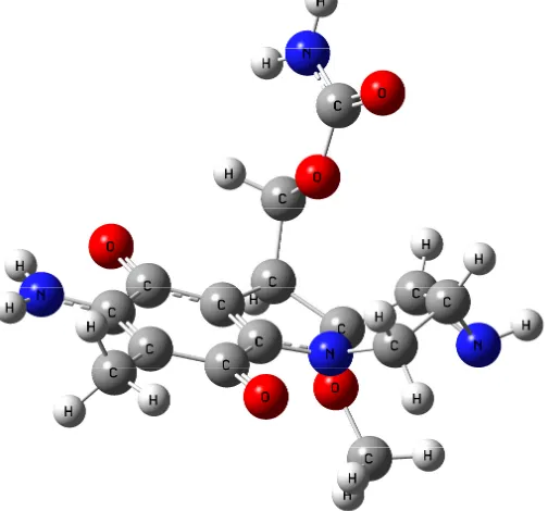
The MC···AT and MC···GC optimized structures were determined at the B3LYP/ 6-31G* level, depicted in Figures 6 and 7, respectively. The atom charge differences of MC, GC and AT are presented in Figures 3, 4 and 5. In line with Figures 2 and 3, the charge difference was higher in the planar area (quinine cycle) in comparison with the charge difference in other parts. For example, the atom charges in the 2nd and 5th position of the MC···GC shifted from 0.392 and 0.338 to 0.449 and 0.408, respectively.
Figure 6. The optimized MC···GC structures
[image:8.612.174.393.426.677.2]
The considerable charge differences are presented in dash marks (Figures 4, 5), owing to the GC-MC intercalation. These changes could also be observed for AT. Because the MC heteroatoms only interacted with the GC and AT heteroatoms (shown in the zone, including hydrogen), the other GC or AT base pair heteroatoms illustrated no remarkable charge changes. Also, it was observed that the hydrogen bonding had become weak in the base pairs. This observation was proved by the increase in the GC hydrogen charges (i.e. H12 atom charge shifted from 0.282 to 0.419), revealing that a weak hydrogen bonding had been formed between these atoms and the heteroatoms in the drug, and the increase in their bond length (i.e. the R bond length (12,22) shifted from 1.629Å to 1.833Å). The interaction with the MC molecule changed the bond angles of the base pairs (in the mentioned area), i.e. in GC, A(11,15,24) from 178.0 to 156.4. From the acquired results associated with the MC intercalation with the base pairs, we concluded that the drug caused changes in the DNA base pairs molecular structures. Consequently, the drug design leading to the highest changes was selected in order to the most effect on the DNA molecular structure.
The polarizability and the dipole moment were computed to obtain information about the electron distribution in the intercalator and the base pairs. Polarizability is a property, depending on the second derivative of the energy with respect to an electric field. On the other hand, dipole moment, being the first derivative of the energy with reference to an applied electric field, is an asymmetry measure in the molecular charge distribution. The most important polarizability and dipole moment values are displayed in Table 2, confirming the existence of the dispersion and electrostatic interactions between DNA base pairs and MC. As a result, the dispersion energy would always be remarkable in the course of interaction between MC and the base pairs.
As it is shown in Table 2, the polarizabilty and dipole moment values are notable, verifying that both the dispersion energy and the electrostatic interactions affect considerably the intercalator and the base pairs interactions. Therefore, a drug should be designed with high (as high as possible) polarizability and dipole moment values to reduce the interactions between DNA and the drug. Furthermore, the dependence of the stacking interaction energy on the vertical distance between the intercalator and the base pairs was investigated. The interaction energies were corrected for the basis set superposition error using the counterpoise method [51,52].
-8 -4 0 4 8 12
2.5 3.5 4.5 5.5
E
(k
ca
l/m
ol
)
[image:9.612.214.432.515.692.2]r(Å)
-9 -8 -7 -6 -5 -4 -3
2 3 4 5 6
E
(k
ca
l/m
ol
)
[image:10.612.198.414.76.261.2]r(Å)
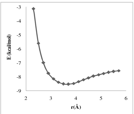
Figure 9. The AT···MC stabilization energies (∆E)
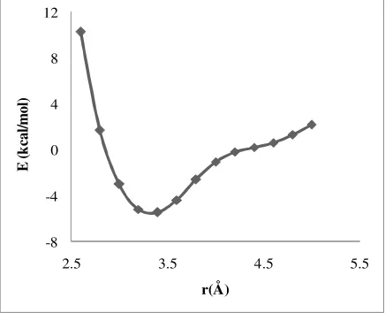
The computed interaction energies are listed in Figures 8 and 9 for MC with GC and AT, respectively. Based on these Figures, the minimum values of the potential energy curve were found to be at 3.4 Å for GC···MC and 3.7 Å for AT···MC. The stabilization energy values (energy necessary to separate MC and the AT pair to infinity) of GC···MC and AT···MC were equal to 5.44 kcal/mol and -8.53 kcal/mol, respectively.
Apparently, the GC···MC stabilization energy was greater than that of AT···MC, due to the higher GC polarizability and the dipole moment values. In addition, because of the higher interaction energy, the distance between GC and the intercalator was less than the distance of AT. From the obtained resulting data, it became obvious that the distance between the DNA base pairs molecules and the drug diminished as the interaction energy increased.
4. CONCLUSIONS
1. In this research, it was demonstrated that MC is a good electron acceptor with high polarizability and dipole moment while the AT and GC base pairs are good electron donors which are favorable for the aromatic stacking interactions between these two systems.
2.
It was also found that the theoretical procedures could properly examine the dispersion and the polarization effect. Subsequently, they could be used for the study of the intercalation processes.
4. It should also be mentioned that the applied DFTB method includes the empirical London dispersion energy term which was found to be very suitable and accurate for the H-bonded and stacked DNA base pairs calculations.
ACKNOWLEDGEMENTS
We gratefully acknowledge generous allocations of computing from the Institute of Petroleum Engineering, University of Tehran for Advanced Computing and Supercomputing Facilities.
.
References
1. J. Cummings, V. J. Spanswick and J. F. Smyth, C. Eur. J. Cancer., 31 (1995) 1928 2. D. Marın, P. Perez, C. Teijeiro and E. Palecek, Biophys. Chem., 75 (1998) 87
3. M. Tomasz, S. Neidle and M. J. Waring, The mitomycins: natural cross-linkers of DNA. In: Molecular Basis of Drug-DNA Antitumor Action, (Eds.) Macmillan Press, London, 2 (1993) 312 4. C. Teijeiro, P. Perez, D. Marin and E. Palecek, Bioelectrochem. Bioenerg., 38 (1995) 77
5. D. Pang and H. D. Abruna, Anal. Chem., 70 (1998) 3162
6. H. Fritzsche, A. Akhebat, E. Taillandier, K. Rippe and T. M. Jovin, Nucleic Acids Res., 21 (1993) 5085
7. P. J. Gane and P. M. Dean, Curr. Opin. Struc. Biol., 10 (2000) 401 8. D. E. Graves and L. M.Velea, Curr. Org. Chem., 4 (2000) 915 9. E. Palecek and M. Fojta, Anal. Chem., 73 (2001) 74A
10.A. Erdem and M. Ozsoz, Electroanalysis, 14 (2002) 965
11.C. Chipot, R. Jaffe, B. Maigret, D. A. Pearlman and P. A. Kollman, J. Amer. Chem. Soc., 118 (1996) 11217
12.S. Berski and Z. Latajka, J. Comput. Chem., 21 (1997) 347
13.R. E. Holmlin, P. J. Dandliker and J. K. Barton, Angew Chem. Int. Ed., 36 (1997) 2714 14.J. Norberg and L. Nilsson, Biophys. J.,74 (1998) 394
15.J. Sponer, J. Leszczynski and P. Hobza, J. Comput. Aided. Mol. Des., 17 (1996) 841
16.I. Saito, M. Takayama, H. Sugiyama, K. Nakatani, A. Tsuchida and M. Yamamoto, J. Amer. Chem. Soc., 117 (1995) 6406
17. C. Alhambra, F. J. Luque, F. Gago and M. Orozco, J. Phys. Chem. B, 101 (1997) 3846
18.P. Hobza, M. Kabelac, J. Sponer, P. Mejzlik and J. Vondrasek, J. Comput. Chem., 18 (1997) 1136
19.P. Hobza, J. Sponer and M. Polasek, J. Amer. Chem. Soc., 117 (1995) 792
20.D. Reha, M. Kabelac, F. Ryjacek, J. Sponer, J. E. Sponer, M. Elstner, S. Suhai and P. Hobza, J. Amer. Chem. Soc., 124 (2002) 3366
21. D. A. Bondarev, W. J. Skawinski and C. A. Venanzi, J. Phys. Chem.B, 104 (2000) 815 22.X. S. Xiao, S. Antony, Y. Pommier and M. Cushman, J. Med. Chem., 48 (2005) 3231 23.J. Sponer, J. Leszczynski and P. Hobza, J. Phys. Chem., 100 (1996) 1965
24.Z. W. Zhu and N. Q. Li, Microchem. J., 59 (1998) 307
25.S. Riahi, A. B. Moghaddam, M. R. Ganjali and P. Norouzi, J. Theor. Comput. Chem. (JTCC), 6 (2007) 331
26.S. Riahi, M. R. Ganjali, A. B. Moghaddam and P. Norouzi, J. Theor. Comput. Chem. (JTCC), 6 (2007) 255
28.S. Riahi, A. B. Moghaddam, M. R. Ganjali and P. Norouzi, J. Mol. Struct. (THEOCHEM), 814 (2007) 131
29.S. Riahi, M. R. Ganjali and P. Norouzi, J. Theor. Comput. Chem. (JTCC), 7 (2008) 317
30.S. Riahi, M.R Ganjali, M.Hariri, S. Abdolahzadeh, P.Norouzi Spectrochim. Acta Part A., 74 (2009) 253
31.S. Riahi, M. R.Ganjali, P. Norouzi and F. Jafari, Sens. Actuators B, 132 (2008) 13 32.S. Riahi, P. Pourhossien and M. R. Ganjali, Petrol. Sci. Technol., 28 (2010) 68 33.M. R. Ganjali, P. Norouzi, B. Akabri-Adergani,S. Riahi, Anal. Lett, 10 (2007) 1923
34.S. Riahi, M. R. Ganjali, R. Dinarvand, S. Karamdoust, K. Bagherzadeh, P. NorouziChem. Bio. Drug Des., 71 (2008) 474
35.M.R. Ganjali, T. Razavi, R. Dinarvand, S. Riahi, P. Norouzi, Int. J. Electrochem. Sci., 3 (2008) 1543
36.
S. Riahi
,
M. F. Mousavi, M. Shamsipur, H. Sharghi,
Electroanalysis
, 15 (2003) 1561
37.S. Riahi, A. Beheshti, M. R. Ganjali, P. Norouzi, Spectrochim. Acta Part A., 74 (2009) 1077 38.F. Faridbod, M.R. Ganjali, R. Dinarvand, S. Riahi, P. Norouzi, J. Food. Drug. Anal, 17 (2009)264.
39.M.R. Ganjali, T. Razavi, R. Dinarvand, S. Riahi, P. Norouzi, Int. J. Electrochem. Sci., 3 (2008) 1543
40.M. R. Ganjali, M. Hariri, S. Riahi, P. Norouzi, M. Javaheri,Int. J. Electrochem. Sci., 4 (2009) 295 41.M.R. Ganjali, T. Razavi, F. Faridbod, S. Riahi, P. Norouzi, Curr. Pharm. Anal, 5 (2009) 28 42.S. Riahi, A. B. Moghaddam, M. R. Ganjali, P. Norouzi Int. J. Electrochem. Sci., 4 (2009) 122 43.M. Elstner, D. Porezag, G. Jungnickel, J. Elsner, M. Haugk, T. Frauenheim, S. Suhai and G.
Seifert, Phys. Rev., 58 (1998) 7260
44.M. R. Ganjali, P. Norouzi, F. Faridbod, S. Riahi, J. Ravanshad, J. Tashkhourian, M. Salavati-Niasari and M. Javaheri, a. IEEE Sens. J., 7 (2007) 544
45.M. R. Ganjali, P. Norouzi, F. Sadat Mirnaghi, S. Riahi and F. Faridbod, IEEE Sens. J., 7 (2007) 1138
46.M. Elstner, P. Hobza, T. Frauenheim, S. Suhai and E. Kaxiras, J. Chem. Phys., 114 (2001) 5149 47.T. M. El-Gogary and G. Koehler, J. Mol. Struct. (THEOCHEM), 808 (2007) 97
48.M. J. Frisch, G. W. Trucks, H. B. Schlegel, G. E. Scuseria, M. A. Robb and et al, Gaussian Inc. Pittsburgh, PA. 1998
49. F. B. Van Duijneveldt, J. G. C. M. van Duijneveldt-van de Rijdt and J. H. van Lenthe, Chem. Rev., 94 (1994) 1873
50. P. Hobza and R. Zahradnik, Intermol. Complex, Elsevier, Amsterdam, 1988
51.M. J. Frisch, J. E. Del Bene, J. S. Binkley and H. F. Schaefer, J. Chem. Phys., 84 (1986) 2279 52.D. W. Schwenke and D. G. Truhlar, J. Chem. Phys., 82 (1985) 2418
![Table 2. Dipole moment [D], polarizibility [B3], HOMO and LUMO energies (in eV) of the drug, the bases and the base pairs](https://thumb-us.123doks.com/thumbv2/123dok_us/1942907.154356/7.612.88.370.330.430/table-dipole-moment-polarizibility-homo-lumo-energies-bases.webp)