“A COMPARATIVE STUDY OF RIPASA AND MODIFIED ALVARADO SCORING SYSTEMS FOR THE DIAGNOSIS OF
ACUTE APPENDICITIS”
Dissertation submitted
in partial fulfilment of the regulations for the award of the degree of
M.S. DEGREE BRANCH – I GENERAL SURGERY
Of
THE TAMIL NADUDr.M.G.R. MEDICAL UNIVERSITY
ESIC Medical College & PGIMSR, K.K.Nagar, Chennai- 600078.
I solemnly declare that this dissertation entitled “A COMPARATIVE STUDY OF RIPASA AND MODIFIED ALVARADO SCORING SYSTEMS FOR THE DIAGNOSIS OF ACUTE APPENDICITIS” is a bonafide and genuine research work carried out by me under the guidance of
Prof.R.Anbazhakan, Professor and Head, Department of General Surgery,
ESIC Medical College & PGIMSR, K.K.Nagar, Chennai.
This dissertation is being submitted to The Tamil Nadu Dr.M.G.R.
Medical University, Chennai, towards partial fulfilment of requirements of
the degree of MS (General Surgery) examination to be held in April 2016.
SIGNATURE OF THE CANDIDATE
Dr.SOUNDHARYA.S
M.S. Postgraduate
Dept. of General Surgery
ESIC Medical College & PGIMSR K.K. Nagar, Chennai- 78.
DATE:
I hereby declare that Tamilnadu Dr. M.G.R. Medical University,
Chennai, shall have the rights to preserve, use and disseminate this
dissertation/thesis in print/electronic format for academic/research purpose.
Dr.SOUNDHARYA.S
M.S. Postgraduate
Dept. of General Surgery
ESIC Medical College & PGIMSR K.K. Nagar, Chennai- 78.
DATE:
PLACE: Chennai
This is to certify that the dissertation entitled “A COMPARATIVE STUDY OF RIPASA AND MODIFIED ALVARADO SCORING SYSTEMS FOR THE DIAGNOSIS OF ACUTE APPENDICITIS”
submitted by Dr.Soundharya.S appearing for M.S. Degree Branch-I General
Surgery examination in April 2016 is a bonafide research work done by her
under my direct guidance and supervision in partial fulfilment of the
regulations of the Tamil Nadu Dr.M.G.R University, Chennai. I forward this
to the The Tamil Nadu Dr.M.G.R. University, Chennai, Tamilnadu, India.
Dr. R. Anbazhakan, M.S., F.I.A.S
Prof.& Head
Dept. Of General Surgery
ESIC Medical College & PGIMSR K.K. Nagar, Chennai- 78
Date:
This is to certify that the dissertation entitled “A COMPARATIVE STUDY OF RIPASA AND MODIFIED ALVARADO SCORING SYSTEMS FOR THE DIAGNOSIS OF ACUTE APPENDICITIS”
submitted by Dr.Soundharya.S appearing for M.S. Degree Branch-I General
Surgery examination in April 2016 is a bonafide research work done by her
under my direct guidance and supervision in partial fulfilment of the
regulations of the Tamil Nadu Dr.M.G.R University, Chennai. I forward this
to the Tamil Nadu Dr.M.G.R. University, Chennai, Tamil Nadu, India.
Dr. Uday Shamrao Kumbhar
M.S., F.M.A.S., F.I.A.G.E.S Associate Professor
Dept. Of General Surgery
ESIC Medical College & PGIMSR K.K. Nagar, Chennai- 78
Date:
This is to certify that the dissertation entitled “A COMPARATIVE
STUDY OF RIPASA AND MODIFIED ALVARADO SCORING
SYSTEMS FOR THE DIAGNOSIS OF ACUTE APPENDICITIS” is a
bonafide research work done by Dr.Soundharya.S, post graduate resident in
M.S.(General Surgery), ESIC Medical College & PGIMSR, K.K. Nagar,
Chennai- 78 under the direct guidance and supervision of Dr.R.Anbazhakan,
Prof.& Head, Dept. of General Surgery, ESIC Medical College & PGIMSR,
K.K.Nagar, Chennai- 78 in partial fulfilment of the requirements for the
degree of M.S. General Surgery of the Tamil Nadu Dr.M.G.R Medical
University, Chennai. I forward this to The Tamil Nadu Dr.M.G.R Medical
University, Chennai, Tamil Nadu, India.
Dr. Srikumari Damodaram, M.S., M.Ch(SGE), M.A.M.S., F.A.C.S., F.I.C.S., F.M.M.C
Dean, ESIC Medical College & PGIMSR, K.K.Nagar, Chennai- 78
Date:
At the outset, before beginning my dissertation, it deems only fit that I
express my gratitude to all the people who not only provided me with this
opportunity, but also were instrumental in its completion in many different
ways.
Any work I take up in life would be incomplete if I don’t begin it with
acknowledging my biggest support system, my family. It is only because of them I stand today as a doctor, following my dreams to become a surgeon.
Thank you, for never accepting excuses for mediocrity and constantly pushing
me to be better.
I would like to thankDr. R. Anbazhakan, Professor and Head, Dept. of General Surgery who was my guide in conducting this study. It was indeed
my good fortune and honour to be guided by a veteran in the field of Surgery.
His vast clinical experience and academic drive was something that helped
me not only with this thesis, but also during the entire course of my residency.
Thank you, Sir. The knowledge I’ve gained from you ranging from academics
to good bedside manners and clinical knowledge, will always stay with me
and make me a better doctor.
My heartfelt gratitude goes to Dr. Uday Shamrao Kumbhar, Associate Professor and my co-guide. Not only is he a great doctor and teacher, but also
research methodology and writing through the course of this dissertation.
Thank you, Sir. Your patience and the number of hours you put into this
thesis with us will always be appreciated.
I would like to extend a warm gratitude to our Respected Dean,
Dr. Srikumari Damodaram, for her unflinching encouragement and support throughout.
My experience in this college has been one of the best I could’ve asked
for. To be able to do your post-graduation in an institute with wonderful
teachers, who support you and help you better yourself is all one could ask
for. I’m very fortunate to have been blessed with the best of them. And their
help in doing my dissertation, starting from choosing the subject till its
completion has been invaluable. I would like to thank Dr.Bhanumati, Dr. M. S. Viswanathan, Dr. N. Murugesan, Dr. Pankaj Surana, Dr. Madhusudhanan, Dr.Sivakumar, Dr.Prabhakar, Dr.Poornima, Dr. Saravanan, Dr. Ashok for the same.
My thanks goes out to The Chairman and all the members of the
Dept. of Community Medicine, ESIC Medical College & PGIMSR, for her
overwhelming work and support throughout this study, and also for the
immense interest she has taken in teaching us how to analyse the study.
You’re only as good as the people you work with, and for this I cannot
possibly thank my co-postgraduates more. A big thank you to
Dr. Veena Bheeman and Dr. Balasubramanian for being so supportive and helpful. I would also like to thank all my juniors and seniors for their constant
help and cooperation.
Last, but most definitely not the least, I would like to thank the people
that made this whole study possible-the patients. As always, their support and their belief is what makes any study successful, and for this my eternal
gratitude goes out to them.
The making of this dissertation has been a very enriching experience. It
taught us some crucial things like having a research oriented thinking and
about keeping up with the times where evidence based medicine is the norm.
It also taught us discipline, better communication skills, and the attitude of
always trying to do what is best for the patients. It was a wholly educational
Sl. No. CONTENTS PAGE NO.
1. Introduction 1
2. Review of literature 5
3. Aims and Objectives 55
4. Materials and Methods 56
5. Results 58
6. Discussion 73
7. Summary 79
8. Conclusion 81
9. Bibliography 82
10. Annexures 92
Proforma
Informed Consent
SCORING SYSTEMS FOR THE DIAGNOSIS OF ACUTE APPENDICITIS
ABSTRACT
INTRODUCTION
Acute appendicitis is the most common condition encountered in general surgical practice. Alvarado and Modified Alvarado scores (MASS) are the commonly used scoring systems for its diagnosis, but its performance has been found to be poor in certain populations. Hence, we compared the RIPASA score with MASS, to find out which is a better diagnostic tool for acute appendicitis in the Indian population.
METHODS
We enrolled 180 patients who presented with RIF pain in the study. Both RIPASA and MASS were applied to them, but management was carried out as per RIPASA score. Final diagnosis was confirmed either by CT scan, intra-operative finding, or post-operative HPE report. Final diagnosis was analysed against both RIPASA and MASS. Sensitivity, Specificity, Positive Predictive Value, Negative Predictive Value and Diagnostic Accuracy was calculated for both RIPASA and MASS.
RESULTS
It was found that RIPASA was better than MASS in terms of Specificity (96% v/s 89%) and Positive Predictive Value (93% v/s 80%), and also to some extent in terms of Diagnostic Accuracy (75% v/s 71%). Whereas the Sensitivity (49.4% in both) and Negative Predictive Value (69% v/s 67%) were similar in both.
CONCLUSION
RIPASA is a more specific and accurate scoring system in our local population, when compared to MASS. It reduces the number of missed appendicitis cases and also convincingly filters out the group of patients that would need a CT scan for diagnosis (score 5-7.5).
KEYWORDS
INTRODUCTION
The abdomen is commonly compared to a Pandora’s box, and for good
reason. Since the abdomen contains within it innumerable viscera and other
anatomical components, the diseases of the abdomen gives rise to a lot of
clinical curiosity. A meticulous examination of the abdomen and clinical
correlation is one of the most important diagnostic tools and becomes
cornerstone of management in many conditions presenting with abdominal
pain. Despite the vast advances in the medical field in terms of imaging and
other investigation modalities, the importance of clinical examination cannot
be stressed upon enough(1).
Acute appendicitis is one of the commonest causes for acute abdomen
in any general surgical practice(2). From the time that it was first described by
Reginald Heber Fitz in 1886(3), it has remained a topic of serial research
works for various factors ranging from its aetiology, to its management
options.
One of the most researched fields pertaining to appendicitis is the one
involving diagnosis. Over the years various types of investigations including
laboratory and radiological, have been studied in detail with the aid of trials.
These were conducted in the hope of finding the most sensitive test for
diagnosing acute appendicitis. But in spite of the vast advances in the field of
that appendicitis is one condition whose diagnosis relies mainly upon the
clinical features. As quoted by Bailey & Love, “Notwithstanding advances in
modern radiographic imaging and diagnostic laboratory investigations, the
diagnosis of appendicitis remains essentially clinical, requiring a mixture of
observation, clinical acumen, and surgical science”(1).
So much has been stressed about the various methods of diagnosis,
only because the same is extremely important. Appendicitis, which if caught
early and managed appropriately can be the most uneventful surgery, while
the other end of the spectrum is also true, that when missed, appendicitis can
turn into a disease with great morbidity and mortality.
Hence, having understood the importance for early and right diagnosis,
and having understood that clinical evaluation provides the best and most
accurate diagnostic modality for appendicitis, many clinical scoring systems
have been developed over the years(4). This has aided the clinician to a large
extent in coming to the right diagnosis and providing early management.
What began as a single scoring system, evolved into many over the years, as
people constantly made modifications to the existing scoring systems based
on the local demographics or by adding more factors. This brought along the
next problem, of finding the single best scoring system, or the scoring system
with the maximum sensitivity and diagnostic accuracy. As a result, multiple
scoring systems in different parts of the world. To date, the most commonly
used scoring system worldwide is the Alvarado and the Modified Alvarado
scoring systems (MASS).(4) Hence, these have almost been considered as the
undocumented gold standard scoring system among clinicians worldwide. So
much so that any new scoring system that has been developed is usually first
compared to this.
Raja Isteri Pengiran Anak Saleha Appendicitis (RIPASA) score is a
fairly newer scoring system developed in 2008, where a study was done in
RIPAS Hospital, Brunnei Darssalem(5,6), to find a more favourable scoring
system than Alvarado and Modified Alvarado as these were found to have
poor sensitivity and specificity in Middle Eastern and Asian population.
Following the development of it, a randomised control trial was also done at
the same hospital comparing the RIPASA and Alvarado scoring systems and
proving the superiority of the former over the latter.
In the present study, RIPASA and Modified Alvarado scoring systems
(MASS)are compared among the local population in the subcontinent of
India, to find out which scoring system is more relevant and applicable, in
order to aid early diagnosis of acute appendicitis.
Appendicitis is one of the routine conditions evoking emergency
surgery worldwide (2) as also in our hospital. The statistics of appendicitis in
INCIDENCE OF ACUTE APPENDICITIS IN ESIC MEDICAL
COLLEGE & PGIMSR FROM JAN – OCT 2013.
· Total number of patients presenting with Right Iliac Fossa pain – 234
· Total number of patients who underwent emergency
appendicectomy – 151
REVIEW OF LITERATURE
HISTORICAL REVIEW(7)
Appendicitis has been a much studied about topic since the early years.
Some literature is available which dates back the study of appendix to as early
back as the 3rd century, and the Greek rule under Caesar.
The anatomical study dates back to 1492, where Leonardo da Vinci
had delineated the organ clearly in his diagrams. He called it “Orecchio”
which literally means ear, to denote the auricular appendage of the caecum.
In 1521, the appendix was illustrated by the physician-anatomist B D
Carpi.
The name “Vermiform appendix” was first coined by V Vidius in
1530, because of its worm-like appearance.
Supplementary data about the appendicular anatomy was published by
Morgagni in 1719, in his Adversaria Anatomica.
Jean Fernel, a French physician, narrated his first case of perforated
appendix, following an autopsy on a 7- year old girl, in 1544.
Another post-mortem study in a boy suffering from chronic abdominal
In 1711, Lorenz Heister, a pupil of Boerhaave, described a small
abscess adjacent to a blackened appendix in a post-mortem study of an
executed criminal.
In 1759, Mestivier described the post-mortem finding of a 45-year old
man whose death occurred after incision and drainage of an abscess in the
right lower quadrant of the abdomen. He describes the abscess as a
consequence of an appendicular perforation due to a pin. This started a long
course of discussions as foreign bodies were identified as causes of
obstruction and perforation of the appendix.
Faecolith as a cause for perforated appendix, was described as early as
1812 by John Parkinson, found during the autopsy of a 12- year old boy.
Louyer-Villermay demonstrated gangrenous appendices during
autopsies in two young men at a presentation in a medical conference held at
Paris, in 1824.
Encouraged by Louyer-Villermay’s work, Parisian physician Francois
Melier documented six additional descriptions of appendicitis encountered at
autopsy, one of which he even suspected prior to death, in 1827. He then went
on to suggest the possibility of prior diagnosis and surgical removal of the
In 1839, Bright and Addison, the renowned physicians of Guy’s
Hospital, described the symptomatology of appendicitis in their book
“Elements of Practical Medicine”, and also stated that the appendix was the
cause for most of the inflammatory processes in the right iliac fossa.
In spite of the repeated observations of the appendicular pathology in
right lower quadrant inflammatory disease, the most likely cause of persistent
mortality was due to unclear therapeutic implications.
In June 1886, Dr. Reginald H. Fitz, Professor of Pathological Anatomy
at Harvard University, presented a paper about the vermiform appendix being
the most common cause of inflammatory disease of the right lower quadrant
and he also clearly explained most of the clinical features and the importance
of early surgical intervention. It was for the first time the word “Appendicitis”
was used.
THE APPENDIX
EMBRYOLOGY(8)
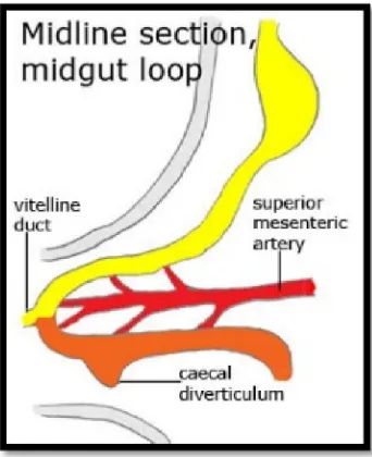
During the sixth week of gestation, when the descent of colon takes
place, appendix arises as an out pouching of the caecal bud (Fig.1). Initially, it
is of the same calibre as the caecum. As the caecum enlarges and attains its
normal position, the appendix gets pushed medially. Sometimes, there can be
which would in turn lead to diagnostic difficulties when there is an
[image:22.595.218.389.167.377.2]appendicular pathology.
Figure 1. Embryology of the Appendix
ANATOMY(9,10)
The vermiform appendix is a worm shaped organ situated at the
posteromedial aspect of the caecum, where the three taenia coli coalesce
about 2cm below the ileocaecal junction.
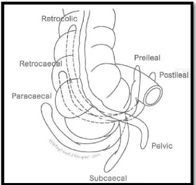
The appendix is notorious for its varied positions (Fig.2), such as
retrocaecal, retrocolic, pelvic, subcaecal, pre-ileal and post-ileal. Of these, the
most common are retrocaecal, retrocolic and pelvic. To trace the appendix,
the anterior taenia coli is identified and traced down the ascending colon and
caecum, and the base of the appendix lies at the point where the three taenia
from 2-20 cm, and its diameter from 5-7mm. It is often relatively longer
during childhood, and may shorten due to atrophy after mid-adult period. The
appendix is connected to the ileal mesentery by a short triangular fold called
the mesoappendix. It extends along the whole viscus, almost up to the tip.
A semilunar fold is present below and posterior to the ileocaecal
opening, and it acts as a valve through which the lumen of the appendix opens
into the caecum. It is called the Valve of Gerlach.
The appendix considered as a vestigial organ all these years, is recently
thought to have an immunological role owing to the numerous patches of
[image:23.595.204.403.428.615.2]lymphoid tissue that it contains.
VASCULAR SUPPLY AND LYMPHATIC DRAINAGE
APPENDICULAR ARTERY
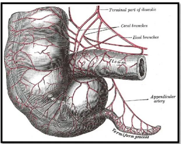
The blood supply to the appendix is from the appendicular artery
(Fig.3), which is a branch of the ileocolic artery. It courses behind the
terminal ileum and into the mesoappendix, close to the base. It gives off a
recurrent branch at this site, which forms an anastomosis at the base, with the
posterior caecal artery. The main appendicular artery itself traverses till the tip
of the appendix. The terminal part which overlies the wall of the appendix
may get thrombosed in acute appendicitis and is the reason for necrosis or
gangrene of the tip of the organ. It is common to find two or more accessory
[image:24.595.164.474.448.695.2]arteries, and this varies from individual to individual.
APPENDICULAR VEINS
The venous drainage of the appendix is by one or more appendicular
veins. These drain into the posterior caecal or ileocolic vein, which in turn
drain into the superior mesenteric vein.
LYMPHATICS
As mentioned earlier, the appendicular wall is rich in lymphatic tissue,
and these drain through numerous (about 8-15 in number) lymphatic vessels
into the mesoappendix. From here, they unite to form three or four larger
vessels and ultimately drain into the lymphatic vessels draining the ascending
colon, and finally terminate in the superior and inferior ileocolic chain of
[image:25.595.187.436.432.697.2]nodes (Fig.4).
INNERVATION
Innervation of the appendix and the overlying visceral peritoneum is
by the sympathetic and parasympathetic nerves from the superior mesenteric
plexus.
IMMUNOLOGICAL FUNCTION(11)
Up until recent times, it had been thought that the appendix was a
vestigial organ and that it had no definite role in the physiology of the human
body. Of late, it has been proposed and widely accepted that the appendix
indeed has a role to play in the immunity of the body. It has been found to
secrete immunoglobulins, especially Immunoglobulin A (IgA) and forms a
part of the gut-associated lymphoid tissue (GALT) system. However
appendicectomy does not predispose to any immunocompromised
manifestations.
ACUTE APPENDICITIS
INCIDENCE(2,12)
Appendicitis is one of the commonest acute conditions manifesting as
pain abdomen in the Emergency room. The life time rate for appendicectomy
is 12% for men and 25% for women, with approximately 7% of all people
Most commonly affected age group is second to fourth decades of life,
with mean age of 31.3 years and median age of 22 years.
Both sexes are affected, with a slight male to female predominance,
about 1.2-1.3:1).
GEOGRAPHIC DISTRIBUTION(2,10,12)
Appendicitis is more frequently seen in USA, Canada, UK, Australia,
New Zealand, and in the white population in South Africa. It is comparatively
rare in Asians and Central Africans. Studies have shown the possibility of the
disease being determined by environmental factors, which is further
strengthened by the fact that when people from latter areas migrate to the
western world or change to a western diet, appendicitis becomes more
prevalent in them. Appendicitis is undoubtedly less common in races that
habitually have bulk cellulose diet.
AETIOLOGY(2)
In spite of the common nature of the condition and the innumerable
studies done, the etiological factors leading up to the condition of appendicitis
still remains unknown and obscure. Universally, it had been rare prior to the
adoption of the western way of living. It has been observed that over the
years, appendicitis has risen from being an insignificant disease to the most
common serious intra-abdominal inflammatory pathology of the western
observed that appendicitis is relatively rare in rural areas and economically
less developed countries, and incidence increases with economic
development, migration to urban areas and western countries. Even though
exact etiological cause is not known, it is clear that many contributory factors
are responsible for the development of appendicitis.
AGE AND SEX
Though the most common age group for appendicitis is the second to
fourth decade, no age is totally immune to the disease. There are case reports
of appendicitis in a new born, and also at the other extreme of age. About
65% of the patients are under the age of 30 years, and only 2% are 60 years
and above. The incidence of appendicitis is maximum between 20 to 30
years(13).
FAMILIAL SUSCEPTIBILITY
There are reported instances of appendicitis occurring in families,
suggesting a possible inherited susceptibility. Reports by Baker(14), Andersson
et al(15) suggested high incidence of appendicitis among immediate family
members. Downs(16) operated upon 16 out of 22 closely related individuals for
appendicitis. In each case, the cause of appendicitis was a fibrous band arising
from the base of the appendix which was attached to the lateral aspect of the
caecum and causing a kinking of the base. Males and females shared the
SEASONAL FACTORS
There may be a possible association between seasonal respiratory
infections and appendicitis, especially in children. An upper respiratory tract
infection could lead to the simultaneous involvement of tonsils and the
lymphoid in the appendix. Origin is postulated to be blood borne(17,18).
RACE AND DIET(19,20,21)
Appendicitis, in general, is associated with a diet that is non-roughage
and high on meat. Racial distribution is mainly due to the associated
economical and diet status of the particular race. The more civilized countries
have been found to have a higher incidence of the disease. Appendicitis has
an interesting national distribution. It is common in highly industrialized
countries like Great Britain, US, France and Germany. It is low in Denmark
and Sweden, but even lower in Spain, Greece, Italy and the rural parts of
Romania. For example, in a study done by Lucas Championnier(21)in
Romania, the incidence of appendicitis in the rural areas was 1 in 22,000,
OTHER MECHANICAL CAUSES FOR THE DISEASE AND ITS
COMPLICATIONS
· FAECOLITHS
Non-calcified inspissated faecal matter is called faecolith. The
presence of a faecolith in the lumen causes mechanical obstruction, and due to
decreased lymphatic and vascular flow, causes ulceration or perforation in the
part of the appendix distal to it. It is a common finding in a large proportion
of appendices found during appendicectomy surgery.(22)
· CONSTIPATION AND PURGATION
In a case of acute appendicitis, administration of purgatives increases
the chance of perforation.
· PARASITES
Most notorious of parasitic infestations are those with round worms,
where they lodge and cause obstruction at the ileocaecal junction or at the
lumen of the appendix and hence cause an acute inflammation that progresses
into perforation. Other parasites known to cause the disease are thread worm
which cause appendicitis either by obstruction of the lumen or by injury to the
· BACTERIAL FACTORS
Bacterial infection of the appendix is a known cause of acute
appendicitis. The pathophysiology is by mucosal erosions of the appendicular
wall, which allows the bacteria to migrate into the submucosal layer, thus
causing an acute infection. The causative bacteria come to affect the appendix
by two possible routes. One is by direct contamination by the resident
variable and mixed flora in the intestine, which is more common, or the other
route is by haematogenous spread.(23,24)
· BANDS AND ADHESIONS
Bands or adhesions can cause kinking of the appendix and produce
appendicitis due to the acute obstruction. These may be bands of congenital
origin which are basically abnormal peritoneal attachments, or adhesions that
are acquired either by repeated infections or post-operatively.
· STRANGULATION WITHIN A HERNIAL SAC
Appendix may be a content in a hernia sac, either by itself or along
with other contents, and this may produce a picture of appendicitis due to
obstruction or strangulation of the hernia sac. Appendix found as a content in
· TRAUMA
Though this is a very rare cause for appendicitis, it is still a
documented possibility and to be considered especially in cases of blunt
trauma to the abdomen in the right iliac fossa. The pathophysiology is that
faecolith gets dislodged during the trauma and causes obstruction in the
appendicular lumen. Cases of post-traumatic appendicitis have been reported
by Birrel, Black and BhajeKar(26).
· SECONDARY TO MALIGNANCY
Carcinoma of the appendix, either primary or secondaries from
elsewhere, can present as acute appendicitis or even as a perforation due to
encroachment of the growth along the wall and obstructing the lumen.
Literature review shows cases that presented as acute appendicitis secondary
to metastasis with primary in the breast.(27,28)
· EPIDEMIC FORM
Acute appendicitis may occur in an epidemic form, where the
causative organism is usually Streptococci, and the portal of entry is through
the nasopharynx(24).
· AMOEBIC APPENDICITIS
Though extremely rare, there have been a few reported cases of
· VASCULAR FACTORS
The main blood supply to the appendix is by the appendicular artery
and it is an end artery. So any cause leading to obstruction of the blood flow
would result in ischemia, inflammation and secondary infection of the
appendix.
PATHOLOGY OF ACUTE APPENDICITIS(30)
GROSS APPEARANCE –
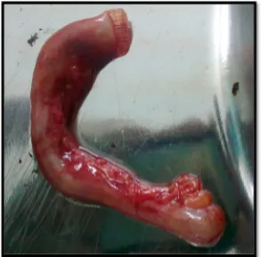
The macroscopic appearance of an appendix during an acute
inflammation (Fig.5) varies from person to person, and the external
appearance mostly depends upon the stage at presentation and the underlying
pathophysiology. Based on these, the gross appearances can range among any
of the following –
· Appendix may appear normal in size and serosa may appear normal with
its shiny appearance
· Patchy hyperemia and suffusion
· Continuous areas of suffusion and congestion
· Dull grey-white appearance with suppurative inflammatory exudates
· Increase in the size of the diameter, usually up to 1cm.
· Focal areas of abscess formation in the mesoappendix
· Focal gangrenous necrosis of the wall
· Area of frank perforation
Figure 5. Gross appearance of appendicitis.
MICROSCOPIC APPEARANCE –
The microscopic appearances in appendicitis (Fig.6) as well vary with
stage and pathophysiology of the disease, but the minimum criteria to
diagnose acute appendicitis microscopically
are-· Mucosal congestion and edema
· Infiltrate of polymorphonuclear inflammatory cells in the mucosa
· Focal small areas of mural ulceration
· With or without crypt abscesses
· Micro-abscesses, submucosal involvement of the inflammatory cell
Figure 6. Microscopic appearance of appendicitis
PATHOGENESIS OF ACUTE APPENDICITIS(9,13,30)
Appendicitis begins due to obstruction of the lumen (Fig.7), due to
various causes as described earlier. This leads to a closed loop obstruction. As
there is continued mucosal secretion from the obstructed part, this leads to
increased and rapid distension of the appendix. The distension stimulates the
visceral afferent nerve fibres, and is responsible for the vague/diffuse pain in
the periumbilical region. On progress of the condition, it leads to rapid
multiplication of bacteria from superadded infection, which leads to further
distension. This causes reflex nausea and vomiting. Due to the distension,
there is capillary and venous occlusion, and due to involvement of the serosa
and parietal peritoneum, pain migrates to right iliac fossa. On further arterial
occlusion, there is increased bacterial invasion, which leads to peritonitis and
manifests as fever, tachycardia and leucocytosis. The greater omentum
attempts to limit the peritonitis by localizing the spread of peritoneal
Two types of acute appendicitis have been recognized. These are –
1. NON-OBSTRUCTIVE ACUTE APPENDICITIS
The process of inflammation usually begins in the mucosa, sometimes
in the lymphatic follicles. Once infection reaches the submucosa, it progresses
rapidly. The organ turns red, inflamed and haemorrhages into the mucous
membrane. If left untreated, the tip may become gangrenous, because at this
part the artery is intramural and is more prone to occlusion by inflammation
or thrombosis.
But in non-obstructive appendicitis per se, progress is slower, allowing
time for the omentum to form a protective barrier and localize the peritonitis.
In most of the cases, infection does not even cross the mucosal layer (i.e.
catarrhal inflammation).
Non-obstructive appendicitis terminates in any of the following ways –
Resolution, Ulceration, Suppuration, Fibrosis, or Gangrene. The former are
more common than the latter.
2. OBSTRUCTIVE APPENDICITIS
The pathogenesis and pathological features of obstructive appendicitis
have already been discussed in detail. It is important to remember the fact that
the consequence of an appendicitis in an obstructive pathology depend on the
1. The contents in the lumen
2. Degree of obstruction
3. Continued secretion by the mucosa
[image:37.595.186.420.228.477.2]4. Inelastic character of the serosa
Figure 7. Faecolith causing obstructive appendicitis
TYPES OF APPENDICITIS
a) Acute catarrhal appendicitis
b) Acute focal appendicitis
c) Acute suppurative appendicitis
d) Gangrenous appendicitis
A pathological study was done by Martin Brumer in 1970(3) in 404
appendicectomy specimens and the incidence was as follows –
a) Normal – 27%
b) Recurrent – 23%
c) Acute – 28.8%
d) Suppurative – 35.2%
e) Gangrenous – 9.9%
f) Perforated – 8.4%
CLINICAL FEATURES(1,9,10,13)
SYMPTOMS
· PAIN ABDOMEN
Pain abdomen is the presenting feature in acute appendicitis. In
majority of the cases, it is classical and almost diagnostic, with the patient
complaining of acute onset pain abdomen, initially periumbilical and colicky,
and later on migrates to Right Iliac Fossa (RIF). But there can be atypical
presentations of pain abdomen and these are usually associated with the
variable anatomical positions. In retrocaecal position of the appendix, patient
complains of pain either in the flank or back, in pelvic appendix, patient may
have pain in the suprapubic region and in the retroileal appendix, patient may
· ANOREXIA
Loss of appetite is a common complaint second to pain abdomen.
· VOMITING
Vomiting is seen in 3/4th of the patients, and it occurs following pain
abdomen. It is due to reflex pyloric spasm, neural stimulation or due to ileus.
· FEVER
Fever occurs following vomiting.
MURPHY’S TRIAD is constituted by Pain Vomiting Fever
· OTHERS
Other associated but infrequent symptoms seen are diarrhoea,
obstipation, abdominal mass
SIGNS
· GENERAL CONDITION: Patient could be toxic, dehydrated based on
the severity of the disease. He may have fever and tachycardia.
· Mc BURNEY’S SIGN: It is described as the point of maximum
tenderness. It lies at the junction between the medial 2/3rd and the lateral
1/3rd along the imaginary line that joins the umbilicus and the right
anterior superior iliac spine. It was first described by Mc Burney in 1890.
Figure 8.1. McBurney's Sign
· BLUMBERG SIGN: Commonly known as Rebound tenderness. A hand
is placed on the right iliac fossa and progressively pressed with each
movement of expiration. It is then released suddenly. If the sign is
positive, the patient will wince or cry with pain. This indicates
inflammation of the parietal peritoneum. (Fig. 8.2)
· ROVSING’S SIGN:When pressure is applied on the abdomen in the left
iliac fossa, it causes pain in the right iliac fossa. Many theories have been
postulated for this- Williams said pressure on the left side of the colon,
forces gas into the caecum; Yashi et al (1958) disproved the involvement
of colonic gas by putting a cannula into the caecum. Sheperd suggested
adhesions of inflamed appendix to the pelvic colon as a cause for this sign.
(Fig. 8.3)
Figure 8.3. Rovsing's sign
· COPE’S SIGN: Pain in the right hypogastrium on flexion and internal
rotation of the right thigh, also known as Obturator Internus test. The
cause for the pain is that the inflamed appendix overlying the obturator
internus and iliacus, causes spasm in these muscles and stretching of these
Figure 8.4. Cope's (Obturator Internus) Sign
· PSOAS SIGN:Pain elicited upon extension of right thigh, due to
[image:42.595.159.463.93.301.2]irritation of psoas muscle, as seen in retrocaecal appendicitis. (Fig.8.5)
· HYPERAESTHESIA IN SHERREN’S TRIANGLE: Sherren’s triangle
is bounded by lines joining the umbilicus, Right anterior superior iliac
spine and pubic symphysis. Skin overlying this triangle is gently struck
and on comparing to the opposite side, hyperaesthesia is elicited. (Fig. 8.6)
Figure 8.6. Hyperaesthesia in Sherren's triangle
· BALDWIN’S TEST: It is positive in retrocaecal appendicitis. Light
pressure is applied and maintained over the point of maximum tenderness
in the right flank, and the patient is asked to raise his right lower limb
keeping the knee extended. Beyond a point, patient will be unable to raise
his leg due to pain and drops it.
· POINTING TEST:Patient is asked to point the site of maximum pain on
coughing, using one finger. If it corresponds with the site of maximum
· ALDER’S TEST: Also known as shifting tenderness. The point of
maximum tenderness is marked in supine position, and patient is asked to
turn to left lateral position, and point of maximum tenderness is marked
again. If there is a shift in the marked point, then it is unlikely to be due to
appendicitis. This test is useful in differentiating appendicitis from
mesenteric lymphadenitis and painful uterine conditions in pregnancy.
· RECTAL EXAMINATION: It is a must in all cases of appendicitis.
Tenderness on right lateral wall, especially when compared to posterior
and left lateral wall is a significant sign. Sometimes, this may be the only
positive sign in case of a pelvic appendicitis.
DIFFERENTIAL DIAGNOSIS(1,9,10,13)
1. PRESCHOOL CHILDREN
· Intussusception
· Meckel diverticulitis
· Acute gastroenteritis
2. SCHOOL AGE CHILDREN
· Gastroenteritis
· Functional pain
· Constipation
3. ADOLESCENT BOYS/YOUNG ADULT MEN
· Crohn’s disease
· Ulcerative colitis
· Epididymitis
4. ADOLESCENT GIRLS/YOUNG ADULT WOMEN
· Pelvic inflammatory disease
· Ruptured ovarian cyst
· Ovarian torsion
· Urinary tract infection
5. ELDERLY
· Malignancy in GIT
· Diverticulitis
· Perforated ulcer
INVESTIGATIONS(1,9,10,13)
1. LABORATORY TESTS
· Complete blood count – Leucocyte count >10,000/mm3, Left shift of
neutrophils with normal total white blood cell count
· C-reactive protein- elevated
2. RADIOGRAPHIC INVESTIGATIONS
· Plain X-ray of the abdomen- may show an appendiceal calculus,
Sentinel loop (dilated, atonic ileum with air-fluid level), Caecal dilatation,
Blurring of the right psoas outline, Widening/blurring of the preperitoneal
fat line, Haziness in right lower quadrant due to fluid and edema, Scoliosis
with concavity to the right, Right lower quadrant mass causing indentation
at the caecum, and rarely gas in the appendix.
· Ultrasonography(31)- Findings of non-compressible, non-peristaltic
tubular structure, with a blind ending, diameter of 6mm or more,
appendicolith causing an acoustic shadow. Other supportive features are
high echogenicity, surrounding fluid or abscess, non-compressible
surrounding fat, caecal pole edema. (Fig. 9.1)
Ultrasonography has a sensitivity of about 88% and specificity of 93% for
diagnosing appendicitis(31). But there are certain disadvantages–
Ø It is user-dependent.
Ø False negative results seen in appendicitis of the appendiceal tip, gangrenous, perforated appendix, retrocaecal appendicitis, gas-filled
appendix.
Figure 9.1. USG in acute appendicitis
· COMPUTERIZED TOMOGRAPHY(32)- CT scan findings of
appendicitis include appendicular diameter more than 6 mm, appendicolith
may be picked up, failure of the appendix to fill with oral contrast, air up
to its tip and enhancement of its wall with IV contrast, inflammatory
changes such as fat attenuation, inflammatory phlegmon, fluid, abscess,
lymphadenopathy, caecal thickening, extraluminal gas, arrow-head sign
(where the lumen of the caecum points towards the opening of the
appendix). (Fig. 9.2)Sensitivity and specificity of CT scan comes close to
Figure 9.2. CT in acute appendicitis
· NUCLEAR MEDICINE(33)- Two types of imaging studies are useful in
evaluation of patients with suspected appendicitis- Radiolabelled white
blood cells (Tc99m WBC) and immunoglobulin G (Tc99m IgG). These
studies localize leucocytes/IgG at the site of appendiceal inflammation
with the use of scintigraphy. (Fig. 9.3)
[image:48.595.117.507.500.701.2]· DIAGNOSTIC LAPAROSCOPY- This is especially useful in equivocal
cases. Paterson Brown et al, in a study found that diagnostic laparoscopy
reduced the number of unnecessary appendicectomies significantly,
[image:49.595.180.467.224.386.2]especially in female patients.(36)(Fig. 9.4)
Figure 9.4. D-lap in acute appendicitis
CLINICAL SCORING SYSTEMS(34)
As discussed before, even with the advances in medicine and imaging
techniques, appendicitis is one condition which still relies upon clinical
examination as a main resort of diagnosis. To aid this, over the years several
scoring systems have been developed taking into account various symptoms,
signs and some basic laboratory investigations. Many studies have been done
worldwide to check the sensitivity and specificity of each of these clinical
scoring systems in the diagnosis of acute appendicitis. Though the most
famous one is the Alvarado scoring system, there is no one universally
accepted scoring system used for diagnosis so far. Some of the scoring
1. ALVARADO SCORING SYSTEM(35)
FEATURE SCORE
Migratory pain 1
Anorexia 1
Nausea 1
Tenderness in RIF 2
Rebound tenderness 1
Elevated temperature 1
Leucocytosis 2
Shift of WBC count to left 1
TOTAL 10
Score <5 – Appendicitis unlikely
5-6 – Appendicitis possible
7-8 – Appendicitis likely
2. MODIFIED ALVARADO SCORING SYSTEM (MASS)(36)
SYMPTOMS SCORE
Migratory RIF pain 1
Nausea/Vomiting 1
Anorexia 1
SIGNS
Tenderness in RIF 2
Rebound tenderness in RIF 1
Elevated temperature 1
LABORATORY FINDINGS
Leucocytosis 2
TOTAL 9
Score <5 – Unlikely to be appendicitis
5-6 – Low Probability to be appendicitis
6-7 – High Probability to be appendicitis
3. RIPASA SCORING SYSTEM(5)
PATIENT’S DEMOGRAPHIC SCORE
Female 0.5
Male 1.0
Age< 39.9 years 1.0
Age> 40 years 0.5
SYMPTOMS
RIF pain 0.5
Pain migration to RIF 0.5
Anorexia 1.0
Nausea & vomiting 1.0
Duration of symptoms < 48 hrs 1.0
Duration of symptoms > 48 hrs 0.5
SIGNS
RIF tenderness 1.0
Guarding 2.0
Rebound tenderness 1.0
Rovsing’s sign 2.0
Fever>370C , <390C 1.0
INVESTIGATIONS
Raised WBC count 1.0
Negative urinalysis 1.0
ADDITIONAL SCORES
Foreign NRIC 1.0
Score <5 – Unlikely to be appendicitis
5-7.5 – Low Probability to be appendicitis 7.5-12 – High Probability to be appendicitis
4. PAEDIATRIC APPENDICITIS SCORE(37)
SYMPTOM/SIGN SCORE
Anorexia 1
Pyrexia 1
Nausea/Vomiting 1
Migration of pain 1
Raised WBC count 1
Raised neutrophil count 1
RIF tenderness 2
Cough/percussion/hopping tenderness 2
Score <5 – Appendicitis unlikely
5 – Appendicitis possible
5. TZANAKI SCORING(38)
FEATURE SCORE
Presence of right lower abdominal tenderness 4
Rebound tenderness 3
Lab findings- WBC>12,000 cells/cumm 2
USG- positive findings 6
TOTAL 15
Score <8 – Appendicitis unlikely
>8 – Appendicitis likely
6. LOW RISK FOR APPENDICITIS SCORE (KHARBANDA)(39)
DIAGNOSTIC FEATURE SCORE
Absolute neutrophil count >6.75 6
Rebound pain/pain with percussion 2
Unable to walk/walks with limp 1
Nausea 2
H/o migratory pain 1
H/o focal RLQ pain 2
TOTAL 14
Score <5 – Low risk for appendicitis
7. LINTULA SCORE(40)
DIAGNOSTIC
CRITERIA RESPONSE SCORE
Gender Male
Female
2 0
Intensity of pain Severe
Mild to moderate
2 0
Relocation of pain Yes
No 4 0 Vomiting Yes No 2 0
Pain in RLQ Yes
No
4 0
Fever >37.50C Yes
No 3 0 Guarding Yes No 4 0
Bowel sounds Absent/tinkling/high pitched Normal 4 0 Rebound tenderness Yes No 7 0
TOTAL SCORE 32
Score >21 – High risk for appendicitis
15-21 – Moderate risk for appendicitis
8. ESKELINEN SCORE(41)
DIAGNOSTIC CRITERIA RESPONSE
Tenderness in RLQ Yes-2, No-1
Rigidity Yes-2, No-1
WBC> 10,000 Yes-2, No-1
Rebound tenderness Yes-2, No-1
Pain in RLQ at presentation Yes-2, No-1
Duration of pain > 48 hours Yes-2, No-1
Final scoring is done with a computer program using complex calculations.
9. OHMANN SCORE(43)
DIAGNOSTIC CRITERIA SCORE
Tenderness in RLQ 4.5
Rebound tenderness 2.5
No micturition difficulties 2.0
Steady pain 2.0
WBC>10 1.5
Age<50 1.5
Relocation of pain to RLQ 1.0
Rigidity 1.0
TOTAL 16
10. FENYO-LINDBERG SCORE(42)
DIAGNOSTIC CRITERIA RESPONSE VALUE
Sex Male Female +8 -8 WBC >14 9-13.9 <8.9 +10 +2 -15
Duration of pain (hrs)
<24 24-48 >48 +3 0 -12
Progression of pain Yes
No
+3 -4
Relocation of pain Yes
No +7 -9 Vomiting Yes No +7 -5
Aggravation by coughing Yes
No
+4 -11
Rebound tenderness Yes
No +5 -10 Rigidity Yes No +15 -4
Tenderness outside RLQ Yes
No
-6 +4
Constant -10
11. CHRISTIAN SCORE(44)
DIAGNOSTIC CRITERIA – 1 point for each
Abdominal pain on history, occurring within 48 hours of presentation Vomiting- 1 or more episode
RLQ tenderness on examination
Low grade fever – defined as <= 38.8 C
Polymorphonuclear leucocytosis – defined as WBC > 10,000 AND neutrophils > 75%
Score >4 – Surgery
≤3 - Observation
TREATMENT(1,9,10,13):
The treatment options available for appendicitis vary depending upon a
number of factors such as time of presentation, clinical picture, obstructive or
non-obstructive pathology amongst others. The patient can be conservatively
managed in case of non-obstructive pathology, resolving symptoms.
Treatment is by Fluid management, IV antibiotics and supportive therapy.
Patient will be on close follow up and in case of non-resolution or recurrence,
may be taken up for surgery (either emergency or interval). The surgical
management of an uncomplicated appendicitis is Appendicectomy, which can
be done by various techniques ranging from open surgery, conventional
laparoscopy, single incision laparoscopic surgery, robotic surgery to NOTES
initially managed conservatively by Ochsner Sherren regimen, with IV fluids,
IV antibiotics and close follow up of the size of the mass and patient may be
taken up for an interval appendicectomy. The management of an appendicular
abscess is by drainage, as in any other abscess in the body.
No matter what the final path of management, the importance of
hydration and IV antibiotics cannot be stressed upon enough.
CLINICAL SCORING SYSTEMS
What are Clinical Scoring Systems?(34)
Clinical Prediction Rules (CPR) are defined as decision-making tools,
which include 3 or more variables obtained from the history, physical
examination or basic diagnostic tests in order to assist the clinician in decision
making.(45)
In recent times, as there is a quest to improve diagnostic accuracy,
there has been an increase in the use of CPRs. These use specific criteria in
order to establish probabilities of outcomes or to aid in assisting management
decisions. There are 3 types of CPRs(46)
-· Diagnostic CPRs - factors related to arriving at a clinical diagnosis
· Prognostic CPRs - for predicting outcomes
The format of a CPR can be of two types.
1. Some require fulfilment of a complete set of criteria in order to direct
management.
2. Some assign values to weighted criteria, the summation of which provides
a score.
The second type, known as Clinical Scoring Systems (CSSs), have
different types too.
1. Dichotomous type- utilizing a cut-off value above which an action is
recommended or an outcome is expected. For example, surgical
intervention may be recommended for a certain validated score over 6.
2. Continuous type- to provide graded risk stratification. A simple
example may stratify a patient to low risk of a disease process for
scores of 1-2, moderate risk for scores of 3-5 and high risk for scores
of 6-7.
While many CSSs exist, not all have been appropriately developed or
evaluated. In the process of evaluation, one must consider several factors
including
· internal validity
· external validity
· sensibility
· potential impact
Why use Clinical Scoring Systems?
Problems still remained in the early diagnosis of appendicitis, as no
one investigation was gold standard for it. Hence it was understood that no
imaging is superior to a good clinical examination and a clinical diagnosis
when it came to appendicitis. To aid the clinician in the diagnosis for a
suspected case of appendicitis, many clinical scoring systems have been
developed over the past 3 decades.
The basis of all medical diagnoses and decisions depend upon the
ability of a clinician to assess the potential risk and benefit, along with sound
clinical knowledge. This helps in making wise, educated decisions, which is
the cornerstone of good medical practice. Practice variation can result in
patient outcome differences, but standardization of practice based on the best
evidence can result in improved care. Numerous studies have demonstrated
the efficacy of Evidence Based Clinical Algorithms (EBCA) such as
pathways and protocols in reducing delays in time-sensitive medication
administration, deciding on surgery, and reducing mortality(47). Integrating
CSSs into EBCA is key to standardizing patient care and this will help in
ALVARADO SCORING AND MODIFIED ALVARADO SCORING
SYSTEMS
(MASS)-In 1986, Alvarado published what is now one of the most well-known
and studied appendicitis scores(35). A retrospective study was done on 305
patients admitted for suspected appendicitis. Clinical and laboratory findings
were compared in relation to pathologically proven acute appendicitis. 277
patients were eligible for analysis. Eight criteria were chosen for inclusion in
the diagnostic score. As Right Lower Quadrant (RLQ) Pain and Left Shift
were found to be the most prevalent, they received 2 points each, while each
of the remaining criteria were given 1 point. This initial study included an age
range of 4 to 80 years (mean 25.3). An Alvarado Score of ≥7 was considered
high risk for appendicitis. It was found to have a sensitivity of 81% and a
specificity of 74%.
Since then, numerous studies have been done world across to
check the Alvarado scoring in various populations.
Bond et al prospectively studied 187 children aged 2 – 17 years with
suspected appendicitis. They used Alvarado’s cut-off score and found a
sensitivity and specificity of 90% and 72% respectively, with a negative
appendectomy rate of 17%. Lower cut-off scores (5 or 6) demonstrated
improved sensitivity, but corresponding reductions in specificity, as
Hsiao et al conducted a retrospective study and confirmed Alvarado’s
data showing that RLQ tenderness and a left shift were the most prevalent
signs in those with pathologically proven appendicitis. Patients with Alvarado
Scores ≥7 were statistically more likely to have appendicitis than controls.
Overall sensitivity and specificity for an Alvarado Score >=7 were 60% and
61% respectively.(49)
Rezak et al, in their retrospective study, founda higher sensitivity and
specificity- 92% and 82% respectively. This study also suggested that a 27%
reduction in CT scanning would have occurred, if patients with scores >7 had
been managed directly by appendectomy without CT evaluation.(50)
In a mixed pediatric-adult population, Owen et al prospectively
evaluated 215 patients and found the sensitivity and specificity were 93% and
81%.(51)
Shreef et al recently in 2010, performed a dual-centre prospective
study, reviewing 350 patients. Interestingly, their reported statistical analysis
was based on an Alvarado threshold of 6, and was based upon 2 different
outcomes; 1) performance of appendectomy and 2) histology. Using the
standard threshold of 7 and including all comers related to histologic
diagnosis, the sensitivity and specificity were 86% and 83% respectively.(52)
Several attempts have been made to modify the Alvarado Score to
Macklin et al sought to simplify the Alvarado Score by eliminating the
criteria for left shift (Modified Score total 9), as done by Kalan in a mixed
adult/paediatric study. Children aged 4-14 years were enrolled, demonstrating
sensitivity and specificity of 76.3% and 78.8% respectively using a cut-off
score of 7 or higher to predict histological appendicitis. Kalan’s study was
limited to 11 children, all of which had modified Alvarado Scores ≥7 and
corresponding appendicitis. Obviously these numbers are too small to draw
any conclusions.(53)
Sooriakumaran et al further modified the score by decreasing the value
of leukocytosis, to make a total score of 8. This score was then compared to
clinical assessment by Emergency Physicians, and found wanting. However,
one must be cautious, as only 3 children were included, and due to the change
in total score, the threshold value was tested at 5.(54)
Significant changes to the Alvarado Score were suggested by
Impellizzeri et al. who studied 156 patients, replacing anorexia with an
elevated fibrinogen level (>400mg/dL), changing migration of pain to length
of pain (although not defined), combining RLQ pain and rebound into one
criteria, and decreasing the temperature cut-off to 37 C. Of note, the diagnosis
of appendicitis was made on surgical report, not pathologic diagnosis. The
authors suggest the above modifications would have decreased admission
OTHER SCORING SYSTEMS
Madan Samuel introduced the Paediatric Appendicitis Score (PAS)
in 2002.Evaluating 1170 children with suspected appendicitis, Samuel
compared historical, clinical and laboratory features in children with
appendicitis (n=734) and those without appendicitis (n=436). 8 variables were
included in a diagnostic model out of 10 points, with greater weight attributed
to RLQ pain and rebound tenderness. Samuel concludes that a score of 6 or
greater shows a high probability of acute appendicitis.(37)
Increased concerns related to radiation exposure from imaging studies
have put pressure on clinicians to accurately decide which children with
abdominal pain should be admitted and observed or discharged without a CT
evaluation. Kharbanda et al derived and validated a score to do that- Low
risk appendicitis score. Kharbanda et al prospectively enrolled 767 patients
with suspected appendicitis who were evaluated by a surgeon. Using 6
weighted predictors of appendicitis determined for a total score of 14, patients
with a score of <=5 were highly unlikely to have appendicitis (sensitivity
99%, NPV 98%).(39)
The Lintula Score relies on clinical data alone. There are no
laboratory results required. Lintula et al first prospectively evaluated 35
maximum value of 32. A high risk threshold was established at >=21, while
low risk was ≤15.(40)
The Eskelinen Score is relatively complex to perform, (requiring
factor multiplication) and was originally designed for use within a computer
program.(41)
The Fenyo-Lindberg Scoreappears to be one of the most complex,
incorporating criteria with multiple levels of response that both add to and
subtract from the total score. In 1987, Fenyo prospectively evaluated 259
adult patients with suspected appendicitis. The resulting score was further
validated in 830 patients, of which 256 had proven appendicitis. Sensitivity,
Specificity, PPV and NPV were 90%, 91%, 83% and 95% respectively.(42)
In 1999, Ohmann prospectively validated his own score in a
multi-centre, multi-phase trial. Subjects evaluated during phase 1 (n=870) received
surgical intervention based on surgeon assessment, while those in phase 2 (n=
614) received computer-assisted diagnostic support using theOhmann Score.
The authors found a statistically significant improvement in specificity, PPV
and accuracy in the phase 2 Score group, along with a decrease in the number
of delayed diagnoses, which was defined as appendectomy on the second day
Probably the simplest of the group, the Christian Score uses a mere 5
criteria. The case group of 58 subjects with suspected appendectomy had
surgical intervention if ≥4 criteria were met. Fifty-nine appendectomy
controls had intervention based solely on surgical staff assessment. Ages
ranged from 7 to 56 years. The negative appendectomy rate was significantly
less in the Score group than that of the control.(44)
RIPASA SCORING
What is probably the newest member to the group of appendicitis
scores is the RIPASA Score, named after its hospital of origin in Brunei. A
mixed population of 400 adults and children who had an appendectomy were
retrospectively identified, the records of 312 were used to derive the score.
Individual criteria were weighted (0.5, 1, 2) based on probabilities and a panel
of staff surgeons. The resulting maximal RIPASA score is 16 - a threshold of
7.5 proving a sensitivity of 88% and specificity of 67%. PPV and NPV were
93% and 53%, while accuracy was 81%. Using the score, an absolute
reduction in negative appendectomies of 9% would have occurred.(5,6)
Chong et al continued to evaluate their new score by prospectively
enrolling 200 adults and children in a comparison of the RIPASA and
Alvarado Scores. In this group of patients, the RIPASA was statistically
71%) and accuracy (92% vs. 87%). Specificity, PPV and negative
appendectomy rates were similar between the 2 scores.(56)
Several other CSSs have been developed for patients with suspected
appendicitis, but do not appear to have been formally evaluated in detail.
Some of these include the Teicher Score, Arnbjornsson Score, Izbicki Score,
AIMS AND OBJECTIVES
To assess the RIPASA scoring system and the Modified Alvarado
Scoring System (MASS) for the diagnosis of Acute Appendicitis, and
compare them with respect to
· Sensitivity
· Specificity
· Positive Predictive Value (PPV)
· Negative Predictive Value (NPV)
MATERIALS & METHODS
After consultation with the statistician, the sample size was calculated
with the following formula and set as 180.
n = t2 x p(1-p) m2 INCLUSION CRITERIA:
· All patients presenting with Right Iliac Fossa (RIF) pain
EXCLUSION CRITERIA:
· Critically ill patients
· Pregnancy
· K/c/o Tuberculosis
· Age group <5 and >50 years
This is a cross-sectional, comparative study conducted at ESIC
Medical College & PGIMSR, K.K.Nagar, Chennai-78 for a period of 1 ½
years, from November 2013 to May 2015. The first 180 patients who
presented to the Surgery OPD and Emergency Department with RIF pain
were included in the study. Relevant history, examination and laboratory
investigations done. Patients were scored according to both Modified
Alvarado Scoring System (MASS) and RIPASA Scoring, and both were
documented in the proforma. In both groups after final scoring, patients were
CATEGORY RIPASA MASS
D (Definite) >12 >8
HP (High Probability) 7.5-12 6-7
LP (Low Probability) 5-7.5 5-6
U (Unlikely) <5 <5
After this, the management of the patient was carried out according to
the RIPASA Scoring system.
· Patients who fell under HP/D category, were taken up for surgery
immediately.
· Patients who fell under LP category were subjected to CT scanning for
diagnosis.
· Patients who fell under U category were worked up for other causes of
pain abdomen, other than appendicitis, by means of imaging and other
appropriate laboratory studies.
Conservatively managed patients were discharged and followed up in
the OPD, while for the patients who were operated upon directly, diagnosis
was confirmed by intraoperative findings and HPE report. With the final
diagnosis confirmation got from either CT scan or Intra-operative finding, or
Post-operative HPE report, an analysis was done comparing both RIPASA
RESULTS
In the present study, patients of age group 5-50 years were included, with
the mean age being 28+/- 11.6 years. The maximum number of patients
belonged to the 2nd and 3rd decades (Fig.10.1). 31% of the patients belonged
to the 25-35 years age group, followed by 26% belonging to 15-25 years age
group, while only 9% belonged to the age group above 45 years. Both sexes
were affected with a slight male preponderance (57% males and 43%
[image:75.595.122.490.357.564.2]females).(Fig.10.2)
Figure 10.1. Age-wise distribution in the study 0%
5% 10% 15% 20% 25% 30% 35%
5--15 15--25 25--35 35--45 45--50
Figure 90.2. Gender distribution in the study
As planned, RIPASA and MASS was applied to all the 180 patients
who presented with RIF pain.
Analysis of RIPASA SCORING(Fig.
10.3)-82% belonged to the age group below 40 years, and 18% above.
Gender differentiation was 57% male and 43% female. 30% presented within
48 hours of onset of symptoms and 70% after. 100% of the patients had RIF
pain, as was the inclusion criteria of the study. 81% of them had RIF
tenderness, 57% had a negative urinalysis, 53% had fever and 47% had a
raised TC. 48% of the patients had nausea or vomiting.
43% 57%
Gender distribution in the study
Figure 10.3. Parameters of RIPASA score in the sample of present study
· Positive score
· Negative Score
Finally, out of the total score, the patients were categorized under 4
categories. 4% of the patients had a score of >12 and were categorized as D,
21% with a score of 7.12 fell under the category HP, 39% had a score of
5-7.5 and were categorized as LP and 36% with a score <5 were termed U.
(Fig.10.4)
0% 10% 20% 30% 40% 50% 60% 70% 80% 90% 100%
Figure 10.4. Categories in final score of RIPASA
D- Definite, HP- High Probability, LP- Low Probability, U- Unlikely
For all the 180 patients, MASS was also applied.
Analysis of
MASS(Fig.10.5)-81%,53%,47% and 48% had RIF tenderness, fever, raised TC and
nausea/vomiting respectively. 23% patients had migratory pain and anorexia
and about 17% had rebound tenderness.
0 5 10 15 20 25 30 35 40
D HP LP U
Figure 10.5. Parameters of MASS in the sample of present study
· Positive score
· Negative Score
With the final score, patients were classified into 4 categories. 12% with score >8 fell under D,16% with 6-7 were under HP,19% with score 5-6 were under LP, and 53% with score <5 were under U. (Fig. 10.6)
Figure 10.6. Categories in final score of MASS
D- Definite, HP- High Probability, LP- Low Probability, U- Unlikely 0% 10% 20% 30% 40% 50% 60% 70% 80% 90%
Modified Alvarado Scoring System
0% 10% 20% 30% 40% 50% 60%
D HP LP U
[image:79.595.126.485.459.670.2]As decided in the protocol, plan of management was carried out as per
RIPASA score. Patients with U were subjected to USG scanning and other
investigations to find out cause for pain abdomen and were either
conservatively managed or referred to other specialist departments based on
the diagnosis. Patients with LP were subjected to CECT Abdomen since it has
a high sensitivity and specificity for diagnosis of appendicitis.(57) The findings
in the CT scan among the LP patients were as follows- Among the 71 patients
who fell under LP category of RIPASA, 59% were diagnosed with
appendicitis (A) and 41% had other non-appendiceal (NA) causes of pain
[image:80.595.124.489.401.618.2]abdomen. (Fig. 10.7)
Figure 10.7. CECT results in LP cases of RIPASA A-Appendicitis, NA-Non-Appendiceal cause
The total number of cases that underwent surgery (S), conservative
management(C) and referrals (R) according to their categories are as
follows-0 10 20 30 40 50 60
A NA


