Copyright © 2001, American Society for Microbiology. All Rights Reserved.
Ex Vivo Stimulation and Expansion of both CD4
⫹
and CD8
⫹
T Cells from Peripheral Blood Mononuclear Cells of Human
Cytomegalovirus-Seropositive Blood Donors by Using a
Soluble Recombinant Chimeric Protein, IE1-pp65
JOCELYN VAZ-SANTIAGO,1JACQUELINE LULE´,2PIERRE ROHRLICH,3CE´LINE JACQUIER,1
NICOLAS GIBERT,2EMMANUELLE LE ROY,2DIDIER BETBEDER,1JEAN-LUC DAVIGNON,2
ANDCHRISTIAN DAVRINCHE2*
Inserm U395, IFR 30, UPS, CNRS, CHU, 31024 Toulouse Ce´dex,2Biovector Therapeutics, 31676 Labe`ge Ce´dex,1
and Unite´ d’Immunite´ Cellulaire Anti-Virale, Institut Pasteur, 75724 Paris,3France
Received 6 March 2001/Accepted 29 May 2001
The transfer of anti-human cytomegalovirus (HCMV) effector T cells to allogeneic bone marrow recipients results in protection from HCMV disease associated with transplantation, suggesting the direct control of
CMV replication by T cells. IE1 and pp65 proteins, both targets of CD4ⴙand CD8ⴙT cells, are considered the
best candidates for immunotherapy and vaccine design against HCMV. In this report, we describe the purification of a 165-kDa chimeric protein, IE1-pp65, and its use for in vitro stimulation and expansion of
anti-HCMV CD4ⴙand CD8ⴙT cells from peripheral blood mononuclear cells (PBMC) of HCMV-seropositive
donors. We demonstrate that an important proportion of anti-HCMV CD4ⴙ T cells was directed against
IE1-pp65 in HCMV-seropositive donors and that the protein induced activation of HLA-DR3-restricted
anti-IE1 CD4ⴙT-cell clones, as assessed by gamma interferon (IFN-␥) secretion and cytotoxicity. Moreover, soluble
IE1-pp65 stimulated and expanded anti-pp65 CD8ⴙT cells from PBMC of HLA-A2, HLA-B35, and HLA-B7
HCMV-seropositive blood donors, as demonstrated by cytotoxicity, intracellular IFN-␥labeling, and
quanti-tation of peptide-specific CD8ⴙcells using an HLA-A2–peptide tetramer and staining of intracellular IFN-␥.
These results suggest that soluble IE1-pp65 may provide an alternative to infectious viruses used in current adoptive strategies of immunotherapy.
Human cytomegalovirus (HCMV) infection is common and usually well controlled. Immunocompromised patients such as those undergoing bone marrow transplantation and infected newborns are especially vulnerable to HCMV disease (5, 20, 23).
The immune control of HCMV replication appears to be
mainly mediated by cellular immune responses. CD4⫹ and
CD8⫹T lymphocytes have been proposed to play a major role
in the control of viral replication and in protection from dis-ease. The contribution of anti-IE1- and anti-pp65-specific T-cell precursors to the total anti-HCMV immunity is now well established (7, 8, 15, 19, 34). IE1 is the major protein produced in the immediate-early phase of the HCMV replication cycle, and the matrix protein pp65 has been shown to be internalized immediately after the viral input without de novo synthesis and
then to be available for presentation to specific CD8⫹cytotoxic
T lymphocytes (CTL) (2, 19). The establishment of a rapid T-cell response before the synthesis of new infectious virions could provide an efficient means to avoid spreading of the
virus. This strongly supports the idea that both CD4⫹ and
CD8⫹T cells directed against IE1 and pp65 could be critical
for the generation of effective vaccines against HCMV and in anti-HCMV cell therapy.
The transfer of anti-HCMV effector T cells to allogeneic
bone marrow recipients results in protection from HCMV dis-eases associated with transplantation. The procedure is based on the use of HCMV-infected autologous fibroblasts to stim-ulate anti-HCMV-specific T cells in vitro (33). The authors
showed that persistence of cytotoxic CD8⫹T cells in recipients
was facilitated by a simultaneous recovery of CD4⫹helper T
cells after bone marrow transplantation (33). More recently, the use of Epstein-Barr virus (EBV)-transformed B cells trans-duced with a recombinant retrovirus expressing pp65 has been suggested to allow the concomitant expansion of both anti-EBV- and anti-HCMV-specific T cells (31). A similar strategy used autologous B lymphoblastoid cells stably transfected with cDNA coding for either pp65 or IE1 for the generation of
specific CD8⫹T-cell clones (26).
Our approach to circumvent the use of infectious virus in ex vivo expansion protocols for cellular immunotherapy is based on a procedure allowing the simultaneous triggering of the anti-IE1 and -pp65 responses by means of a recombinant chi-meric protein, IE1-pp65.
In this paper, we report the construction and purification of a recombinant IE1-pp65 protein from insect cells. We
demon-strate that an important proportion of anti-HCMV CD4⫹T
cells was directed against IE1-pp65 in HCMV-seropositive do-nors and that the protein induced activation of
HLA-DR3-restricted anti-IE1 CD4⫹ T-cell clones, as assessed through
gamma interferon (IFN-␥) secretion and cytotoxicity.
More-over, soluble IE1-pp65 was able to stimulate and to expand
anti-pp65 CD8⫹T cells from peripheral blood mononuclear
* Corresponding author. Mailing address: INSERM U395, IFR 30, UPS, CNRS, CHU, BP 3028, 31024 Toulouse Ce´dex, France. Phone: 33 5 62 74 83 85. Fax: 33 5 62 74 83 86. E-mail: davrinch@purpan .inserm.fr.
7840
on November 9, 2019 by guest
http://jvi.asm.org/
transfected U373MG astrocytoma cells (8) using a reverse transcriptase-PCR Superscript kit (Gibco). Primers corresponding to the 5⬘and 3⬘ends of IE1 cDNA were GATCCGGATCCATGGAGTCCTCTGCCAAGAGA, with a BamHI restriction site, and CCCGGGAATTCCTGGTCAGCCCTTGCTTCTA AGT, with anEcoRI restriction site, respectively. pp65 cDNA (UL83; HCMV AD169 accession number NC001347) was obtained from viral DNA as follows. MRC5 fibroblasts were infected at a multiplicity of infection of 5 with HCMV AD169 and maintained in culture until a cytopathic effect appeared. Superna-tants containing HCMV virus were heat inactivated for 30 min at 60°C, and viral particles were sedimented through centrifugation at 100,000⫻gfor 30 min at 4°C. Pellets were treated with proteinase K (250g in 10 mM Tris-Cl [pH 7.5]–1 mM EDTA–2% sarcosyl lysis buffer) for 30 min at room temperature. Viral DNA was phenol-chloroform extracted and solubilized in distilled water. Re-verse transcriptase-PCR was performed on viral DNA with the following prim-ers, at the 5⬘and 3⬘ends, respectively: CCCGGGAATTCATGGCATCCGTA CTGGGTCCC, with anEcoRI restriction site, and GAATTCGGATCCTCAA CCTCGGTGCTTTTTGG, with aBamHI restriction site. IE1 (1,480 bp) and pp65 (1,665 bp) PCR fragments were submitted to dideoxy sequencing using standard procedures to assess whether the sequence was in agreement with those reported in a data bank. Then they were digested with BamHI andEcoRI enzymes and cloned into the pUC18 plasmid using standard methods: the puri-fied IE1 and pp65 fragments (JetSorb; Genomed) were ligated with T4 DNA ligase (Gibco) at a 1:1 molar ratio and the resulting 3,147-bp fragment corre-sponding to the IE1-pp65 cDNA was purified from an agarose gel using JetSorb. Cloning under the control of the polyhedrin promoter and downstream of a His6
tag sequence was done into theBglII site of the pAcHTL-B plasmid (BD-Pharmingen-Biosciences, Pont de Claix, France). The cloned IE1-pp65 fragment was submitted to dideoxy sequencing. Sf9 insect cells (BD-Pharmingen-Bio-sciences) were grown in TMN-FH medium (BD-Pharmingen-Bio(BD-Pharmingen-Bio-sciences) con-taining 10% fetal calf serum (FCS) at 2⫻106cells/ml. Recombinant
baculovi-ruses were prepared using a Pharmingen kit. All the reagents were from BD-Pharmingen-Biosciences unless otherwise stated. Cotransfection of Sf9 cells and amplification of the viruses were done according to the manufacturer’s instruc-tions.
Production and purification of IE1-pp65 protein.Sf9 cells grown in 150-cm2
flasks were infected with recombinant viruses and recovered 5 days later. Cells were pelleted, washed with phosphate-buffered saline (PBS), and lysed with lysis buffer supplemented with a protease inhibitor cocktail (Sigma, Saint Quentin Fallavier; France). Cells were sonicated and centrifuged for 30 min at 40,000⫻
g, and the supernatant was filtered through 0.45-m-pore-size units (Nalgene). The cell lysate was submitted to Ni2⫹affinity column chromatography. The His
6
IE1-pp65 protein was eluted in elution buffer containing 0.5 M imidazole and filtered by PD10 column chromatography (Pharmacia). The protein was recov-ered in PBS (1 part) diluted in distilled water containing 10% glycerol (3 parts). Fractions were quantitated using a MicroBCA kit (Pierce) and stored at⫺20°C until use or submitted to sodium dodecyl sulfate–10% polyacrylamide gel elec-trophoresis (SDS–10% PAGE). The protein was visualized either by Coomassie blue staining or by Western blotting on nitrocellulose membranes (Hybond C; Amersham). Blots were revealed with IE1 and pp65 monoclonal anti-bodies, both from Argene (France).
Donors and HLA typing.For ex vivo stimulations of major histocompatibility complex (MHC) class I-restricted PBMC, HCMV-seropositive donors V (HLA-A2, -B35), P (HLA-A2), and M (HLA-B35, -B7) were used. PBMC from random healthy blood donors were also used for the screening of CD4⫹T-lymphocyte reactivity to IE1-pp65. Informed consent was obtained from donors. HLA typing was performed by the Laboratoire Central d’Immunologie (E. Ohayon, Rangueil Hospital, Toulouse, France).
Stimulation of HCMV-specific CD4ⴙT cells and determination of cell fre-quency.PBMC from healthy HCMV-seropositive blood donors were collected and stored in liquid nitrogen. PBMC were thawed on the day of testing and resuspended in RPMI 1640-glutamax (Life Technologies) containing 1 mM sodium pyruvate, 100 U of penicillin/ml, 100g of streptomycin/ml, and 10% FCS in 5-ml polystyrene tubes (Falcon). Cells (2⫻106) were incubated in 200l
of medium without antigen (Ag) or medium containing either HCMV Ag (IE1-pp65; 20g/ml), total HCMV Ag, or control Ag (Bio-Whitaker; 120l/ml) at 37°C under a humidified 5% CO2atmosphere (5° slant). After 3 h, 1,600l of
medium containing 12.5g of brefeldin A (Sigma)/ml was added. After an additional 13 h of incubation, cells were washed in cold PBS, incubated for 10 min at 37°C in PBS containing 0.5% bovine serum albumin (BSA) and 1 mM EDTA, and then washed with PBS containing 0.1% sodium azide (PBS-NaN3).
Determination of intracellular IFN-␥production by CD4⫹CD69⫹cells was adapted from the method developed by Waldrop et al. (32) and Kern at al. (14) and was performed as follows. Surface staining was performed for 30 min at 4°C in the dark with Quantum Red-conjugated anti-CD4 (Sigma) and phycoerythrin (PE)-conjugated anti-CD69 (Beckman Coulter) monoclonal antibodies. Cells were fixed with 4% paraformaldehyde (PFA) for 5 min at 37°C and then washed in PBS-NaN3prior to permeabilization (permeabilization solution; Becton
Dick-inson) according to the manufacturer’s instructions. Cells were intracellularly stained with fluorescein isothiocyanate (FITC)-conjugated anti-IFN-␥(Becton Dickinson) for 30 min at 4°C and then washed with PBS and analyzed on a Beckman Coulter (Fullerton, Calif.) XL apparatus. List mode acquisition of 250,000 events was performed. The percentage of IFN-␥-positive cells was cal-culated by gating on CD4⫹and CD69⫹populations. Percentages were consid-ered positive when they were at least 2.5 times above the background values obtained with control Ag.
Activation of anti-IE1 CD4ⴙT-cell clones.IE1-specific CD4⫹T-cell clones were obtained from HCMV-positive donors as described previously (18). Cells were cultured in RPMI 1640-Glutamax medium supplemented with 1 mM so-dium pyruvate, 100 U of penicillin/ml, 100g of streptomycin/ml, and 10% AB human serum from pooled blood (RPMI-HS). Restimulation was performed every 7 to 10 days using allogeneic irradiated PBMC (30 Gy) in the presence of phytohemagglutinin (1g/ml) and interleukin-2 (IL-2) (20 U/ml). FzD3, FzF5, and FzD11 DR3-restricted CD4⫹T-cell clones were used in the experiments.
T-cell proliferation assay.PBMC (2⫻105) were incubated in 96-well
U-bottomed plates in RPMI-HS (200l) in triplicate, either in the absence of Ag or in the presence of IE1-pp65 (10g/ml) or pokeweed mitogen. On day 6, cultures were pulsed overnight with [3H]thymidine ([3H]TdR) (Amersham) (1 Ci/well). The [3H]TdR incorporation was determined in a beta counter and
expressed as the mean of triplicates.
IFN-␥production and ELISA.U373MG-CIITA cells (3⫻104per well) were
seeded in triplicate in 96-well culture plates and then incubated with HCMV (Towne) for 12 h. In separate wells, U373MG-CIITA cells were incubated in triplicate with either IE1 (91-110) peptide, IE1-pp65, or medium alone for 12 h. Cells were then fixed with 0.05% glutaraldehyde, washed three times in medium, and incubated with IE1-specific T-cell clone FzD11 (2⫻104cells/well) for 24 h.
The supernatant from triplicate wells was then collected and pooled. Samples were stored at ⫺80°C until a IFN-␥ enzyme-linked immunosorbent assay (ELISA) was performed.
Supernatants of cultured IE1-specific T-cell clones were collected and kept at
⫺80°C until cytokine determination. IFN-␥was measured using a Medgenix screening line ELISA (Fleurus, Belgium).
Stimulation of anti-pp65 CD8ⴙ T cells from HCMV-seropositive donor PBMC.PBMC (4⫻106) were incubated in 24-well plates in RPMI-HS with
different Ag as indicated. On days 3 and 7, IL-7 (100 U/well; Biosource or
on November 9, 2019 by guest
http://jvi.asm.org/
Sanofi-Synthe´labo, Labe´ge, France) was added. On day 12, restimulation was performed using autologous irradiated PBMC (20 Gy) in the presence of the same Ag as on day 1. On days 13 and 15, IL-2 (20 U/well) and IL-7 (100 U/well) were added. A chromium release assay was then performed at the times indi-cated.
Chromium release assays. (i) CD8ⴙeffectors. HLA-matched EBV-trans-formed B cells were used as targets. Target cells (5⫻105/ml) were seeded in
24-well plates in RPMI–10% FCS and incubated for 18 h with either IE1-pp65 or peptides as indicated.
(ii) CD4ⴙeffectors.HLA-matched EBV-transformed B cells and U373MG-CIITA cells (12) were used as targets. Target cells (5⫻105/ml) were seeded in
24-well plates in RPMI–10% FCS and incubated for 18 h with either IE1-pp65 (1M) or the IE1 (91-110) relevant peptide or medium alone, as indicated.
For both CD4⫹(FzD3 and FzF5 CD4⫹T-cell clones) and CD8⫹assays, targets were labeled at 100Ci per well with [51Cr]Na
2CrO4(313 mCi/mg; ICN)
for 2 h and washed three times in RPMI-FCS. The effector cells were incubated with 5⫻103target cells at various effector-to-target ratios in triplicate using
96-well U-bottomed microtiter plates for 5 h. Percent specific51Cr release was
calculated as follows: [(cpm for experimental release minus cpm for spontaneous release)/(cpm for maximal release minus cpm for spontaneous release)]⫻100, where “cpm” is counts per minute. Spontaneous release was always less than 25% of the maximal value. The standard deviation for triplicates was less than 5%.
Determination of peptide-specific CD8ⴙT-cell frequency. (i) Cell staining with HLA-A2–N9V tetramer.HCMV pp65-derived (N9V) and melanoma-de-rived (GP100-154) peptides were used to synthesize tetrameric complexes as described previously (4). Briefly, purified HLA heavy chain containing a BirA enzymatic biotinylation site and human2-microglobulin were folded by mixture with the purified peptide. The 45-kDa refolded product was isolated by fast protein liquid chromatography and then biotinylated with the BirA enzyme (a kind gift from F. Romagne, Immunotech, France). PE-conjugated streptavidin (Sigma) was added in a 1:4 molar ratio and the tetrameric product was concen-trated to 1 mg/ml. Both GP 100 and N9V PE-labeled tetramers were used at a 20-g/ml final concentration for analysis of N9V-specific T cell frequency. Cells were analyzed using a Beckman Coulter apparatus.
Labeling of IFN-␥ⴙCD8ⴙcells.PBMC (107/ml) in RPMI containing 0.1%
BSA were stimulated with the appropriate peptide (10g/ml) for 1 h and then incubated for a further 5 h in RPMI supplemented with 12.5% FCS and 12.5 mM brefeldin A. Cells were sequentially washed with cold PBS, 1 mM EDTA, and PBS–3% FCS and then used for labeling with FITC-conjugated anti-CD8 mono-clonal antibodies (Dako, Trapes, France). Labeled cells were then washed in PBS and fixed in PBS containing 4% paraformaldehyde prior to permeabilization with Becton Dickinson permeabilization solution (BD-Pharmingen-Biosciences). After storage for 10 min in the dark at room temperature, cells were washed in PBS–0.5% BSA and then incubated with PE-labeled anti-IFN-␥ (BD-Pharmin-gen-Biosciences) for 30 min in the dark. Samples were washed, suspended in PBS–1% formaldehyde, and analyzed on a Coulter EPICS Elite cell sorter.
RESULTS
Purification and characterization of IE1-pp65.Based on the
assumption that the association of IE1 and pp65 may provide
a very efficient means to expand both CD4⫹ and CD8⫹ T
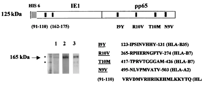
lymphocytes against HCMV in ex vivo procedures, we investi-gated the construction of a chimeric protein rather than the separate production of both antigens. Figure 1 shows the SDS-PAGE profile of the purified IE1-pp65 protein produced in insect cells as described in Materials and Methods. Despite a calculated 125-kDa molecular size the purified protein mi-grated at a position corresponding to about 165 kDa. This one-step procedure allowed us to obtain about 60 mg of puri-fied protein from 1 liter of insect cells grown in plastic culture flasks and to circumvent the difficulties we usually encountered in the separate purification procedures of pp65 and IE1. The protein was specifically recognized in Western blotting exper-iments with both anti-IE1 and anti-pp65 monoclonal antibod-ies as indicated (Fig. 1).
Induction of proliferation of PBMC and frequency of CD4ⴙ
T cells producing intracellular IFN-␥in response to IE1-pp65.
We first tested whether IE1-pp65 was capable of inducing proliferation of PBMC from HCMV-positive blood donors. As shown in Fig. 2, proliferation was observed in PBMC from blood donors. Proliferation assays using PBMC from HCMV-seronegative blood donors were consistently negative (data not shown).
We then assessed whether CD4⫹T cells from latently
in-fected healthy blood donors were activated by IE1-pp65. PBMC from randomly selected blood donors were incubated in the presence of control Ag, purified IE1-pp65, or total HCMV Ag and were analyzed by flow cytometry for
intracel-lular IFN-␥production.
Data obtained from each individual are shown in Fig. 3.
HCMV-positive (number⫽15) and control HCMV-negative
(number⫽3) blood donors were studied. Each dot represents
[image:3.612.133.472.71.215.2]an experimental value obtained from a single blood donor. Values obtained with different antigens for each individual blood donor are artificially connected with a line. As shown in Fig. 3, 100% (15 of 15) of the HCMV-positive blood donors
FIG. 1. Production and purification of IE1-pp65 fusion protein. Sf9 insect cells were infected with IE1-pp65 recombinant baculoviruses. The recombinant Ag was purified through Ni2⫹chromatography and analyzed by SDS-PAGE. The gel was submitted to either Coomassie blue staining (1) or immunoblotting with anti-IE1 (2) and anti-pp65 (3) monoclonal antibodies. Molecular size standards (175 and 83 kDa) migrated as indicated (ⴱ) at the left end of the gel. The apparent molecular size of IE1-pp65 was 165 kDa, compared to a theorical 125 kDa. Positions and sequences of MHC class I and class II epitopes used in further functional experiments are as indicated on the schematic representation of IE1-pp65.
on November 9, 2019 by guest
http://jvi.asm.org/
responded to total HCMV Ag (mean⫽ 0.36; range, 0.03 to
10.2). Seventy-three percent (11 of 15) (mean⫽0.31; range, 0
to 1.2) responded to IE1-pp65. In 6 out of 15 blood donors the
percentages of IFN-␥⫹CD4⫹T cells obtained with IE1-pp65
were equal to or above those obtained with total HCMV Ag.
These results confirm that the frequencies of CD4⫹ T cells
against total HCMV Ag are high (8, 32) and suggest that they are dominated by responses to two major proteins, IE1 and pp65.
Activation of IE1-specific CD4ⴙ T-cell clones by purified
IE1-pp65.The efficiency of activation of clonal CD4⫹T cells
by the IE1-pp65 protein was evaluated using IE1-specific T-cell clones. Cytotoxicity was tested on EBV-transformed B cells and U373MG-CIITA cells pulsed with Ag. As shown in Fig.
4A, IE1-specific clonal FzD3 and FzF5 CD4⫹T cells (7)
effi-ciently lysed HLA-DR3 (Steinlin) EBV B cells pulsed with the relevant IE1 (91-110) peptide. Likewise, IE1-specific FzD3 and
FzF5 CD4⫹T-cell clones lysed U373MG-CIITA cells pulsed
with IE1 (91-110) peptide or with IE1-pp65.
Figure 4B shows that the FzD11 CD4⫹ T-cell clone
pro-duced IFN-␥in response to IE1-pp65-pulsed U373MG-CIITA
cells as antigen-presenting cells. Peptide IE1 (91-110) was used
as a positive control. IFN-␥production was Ag dose
depen-dent. Likewise, U373MG-CIITA cells infected with HCMV
induced IFN-␥production by the FzD11 CD4⫹T-cell clone.
The amount of IFN-␥produced corresponded to an ⬃1 nM
concentration of protein.
Stimulation of CD8ⴙT lymphocytes from
[image:4.612.61.285.70.231.2]HCMV-seroposi-tive donors with IE1-pp65 and expansion of anti-pp65 CTL. Restimulation of anti-pp65 CTL from HCMV-positive donors
FIG. 2. Induction of proliferation of PBMC by purified IE1-pp65. PBMC from HCMV-seropositive blood donors were cultured for 6 days either in the absence of stimulus (none) or in the presence of IE1-pp65 (10g/ml) or pokeweed mitogen (PWM) as a control. Pro-liferation was measured by evaluation of [3H]TdR incorporation.
[image:4.612.334.523.79.372.2]FIG. 3. Frequency of CD4⫹T cells producing intracellular IFN-␥ in response to IE1-pp65. PBMC from HCMV-seropositive blood do-nors were incubated for 16 in the presence of IE1-pp65 purified pro-tein. Total HCMV Ag (positive control) and negative control Ag were used for comparison. Cells were then collected, permeabilized, and analyzed for intracellular IFN-␥ production by flow cytometry. To follow the data from each individual, values obtained with different Ag for each blood donor are artificially connected with a line. Statistical analysis was performed using the EPI INFO 6 software (Centers for Disease Control and Prevention).
FIG. 4. Activation of CD4⫹T-cell clones in the presence of IE1-pp65 fusion protein. (A) U373MG-CIITA and EBV B cells were incubated in the presence of either IE1-pp65 or IE1 (91-110) peptide, as indicated, for 16 h. Cells were then washed and incubated in the presence of FzD3 (lighter gray) and FzF5 (darker gray) IE1-specific T-cell clones in a classical51Cr release cytotoxicity assay (effector-to-target ratio⫽10). (B) U373MG-CIITA cells were either infected with HCMV (multiplicity of infection⫽2) (Œ) or incubated in the presence of IE1-pp65 (■) or IE1 (91-110) peptide (}) at the indicated concen-trations for 12 h and fixed with glutaraldehyde. IE1-specific CD4⫹ T-cell clone FzD11 was added to the culture for 24 h and IFN-␥in the supernatant was measured.
on November 9, 2019 by guest
http://jvi.asm.org/
[image:4.612.56.291.444.630.2]by using synthetic peptide is well documented. We recently
reported the expansion of CD8⫹ T cells by using HLA-A2
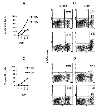
(N9V) and HLA-B35 (I9Y) binding peptides (2). These spe-cific cells were able to kill HCMV-infected targets. Figure 5A shows that HLA-A2-restricted CTL from donor V (HLA-A2, -B35) stimulated with N9V were able to specifically kill N9V-pulsed but not S9V-N9V-pulsed targets. We then assessed whether IE1-pp65 could induce restimulation of anti-pp65 CTL. Figure 5C shows that PBMC with anti-N9V specificity from donor V were stimulated following incubation with IE1-pp65. In con-trast, restimulation of CTL directed against the S9V peptide was significantly lower than that observed with N9V. Then the percentage of peptide-specific CTL was determined by flow cytometry, using the tetrameric N9V–HLA-A2 complex.
Stain-ing of anti-N9V CD8⫹T cells is shown for both N9V
peptide-and IE1-pp65-based restimulation protocols in Fig. 5B peptide-and D, respectively. The percentage of anti-N9V CTL was evaluated after 10 days (panels a) and 26 days (panels b) of culture as
indicated. Starting percentages of anti-N9V CD8⫹T cells in
donor V were 0.08% versus 0.02% using the GP100 tetramer (data not shown). Figure 5 shows that, from day 10 to day 26
of culture, anti-N9V CD8⫹ T cells increased from 2.5% (B,
panel a) to 4.9% (B, panel b) for peptide-raised CTL and from 0.46% (D, panel a) to 1.32% (D, panel b) for IE1-pp65-raised
CTL. These figures are reflected in51Cr release assays which
show a higher percentage of lysis at a given effector-to-target ratio with peptide-derived CTL than with IE1-pp65-derived CTL (Fig. 5A and B). This discrepancy may be explained by a competitive interaction between multiple processed peptides derived from IE1-pp65 for binding with HLA-A2 molecules. This competition presumably would not occur when using N9V peptide, which is directed to the surface HLA-A2. Using the
same approach, we assessed whether IE1-pp65-derived CD8⫹
[image:5.612.137.463.70.435.2]T cells from donor V contained HLA-B35-restricted CTL di-rected against peptide I9Y. Figure 6A shows that at the time of blood drawing, which differs from that of Fig. 5, IE1-pp65- as well as I9Y-raised CTL were able to kill I9Y-pulsed targets. It is noteworthy that even though IE1-pp65 was used at a much
FIG. 5. Stimulation of HCMV-seropositive PBMC with IE1-pp65 allowed expansion of HLA-A2-restricted anti-pp65 CTL. PBMC from donor V (HLA-A2, -B35) were stimulated in vitro with either N9V peptide (A) or IE1-pp65 (C). Then, anti-N9V CTL cytotoxicity was determined by using HLA-matched EBV-transformed B cells incubated with N9V or S9V peptides (100 nM) as targets in a51Cr release assay. The percentage of N9V-derived (B) and IE1-pp65-derived (D) anti-N9V CTL was determined by flow cytometry after 10 days (panels a) and 26 days (panels b) of culture by using double labeling with anti-CD8 and HLA-A2–N9V tetramer. HLA-A2–GP100 tetramer was used as a negative control. E/T, effector-to-target ratio.
on November 9, 2019 by guest
http://jvi.asm.org/
lower concentration (40 nM) than the peptide (5M), about 10 times fewer effector cells were necessary to obtain an iden-tical percentage of lysis.
To confirm that the IE1-pp65 protein was able to expand
anti-pp65 CD8⫹ CTL regardless of HLA restriction, PBMC
from donor M (HLA-B7, -B35) were used in restimulation protocols. To this end, PBMC were restimulated with IE1-pp65 at day 0 and day 14 and then collected at day 26. Then
51Cr release CTL assays were performed using HLA-B7
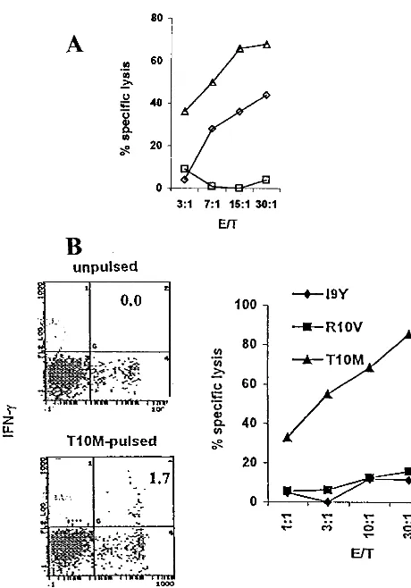
tar-gets pulsed either with I9Y, an HLA-B35 binding peptide, or with R10V and T10M, two HLA-B7 binding peptides. Figure 6B shows that only targets pulsed with the HLA-B7-matched peptide T10M were killed. This indicated that the other known HLA-B7 epitope, R10V, was not immunogenic in donor M at the time of blood drawing. In addition, we did not succeed in
Fig. 6B show that CD8⫹IFN-␥⫹cells were observed only when
effector cells had been restimulated with T10M (1.7%) com-pared to unrestimulated cells (0.0%).
The capacity of IE1-pp65 to sensitize EBV B target cells to
cytotoxicity by CD8ⴙT cells is not due to contaminating
pep-tides.Finally, even though the procedure for IE1-pp65
purifi-cation included affinity and gel filtration chromatography, both excluding contaminating peptides, we assessed whether the capacity of IE1-pp65 to restimulate CTL was not due to a bystander effect involving direct binding of contaminating epitopes. EBV B cells that were pulsed with 100 nM purified IE1-pp65 were not sensitive to lysis by HLA-A2-restricted anti-N9V CTL (data not shown), suggesting that the protein has to be processed for stimulation.
DISCUSSION
Since it has been shown that IE1 and pp65 are targets for
CD4⫹and CD8⫹T cells, we suggested that the construction of
a chimeric protein, IE1-pp65, could be beneficial to in vitro restimulation and expansion of anti-HCMV precursors. In this study we report the production of a recombinant fusion pro-tein, IE1-pp65, and its purification from insect cells infected with recombinant IE1-pp65 baculovirus. We show that this
recombinant protein specifically stimulates both CD4⫹ and
CD8⫹ T cells and allows expansion of CD8⫹ T cells from
PBMC.
It appears from our present data that the frequency of CD4⫹
T cells against IE1-pp65 is an important component of the
CD4⫹T-cell response against CMV total Ag. The high
fre-quency of CD4⫹T cells against CMV Ag has been previously
suggested from bulk culture (3, 7) and demonstrated in limit-ing dilutions (8) and in flow cytometry assays uslimit-ing synthetic peptides and detection of intracellular cytokines (32). How-ever, limiting dilution analyses are thought to underestimate the frequencies of specific precursors when compared with techniques using the intracellular detection of cytokines (14, 15, 32). On the other hand, the use of a synthetic peptide, although powerful to identify epitopes, cannot stimulate the response against the whole spectrum of potential epitopes of a protein in a single bulk culture. Although our present experi-ments do not assess the response to single proteins (i.e., IE1 versus pp65), they allow us to compare the response to two soluble recombinant proteins on the one hand and the whole range of CMV proteins on the other hand.
The recombinant chimeric IE1-pp65 protein induced the
proliferation and production of IFN-␥by CD4⫹T cells.
There-FIG. 6. Stimulation of HLA-B35- and HLA-B7-restricted CTL from PBMC using IE1-pp65. PBMC from donor V (HLA-A2, -B35) and donor M (HLA-B7, -B35) were stimulated at days 0 and 14 with either HLA-A2 binding N9V (䊐) or HLA-B35 binding I9Y ({) pep-tides or IE1-pp65 (‚) for donor V and IE1-pp65 for donor M. At day 26, effectors were incubated with HLA-B35 EBV-transformed B cells incubated with I9Y for donor V (A) and with HLA-B7 EBV-trans-formed B cell targets pulsed with either R10V or T10M (both HLA-B7 binding peptides) or with I9Y as indicated for donor M (B). Cytotox-icity was determined in a51Cr release assay at different effector-to-target (E/T) ratios in the presence of a 100 nM final concentration of the respective peptide. At the same time, the frequency of anti-T10M CTL from donor M was evaluated by flow cytometry from the per-centage of IFN-␥⫹CD8⫹double-labeled cells contained in PBMC (B). The figure is representative of three different experiments.
on November 9, 2019 by guest
http://jvi.asm.org/
[image:6.612.59.289.69.398.2]fore, it is possible to use this recombinant protein to activate
CD4⫹T cells specific for the separate proteins, for example
with the goal of using expanded populations for cellular ther-apy. The derivation of clones will be particularly useful because of their targeted specificity to chosen Ag without potentially harmful reactivity against self-derived Ag. Intracellular
stain-ing for IFN-␥using IE1-pp65 did not allow us to determine
whether CD4⫹T cells activated in PBMC were specific for IE1
or pp65. However, we could demonstrate that the IE1-specific
CD4⫹T-cell clones used in this study, although they were not
derived from the chimera protein, responded strongly to
IE1-pp65 both by cytotoxic activity and IFN-␥production.
Con-versely, the specificity of CD4⫹ T-cell clones obtained using
IE1-pp65 may be tested with separate reagents expressing one of the proteins separately. Most remarkably, infected
U373MG-CIITA cells induced IFN-␥production by an
IE1-specific CD4⫹clone, FzD11 (Fig. 4B), and by others (data not
shown). This observation needs to be extended to macro-phages and dendritic cells which have been shown to be in-fected in vitro using clinical isolates (12). It suggests the
po-tential in vivo reactivity of IE1-specific CD4⫹ T cells to
HCMV-infected or -reactivating (29) cells in vivo.
It is well established that the in vitro restimulation of MHC
class II-restricted CD4⫹effector T cells from PBMC occurs in
the presence of exogenous antigen. Other reports have shown that in viral infections, endogenous Ag can be presented to
MHC II-restricted CD4⫹T cells (13, 16, 21). This possibility
will be investigated for the presentation of nuclear Ag IE1. Increasing numbers of studies show that MHC class I presen-tation of exogenous antigen also occurs but is restricted to dendritic cells (for a review, see reference 24). Surprisingly, the use of soluble IE1-pp65 allowed restimulation of CTL from PBMC of different HLA donors. According to published data showing that dendritic cells have the capacity to deliver exog-enous Ag in the cytosolic pathway, as we recently demon-strated using pp65-positive apoptotic bodies (2), we propose that peripheral dendritic cells may be responsible for IE1-pp65 uptake and presentation to anti-pp65 CTL. The identity of
dendritic subsets such as CD11c⫹ cells (17) and molecular
mechanisms involved in Ag delivery are not within the scope of this paper and remain to be explored. The use of IE1-pp65 is particularly relevant if we consider that some donors are not responsive to known dominant epitopes as described recently for the HLA-A2 binding peptide N9V (30). Furthermore, such donors may respond to subdominant HLA-A2 epitopes (30) or to unpredicted peptides as shown in a report by Kern and colleagues (15). As there is no fully reliable method to predict epitopes from peptide sequences, as previously shown by oth-ers (15, 30), the use of an entire protein as opposed to peptides stands a better chance to expand CTL. Since pp65 and IE1 variabilities are low among different wild-type strains of HCMV (6, 25, 30) we suspect that IE1-pp65 may target any HCMV strain independently of HLA haplotype.
It remains to be determined whether our procedure using IE1-pp65 would also induce stimulation of anti-IE1 CTL. This would be of interest since, as recently reported, frequencies of
CD8⫹T cells directed against IE1 and pp65 seem to be of
similar magnitude (11, 15, 27). This could exclude disadvan-tages of using isolated IE1 or pp65 since it has been shown recently that some individuals were often reactive to only one
of the two proteins but not both (15, 26). Moreover, the ben-efits of using IE1-pp65 are high compared with the use of HCMV-infected feeder cells if we consider that IE1 processing could be blocked by neosynthesized pp65 (10) and that the viral US proteins may exert a blockade of Ag presentation, as extensively reviewed previously (22).
In conclusion, the present work shows that both IE1 and pp65 are available within the chimera protein as in vitro targets for T-lymphocyte responses. Production of the IE1-pp65 pro-tein under good manufacturing practice conditions will allow in vitro amplification of cells specific for these HCMV proteins which are major targets of the immune system. In conclusion, our approach may provide an alternative to the use of infec-tious virus in cell immunotherapy.
ACKNOWLEDGMENTS
This work was supported by institutional grants from INSERM, the Midi Pyre´ne´es region, and Biovectors Therapeutics. J.-L.D. was sup-ported by Association pour la Recherche sur le Cancer (ARC) and the Etablissement Franc¸ais des Greffes. P.R. was supported by a grant from Assistance Publique des Hoˆpitaux de Paris.
We acknowledge Georges Cassar for technical assistance in flow cytometry and Sylvie Darche (Institut Pasteur, Paris) for tetramer technology. We thank Sanofi-Synthelabo (Labe`ge, France) for supply-ing us with recombinant IL-2 and IL-7.
REFERENCES
1.Alp, N. J., T. D. Allport, J. Van Zanten, B. Rodgers, J. G. Sissons, and L. K. Borysiewicz.1991. Fine specificity of cellular immune responses in humans to human cytomegalovirus immediate-early 1 protein. J. Virol.65:4812–4820. 2.Arrode, G., C. Boccaccio, J. Lule, S. Allart, N. Moinard, J. P. Abastado, A. Alam, and C. Davrinche.2000. Incoming human cytomegalovirus pp65 (UL83) contained in apoptotic infected fibroblasts is cross-presented to CD8⫹T cells by dendritic cells J. Virol.74:10018–10024.
3.Beninga, J., B. Kropff, and M. Mach.1995. Comparative analysis of fourteen individual human cytomegalovirus proteins for helper T cell response. J. Gen. Virol.76:153–160.
4.Bousso, P., A. Casrouge, J. D. Altman, M. Haury, J. Kanellopoulos, J. P. Abastado, and P. Kourilsky.1998. Individual variations in the murine T cell response to a specific peptide reflect variability in naive repertoires. Immu-nity9:169–178.
5.Britt, W.1996. Cytomegalovirus, p. 2493–2523.InB. N. Fields, D. M. Knipe, and P. M. Howley (ed.), Fields virology. Lippincott-Raven Publishers, Phil-adelphia, Pa.
6.Brytting, M., J. Wahlberg, J. Lundeberg, B. Wahren, M. Uhlen, and V. A. Sundqvist.1992. Variations in the cytomegalovirus major immediate-early gene found by direct genomic sequencing. J. Clin. Microbiol.30:955–960. 7.Davignon, J. L., P. Castanie, J. A. Yorke, N. Gautier, D. Clement, and C.
Davrinche.1996. Anti-human cytomegalovirus activity of cytokines produced by CD4⫹T-cell clones specifically activated by IE1 peptides in vitro. J. Virol. 70:2162–2169.
8.Davignon, J. L., D. Clement, J. Alriquet, S. Michelson, and C. Davrinche. 1995. Analysis of the proliferative T cell response to human cytomegalovirus major immediate-early protein (IE1): phenotype, frequency and variability. Scand. J. Immunol.41:247–255.
9.Gavin, M. A., M. J. Gilbert, S. R. Riddell, P. D. Greenberg, and M. J. Bevan. 1993. Alkali hydrolysis of recombinant proteins allows for the rapid identi-fication of class I MHC-restricted CTL epitopes. J. Immunol.151:3971–3980. 10.Gilbert, M. J., S. R. Riddell, B. Plachter, and P. D. Greenberg.1996. Cyto-megalovirus selectively blocks antigen processing and presentation of its immediate-early gene product. Nature383:720–722.
11. Gyulai, Z., V. Endresz, K. Burian, S. Pincus, J. Toldy, W. I. Cox, C. Meri, S. Plotkin, and K. Berencsi.2000. Cytotoxic T lymphocyte (CTL) responses to human cytomegalovirus pp65, IE1-Exon4, gB, pp150, and pp28 in healthy individuals: reevaluation of prevalence of IE1-specific CTLs. J. Infect. Dis. 181:1537–1546.
12. Jahn, G., S. Stenglein, S. Riegler, H. Einsele, and C. Sinzger.1999. Human cytomegalovirus infection of immature dendritic cells and macrophages. Intervirology42:365–372.
13. Jaraquemada, D., M. Marti, and E. O. Long.1990. An endogenous process-ing pathway in vaccinia virus-infected cells for presentation of cytoplasmic antigens to class II-restricted T cells. J. Exp. Med.172:947–954.
14. Kern, F., I. P. Surel, C. Brock, B. Freistedt, H. Radtke, A. Scheffold, R. Blasczyk, P. Reinke, J. Schneider-Mergener, A. Radbruch, P. Walden, and
on November 9, 2019 by guest
http://jvi.asm.org/
19. McLaughlin-Taylor, E., H. Pande, S. J. Forman, B. Tanamachi, C. R. Li, J. A. Zaia, P. D. Greenberg, and S. R. Riddell.1994. Identification of the major late human cytomegalovirus matrix protein pp65 as a target antigen for CD8⫹virus-specific cytotoxic T lymphocytes. J. Med. Virol.43:103–110. 20.Mocarski, E. E.1996. Cytomegaloviruses and their replication, p. 2447–2492. InB. N. Fields, D. M. Knipe, and P. M. Howley (ed.), Fields virology. Lippincott-Raven Publishers, Philadelphia, Pa.
21. Munz, C., K. L. Bickham, M. Subklewe, M. L. Tsang, A. Chahroudi, M. G. Kurilla, D. Zhang, M. O’Donnell, and R. M. Steinman. 2000. Human CD4(⫹) T lymphocytes consistently respond to the latent Epstein-Barr virus nuclear antigen EBNA1. J. Exp. Med.191:1649–1660.
22. Ploegh, H. L.1998. Viral strategies of immune evasion. Science280:248–253. 23. Plotkin, S. A.1994. Vaccines for varicella-zoster virus and cytomegalovirus:
recent progress. Science265:1383–1385.
24. Reimann, J., and R. Schirmbeck.1999. Alternative pathways for processing exogenous and endogenous antigens that can generate peptides for MHC class I-restricted presentation. Immunol. Rev.172:131–152.
25. Retiere, C., B. M. Imbert, G. David, P. Courcoux, and M. M. Hallet.1998. A polymorphism in the major immediate-early gene delineates groups among cytomegalovirus clinical isolates. Virus Res.57:43–51.
rus protein pp65 that are conserved between eight strains of the virus. J. Immunol.163:5512–5518.
31. Sun, Q., K. E. Pollok, R. L. Burton, L. J. Dai, W. Britt, D. J. Emanuel, and K. G. Lucas.1999. Simultaneous ex vivo expansion of cytomegalovirus and Epstein-Barr virus-specific cytotoxic T lymphocytes using B-lymphoblastoid cell lines expressing cytomegalovirus pp65. Blood94:3242–3250.
32. Waldrop, S. L., C. J. Pitcher, D. M. Peterson, V. C. Maino, and L. J. Picker. 1997. Determination of antigen-specific memory/effector CD4⫹T cell fre-quencies by flow cytometry: evidence for a novel, antigen-specific homeo-static mechanism in HIV-associated immunodeficiency. J. Clin. Investig. 99:1739–1750.
33. Walter, E. A., P. D. Greenberg, M. J. Gilbert, R. J. Finch, K. S. Watanabe, E. D. Thomas, and S. R. Riddell.1995. Reconstitution of cellular immunity against cytomegalovirus in recipients of allogeneic bone marrow by transfer of T-cell clones from the donor. N. Engl. J. Med.333:1038–1044. 34. Wills, M. R., A. J. Carmichael, K. Mynard, X. Jin, M. P. Weekes, B. Plachter,
and J. G. Sissons.1996. The human cytotoxic T-lymphocyte (CTL) response to cytomegalovirus is dominated by structural protein pp65: frequency, spec-ificity, and T-cell receptor usage of pp65-specific CTL. J. Virol.70:7569– 7579.