Copyright © 2002, American Society for Microbiology. All Rights Reserved.
Modulation of the Cell Division Cycle by Human Papillomavirus
Type 18 E4
Tomomi Nakahara,† Akiko Nishimura,‡ Masakazu Tanaka, Takaharu Ueno,
Akinori Ishimoto, and Hiroyuki Sakai*
Laboratory of Gene Analysis, Department of Viral Oncology, Institute for Virus Research, Kyoto University, Sakyo-Ku, Kyoto 606-8507, Japan
Received 11 March 2002/Accepted 20 July 2002
The life cycle of human papillomaviruses (HPVs) is tightly coupled to the differentiation program of their host epithelial cells. HPV E4 gene expression is first observed in the parabasal layers of squamous epithelia, suggesting that the E4 gene product contributes to the mechanism of differentiation-dependent virus replica-tion, although its biological function remains unclear. We analyzed the effect of HPV type 18 E4 on cell proliferation and found that E4 expression induced cell cycle arrest at the G2/M boundary. The functional
region of E4 necessary for the growth arrest activity was located in the central portion of the molecule, and this activity was independent of the E4-mediated collapse of cytokeratin intermediate filament structures.
Human papillomaviruses (HPVs) infect epithelial cells, causing hyperproliferative lesions such as warts and condylo-mas. Over 70 HPV types have been identified (12). According to their tissue tropism, they are categorized into two major groups, the cutaneous and mucosal HPVs. The mucosal HPVs are further grouped into high-risk and low-risk types. Lesions caused by high-risk HPVs have a propensity to progress to malignant tumors, most prominently cervical carcinomas. In contrast, lesions caused by low-risk HPVs have a much lower risk for malignant progression (51).
The HPV E6 and E7 oncoproteins are known to play key roles in HPV-associated cancer formation (21, 22, 24, 33, 34). This fact is supported by the finding that most HPV-positive cancer cells maintain the expression of E6 and E7 (4, 52). E6 forms a complex with p53 and, in combination with the cellular ubiquitin ligase E6AP, induces the degradation of p53 through the proteasome (25, 48, 49, 60). E7 binds to and functionally inactivates pRB (20, 35). Both p53 and pRB play critical roles as regulators of the cell cycle and apoptosis. By inhibiting these and other regulatory mechanisms, HPV E6 and E7 allow for the accumulation of genetic mutations and the survival of mu-tated cells (8, 42, 61). E6 and E7 expression also contributes to the immortalization of infected cells; E6 can enhance telo-merase activity through an unknown mechanism (29, 57), whereas E7 inhibits a p16ink4A-dependent pathway that limits
cellular proliferation in epithelial cells (28). It is generally believed that these functions of the viral oncoproteins contrib-ute in important ways to HPV-induced cancer formation. How-ever, it is likely that additional viral genes also contribute to the establishment of hyperproliferative potentially precancerous lesions caused by high-risk HPVs. In addition, the relatively
low incidence of malignant progression of high-risk HPV-pos-itive lesions indicates that additional mutations of cellular genes may also be necessary for malignant progression.
Studies of the viral life cycle are important for understand-ing the process of HPV-induced cancer formation. HPV in-fects the basal layer cells of cutaneous or mucosal membranes. The HPV genome is maintained in an episomal state and at a low copy number in basal cells. The basal cells divide, and the descendant cells move to the upper layers of the epithelium and undergo a program of terminal differentiation. The HPV genome is amplified to a high copy number in differentiated cells, late genes are expressed, and virus particle formation is observed. This tight association between viral replication and the host cell differentiation program is a prominent feature of the HPV life cycle. Several reports have described the regula-tory mechanisms of viral replication and how these are con-nected to the cellular differentiation program. Certain tran-scription factors, including YY1, skn-1, and CDP, have been implicated in differentiation-dependent HPV gene expression (1–3, 30, 38, 63).
The expression profile of the HPV E4 gene is clearly linked to cellular differentiation status (6, 9, 10, 13, 14, 36, 39). The E4 open reading frame is located in the region of early viral genes, such as E1 and E2, even though other studies have suggested that its expression pattern is more characteristic of a late gene. E4 expression is first detected in the parabasal layers, where vegetative HPV DNA replication is initiated. The E4 protein is encoded by a spliced E1∧E4 mRNA and is expressed as a fusion protein with the N terminus of the E1 protein. It has been reported that in HPV type 1 (HPV1)-infected warts, the E4 protein accumulates at high levels and constitutes approx-imately 20% of the total proteins (6, 13). Since the expression of E4 is tightly linked to host cell differentiation, it likely plays an important role in the viral life cycle (13, 26). Several bio-logical activities of E4 have been reported; the best defined is its association with cytokeratins and the concomitant destabi-lization of cytokeratin networks (17, 41, 44, 47, 54). It has been proposed that this activity of E4 may contribute to the efficient transmission of HPV, as progeny virus is shed within terminally
* Corresponding author. Mailing address: Laboratory of Gene Anal-ysis, Department of Viral Oncology, Institute for Virus Research, Kyoto University, Sakyo-Ku, Kyoto 606-8507, Japan. Phone: 81-75-751-4010. Fax: 81-75-751-3995. E-mail: [email protected].
† Present address: McArdle Laboratory for Cancer Research, Uni-versity of Wisconsin—Madison, Madison, WI 53706.
‡ Present address: Department of Pathology, Harvard Medical School, Boston, MA 02115.
10914
on November 8, 2019 by guest
http://jvi.asm.org/
differentiated epithelial squamae (17). These studies, however, do not rule out the possibility that E4 may have additional biological activities that also contribute to the viral life cycle. In this report, high-risk HPV-derived E4 proteins were ex-pressed in cultured cells, and their effect on cell growth was analyzed. E4 expression suppressed cell growth and arrested cell cycle progression at the G2/M boundary. No apoptosis was
observed, however. Moreover, this activity of E4 was indepen-dent of its ability to disrupt cytokeratin networks. Our findings suggest that E4 may also contribute to the regulation of the viral life cycle by modulating the host cell division cycle.
MATERIALS AND METHODS
Cell cultures and transfection.HeLa, CV1, and C33A cells were maintained in Dulbecco’s modified Eagle medium supplemented with 10% heat-inactivated fetal bovine serum. DNA transfections were performed by using a standard calcium phosphate precipitation method (37). Cells (2⫻105) were seeded in a 6-cm dish 1 day prior to transfection. Plasmid and carrier DNAs (total, 10g) were incubated with 500l of HEPES-buffered saline transfection buffer (140 mM NaCl, 0.75 mM Na2HPO4, 25 mM HEPES, 110 mM CaCl2[pH 6.90]) for 30 min at room temperature and then added to a culture dish. At 20 h after transfection, cells were washed once with phosphate-buffered saline (PBS), and fresh growth medium was added.
Plasmids.The E1∧E4 cDNA expression plasmid was created by joining the corresponding E1 and E4 sequences by PCR. The information for the HPV16 E1∧E4 (16E4) and HPV18 E1∧E4 (18E4) mRNA structures was obtained from the HPV database (18). The E1∧E4 DNA was cloned into plasmid pCMV-FLAG1 (Stratagene Inc., San Diego, Calif.) to fuse a FLAG epitope tag to the N terminus. The FLAG-E1∧E4 sequence was subsequently transferred to a pCMV4 expression plasmid (37). A series of truncation mutations were intro-duced by using a PCR-mediated mutagenesis strategy (11); see Fig. 4A for the individual E1∧E4 mutants. A commercially available green fluorescent protein (GFP) expression plasmid, pGreenLantern-1 (Invitrogen Corp., Carlsbad, Cal-if.), was used to mark transfected cells. For transfection experiments, herring sperm DNA (Roche Diagnostics GmbH, Mannheim, Germany) was used as carrier DNA. The human immunodeficiency virus type 1 (HIV-1) Vpr gene was amplified from an HIV-1 LAI DNA clone (40) by PCR, and a FLAG-Vpr expression plasmid was constructed as described for the FLAG-E1∧E4 expres-sion plasmid.
Immunofluorescence microscopy and immunoblotting.The transfected cells were fixed with 3% paraformaldehyde and permeabilized with 0.1% Triton X-100. FLAG-tagged E4 proteins were detected with an anti-FLAG polyclonal antibody (Sigma, St. Louis, Mo.) and Alexa Fluor 488 goat anti-rabbit immuno-globulin G (Molecular Probes, Eugene, Oreg.). Cytokeratins were detected with an anticytokeratin 8/18 monoclonal antibody (MAb) (Progen Biotechnik GmbH, Heidelberg, Germany) and Alexa Fluor 546 goat anti-mouse immunoglobulin G (Molecular Probes). Nuclei were visualized by staining with 4⬘,6⬘ -diamidino-2-phenylindole dihydrochloride (DAPI) (Nacalai Tesque, Inc., Kyoto, Japan).
Whole-cell extracts were prepared from transfected cells with radioimmuno-precipitation (RIPA) buffer (150 mM NaCl, 50 mM Tris-HCl [pH 8.0], 0.1% sodium dodecyl sulfate [SDS], 1% Nonidet P-40, 0.5% sodium deoxycholate, 1 mM dithiothreitol, 16g of benzamidine HCl/ml, 10g of phenanthroline/ml, 10g of aprotinin/ml, 10g of leupeptin/ml, 10g of pepstatin A/ml, 1 mM phenylmethylsulfonyl fluoride). A 500-l portion of RIPA buffer was added directly to a culture dish and incubated for 20 min at 4°C with rocking. The cell lysate was transferred to a microcentrifuge tube and centrifuged at 12,000⫻gfor 10 min at 4°C. The supernatant was used as the detergent-soluble fraction. The pellet was resuspended in 500l of SDS-polyacrylamide gel electrophoresis (PAGE) sample buffer (50 mM Tris-HCl [pH 6.8], 2% SDS, 100 mM dithio-threitol, 10% glycerol, 0.1% bromophenol blue) and used as the non-deter-gent-soluble fraction. Both fractions were analyzed by SDS–15% PAGE and transferred to a Hybond-P polyvinylidene difluoride membrane (Amersham Pharmacia Biotech, Ltd., Little Chalfont, England). An anti-FLAG MAb M5 (Sigma) was used for the detection of FLAG-tagged proteins. For visualization, a chemiluminescence detection reagent (Lumi-Light Western blotting substrate; Roche Diagnostics GmbH) was used.
Flow cytometry.Cells were cotransfected with E4 or Vpr expression plasmid (2g) and GFP expression plasmid pGreenLantern-1 (0.5g), fixed with 1% paraformaldehyde–ethanol for 10 min at various times after transfection, and treated with 0.5 mg of RNase A/ml–0.1% Triton X-100 in PBS for 30 min at
37°C. Cells were stained with 0.1 mg of propidium iodide (PI)/ml–PBS and analyzed by flow cytometry (FACScan; Becton Dickinson and Company, Frank-lin Lakes, N.J.). To selectively analyze transfected cells, only GFP-positive cells were counted for PI staining.
The detection of apoptotic cells was performed with an Annexin-V-FLUOS staining kit (Roche Diagnostics GmbH).
Growth suppression assays.HeLa cells (2⫻105) transfected with 2g of E4 or Vpr expression plasmid, 0.5g of pGreenLantern-1, and 7.5g of carrier DNA were seeded at a density of 0.5⫻105cells/6-cm dish at 24 h after trans-fection. Cell numbers per dish were determined at various times after transfec-tion. Only live cells, determined by trypan blue exclusion, were counted.
RESULTS
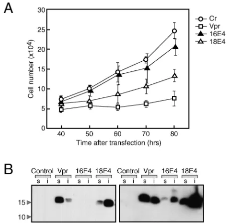
[image:2.587.307.534.74.297.2]18E4 expression induces cellular growth suppression.The HPV E4 gene product is expressed as an E1∧E4 fusion protein containing the 5 amino-terminal amino acids of E1 at its N terminus (18). To investigate the biological activity of high-risk HPV E1∧E4 proteins, we constructed FLAG-tagged E1∧E4 expression plasmids for HPV16 and HPV18. HeLa cells were transfected with these E1∧E4 expression plasmids, and the growth of the transfected cells was analyzed (Fig. 1A). In each experiment, transfection efficiency was monitored by cotrans-fection of a GFP expression plasmid, and over 80% of the cells were confirmed to be GFP positive. Cell growth was dimin-ished in 18E4-expressing cells compared to control transfected cells. Similar results were obtained for the expression of an
FIG. 1. Growth-inhibitory effect of HPV E4. (A) HeLa cells (2⫻ 105) were transfected with 2g of expression plasmid for 16E4, 18E4,
or HIV-1 Vpr. A GFP expression plasmid, pGreenLantern-1 (Cr), was cotransfected to distinguish the transfected cells. GFP-positive cells were counted at the indicated times after transfection. Transfection efficiency was over 80%. The results shown are derived from an ex-periment performed in triplicate; error bars indicate standard devia-tions. (B) At 48 h after transfection, cell extracts were prepared with RIPA buffer. Both detergent-soluble (s) and non-detergent-soluble (i) fractions were analyzed by SDS-PAGE, and E4 and Vpr were detected by immunoblot analysis with an anti-FLAG antibody. The two panels represent short (left) and long (right) exposures of the same film. The positions of molecular weight markers (in thousands) are indicated on the left.
on November 8, 2019 by guest
http://jvi.asm.org/
untagged E1∧E4 protein (data not shown).16E4 also showed weak but significant activity for suppressing cell growth. As a positive control for growth suppression, we expressed a FLAG-tagged HIV-1 Vpr regulatory protein which was previously reported to interfere with cellular proliferation (31). The HIV-1 Vpr protein was able to induce growth suppression to a similar extent as 18E4 (Fig. 1A).
Next, we analyzed the levels of expression of the 16E4, 18E4, and HIV-1 Vpr proteins by immunoblot analysis with FLAG epitope-specific antibodies. 18E4 and HIV-1 Vpr were ex-pressed at similar levels, whereas the steady-state levels of 16E4 appeared much lower (Fig. 1B), a finding which might account for its weak growth-inhibitory activity (Fig. 1A). As expected, the Vpr protein was localized to the nucleus (data not shown) and was recovered in the detergent-soluble fraction (Fig. 1B). In contrast, 18E4 and 16E4 were mostly cytoplasmic (data not shown) and were detected in the non-detergent-soluble fraction (Fig. 1B).
18E4 expression induces G2cell cycle arrest.We next ana-lyzed whether 16E4 and 18E4 caused growth arrest at a specific phase of the cell cycle. It has been reported that HIV-1 Vpr induces growth arrest at the G2phase of the cell cycle (23, 27).
HeLa cells transfected with the Vpr expression plasmid exhib-ited an increased proportion of cells in G2/M at 40 h after
transfection, consistent with G2 arrest (Fig. 2A). This effect
was even more dramatic at later times. Like Vpr-expressing cells, HeLa cells transfected with 18E4 also exhibited a dra-matic increase in the G2/M population (Fig. 2A). As expected,
HeLa cells transfected with 16E4 showed a less dramatic in-crease in the G2/M population, particularly at early times after
transfection, but this increase became more noticeable at later times (Fig. 2A). The difference between 16E4 and 18E4 is most likely a consequence of the lower steady-state levels of 16E4 in the transfected HeLa cells (Fig. 1B). These results suggest that 18E4 is expressed at higher levels and therefore that its bio-logical activities are more noticeable. Hence, we focused most-ly on 18E4 for the rest of our studies. The results suggest that, like the expression of HIV-1 Vpr protein, the expression of high-risk HPV E1∧E4 proteins can induce growth arrest in cells at the G2/M boundary of the cell division cycle.
The HeLa cell line is a cervical cancer cell line that contains integrated copies of HPV18, and the HPV18 E6 and E7 on-coproteins are expressed in these cells (5, 50). High-risk HPV E6 and E7 proteins are known to subvert G1/S as well as G2/M
checkpoint control, raising the possibility that these viral onco-proteins may somehow mask additional effects of E4 expres-sion (19, 32, 53, 58, 59, 62). To clarify this issue, we analyzed the effects of E4 expression in two other, HPV-negative epi-thelium-derived cell lines: C33A, an HPV-negative human
cer-FIG. 2. Cell cycle analysis of E4- and Vpr-expressing cells. (A) HeLa cells were transfected with 2g of Vpr or the indicated E4 expression plasmid in combination with pGreenLantern-1. Cells were collected at the indicated times after transfection, and the DNA con-tents were analyzed by flow cytometry (FACScan). Nuclei were stained
with PI. Only the population of transfected GFP-positive cells was counted. Closed and open arrows indicate peaks corresponding to G1
and G2/M phases, respectively. The ratio of G2/M to G1is shown in
each panel. (B) CV1 and C33A cells were transfected with an 18E4 expression plasmid, and the cell cycle profiles were analyzed as de-scribed for panel A. Cr, control. (C) Induction of apoptosis by E4 or Vpr expression, as determined by Annexin V staining at 50 h after transfection. The samples were costained with PI for the detection of necrotic cells. Apoptotic cells are positive for Annexin V staining (FL1-H) and negative for PI staining (FL2-H).
on November 8, 2019 by guest
http://jvi.asm.org/
vical carcinoma cell line, and CV1, a monkey kidney-derived cell line. 18E4 induced G2arrest in both cell lines, indicating
that the G2/M growth arrest effect observed was independent
of E6 and E7 functions (Fig. 2B).
In addition to inducing G2arrest, Vpr is also known to elicit
an apoptotic response (55, 56). Vpr expression increased the Annexin V-positive cell population in HeLa cells. The samples were costained with PI to distinguish apoptotic cells from ne-crotic cells. Vpr-induced Annexin V-positive cells were nega-tive for PI staining, indicating that Vpr induced apoptotic cell death (Fig. 2C). In contrast to the expression of Vpr, the expression of 18E4 did not increase the incidence of apoptosis in HeLa cells. It has been reported that the mechanisms by which Vpr induces G2 arrest and apoptosis are linked (55).
Since 18E4 induced an increase in the G2/M population almost
as efficiently as HIV-1 Vpr (Fig. 1) but exhibited no proapo-ptotic activity, these results suggest that the mechanism of G2
cell cycle arrest induction by E1∧E4 is different from that of Vpr.
Under normal conditions, cell cycle arrest at the G2/M
boundary is induced in response to DNA damage and unfa-vorable environmental conditions. If high-risk HPV E1∧E4 expression induces similar G2 arrest, then cells will be
main-tained at the G2 phase without DNA synthesis or nuclear
division. Microscopic observations of 18E4-transfected HeLa and CV1 cells at 70 h after transfection revealed that the cells and the nuclei were frequently enlarged. Especially with HeLa cells, cells containing several gigantic nuclei and micronuclei were observed (Fig. 3). These results suggest that 18E4-ex-pressing cells may continue to synthesize DNA and undergo aberrant nuclear division. This notion is supported by the FACScan results shown in Fig. 2A, where a cell population with a greater than 4N DNA content was observed upon ex-pression of 18E4 in HeLa cells.
The central region of the 18E4 protein is necessary for G2/M
cell cycle arrest.Next, we mapped the domain(s) in the 18E4 protein that is required for the G2arrest effect. Thus far, most
studies on E4 proteins have been performed with HPV1 and
HPV16 (18). Mapping studies with16E4 have indicated that the N-terminal region is required for an association with cyto-keratin, whereas the C-terminal region is involved in the col-lapse of the cytokeratin intermediate filament (IF) network as well as E4 oligomerization (16, 43, 45). The functional motifs of 18E4 were extrapolated by sequence comparison with 16E4 and are shown in Fig. 4A. Given the importance of the N- and C-terminal domains of 16E4, we constructed mutant 18E4 expression plasmids with serial deletions at the N and C ter-mini.
E4 is a relatively small protein, consisting of approximately 100 amino acids, and deletions introduced into such a small protein may severely affect protein stability. Therefore,
steady-FIG. 3. Morphology of E4-expressing cells. The cells were trans-fected with 4g of an 18E4 expression plasmid, fixed with 50% ac-etone–methanol at 70 h after transfection, and stained with methylene blue. Nuclei were visualized by DAPI staining. Control cells were transfected with the empty expression plasmid.
FIG. 4. Identification of the region within the 18E4 protein that is necessary for G2/M arrest. (A) Schematic representation of the mutant
18E4 proteins tested. The truncated regions of the FLAG-tagged 18E4 protein are represented by black-gray bars. The motifs and functional regions reported for 16E4 are indicated (45). The corresponding re-gions in 18E4 are represented by gray boxes, and the amino acid sequence alignment of the conserved regions is presented. (B) Steady-state levels of mutant 18E4 proteins expressed in transfected cells. Both detergent-soluble (s) and non-detergent-soluble (i) fractions were analyzed by SDS-PAGE, followed by immunoblot analysis with an anti-FLAG antibody. The two panels represent short (upper) and long (lower) exposures of the same film. The positions of molecular weight markers (in thousands) are indicated on the left. (C) Cell cycle profiles of HeLa cells expressing the indicated 18E4 mutants. Cells were transfected with 4g of an E4 expression plasmid and pGreen-Lantern-1. FACScan analysis was performed as described in the legend to Fig. 2A. Cr, control; WT, wild type.
on November 8, 2019 by guest
http://jvi.asm.org/
state levels of the mutant 18E4 proteins were analyzed prior to functional analyses. HeLa cells were transfected with the mu-tant FLAG-18E4 plasmids, and steady-state levels of the E4 proteins in both detergent-soluble and non-detergent-soluble fractions were determined by immunoblot analysis with an anti-FLAG MAb (Fig. 4B). Only amino-terminal mutant N⌬20 was expressed at levels similar to those of wild-type 18E4 protein. Steady-state levels of amino-terminal mutants N⌬5 and N⌬10 were reduced but, like the N⌬20 mutant, these mutants showed distributions in detergent-soluble and non-detergent-soluble fractions similar to those of wild-type 18E4 protein. Steady-state levels of the N⌬30 mutant and all the C-terminal deletion mutants were dramatically reduced, and these proteins were mainly detected in the detergent-soluble fraction, suggesting that the intracellular localization of these mutant proteins is altered compared to that of wild-type 18E4 protein.
To analyze which region(s) of E4 may be essential for the induction of G2 arrest, we expressed the mutant proteins in
HeLa cells and analyzed cell cycle distribution by FACScan analysis at 50 h posttransfection. Except for the expression of the N⌬30 and C⌬60 mutants, the expression of each of the other mutant 18E4 proteins induced a marked increase in the G2/M population similar to that seen with wild-type 18E4
pro-tein (Fig. 4C). The effect of C⌬70 was relatively weak, but this result is difficult to interpret due to the low level of expression of this mutant. When we transfected larger amounts of expres-sion plasmids, we observed a more dramatic increase in the G2/M population with C⌬60 but not with N⌬30 (data not
shown). However, from these results it is not possible to dis-criminate whether N⌬30 lacks the essential domain to induce G2 arrest or whether the level of expression of the mutant
protein was too low to exhibit its G2arrest effect. Nevertheless,
these results indicate that the domain of 18E4 essential to induce G2/M arrest is minimally contained within amino acids
21 to 59 of 18E4.
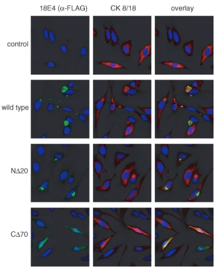
Cytokeratin association is dispensable for E4-mediated G2
arrest.In contrast to wild-type 18E4 and the N⌬5, N⌬10, and N⌬20 mutants of 18E4, which were detected in the non-deter-gent-soluble fraction, the C⌬70 and C⌬80 mutants were found mostly in the detergent-soluble fraction (Fig. 4C). Since each of these mutants was similarly active in inducing G2/M arrest,
it appears unlikely that the ability of E4 to form non-detergent-soluble complexes is linked to its growth-inhibitory function. To analyze this notion in more detail, we performed an immu-nofluorescence analysis of cytokeratin 8/18 and E4 in HeLa cells transfected with a control vector or a vector expressing wild-type 18E4 or the N⌬20 or C⌬70 mutant (Fig. 5). Cyto-plasmic aggregates presumably corresponding to the non-de-tergent-soluble E4 material were detected in cells expressing wild-type 18E4. Cytokeratin 8/18 staining of normal cells re-vealed a filamentous network throughout the cytoplasm. In 18E4-expressing cells, the staining pattern for cytokeratin 8/18 was markedly different, suggesting that the IF network might have collapsed, presumably due to 18E4 expression. In support of this notion, cytokeratin 8/18 and 18E4 colocalized in cyto-plasmic aggregates, as was previously reported for 16E4 (17). Although the N⌬20 mutant formed aggregates similar to those seen with wild-type 18E4, there was less dramatic colocaliza-tion with cytokeratin 8/18-positive structures. The C⌬70
mu-tant displayed a diffuse cytoplasmic and perinuclear staining pattern, similar to that of cytokeratin 8/18 in normal cells. The cytokeratin IF network was maintained intact in C⌬ 70-express-ing cells. These results are consistent with studies on 16E4 that mapped the domains for multimerization to the C terminus and the ability to interact with cytokeratins to the N terminus (45). These results indicate that neither the ability of E4 to form oligomeric aggregates nor its association with cytokera-tins is required for the ability of E4 to arrest cells in G2/M. The
failure of the C⌬70 mutant to induce the formation of E1∧E4 aggregates and/or collapse of the cytokeratin network may be attributable to the low levels of expression of this mutant. Regardless, since this mutant still efficiently induced G2/M
arrest, the formation of E1∧E4 aggregates and/or collapse of the cytokeratin network are not necessary prerequisites for the E1∧E4-mediated induction of G2/M growth arrest.
DISCUSSION
[image:5.587.314.527.70.335.2]The life cycle of papillomaviruses is tightly associated with the differentiation program of epithelial cells via mechanisms that remain to be fully investigated. The ability of HPVs to induce hyperproliferation of epithelial cells is an important aspect of the oncogenic activities of high-risk HPVs. To fully understand the molecular mechanisms that contribute to this activity of HPVs, it is important to investigate the cross talk between HPVs and their epithelial host cells on a molecular
FIG. 5. Immunofluorescence analysis of 18E4 proteins and cyto-keratin 8/18 in transfected cells. Cells transfected with wild-type, N⌬20, and C⌬70 18E4 expression plasmids (5g) were fixed at 48 h after transfection. E4 proteins (left panels) and cytokeratin (CK) (mid-dle panels) were detected by using anti-FLAG and anticytokeratin 8/18 antibodies, respectively. Nuclei (right panels) were visualized by DAPI
staining.
on November 8, 2019 by guest
http://jvi.asm.org/
level. The expression of the E4 gene product is coupled to the keratinocyte differentiation program, and it has been proposed that E4 contributes to the viral life cycle by interacting with cytokeratins and inducing collapse of the IF network (17, 43). This process presumably facilitates the egress of progeny HPVs when they are shed within the terminally differentiated keratinocyte squamae. In this report, we have analyzed the effect of E4 expression on cell growth. These studies revealed that 18E4 can induce G2/M growth arrest. Since this effect was
observed in HPV18-positive and -negative cells, the E6 and E7 oncoproteins do not interfere with this activity of E4. The growth arrest activity of 18E4 was independent its ability to induce collapse of the cytokeratin IF network.
Both 16E4 and 18E4 induced G2/M arrest in HeLa cells,
suggesting that growth inhibition may be an activity shared by other HPV E4 proteins. Compared to that of 18E4, the activity of 16E4 appeared weaker, but this finding may be a conse-quence of the lower levels of 16E4 expression in HeLa cells. Additional experiments will be necessary to define the molec-ular targets of 18E4 that mediate this G2/M arrest.
The 18E4 protein was as efficient as HIV-1 Vpr in suppress-ing the growth of HeLa cells. However, in contrast to that of Vpr, 18E4-mediated growth suppression was not accompanied by apoptosis. Since the p53 pathway in HeLa cells is function-ally compromised by the expression of HPV18 E6, Vpr-in-duced apoptosis is likely independent of p53. Although the mechanism is largely unknown, it has been suggested that the abilities of Vpr to induce G2/M arrest and apoptosis may be
functionally related (55). Since no apoptosis was observed in 18E4-expressing cells, it may be concluded that HIV-1 Vpr-induced G2/M arrest and 18E4-induced G2/M arrest are
me-diated by different mechanisms. Alternatively, it is possible that 18E4 contains additional antiapoptotic activities that prevent G2/M-arrested cells from being eliminated by apoptosis (46).
18E4-expressing HeLa cells showed marked nuclear en-largement and multinucleation, and FACScan analysis re-vealed the appearance of a hyperploid cell population. This finding suggests that a small percentage of 18E4-expressing HeLa cells, after arresting at the G2/M boundary for various
amounts of time, will undergo additional rounds of DNA syn-thesis and nuclear division in the absence of cellular division. It is interesting that high-risk HPV E6 and E7 can also induce aberrant DNA synthesis in G2/M-arrested epithelial cells (58).
Moreover, functional disruption of p53 or p21cip1/WAF1by
high-risk HPV E6 also causes DNA rereplication in G2/M-arrested
cells (7). Hence, it is possible that the observed multinucle-ation and the appearance of hyperploid cells are a manifesta-tion of compromised G2/M checkpoint control caused by
HPV18 E6 and/or E7 proteins that are synthesized from the integrated HPV18 genomes in HeLa cells. HPV E4 gene ex-pression in an HPV-associated lesion is first detected in the parabasal layers, and it is expected that the other viral genes, including those for E6 and E7, are coexpressed in these cells. Hence, the cooperative interaction between E4-expressing cells and E6- and E7-expressing cells that likely causes the endoreduplication and multinucleation observed in 18E4-ex-pressing HeLa cells may be physiologically significant.
Our studies showed that the N- and C-terminal domains of 18E4 fulfill functions similar to those in 16E4. The N-terminal domain contributes to the ability of E4 to interact with
cyto-keratins, whereas the C-terminal domain is necessary for E4 to form cytoplasmic aggregates and to cause the collapse of the cytokeratin IF network structure (43, 45). The C-terminal do-main of E4 is also the site of interaction for a recently identi-fied putative RNA helicase, E4-DBP (15). Our analysis showed that the C- and N-terminal domains were largely dispensable for the growth-suppressing activity of E4. Our mapping exper-iments revealed that a sequence of approximately 40 amino acid residues located in the central region of E4 may constitute a novel domain necessary for E4-mediated G2/M growth
ar-rest. Additional mutagenesis experiments with this domain will be necessary to map the structural determinants of this novel activity of E4 in greater detail and to perform a targeted search for cellular factors that can interact with this region of E4.
ACKNOWLEDGMENTS
We thank Atsue Ueda for technical assistance and manuscript prep-aration. We are grateful to Karl Mu¨nger (Harvard Medical School) for critical review of the manuscript.
This research was supported in part by grants to H.S. from the Japanese Ministry of Education, Culture, Sports, Science and Tech-nology.
REFERENCES
1.Ai, W., J. Narahari, and A. Roman.2000. Yin yang 1 negatively regulates the differentiation-specific E1 promoter of human papillomavirus type 6. J. Vi-rol.74:5198–5205.
2.Ai, W., E. Toussaint, and A. Roman.1999. CCAAT displacement protein binds to and negatively regulates human papillomavirus type 6 E6, E7, and E1 promoters. J. Virol.73:4220–4229.
3.Andersen, B., A. Hariri, M. R. Pittelkow, and M. G. Rosenfeld.1997. Char-acterization of Skn-1a/i POU domain factors and linkage to papillomavirus gene expression. J. Biol. Chem.272:15905–15913.
4.Baker, C. C., W. C. Phelps, V. Lindgren, M. J. Braun, M. A. Gonda, and P. M. Howley.1987. Structural and transcriptional analysis of human papil-lomavirus type 16 sequences in cervical carcinoma cell lines. J. Virol.61:
962–971.
5.Boshart, M., L. Gissmann, H. Ikenberg, A. Kleinheinz, W. Scheurlen, and H. zur Hausen.1984. A new type of papillomavirus DNA: its presence in genital cancer biopsies and in cell lines derived from cervical cancer. EMBO J.
3:1151–1157.
6.Breitburd, F., O. Croissant, and G. Orth.1987. Expression of human pap-illomavirus type 1 E4 gene products in warts, p. 115–122.InB. M. Steinberg, J. L. Brandsma, and L. B. Taichman (ed.), Papillomavirus: cancer cells. Cold Spring Harbor Laboratory Press, Cold Spring Harbor, N.Y.
7.Bunz, F., A. Dutriauux, C. Lengauer, T. Waldman, S. Zhou, J. P. Brown, J. M. Sedivy, K. W. Kinzler, and B. Vogelstein.1998. Requirement for p53 and p21 to sustain G2arrest after DNA damage. Science282:1497–1501. 8.Butz, K., C. Denk, A. Ullmann, M. Scheffner, and F. Hoppe-Seyler.2000.
Induction of apoptosis in human papillomavirus positive cancer cells by peptide aptamers targeting the viral E6 oncoprotein. Proc. Natl. Acad. Sci. USA97:6693–6697.
9.Chow, L. T., M. Nasseri, S. M. Wolinsky, and T. R. Broker.1987. Human papillomavirus type 6 and 11 mRNAs from genital condylomata acuminata. J. Virol.61:2581–2588.
10.Chow, L. T., S. S. Reilly, T. R. Broker, and L. B. Taichman.1987. Identifi-cation and mapping of human papillomavirus type 1 RNA transcripts recov-ered from plantar warts and infected epithelial cell cultures. J. Virol.61:
1913–1918.
11.Cormack, B.1987. Mutagenesis of cloned DNA, p. 8.5.1–8.5.9.InF. M. Ausubel, R. Brent, R. E. Kingston, D. D. Moore, J. G. Seidman, J. A. Smith, and K. Struhl (ed.), Current protocols in molecular biology. John Wiley & Sons, Inc., New York, N.Y.
12.de Villiers, E. M.1994. Human pathogenic papillomavirus types: an update. Curr. Top. Microbiol. Immunol.184:1–12.
13.Doorbar, J., D. Campbell, R. J. Grand, and P. H. Gallimore.1986. Identi-fication of the human papilloma virus-1a E4 gene products. EMBO J.5:355– 362.
14.Doorbar, J., I. Coneron, and P. H. Gallimore.1989. Sequence divergence yet conserved physical characteristics among the E4 proteins of cutaneous hu-man papillomaviruses. Virology172:51–62.
15.Doorbar, J., R. C. Elston, S. Napthine, K. Raj, E. Medcalf, D. Jackson, N. Coleman, H. M. Griffin, P. Masterson, S. Stacey, Y. Mengistu, and J. Dun-lop.2000. The E1E4 protein of human papillomavirus type 16 associates with
on November 8, 2019 by guest
http://jvi.asm.org/
a putative RNA helicase through sequences in its C terminus. J. Virol.74:
10081–10095.
16.Doorbar, J., S. Ely, N. Coleman, M. Hibma, D. H. Davies, and L. Crawford.
1992. Epitope-mapped monoclonal antibodies against the HPV16E1-E4 pro-tein. Virology187:353–359.
17.Doorbar, J., S. Ely, J. Sterling, C. McLean, and L. Crawford.1991. Specific interaction between HPV-16 E1-E4 and cytokeratins results in collapse of the epithelial cell intermediate filament network. Nature352:824–827. 18.Doorbar, J., and G. Myers.1996. The E4 protein, p. III58–III80.InG. L.
Myers et al. (ed.), The human papillomaviruses. 1996 Compendium. Los Alamos National Laboratory, Los Alamos, N.Mex.
19.Duensing, S., L. Y. Lee, A. Duensing, J. Basile, S. Piboonniyom, S. Gonzalez, C. P. Crum, and K. Munger.2000. The human papillomavirus type 16 E6 and E7 oncoproteins cooperate to induce mitotic defects and genomic in-stability by uncoupling centrosome duplication from the cell division cycle. Proc. Natl. Acad. Sci. USA97:10002–10007.
20.Dyson, N., P. M. Howley, K. Munger, and E. Harlow.1989. The human papilloma virus-16 E7 oncoprotein is able to bind to the retinoblastoma gene product. Science243:934–937.
21.Halbert, C. L., G. W. Demers, and D. A. Galloway.1991. The E7 gene of human papillomavirus type 16 is sufficient for immortalization of human epithelial cells. J. Virol.65:473–478.
22.Hawley-Nelson, P., K. H. Vousden, N. L. Hubbert, D. R. Lowy, and J. T. Schiller.1989. HPV16 E6 and E7 proteins cooperate to immortalize human foreskin keratinocytes. EMBO J.8:3905–3910.
23.He, J., S. Choe, R. Walker, P. Di Marzio, D. O. Morgan, and N. R. Landau.
1995. Human immunodeficiency virus type 1 viral protein R (Vpr) arrests cells in the G2phase of the cell cycle by inhibiting p34cdc2activity. J. Virol.
69:6705–6711.
24.Hudson, J. B., M. A. Bedell, D. J. McCance, and L. A. Laimins.1990. Immortalization and altered differentiation of human keratinocytes in vitro by the E6 and E7 open reading frames of human papillomavirus type 18. J. Virol.64:519–526.
25.Huibregtse, J. M., M. Scheffner, and P. M. Howley.1991. A cellular protein mediates association of p53 with the E6 oncoprotein of human papilloma-virus type 16 or 18. EMBO J.10:4129–4135.
26.Jareborg, N., and S. Burnett.1991. Immunofluorescent detection of bovine papillomavirus E4 antigen in the cytoplasm of cells permissive in vitro for viral DNA amplification. J. Gen. Virol.72:2269–2274.
27.Jowett, J. B., V. Planelles, B. Poon, N. P. Shah, M. L. Chen, and I. S. Chen.
1995. The human immunodeficiency virus type 1vprgene arrests infected T cells in the G2⫹M phase of the cell cycle. J. Virol.69:6304–6313. 28.Kiyono, T., S. A. Foster, J. I. Koop, J. K. McDougall, D. A. Galloway, and
A. J. Klingelhutz.1998. Both Rb/p16INK4ainactivation and telomerase ac-tivity are required to immortalize human epithelial cells. Nature396:84–88. 29.Klingelhutz, A. J., S. A. Foster, and J. K. McDougall.1996. Telomerase activation by the E6 gene product of human papillomavirus type 16. Nature
380:79–82.
30.Kukimoto, I., and T. Kanda.2001. Displacement of YY1 by differentiation-specific transcription factor hSkn-1a activates the P670promoter of human papillomavirus type 16. J. Virol.75:9302–9311.
31.Macreadie, I. G., L. A. Castelli, D. R. Hewish, A. Kirkpatrick, A. C. Ward, and A. A. Azad.1995. A domain of human immunodeficiency virus type 1 Vpr containing repeated H(S/F)RIG amino acid motifs causes cell growth arrest and structural defects. Proc. Natl. Acad. Sci. USA92:2770–2774. 32.Martin, L. G., G. W. Demers, and D. A. Galloway.1998. Disruption of the
G1/S transition in human papillomavirus type 16 E7-expressing human cells is associated with altered regulation of cyclin E. J. Virol.72:975–985. 33.Matlashewski, G., J. Schneider, L. Banks, N. Jones, A. Murray, and L.
Crawford.1987. Human papillomavirus type 16 DNA cooperates with acti-vated ras in transforming primary cells. EMBO J.6:1741–1746.
34.Munger, K., W. C. Phelps, V. Bubb, P. M. Howley, and R. Schlegel.1989. The E6 and E7 genes of the human papillomavirus type 16 together are necessary and sufficient for transformation of primary human keratinocytes. J. Virol.
63:4417–4421.
35.Munger, K., B. A. Werness, N. Dyson, W. C. Phelps, E. Harlow, and P. M. Howley.1989. Complex formation of human papillomavirus E7 proteins with the retinoblastoma tumor suppressor gene product. EMBO J.8:4099–4105. 36.Nasseri, M., R. Hirochika, T. R. Broker, and L. T. Chow.1987. A human papilloma virus type 11 transcript encoding an E1-E4 protein. Virology
159:433–439.
37.Nishimura, A., T. Ono, A. Ishimoto, J. J. Dowhanick, M. A. Frizzell, P. M. Howley, and H. Sakai.2000. Mechanisms of human papillomavirus E2-mediated repression of viral oncogene expression and cervical cancer cell growth inhibition. J. Virol.74:3752–3760.
38.O’Connor, M. J., W. Stunkel, C. H. Koh, H. Zimmermann, and H. U. Bernard.2000. The differentiation-specific factor CDP/Cut represses tran-scription and replication of human papillomaviruses through a conserved silencing element. J. Virol.74:401–410.
39.Palefsky, J. M., B. Winkler, J. P. Rabanus, C. Clark, S. Chan, V. Nizet, and G. K. Schoolnik.1991. Characterization of in vivo expression of the human
papillomavirus type 16 E4 protein in cervical biopsy tissues. J. Clin. Investig.
87:2132–2141.
40.Peden, K.1991. Changes in growth properties on passage in tissue culture of viruses derived from infectious molecular clones of HIV-1 LAI. Virology
185:661–672.
41.Pray, T. R., and L. A. Laimins.1995. Differentiation-dependent expression of E1-E4 proteins in cell lines maintaining episomes of human papilloma-virus type 31b. Virology206:679–685.
42.Reznikoff, C. A., C. Belair, E. Savelieva, Y. Zhai, K. Pfeifer, T. Yeager, K. J. Thompson, S. DeVries, C. Bindley, M. A. Newton, et al.1994. Long-term ge-nome stability and minimal genotypic and phenotypic alterations in HPV16 E7-, but not E6-, immortalized human uroepithelial cells. Genes Dev.8:2227–2240. 43.Roberts, S., I. Ashmole, L. J. Gibson, S. M. Rookes, G. J. Barton, and P. H. Gallimore.1994. Mutational analysis of human papillomavirus E4 proteins: identification of structural features important in the formation of cytoplas-mic E4-cytokeratin networks in epithelial cells. J. Virol.68:6432–6445. 44.Roberts, S., I. Ashmole, G. D. Johnson, J. W. Kreider, and P. H. Gallimore.
1993. Cutaneous and mucosal human papillomavirus E4 proteins form in-termediate filament-like structures in epithelial cells. Virology197:176–187. 45.Roberts, S., I. Ashmole, S. M. Rookes, and P. H. Gallimore.1997. Mutational analysis of the human papillomavirus type 16 E1-E4 protein shows that the C terminus is dispensable for keratin cytoskeleton association but is involved in inducing disruption of the keratin filaments. J. Virol.71:3554–3562. 46.Rogel-Gaillard, C., F. Breitburd, and G. Orth.1992. Human papillomavirus
type 1 E4 proteins differing by their N-terminal ends have distinct cellular localizations when transiently expressed in vitro. J. Virol.66:816–823. 47.Rogel-Gaillard, C., G. Pehau-Arnaudet, F. Breitburd, and G. Orth.1993.
Cytopathic effect in human papillomavirus type 1-induced inclusion warts: in vitro analysis of the contribution of two forms of the viral E4 protein. J. Investig. Dermatol.101:843–851.
48.Scheffner, M., J. M. Huibregtse, R. D. Vierstra, and P. M. Howley.1993. The HPV-16 E6 and E6-AP complex functions as a ubiquitin-protein ligase in the ubiquitination of p53. Cell75:495–505.
49.Scheffner, M., B. A. Werness, J. M. Huibregtse, A. J. Levine, and P. M. Howley.1990. The E6 oncoprotein encoded by human papillomavirus types 16 and 18 promotes the degradation of p53. Cell63:1129–1136.
50.Schwarz, E., U. K. Freese, L. Gissmann, W. Mayer, B. Roggenbuck, A. Stremlau, and H. zur Hausen.1985. Structure and transcription of human papillomavirus sequences in cervical carcinoma cells. Nature314:111–114. 51.Shah, K. V., and P. M. Howley.1996. Papillomavirus, p. 2077–2109.InB. N.
Fields, D. M. Knipe, and P. M. Howley (ed.), Virology, 3rd ed. Lippincott-Raven Press, Ltd., New York, N.Y.
52.Shirasawa, H., Y. Tomita, S. Sekiya, H. Takamizawa, and B. Simizu.1987. In-tegration and transcription of human papillomavirus type 16 and 18 se-quences in cell lines derived from cervical carcinomas. J. Gen. Virol.68:583–591. 53.Slebos, R. J., M. H. Lee, B. S. Plunkett, T. D. Kessis, B. O. Williams, T. Jacks, L. Hedrick, M. B. Kastan, and K. R. Cho.1994. p53-dependent G1 arrest involves pRB-related proteins and is disrupted by the human papillo-mavirus 16 E7 oncoprotein. Proc. Natl. Acad. Sci. USA91:5320–5324. 54.Sterling, J. C., J. N. Skepper, and M. A. Stanley.1993. Immunoelectron
mi-croscopical localization of human papillomavirus type 16 L1 and E4 proteins in cervical keratinocytes cultured in vivo. J. Investig. Dermatol.100:154–158. 55.Stewart, S. A., B. Poon, J. B. Jowett, and I. S. Chen.1997. Human immu-nodeficiency virus type 1 Vpr induces apoptosis following cell cycle arrest. J. Virol.71:5579–5592.
56.Stewart, S. A., B. Poon, J. Y. Song, and I. S. Chen.2000. Human immuno-deficiency virus type 1 Vpr induces apoptosis through caspase activation. J. Virol.74:3105–3111.
57.Stoppler, H., D. P. Hartmann, L. Sherman, and R. Schlegel.1997. The human papillomavirus type 16 E6 and E7 oncoproteins dissociate cellular telomerase activity from the maintenance of telomere length. J. Biol. Chem.
272:13332–13337.
58.Thomas, J. T., and L. A. Laimins.1998. Human papillomavirus oncoproteins E6 and E7 independently abrogate the mitotic spindle checkpoint. J. Virol.
72:1131–1137.
59.Thompson, D. A., G. Belinsky, T. H. Chang, D. L. Jones, R. Schlegel, and K. Munger.1997. The human papillomavirus-16 E6 oncoprotein decreases the vigilance of mitotic checkpoints. Oncogene15:3025–3035.
60.Werness, B. A., A. J. Levine, and P. M. Howley.1990. Association of human papillomavirus type 16 and 18 E6 proteins with p53. Science248:76–79. 61.White, A. E., E. M. Livanos, and T. D. Tlsty.1994. Differential disruption of
genomic integrity and cell cycle regulation in normal human fibroblasts by the HPV oncoproteins. Genes Dev.8:666–677.
62.Xiong, Y., D. Kuppuswamy, Y. Li, E. M. Livanos, M. Hixon, A. White, D. Beach, and T. D. Tlsty.1996. Alteration of cell cycle kinase complexes in human papillomavirus E6- and E7-expressing fibroblasts precedes neoplastic transformation. J. Virol.70:999–1008.
63.Yukawa, K., K. Butz, T. Yasui, H. Kikutani, and F. Hoppe-Seyler.1996. Regulation of human papillomavirus transcription by the differentiation-dependent epithelial factor Epoc-1/skn-1a. J. Virol.70:10–16.