i
VALIDATION OF MODIFIED POSSUM SCORING
SYSTEM IN PERFORATIVE PERITONITIS
Dissertation submitted to
The Tamil Nadu M.G.R Medical University
Chennai- 600032
In partial fulfillment of the
Regulations of the award of degree of
M.S. General Surgery
Department of General Surgery
Coimbatore Medical College Hospital
Coimbatore - 641018
ii
DECLARATION BY THE CANDIDATE
I hereby declare that this dissertation entitled “VALIDATION OF
MODIFIED POSSUM SCORING SYSTEM IN PERFORATIVE
PERITONITIS” is a bonafide and genuine research work carried out by me
under the guidance of Prof. Dr. D.N. RENGANATHAN M.S, Professor of
Surgery, Department of General Surgery, Coimbatore Medical College and
Hospital, Coimbatore.
DATE: Signature of the candidate
iii
CERTIFICATE
This is to certify that the dissertation entitled “VALIDATION OF
MODIFIED POSSUM SCORING SYSTEM IN PERFORATIVE
PERITONITIS” is a bonafide research work done by Dr. Ambarish Chatterjee
in partial fulfilment of the requirement for the Degree of M.S. in General
Surgery.
Signature of the Guide Signature of H.O.D
PROF. DR. D. N. RENGANATHAN M.S PROF. DR. V. ELANGO M.S
Professor of Surgery, Head of Department of Surgery,
Department of General Surgery, Coimbatore Medical College
Coimbatore Medical College and Hospital, and Hospital,
Coimbatore. Coimbatore.
DATE: Signature of Dean
PLACE: COIMBATORE Coimbatore Medical College and
vii
ACKNOWLEDGEMENT
First I thank God for the strength and all the blessings I have received.
It gives me immense pleasure to express a deep sense of gratitude and
indebtedness that I feel towards my teacher and guide, whose ceaseless support
helped me through the entire course of this research and the dissertation,
Professor DR. D. N. RENGANATHAN M.S, Department of Surgery,
Coimbatore Medical College and Hospital, Coimbatore. I am grateful to him for
the valuable suggestions, guidance, great care and attention to minute details
that he has so willingly shown in the preparation of this dissertation.
I owe great respect and gratitude to Professor DR. V. ELANGO M.S,
the Head of Department of General Surgery and also to
Professor DR. P.V. VASANTHA KUMAR M.S Retired Professor and HOD.
I deeply thank DR. V.S. VENKADESAN M.S, D.A, Senior Assistant
Professor, and DR. S. DURAIRAJ M.S Assistant Professor and
DR. R. NARAYANA MOORTHY M.S Assistant Professor of Surgery
scholarly suggestions and all round encouragement.
I acknowledge my gratitude to our Registrar DR. T. SRINIVASAN M.S
and all my Assistant Professors of Department of Surgery for their
encouragement and support.
I am thankful to ETHICS COMMITTEE of Coimbatore Medical
viii
I am extremely grateful to DR. R. VIMALA M.D (PATH), Dean,
Coimbatore Medical College and Hospital, Coimbatore for her valuable help
and co-operation.
I thank my parents, Mr. Soumitra Chatterjee and Mrs. Shruti Chatterjee
and my sister, Ms. Asmita Chatterjee whose support, prayers and sacrifices
sailed me through this endeavour. I also thank my beloved wife,
Dr. Hemangi Chatterjee for the unconditional love and support.
I thank all my post graduate colleagues for their whole hearted support
during this study period.
DATE: Signature of the candidate
ix
CONTENTS
Contents
Page No.
1. INTRODUCTION 1
2. AIMS AND OBJECTIVE 4
3. REVIEW OF LITERATURE 5
4. METHODOLOGY 32
5. RESULTS 39
6. DISCUSSION 77
7. SUMMARY 80
8. CONCLUSION 82
9. BIBLIOGRAPHY 84
10. PHOTOGRAPHS 40-42
11. ANNEXURE
PROFORMA I
CONSENT FORM Iii
MASTER CHART Viii
x
LIST OF TABLES
Sr. No. TABLES PAGE No.
1 Indications 39
2 Types of surgeries 43
3 List of complications 45
4 Comparison of observed and expected mortality rates 47
5 Comparison of observed and expected morbidity rates 49
6 Comparison of mean total POSSUM score and outcome 51
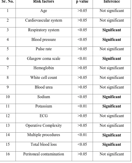
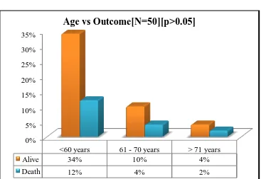
7 Risk factors 52
8 Age 54
9 Cardiovascular system 55
10 Respiratory System 56
11 Blood pressure 57
12 Pulse rate 58
13 Glasgow coma scale 59
14 Hemoglobin 60
15 White cell count 61
16 Blood urea level 62
17 Serum sodium 63
18 Serum potassium 64
xi
20 Operative Complexity 66
21 Multiple procedures 67
22 Total blood loss 68
23 Peritoneal soiling 69
24 Presence of malignancy 70
25 Mode of surgery 71
26 Perforation to operation time 72
27 Co-morbid status 74
28 Predicted risk of mortality 76
xii
LIST OF GRAPHS
Sr. No. GRAPHS PAGE No.
1 Indications 39
2 Types of surgeries 43
3 Outcome of surgery 44
4 Complications 46
5 Individual complications 46
6 Comparison of observed and expected mortality rates 48
7 Comparison of observed and expected morbidity rates 50
8 Comparison of mean total POSSUM score and outcome 51
9 Age 54
10 Cardiovascular system 55
11 Respiratory System 56
12 Blood Pressure 57
13 Pulse rate 58
14 Glasgow coma scale 59
15 Hemoglobin 60
16 White cell count 61
17 Blood Urea level 62
18 Serum sodium 63
xiii
20 Operative Complexity 66
21 Multiple procedures 67
22 Total blood loss 68
23 Peritoneal soiling 69
24 Presence of malignancy 70
25 Mode of surgery 71
26 Perforation to operation time 73
xiv
LIST OF PHOTOGRAPHS
Sr. No. PHTOGRAPHS PAGE No.
1 Perforated duodenal ulcer 40
2 Ileal perforation 40
3 Appendicular perforation with abscess 41
4 Perforated appendix with fecolith 41
xv
ABSTRACT
Background and objectives: Perforative peritonitis carries considerable
morbidity and mortality. Even if patients reach the hospital at the earliest, still
the post operative period is unpredictable most of the times. It therefore
becomes necessary for a scoring system that predicts the post-operative period.
POSSUM (Physiological and Operative Severity Score for the enUmeration of
Mortality and Morbidity) helps in predicting the post-operative morbidity and
mortality in these patients. POSSUM scores are calculated on the basis of 12
physiological factors and 6 operative factors. In our study we have included two
more factors which are specifically important in perforative peritonitis. These
two factors are the perforation to operation time, which is the time duration
between the occurrence of perforation and operation; and the presence of
co-morbidity. The presence of these factors significantly affects the post-operative
status of the patients. Through this prospective study we can predict which
patients are at a higher risk of death or complication and give appropriate
management as necessary.
Methods: In this study, the sample size was 50 patients admitted in Coimbatore
Medical College and Hospital, who are diagnosed with peritonitis due to hollow
viscus perforation. Data was collected based on factors of POSSUM scoring
xvi
uncomplicated and statistical analysis was done by comparing the expected and
observed outcomes.
Results: By applying linear analysis, an observed to expected ratio of 1.005 was
obtained for mortality rate and 1.001 for morbidity rate. There was no statistical
significant difference between the observed and expected mortality rates (χ2
=
3.54, p = 0.316) and morbidity rates (χ2
= 2.40, p = 0.792). It was found to be
comparable to other studies. Two factors were independently studied;
perforation to operation time and presence of co-morbidity. A statistical
significance was established between these two variables and the outcome.
(p<0.05)
Interpretation and Conclusion: POSSUM scoring system is a good indicator
of post operative outcome in surgeries performed for perforative peritonitis and
was applicable in our setup. It may be used in identify high risk patients and
give preferential care to these patients for better outcome. Inclusion of factors
like perforation to operation time and co-morbid status can improve the scoring
system. Hence a modification in the scoring system according to the surgery
will more improve the outcome of the patients and better care can be provided
to them.
Key words: POSSUM; perforative peritonitis; perforation to operation time;
xvii
LIST OF ABBREVATIONS
POSSUM – Physiological and Operative Severity Score for
the enUmeration of Mortality and Morbidity
P-POSSUM – Portsmouth Physiological and Operative Severity
Score for the enUmeration of Mortality and Morbidity
ASA – American Society of Anaesthesiologists
J-POSSUM – Jabalpur Physiological and Operative Severity
Score for the enUmeration of Mortality and Morbidity
APACHE – Acute Physiology and Chronic Health Evaluation
ROC – Receiver Operative Characteristic
χ2
test – Chi square test
SIRS – Systemic inflammatory response syndrome
1
INTRODUCTION
Even in modern era, perforative peritonitis has a high mortality and
morbidity. Peritonitis developing as a result of hollow viscus perforation is a
common condition in a developing country like India. Even if the patient
reaches the hospital in time and is operated, the post operative period is still
unpredictable.
Secondary peritonitis is the consequence of contamination of the
peritoneal cavity due to contents of organ within the peritoneal cavity. Majority
of these episodes are due to lesions in stomach, duodenum, small intestines,
appendix and colon (1). Mortality due to hollow viscus perforation ranges from
10% to 40 %( 2). Due to delay in operative intervention and co-morbidities, there
is significant post operative mortality and morbidity. In surgical practice, where
major invasive procedures are being performed, audits are mandatory for
improving the standard of care as well as indicator for allotting resources (13).
POSSUM would help to identify those patients who are at increased risk
of developing complications and death. POSSUM was developed by Copeland
et al in 1991(14). Numerous scoring systems have been developed such as ASA
(American Society of Anesthesiologist) (15) for general risk prediction,
APACHE III (Acute Physiology and Chronic Health Evaluation III) (16) for
intensive care, Goldman Index (17) for cardiac related complications
2
Ireland) (18, 19). These scoring systems have provided an objective assessment of
patients‟ health and therefore a meaningful comparison can be made. However,
surgeons are more aware of POSSUM than these scoring systems, since ASA is
too simple and highly subjective whereas APACHE is too complex for general
use. For general surgical procedures POSSUM and its subsequent modifications
incorporate physiological, operative and pathological information and provide a
comparison of outcomes between surgeons, units and healthcare systems (20, 21).
POSSUM was developed by Copeland et al from a cohort of 1372
patients in 1991 mainly for surgical audits. It is a scoring system based on 12
pre-operative physiological factors and six operative factors. Each factor is
scored with 4 graded score values; the sum of individual scores was used to
predict 30 days‟ post-operative morbidity and mortality after deriving equations
from logistic regression analysis (14).
The P-POSSUM is a modification of POSSUM, which incorporates the
same variables and grading system, but a uses a different equation, which
provides a better fit to the observed mortality rate (21). It has already been used
in general (22), vascular (23- 26), colorectal (27- 29), esophageal (30) and laparoscopic
(31)
procedures. But the studies mostly have been done in developed countries
where patient characteristics, presentation and hospital resources differ from our
3
Hence, there is a need to validate POSSUM in Indian scenario where
problems like delayed presentation and limited resources can affect the outcome
even with adequate quality care (33- 35).
This study was undertaken to assess the validity of POSSUM scoring
system in patients with perforative peritonitis to analyze the post-operative
outcome in this high risk group.
In our study we have analyzed two more variables; perforation to
operation time and presence of co-morbidity as these factors significantly affect
4
AIMS AND OBJECTIVES
AIM:
To assess the validity of modified POSSUM score in perforative peritonitis.
OBJECTIVES:
1. To assess the validity of POSSUM scoring in predicting post-operative
morbidity and mortality in patients who undergo emergency laparotomy for
perforative peritonitis.
2. To validate two factors; perforation to operation time and co-morbid status in
5
REVIEW OF LITERATURE
Intra-abdominal infections have been well known throughout the history
of surgery. However, only in the last century there has been a significant
progress in the treatment of this disease. Timely surgical intervention has been
one of the major reasons behind this success. However, the reduction in
mortality from 90% to 10 – 20% within a century cannot be credited to surgery
alone (2). Improved antibiotics along with the improvement in post operative
care have been armamentarium in this progress. With improved imaging
techniques, better localization and subsequent drainage of intra-abdominal
abscesses has been possible. Despite this fact; mortality persists with most
patients succumbing from sepsis and MODS. It is now clear that the most
influential factor in managing these cases is early accurate diagnosis and
treatment (2).
ANATOMY OF THE PERITONEAL CAVITY
The peritoneal cavity is divided into greater and lesser sacs which
communicate with each other via the foramen of Winslow. The greater sac due
to anatomic and physiological factors has number of potential sites where fluid
can get accumulated. These are, the right subhepatic space, right and left
6
Right subhepatic space
The space is bounded by the inferior surface of right lobe of liver
superiorly, the hepatic flexure and transverse mesocolon inferiorly, second part
of the duodenum and hepatoduodenal ligament medially and laterally by the
body wall. Posteriorly it opens into the Morison‟s pouch; the most dependent
space in the peritoneal cavity during recumbence and is the most likely site for
fluid collection.
Right subphrenic space
This space is between the right hemi-diaphragm and the superior surface
of the right lobe of liver, medially by the falciform ligament and posteriorly the
right triangular and coronary ligaments.
Left subphrenic space
This is a large space that extends from above the left lobe of liver, is
posterior to spleen and also antero-inferiorly beneath the left lobe of liver. It has
two components; subphrenic and the subhepatic. The subphrenic component lies
between the left hemi-diaphragm and left hepatic lobe and medially by the
falciform ligament; postero-medial border consists of the left triangular
ligament of the liver; laterally it extends between the diaphragm and the spleen.
7
component is defined antero-superiorly by the inferior surface of the left hepatic
lobe and posteriorly by the anterior wall of the stomach and the lesser omentum.
Paracolic gutters
These spaces lay between the body wall and the ascending colon on right
and descending colon on left. On the left, the communication of the paracolic
gutter with the subphrenic space is limited by the phrenocolic ligament and
inferiorly the communication with the pelvis is prevented by sigmoid colon.
However on the right the communications exists between right paracolic gutter,
right subphrenic and subhepatic spaces and the pelvis.
Pelvic cavity
The pelvic space is most dependent space within the peritoneal cavity in
upright and recumbent posture. Anteriorly it is defined by the urinary bladder,
posteriorly by the rectum, bony pelvis and retroperitoneum. In females, the
pelvic cavity is subdivided into anterior compartment, uterovesical pouch and
posterior compartment, rectovesical pouch. The rectovesical pouch is the most
likely location of a pelvic abscess.
Lesser sac
The lesser sac lies posterior to stomach and gastrohepatic ligament.
Superiorly it is bounded by the caudate lobe of the liver, inferiorly the
8
sac. Despite free communication between the greater sac and lesser sac,
infections originating in the greater sac uncommonly extend to the lesser sac.
Infections in this space usually arise from stomach and pancreas.
PHYSIOLOGY OF THE PERITONEAL CAVITY
The peritoneal cavity is lined by a single layer of mesothelial cells with
basement membrane supported by highly vascular connective tissue (3). The
surface area of the peritoneum is averaging 1.8m2 in an adult male (4). With
1mm increase in the thickness of the peritoneum by fluid accumulation, it can
result in the sequestration of about 18 litres of fluid which relates to the massive
9
a closed sac while in female it is continuous with the fallopian tube mucus
membrane.
About 1m2 of peritoneum functions as a passive, semi-permeable
membrane to the diffusion of water, electrolytes and macromolecules. Normally
<50ml of sterile fluid is present in the peritoneal cavity and it closely resembles
lymph fluid with a low specific gravity, low protein content and <3000
cells/mm3. Contrast material injected into the peritoneal cavity in the paracecal
area transmigrates towards the right subphrenic space and pelvis (6).
The cephalad movement is produced by the negative pressure in the subphrenic
space produced by the diaphragmatic motion. Most of the peritoneal fluid is
absorbed via the parietal peritoneal surface into the lymphatic circulation, with
10
of the particulate matters, microorganisms and cells is largely by the
diaphragmatic lymphatics (8). The diaphragmatic lymphatics ultimately drain
into the thoracic duct. Based upon animal models, following intra-peritoneal
injection of bacteria, organisms can be recovered from the right thoracic duct
within 6 minutes and from the blood within 12 minutes (9). The two main
defence mechanisms in clearing bacteriae from the peritoneal cavity is the
diaphragmatic clearance and phagocytosis by resident peritoneal macrophages.
These remain the first line of clearance after bacterial contamination.
LOCAL RESPONSE TO PERITONEAL IRRITATION
The classical response is characterized by hyperaemia of the peritoneum,
influx of fluid, recruitment of phagocytes and by fibrin deposition. The earliest
physiological response is increase in local blood flow and influx of fluid.
Although the classical stimulating agent is endotoxin produced by gram
negative bacteriae; other organisms such as gram positive bacteriae, Bacteroides
species and yeasts also produce similar response. The systemic effects such as
hypotension, fever, leucocytosis, platelet aggregation and shock are brought by
tumour necrosis factor (TNF) and interleukin-1 (IL-1) mainly. Non-infectious
irritants such as gastric juice, pancreatic juice, bile, urine and meconium cause
sterile peritonitis. They initiate inflammatory process by causing mesothelial
11
PERITONEAL HEALING
The rate by which the peritoneal heals is independent of the size of the
peritoneal wound. Within 3 days the wound is covered by connective tissue
cells, and by 5th day new cells resemble normal mesothelium (2). The peritoneal
injury in presence of infection and inflammation results in adhesions. As the
inflammation resolute fibrinous adhesions are degraded and removed by normal
fibrinolytic activity. With persistent infection, the filmy fibrinous adhesions are
transformed to fibrous adhesions by fibroblasts, capillaries and collagen
deposition.
PERITONITIS
Inflammation of the peritoneum can be caused by bacteriae, fungi,
viruses, chemical irritants and foreign bodies. Peritonitis is divided into three
types based on the source and nature of microbial contamination (2).
1. Primary peritonitis: Infection usually mono-microbial, extra-peritoneal
source and without visceral perforation. For example; conditions such as
tuberculosis, alcoholic cirrhosis, nephrotic syndrome, renal failure and
systemic lupus erythematosus.
2. Secondary peritonitis: Most common form, intra-peritoneal source
12
3. Tertiary peritonitis: It develops following treatment of secondary
peritonitis and represents a failure of the host defence response or
superinfection.
SECONDARY PERITONITIS
Secondary peritonitis is due to contamination of the peritoneal cavity
from an organ within the peritoneal cavity. The majority are due to lesions in
the stomach, duodenum, small intestine, colon and appendix. Approximately
10% are caused by complications of abdominal surgery. The mortality rate
ranges from 10% to 40% in these cases (2). The condition related mortality
differs such as that due to perforated duodenal ulcer and perforated appendicitis
is low, 0% to 10%; while due to intestinal perforation and conditions of biliary
tract is higher 20% to 40%. Mortality in peritonitis due to anastomotic leak
approaches 30% (5). The outcome is influenced by factors such as advanced age,
renal, cardiac, hepatic, or pulmonary status, malignancy and diabetes. All these
factors cause threefold increase in mortality. Wittman noted that a delay in 6
hours prior to treatment can increase the mortality rate from 10% to 30% (5).
MANAGEMENT OF SECONDARY PERITONITIS
The primary objectives in the treatment of secondary peritonitis are: (1)
13
contamination, (4) reduction of the bacterial inoculums and (5) metabolic
support.
Resuscitation: The rate at which resuscitation is accomplished is determined by
the degree of hypovolemia and the physiological status of the patient. The
effectiveness of fluid management is estimated by the urine output, pulse rate,
blood pressure and mental status. Central cardiac pressure monitoring catheters,
supplement oxygen, airway protection and a nasogastric tube to decompress in
the presence of ileus. Proton pump inhibitors must be administered to prevent
stress gastric ulcers.
Antibiotic therapy: The initial antibiotic therapy should be empirical. The
microbial contamination depends upon the involved portion of the
gastrointestinal tract. Oesophageal perforations involve gram-positive cocci and
anaerobes. The stomach and duodenum, under normal circumstances are
colonized by lactobacilli and yeast. However, perforations of the stomach and
duodenum usually results in chemical peritonitis due to acid injury rather than
bacterial peritonitis. Perforations of the small intestines and colon result in
polymicrobial contamination due to diverse flora of the intestine. The number of
bacteria per gram of intra luminal contents in the colon varies from 107 in
cecum to 1012 in rectum, the anaerobe to aerobe ratio being 100:1 (10). 76% of
14
Bacteroides fragilis the most common combination (11). Presumptive therapy
should cover both aerobic gram negative rods and anaerobic organisms.
For mild to moderate intra-abdominal infection (2): Second or third
generation cephalosporin or β lactamase inhibitor combination or monobactam
and metronidazole.
For sever intra-abdominal infection without renal dysfunction (2):
Carbapenem or Fluoroquinolone and metronidazole or aminoglycoside and
metronidazole with / without ampicillin.
For severe intra-abdominal infection with renal dysfunction (2):
Carbapenem or fluoroquinolone and metronidazole.
Surgical management: Operative management should be directed to the control
of the source of contamination. It is accomplished by closure of the perforation,
resection of the hollow viscus, or exclusion of the organ from the peritoneal
cavity. The second goal of operative management is to reduce the bacterial
inoculums. The standard procedures include swabbing, debriding fibrin, blood
and necrotic material and copious irrigation of the peritoneal cavity. Special
attention should be given to the peritoneal spaces.
COMPLICATIONS OF PERITONITIS (12): The complications can be divided
15
Systemic complications include:
1. Bacterial / Endotoxic shock
2. SIRS
3. MODS
4. Death
Abdominal complications include:
1. Paralytic ileus
2. Residual or recurrent abscess and / or inflammatory mass 3. Portal pyemia and / or liver abscess
4. Adhesional small bowel obstruction
Even with modern care, the mortality rate following peritonitis is from
10% to 40% (22, 2). Hence, it becomes necessary to identify individuals who are
at high risk for death or complications and give preferential treatment to these
individuals.
In many hospitals, the quality of care is assessed by discussing individual
case or by reviewing a series of patients undergoing particular type of surgery.
Comparisons between different surgeons, units, hospitals and regions are
bedevilled by differences in patient characteristics, presentations, fitness and the
nature of the surgery performed (14).
The Royal College of Surgeons of England defines audit as “the
systematic appraisal of the implementation and outcome of any process in the
context of prescribed targets and standards”. The difficulty that lies in this
definition is in interpreting prescribed targets and standards; it infers that the
16
probably morbidity is as important as mortality while discussing quality of care.
In an audit, it is also important to discuss about individuals in whom deaths or
complications were expected, but did not occur. Hence an audit should include
„surgical successes‟ in addition to mortality and morbidity rates, if it is to be
educational (14).
The ideal scoring system for surgical audit should assess both mortality
and morbidity in addition to surgical success. It should also be quick and easy to
use and should be applicable to all surgical procedures whether elective or
emergency. It should be useful in any hospital setup and provide educational
information. It should also be possible to integrate the scoring system into
pre-existing audit programs with minimum disruption (14).
Copeland G P analyzed 48 physiological and 14 operative factors over a
period of 6 months to reduce the number of variables in an effort to create a
simple, surgeon based risk adjusted scoring system. Finally 12 physiological
and 6 operative factors were produced after further analysis of 35 factors for 6
more months. Multivariate discriminate analysis was then done to obtain
multivariate discrimination function coefficients for each set of variables
producing 12 factors, 4 grade physiological score and logistic regression
17
PHYSIOLOGICAL AND OPERATIVE SEVERITY SCORE FOR THE
ENUMERATION OF MORTALITY AND MORBIDITY (POSSUM)
Physiological factors Operative factors
1 Age (in years) Operative complexity
2 Cardiac signs Multiple procedures
3 Respiratory history Blood loss
4 Blood pressure systolic (mmHg) Peritoneal contamination
5 Pulse (beats/min) Extent of malignant spread
6 Glasgow coma score Elective versus emergency
7 Hemoglobin (gm/100ml)
8 White cell count ( x 1012 / l)
9 Urea (mmol/L)
10 Sodium (mmol/L)
11 Potassium (mmol/L)
12 Electrocardiogram
Physiological scoring:
SCORES
1 2 4 8
Age (in years) ≤ 60 61 – 70 ≥71 -
Cardiac signs
Chest radiograph
No failure Diuretic,
digoxin, antianginal or hypertensive therapy Peripheral edema; warfarin therapy Borderline cardiomegaly Raised jugular venous pressure Cardiomegaly Respiratory history Chest radiograph
No dyspnea Dyspnea on
exertion Mild COAD Limiting dyspnea (one flight) Moderate Dyspnea at rest (rate ≥
18
COAD consolidation
Blood pressure
systolic (mmHg)
110 – 130 131 – 170
100 – 109
≥ 171 90 – 99
≤ 89
Pulse (beats/min) 50 – 80 81 – 100 101 – 120 ≥ 121
Glasgow coma
score
15 12 – 14 9 – 11 ≤ 8
Hemoglobin (gm/100ml)
13 – 16 11.5 – 12.9
16.1 – 17.0
10.0 – 11.4 17.1 – 18.0
≤ 9.9 ≥ 18.1 White cell count
( x 1012 / l)
4 – 10 10.1 – 20.0 ≥ 20.1 -
Urea (mmol/L) ≤ 7.5 7.6 – 10.0 10.1 – 15.0 ≥ 15.1
Sodium (mmol/L) ≥ 136 131 – 135 126 – 130 ≤ 125
Potassium (mmol/L)
3.5 – 5.0 3.2 – 3.4
5.1 – 5.3
2.9 – 3.1 5.4 – 5.9
≤ 2.8 ≥ 6.0
Electrocardiogram Normal - Atrial
fibrillation Rate
(60-90/min)
Any other abnormal rhythm or ≥ 5
ectopic/min, Q waves or ST/T wave
changes
Operative scoring:
Score
1 2 4 8
Operative
severity* Minor Moderate Major Major +
Multiple
procedures 1 - 2 > 2
Total blood
loss (ml) ≤ 100 101 – 500 501 – 999 ≥ 1000
Peritoneal
soiling None
Minimal (serous
fluid)
Local pus Bowel content, pus
or blood
Presence of
malignancy None
Primary only
Nodal
metastases Distant metastases
Mode of
surgery Elective -
Emergency resuscitation of
19
>2 hours possible # Operation < 24
hours after admission
< 2 hours needed)
# indicates that resuscitation is possible even if this period is not actually
utilized.
Surgery of moderate severity includes appendicectomy, cholecystectomy,
mastectomy, transurethral resection of prostate.
Major surgery includes any laparotomy, bowel resection, cholecystectomy with
choledochotomy, peripheral vascular procedure or major amputation.
Major + surgery include any aortic procedure, abdomino-perineal resection,
pancreatic or liver resection, oesophago-gastrectomy.
The scoring system was then studied prospectively on 1,372 patients
undergoing general surgeries using logistic regression analysis to obtain
statistically significant equations.
Physiological score (12-48), Operative score (6-48)
POSSUM equation for morbidity:
Ln R/1- R = - 5.91 + (0.16 x physiological score) + (0.19 x operative
severity score)
POSSUM equation for mortality:
Ln R/1 -R= -7.04+ (0.13 x physiological score) + (0.16 x operative
20
Where R = predicted risk
The predictive values of these equations were assessed and validated by
the determination of receiver operating characteristic curves. It was concluded
by suggesting a wider application of this scoring system to assess its validity in
other surgeries and different setups (13).
Copeland G P (36) used the POSSUM in 344 patients undergoing
reconstructive vascular surgery and assessed its efficiency in comparative audit
between two units. They succeeded in predicting that POSSUM was a better
predictor of adverse outcome following surgery. Estimated mortality rates of
10.2% in unit A (observed 9.4%) and 20.2% in unit B (observed 20.2%) were
obtained and using ROC curves they found that there was no statistical
significant difference between the two units. They concluded that POSSUM
was a better guide for comparing efficiency of quality of care, compared to
crude mortality rates.
Copeland G P (37) after analyzing the basis of comparative audit suggested
POSSUM to fulfill the basic need of providing a good comparative audit for
general surgical patients.
Jones D R (38) compared the efficiency of POSSUM and APACHE II
scoring systems in predicting the adverse outcome in 117 patients in a general
surgery unit, undergoing major surgery (elective and emergency). Pre-operative
and intra-operative data was collected and patients were monitored for
21
complications were seen in 50% patients. ROC curve analysis was performed to
calculate predictive value of POSSUM and APACHE II scoring systems.
POSSUM was better predictor of mortality (area under the curve 0.753) and
morbidity (area under the curve 0.82) as compared with APACHE II (area under
the curve 0.54) and a statistical significance was seen (p < 0.002). Hence
POSSUM scoring system was recommended as an accurate predictor of post
operative outcomes.
Sagar P M (39) validated POSSUM for predicting adverse outcome rate in
colorectal resection and its uses in comparative audit. 248 patients who
underwent colorectal resection in two different units were studied and POSSUM
was applied. Mortality rate predicted by POSSUM in unit A was 5.2%
(observed 6%) and in unit B 9.8% (observed 9%). The observed to expected
ratio were nearly identical in both the units. They therefore validated POSSUM
system in patients undergoing colorectal surgery and also its efficacy in
comparative audit.
Sagar P M (27) used POSSUM to compare adverse outcome following
colorectal resection in 438 patients among five surgeons. While the crude
mortality rates were from 5.6% to 6.9% and morbidity rates were 13.6% to
30.6%, risk adjusted analysis using POSSUM proved no statistically significant
difference and also the overall observed to expected ratio for mortality was 0.87
22
of individual surgeon‟s efficiency is possible as POSSUM is a good predictor of
adverse outcome.
Murray G D (40) suggested that there is a need for statistical remodeling
for predicting the quality of care and comparisons using crude mortality rates
were not a good method.
Whitely MS (22) from Portsmouth University evaluated POSSUM in 1,485
patients undergoing general surgical procedures. Mortality rate was used to
compare the observed and expected rates as there were difficulties in defining
morbidity and collecting data on complications. Mortality is also an objective
measure of surgical outcome. The predicted deaths were 90, while the observed
deaths were 37. They demonstrated an over prediction of by a factor of 2 by
using the POSSUM scoring system and linear analysis as described by Hosmer
and Lemeshow. In order to improve the predictive capability of the scoring
system, they used linear regression analysis to derive a better equation, but
using the same set of variables as described in the original POSSUM scoring
system. For mortality it was,
Log [R/1-R] = (0.1692 x PS) + (0.155 x OS) – 9.065
Where R = risk of mortality.
The new modified Portsmouth POSSUM scoring system was created and
which provided a better fit to the observed mortality rate (O: E ratio 1, χ2 test
23
comparison of POSSUM, which resulted in better application of risk adjusted
scoring system.
Prythech D R (41) prospectively compared POSSUM and P-POSSUM in
10,000 general surgical patients between August 1993 and November 1995.
POSSUM was applied to all 10,000 patients, while the first 1,500 patients were
used to derive a modified P-POSSUM equation, which was then applied
prospectively to the remaining cases. The POSSUM score over predicted the
mortality rate by a factor of 2, the observed mortality rate was 287 deaths and
predicted was 697 deaths, the P-POSSUM scoring system when applied
prospectively on the subsequent 7,500 cases showed an observed to expected
ratios of 0.90 (χ2
= 1.63 5 d. f.,) and 0.85 (χ2 = 1.35 4 d. f). They concluded by
suggesting that application of P-POSSUM scoring system for predicting
mortality and also emphasized the need for evaluation of geographical variation
in predicting the adverse outcome rate.
Wijesinghe (42) compared POSSUM and P-POSSUM for predicting
mortality following vascular surgery in 312 consecutive patients. Data for the
first 30 day post operative days was collected, which showed 41 deaths.
Analysis was done by linear and exponential methods for POSSUM and
P-POSSUM, respectively. Using POSSUM they obtained an observed to expected
ratio of 0.59 with linear analysis and 1.14 with exponential analysis.
P-POSSUM revealed an observed to expected ratio of 0.89 using linear analysis. It
24
concluded that POSSUM and P-POSSUM systems are accurate in predicting the
mortality rate when the correct method of analysis was used for either system
and the scoring systems were valid in general as well as in vascular surgery.
Menon K V (43) analyzed P-POSSUM in the outcome of methicillin
resistant staphylococci aureus infected patients undergoing surgery. In 1132
patients of whom 30 were diagnosed with methicillin resistant staphylococci
aureus. The outcome was compared with non infected group who having similar
predicted mortality rate as per P-POSSUM. There was no statistical difference
between these two groups. They therefore validated P-POSSUM as a means of
standardizing patient data so that comparison could be made amongst diverse
groups of patients.
Midwinter (44) compared POSSUM and P-POSSUM for mortality and
morbidity rates in vascular surgery cases. 221 patients undergoing elective and
emergency vascular surgeries by the same surgeon were studied. Overall
mortality and morbidity rates were 6.6% and 57.6% respectively. The POSSUM
scoring system showed a significant difference between observed and expected
mortality rates (χ2
test = 24.04, 6 d. f., p <0.001), P-POSSUM scoring system
showed better concordance between expected and observed mortality rates (χ2
test = 9 6 d. f., p = 0.17). They thus concluded that P-POSSUM is a better
predictor of post operative mortality rates. They also suggested that widespread
application among different regions is needed to assess its validity and if a good
25
comparative audit as well as. It enables an individual surgeon or unit to assess
the effectiveness of care provided.
Jones H J S and de Cossart L (45) conducted a meta-analysis of the various
scoring systems available for risk scoring in surgical patients by comparing
Goldman cardiac index, ASA, prognostic nutritional index, hospital prognostic
index, APACHE II, POSSUM and P-POSSUM scoring systems. They
suggested that POSSUM and P-POSSUM scoring systems could be used
because of they can be easily applied, with use of routine preoperative
investigations and could serve as an important risk scoring tool.
Treharne G D (25) used the physiological factors of POSSUM scoring
system to compare the outcome among patients undergoing abdominal aortic
aneurysm repair by conventional and endovascular procedures. 104 open
surgery cases and 49 endovascular surgery patients were included in the study.
P-POSSUM scoring system was used to study the two groups of patients to
achieve comparability among the cohorts. Though the indications for the type of
surgery depended upon the physiological status of the patient, using
P-POSSUM scoring they were able to match the two groups. The O: E ratios of
0.75 and 0.86 for open and endovascular groups validated P-POSSUM scoring
system for predicting the mortality rate which allowed the authors to conclude
26
Neary B (26) in a retrospective study with the use of physiological factors
of POSSUM predicted the adverse outcome following intra arterial
thrombolysis of acute leg ischemia, which is a non operative method. They
found that the physiological component of POSSUM accurately predicted the
adverse outcome rate. They suggested application of POSSUM in non operative
cases also.
Tekkis P (28) analyzed mortality in gastrointestinal surgery patients using
POSSUM and P-POSSUM scoring systems. A total of 505 consecutive patients
who underwent major gastrointestinal surgeries (elective 66.1%, emergency
33.9%) were analyzed. The observed mortality rate was 56 deaths, and the
expected mortality rate using POSSUM was 108 deaths, which they found to be
a significant over prediction (χ2 test = 44.82, 4 d.f., p < 0.001). When
P-POSSUM was used, the expected rate was 57 (χ2 test = 3.34, 4 d.f., p < 0.51).
Comparison suggests P-POSSUM as the recommended scoring system.
Bann S D and Sarin S (46) assessed the validity of POSSUM scoring using
the hospital based on protocols for investigations and excluded patients with
incomplete data. They found there was a significant lack of fit to the observed
mortality rate. They suggested clarifications regarding the applicability of
POSSUM and P-POSSUM in general surgical patients.
Yii M K and Ng K J (32) evaluated POSSUM and P-POSSUM scoring
systems for predicting mortality rates in patients undergoing general surgery in
27
their scenario of a developing country. The observed rates among four different
risk subsets were 6.1%, and the POSSUM system predicted 10.5% showing a
significant difference (p < 0.01). The predicted mortality by using the
P-POSSUM was 4.8% which showed a good fit to the observed rate. They
concluded by validating P-POSSUM as an effective tool for predicting the
adverse outcome rate in the Malaysian scenario. They suggested further studies
to validate P-POSSUM, in other developing countries to allow for accurate
comparison of data.
Organ N in a retrospective study, evaluated P-POSSUM scoring in 221
patients who underwent surgery to test its effectiveness in an Australian
scenario. Assessment was done using linear analysis and ROC curves. They
noticed a significant difference between the observed mortality rates (28) and
the predicted rates (49.9). They concluded that there was a high discordance to
warrant the applicability of P-POSSUM for routine assessment of expected
mortality rates. They suggested further studies for local calibration in Australian
conditions to arrive at a more effective risk adjusted scoring.
Copeland G P (47) explained the genesis of the POSSUM and described
the correct method to analyze it. He suggested the usage of POSSUM to identify
high risk patients, who may benefit from preoperative and per-operative
optimization to provide better surgical care to the patients. He concluded by
28
other countries too, to assess the quality of care by using the difference in the O:
E ratio.
Shuhaibar J H (48) compared POSSUM an P-POSSUM scoring in
predicting mortality rates following infra renal abdominal aortic aneurysm
repair surgery. 118 patients were included and outcomes were compared using
POSSUM, P-POSSUM and the length of hospital stay hypothesis. The O: E
ratio was 1.24 for POSSUM and 0.71 for POSSUM. They thus validated
P-POSSUM and P-POSSUM scoring system for prediction of post operative
mortality rate.
Zafirellis K D (30) assessed the applicability of POSSUM scoring system
in predicting mortality rates in patients of esophageal carcinoma, who
underwent esophagectomy. A total of 204 patients were studied retrospectively
and using linear method of analysis they found the observed and expected
mortality rates were 12.7% and 19.1% respectively, depicting a poor assessment
of mortality rate prediction. They thus concluded that POSSUM scoring system
required recalibration to allow a better prediction of mortality rates in their
study group.
Neary W D (49) conducted a meta-analysis of POSSUM and its
modifications using Medline, Cochrane library and Embase databases. A
description of the genesis of POSSUM was described, the method of application
and analysis, the recommended method and also its limitations with regard to its
29
description of the POSSUM system was given and the results in various studies
were described; about missing data and the timing of physiological scoring. The
controversies regarding the recommended investigations were also cleared. The
lack of facilities to determine accurate measurement of the total blood loss was
explained to significantly alter the final score. The applicability of POSSUM in
general surgery patients and its evolution for individual specialties was
described and studies were reviewed. A comparative analysis of POSSUM and
APACHE II was done and the superiority of POSSUM was stressed upon. The
authors validated POSSUM as an important comparative surgical audit tool.
Bennet-Guerrero E (21) used P-POSSUM to compare mortality rates
among surgeries performed in the USA and UK. Prospective analysis of two
cohorts in USA (n=1,056) and UK (n=1539) were done. P-POSSUM predicted
mortality rates showed significant fit to the observed mortality rates in UK (156
and 152) and in USA (82 and 86). They were able to show a better outcome of
surgeries in USA as compared to UK (Odds ratio = 4.5, p < 0.01). They thus
validated P-POSSUM as a predictor of post operative mortality rates and a valid
system in surgical audit to compare outcome among surgical systems in two
different countries.
Brooks M S (50) compared POSSUM, P-POSSUM score and surgical risk
score among 949 patients who underwent general surgical procedures. They
P-30
POSSUM (observed and expected rates being 7.3 and 8.4 respectively) and
surgical risk scoring system (5.9 and 8.4). They concluded by validating both
these scoring systems for predicting post operative mortality.
Tambyraja A L (31) evaluated POSSUM in predicting outcome after
laparoscopic cholecystectomy in 76 patients aged over 80 years. They found an
O: E ratio of 1 for morbidity and 0 for mortality. They concluded by approving
POSSUM and suggested correction for predicting mortality following other
laparoscopic procedures.
Mohil R S (33) studied POSSUM and P-POSSUM scoring for predicting the
adverse outcome rate in patients undergoing emergency laparotomy. 120
patients undergoing emergency laparotomy at Safdarjang Hospital, Delhi, were
studied prospectively to assess the applicability in their setup. All patients were
scored physiologically pre-operatively and then intra-operative scoring was
done, to calculate expected 30 day morbidity and mortality rates. Out of 120
patients, 16 patients (13.3%) died within 30 days of surgery and 62(51.7%)
developed significant complications. On analysis, they found an O: E ratio of
0.62 for POSSUM (χ2 test = 10.79, 9 d. f., p = 0.148) and 0.66 using
P-POSSUM (χ2
= 5.33, 9 d. f., p= 0.619). They concluded that POSSUM and
P-POSSUM scoring systems can accurately predict postoperative mortality rates
even in the Indian scenario, where patients belonged to the socioeconomic strata
31
also be used to help remove any bias in patient selection and serve as an
important method in predicting the post operative adverse outcome.
Parihar V (34) performed a risk adjusted audit of low risk general surgery patients
using POSSUM and P-POSSUM in 788 patients. They found a good fit of
mortality using POSSUM (O: E ratio = 0.94) and P-POSSUM (O: E ratio =
1.525). In order to reduce over prediction in low risk general surgical patients,
they conducted multi variate regression analysis to obtain a new equation called
Jabalpur POSSUM (J-POSSUM). It provided a better fit to the observed
mortality and morbidity rates (O: E ratio = 1.04) in low risk general surgery
patients. They validated POSSUM, P-POSSUM and J-POSSUM scoring system
in predicting adverse outcome in general surgery patients in the Indian setup.
Lam C M (51) validated P-POSSUM scoring system among patients who
underwent hepatectomy for hepatocellular carcinoma in China for predicting
mortality rate. (O: E ratio =1.4 χ2 test = 7.6, 3 d. f., p=0.055).
Gatt M (52) used POSSUM to randomize two groups of patients who
underwent major colonic resection in a randomized controlled trial to evaluate
32
METHODOLOGY
SOURCE OF CLINICAL DATA:
The clinical data for this study were obtained from 50 patients
undergoing emergency laparotomy for perforative peritonitis admitted in
Coimbatore Medical College and Hospital, Coimbatore. Patients were informed
about the study and informed consent obtained.
STUDY PERIOD:
The study was conducted during a time frame of 12 months, from
December 2012 to November 2013 and the period of follow up was 4 weeks
following surgical procedure.
METHOD OF COLLECTING DATA:
Patients admitted in Department of Surgery and scheduled to undergo
emergency laparotomy were selected based on inclusion and exclusion criteria
and scored according to their physiological and operative findings using a
proforma sheet (Annexure). Additional 2 factors were taken into consideration.
They are:
1. Perforation – Operation time, i.e. the time duration between the
occurrence of perforation and the operation being conducted for the same.
2. Any co-morbid status like diabetes mellitus, hypertension, chronic liver
33
Inclusion criteria:
1. Age above 12 years.
2. Patients with established peritonitis following hollow viscus perforation.
3. Patients with intra-peritoneal abscess due to hollow viscus perforation.
Exclusion criteria:
1. Age 12 years and below.
2. Patients undergoing emergency explorative laparotomy due to other
causes like abdominal trauma.
3. Patients with primary peritonitis due to tuberculosis alcoholic cirrhosis,
nephrotic syndrome, cardiac failure or systemic lupus erythematosus.
The study protocol was approved by the College Ethical Committee members.
Scores were allotted to the physiological and operative factors in the
study and the final expected mortality and morbidity rate was calculated.
Complications were assessed by clinical observation. Routine bacteriological
screening and postoperative radiological scanning were not carried out, but
confirmatory bacteriological and radiological tests were done when clinical
34
Physiological scoring:
SCORES
1 2 4 8
Age (in years) ≤ 60 61 – 70 ≥ 71 -
Cardiac signs
Chest radiograph
No failure Diuretic,
digoxin, antianginal or hypertensive therapy Peripheral edema; warfarin therapy Borderline cardiomegaly Raised jugular venous pressure Cardiomegaly Respiratory history Chest radiograph
No dyspnea Dyspnea on
exertion Mild COAD Limiting dyspnea (one flight) Moderate COAD Dyspnea at rest (rate ≥
30/min) Fibrosis or consolidation Blood pressure
systolic (mmHg)
110 – 130 131 – 170
100 – 109
≥ 171 90 – 99
≤ 89
Pulse (beats/min) 50 – 80 81 – 100 101 – 120 ≥ 121
Glasgow coma score
15 12 – 14 9 – 11 ≤ 8
Hemoglobin (gm/100ml)
13 – 16 11.5 – 12.9
16.1 – 17.0
10.0 – 11.4 17.1 – 18.0
≤ 9.9 ≥ 18.1 White cell count
( x 1012 / l)
4 – 10 10.1 – 20.0 ≥ 20.1 -
Urea (mmol/L) ≤ 7.5 7.6 – 10.0 10.1 – 15.0 ≥ 15.1
Sodium (mmol/L) ≥ 136 131 – 135 126 – 130 ≤ 125
Potassium (mmol/L)
3.5 – 5.0 3.2 – 3.4
5.1 – 5.3
2.9 – 3.1 5.4 – 5.9
≤ 2.8 ≥ 6.0
Electrocardiogram Normal - Atrial
fibrillation Rate (60-90/min)
Any other abnormal rhythm or ≥ 5
35
Operative scoring:
Score
1 2 4 8
Operative severity*
Minor Moderate Major Major +
Multiple procedures
1 - 2 > 2
Total blood loss (ml)
≤ 100 101 – 500 501 – 999 ≥ 1000
Peritoneal soiling
None Minimal
(serous fluid)
Local pus Bowel
content, pus or blood
Presence of malignancy
None Primary only Nodal
metastases
Distant metastases
Mode of surgery
Elective - Emergency
resuscitation of >2 hours
possible # Operation < 24 hours after
admission
Emergency (immediate surgery < 2 hours needed)
# indicates that resuscitation is possible even if this period is not actually
utilized.
Surgery of moderate severity includes appendicectomy, cholecystectomy,
mastectomy, transurethral resection of prostate.
Major surgery includes any laparotomy, bowel resection, cholecystectomy with
choledochotomy, peripheral vascular procedure or major amputation.
Major + surgery include any aortic procedure, abdomino-perineal resection,
pancreatic or liver resection, oesophago-gastrectomy.
36
POSSUM equation for morbidity:
Ln R/1- R = - 5.91 + (0.16 x physiological score) + (0.19 x operative
severity score)
POSSUM equation for mortality:
Ln R/1 -R= -7.04+ (0.13 x physiological score) + (0.16 x operative
severity score)
Where R = predicted risk (4)
The patients were then followed up for a period of 4 weeks post
operatively and complications were noted upon the criteria as defined by
POSSUM scoring system (4).
Definitions of complications:
Anastomotic Leak A discharge of the bowel content via
the drain, wound or abnormal orifice.
Cardiac Failure Symptoms or signs of left ventricular
or congestive
Cardiac failure which required an alteration from preoperative therapeutic measures.
Hemorrhage - Deep Postoperative bleeding requiring
re-exploration.
Hemorrhage - Wound Local hematoma requiring
evacuation.
Infection - Deep The presence of an intra-abdominal
37
Infection - Urinary The presence of bacteria greater than
105 / ml and the presence of white cells in the urine, in previously clear urine.
Infection - Wound Wound cellulitis or the discharge of
purulent exudates.
Septicemia Positive blood culture.
Pyrexia of unknown origin Any temperature above 37°C for
more than 24 h occurring after the original pyrexia following surgery (if present) had settled, for which no obvious cause could be found.
Renal function impaired Arbitrarily defined as an increase in
blood urea of > 5 mmol/l from preoperative levels.
Respiratory failure Respiratory difficulty requiring
emergency ventilation.
Deep venous thrombosis and pulmonary embolus
When suspected confirmation should be obtained by venography or
ventilation/perfusion scanning. Alternative is to diagnose at post mortem.
Wound dehiscence A superficial or deep wound
38
Statistical methods:
Using outcome (dead / alive or complicated / uncomplicated) as a
dichotomous dependent comparison between predicted and observed rates of
morbidity and mortality was assessed using χ2 test and statistical significance
was determined. The differences in quantitative variables between groups were
assessed by means of the unpaired t test. A p value of < 0.05 using a two-tailed
test was taken as being of significance for all statistical tests. Logistic
39
RESULTS
Our study included 50 perforative peritonitis patients operated between
December 2012 and November 2013. 39 major surgeries and 11 moderate
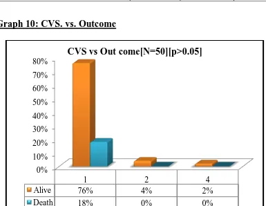
[image:56.595.73.499.541.732.2]surgeries were performed as per operative factors, all as emergency procedures.
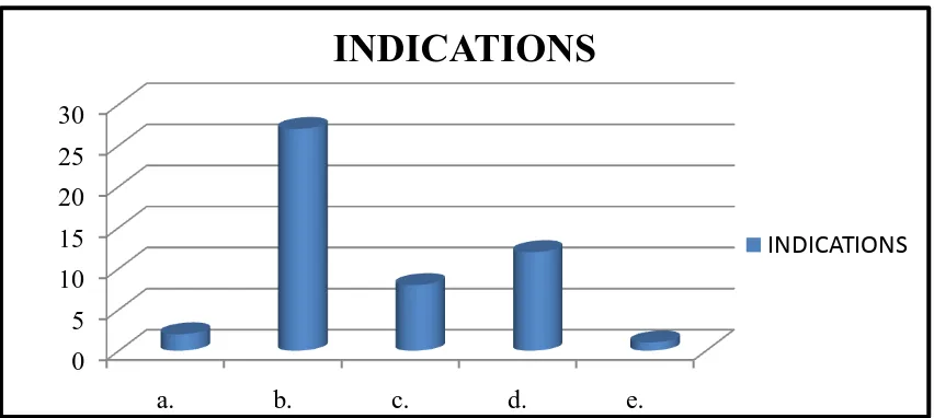
Table 1: Indications
Indications No. of patients
a. Gastric malignancy perforation 2
b. Duodenal and antral perforation 27
c. Ileal perforation 8
d. Appendicular perforation 12
e. Sigmoid volvulus perforation 1
Total 50
Graph 1: Indications
0 5 10 15 20 25 30
a. b. c. d. e.
INDICATIONS
40
PHOTOGRPH 1: PERFORATED DUODENAL ULCER
41
PHOTOGRAPH 3: PERFORATED APPENDICITIS WITH ABSCESS
42
43
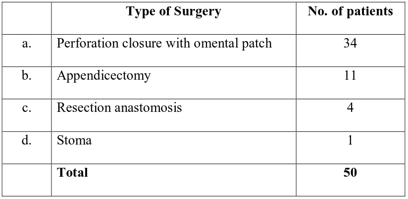
Types of surgeries performed:
39 major surgeries performed were perforation closure, resection
anastomosis, colostomy, ileostomy and feeding jejunostomy and 11 moderate
[image:60.595.92.504.261.462.2]procedures include appendicectomy.
Table 2: Types of surgeries
Type of Surgery No. of patients
a. Perforation closure with omental patch 34
b. Appendicectomy 11
c. Resection anastomosis 4
d. Stoma 1
Total 50
Graph 2: Types of surgeries
0 5 10 15 20 25 30 35
a.
b. c.
d.
34
11
4
1
44
Outcome of surgery:
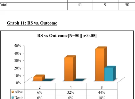
Out of 50 patients studied, death occurred in 9 patients resulting in crude
mortality rate of 18% represented in graph 3.
Out of the remaining 41 patients, 25 patients had at least one
complication, resulting in crude morbidity rate of 61%. The remaining 16
patients showed no evidence of any complication.
Graph 3: Outcome of surgery
41 9
Outcome
Alive
45
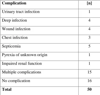
Complications:
The complications during the 4 weeks follow up period were as follows
[image:62.595.131.462.230.542.2]in Table 3.
Table 3: List of complications
Complication [n]
Urinary tract infection 1
Deep infection 4
Wound infection 4
Chest infection 3
Septicemia 5
Pyrexia of unknown origin 1
Impaired renal function 1
Multiple complications 15
No complication 16
Total 50
46
Graph 4: Complications
Graph 5: Individual complications
16
25
Complications
No Complication
Complications
2% 8%
8%
6%
10%
2% 2%
30% 32%
Complication [N=50]
Urinary tract infection[2%] Deep infection[8%] Wound infection[8%] Chest infection[6]% Septicemia[10%] P U O [2%] Impaired renal function[2%]
Mulitple complications [30%]
47
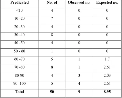
Observed: Expected mortality rates:
Comparison of observed and POSSUM predicted mortality rates was
done using linear analysis is represented in table 4 and graph 6.
An observed to expected ratio (O: E) of 1.005 was obtained and there was
no significant difference between the predicted and observed values (χ2
= 3.54,
[image:64.595.86.510.338.680.2]p = 0.316).
Table 4: O: E mortality rate
Predicated
mortality rate (%)
No. of
Patients
Observed no.
of deaths (O)
Expected no.
of deaths (E)
<10 4 0 0
10 -20 7 0 0
20 -30 4 0 0
30 -40 8 0 0
40 -50 4 0 0
50 - 60 1 0 0
60 -70 5 1 1.7
70 -80 8 1 2.61
80-90 4 3 2.03
90 -100 5 4 2.61
48
Graph 6: O: E ratio
<10 10 -20 20 -30 30 -40 40 -50 50 -60 60 -70 70 -80 80-90 90 -100 Total Observed no.of deaths 0 0 0 0 0 0 1 1 3 4 9 Expected no.of deaths 0 0 0 0 0 0 1.7 2.61 2.03 2.61 8.95
0 1 2 3 4 5 6 7 8 9 10
49
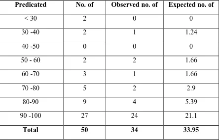
Observed: Expected morbidity rates
Comparison of observed and POSSUM predicted morbidity rates was
done using linear analysis is represented in table 5 and graph 7.
An observed to expected ratio (O: E) of 1.001 was obtained and there was
no significant difference between the predicted and observed values (χ2
= 2.40,
[image:66.595.78.522.337.622.2]p = 0.792).
Table 5: O: E for morbidity rate
Predicated
morbidity rate (%)
No. of
Patients
Observed no. of
complication (O)
Expected no. of
complication (E)
< 30 2 0 0
30 -40 2 1 1.24
40 -50 0 0 0
50 - 60 2 2 1.66
60 -70 3 1 1.66
70 -80 5 2 2.9
80-90 9 4 5.39
90 -100 27 24 21.1
50
Graph 7: O: E for morbidity rate
< 30 30 -40 40 -50 50
-60 60 -70 70 -80 80-90 90
-100 Total Observed no.of complication 0 1 0 2 1 2 4 24 34 Expected no.of complication 0 1.24 0 1.66 1.66 2.9 5.39 21.1 33.95
0 5 10 15 20 25 30 35 40
51
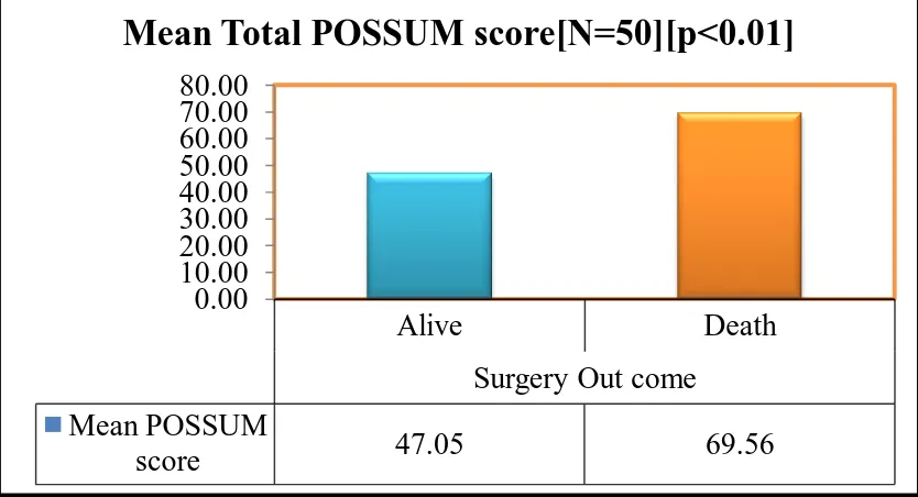
Mean total POSSUM score vs Outcome:
The mean POSSUM score in our study was 51.1. The mean POSSUM
score of patients who survived was 47.05 and those with mortality was 69.56.
[image:68.595.74.491.512.738.2](p < 0.01; Statistically significant)
Table 6: Mean POSSUM vs Outcome
Group No. Of patients Mean total POSSUM score
Alive 41 47.05
Death 9 69.56
Total 50 51.10
Graph 8: Mean POSSUM vs Outcome
Alive Death
Surgery Out come
Mean POSSUM
score 47.05 69.56
0.00 10.00 20.00 30.00 40.00 50.00 60.00 70.00 80.00