virus (HBV) infection. Here, we describe a protocol for efficient and reproducible infection of primary tupaia hepatocytes with HBV. We report that human serum interferes with HBV binding to the hepatocytes, thus limiting the maximum multiplicity of infection. Purification of HBV virions by gradient sedimentation greatly enhances virus binding and infectivity. Covalently closed circular DNA was clearly detectable by Southern blot analysis and newly synthesized single-stranded HBV DNA was visible 2 weeks postinoculation. Primary tupaia hepatocytes are also susceptible to infection with the recently discovered woolly monkey hepatitis B virus (WMHBV) but not to woodchuck hepatitis virus infection. Compared to HBV, WMHBV replicated at a higher rate with single-stranded DNA detectable within the first week postinoculation. Primary tupaia hepatocytes should represent a useful system for studying early steps of HBV and WMHBV infection.
With an estimated 350 million chronically infected people worldwide, hepatitis B virus (HBV) infection represents a ma-jor health care problem. Every year nearly 1 million individuals succumb to HBV-associated liver diseases, such as cirrhosis and hepatocellular carcinoma (9, 18). Although an efficient and safe vaccine is available, HBV is spreading, especially in Asia and Africa (10). Treatment of chronic HBV infection is still unsatisfactory. At present, alpha interferon and lamivu-dine are the only therapeutic options available in clinical prac-tice. However, alpha interferon yields a sustained suppression of viral replication in only about one-third of patients, and lamivudine resistance with mutations in the viral polymerase gene is a frequent event in lamivudine-treated patients (3, 7). Based on transfection studies of hepatoma cell lines, the mech-anisms of HBV replication have been elucidated in great detail (11, 13). By contrast, little is known about the early events of the viral life cycle. Unfortunately, permanent cell lines are not permissive to HBV infection, and primary human hepatocytes are not easily available for in vitro infection studies. Further-more, the quality of liver tissue obtained at surgery for the preparation of primary human hepatocytes is highly variable (5). Therefore, alternative experimental systems for studying HBV infection are urgently needed.
Two reports have described transient HBV infection of the Asian tree shrew,Tupaia belangeri, in vivo (17, 20). Further-more, successful infection of tupaias with human herpes sim-plex virus and hepatitis C virus has been described (2, 19). Tupaias are squirrel-like animals that are closely related to primates and are endemic to subtropical areas of southeast Asia (12). The animals are easily bred in captivity. In addition, cultures of primary tupaia hepatocytes (PTH) can be prepared using well-established liver perfusion protocols.
We previously described HBV infection of PTH (17); infec-tion efficiency was too low, however, to allow a detailed anal-ysis of viral replicative intermediates. Here, we report that human serum inhibits binding of HBV virions to PTH, thus limiting the maximum multiplicity of infection. Purification of viral particles by gradient centrifugation strongly enhances HBV binding to PTH and increases the efficiency of infection. We further show that PTH are also susceptible to infection with the recently discovered woolly monkey hepatitis B virus (WMHBV) (8) and that WMHBV replicates at higher levels in PTH than HBV.
MATERIALS AND METHODS
Animals and preparation of primary hepatocytes.Asian tree shrews (T. be-langeri) were originally obtained from the German Primate Center in Go¨ttingen, Germany. The animals were maintained in the animal facility of the University of Freiburg. Details of breeding conditions are available on request. Primary hepatocytes were prepared by two-step collagenase perfusion as described pre-viously (17) with the following modifications: the liver was perfused for 5 min via the portal vein with Hanks solution containing 5 mM EGTA, followed by per-fusion for 10 min with Hanks solution containing 5 mM CaCl2and 0.5 mg of
collagenase (HepPlus; Serva, Heidelberg, Germany)/ml. The flow rate was 20 ml per min. The cells were seeded onto collagen-coated six-well plates (1 million viable cells per 9.5 cm2) and kept in Hepato-STIM Hepatocyte Defined Medium
(Becton Dickinson, Bedford, Mass.). Hepato-STIM is a serum-free, fully defined medium that was optimized for maintaining primary hepatocytes for up to 3 weeks without loss of cellular function. Primary rat hepatocytes were prepared and cultivated by the identical procedure described for tupaia hepatocytes.
In vitro infection of PTH.HBV infection was performed 1 day after PTH preparation. After the medium was changed, the HBV inoculum was added. HBV-positive serum was obtained from an asymptomatic chronic HBeAg-posi-tive individual. The viral titer was about 109virus genome equivalents (VGE) per
ml, as estimated by comparing the Southern blot hybridization signal of purified serum DNA with known amounts of cloned HBV DNA. Woolly monkey serum was obtained from a colony kept in a European zoo, in which several animals were recently found to be HBsAg positive. The viral sequence is closely related to that reported by Lanford et al. (8) (V. Weich, A. Wahl-Feuerstein, and M. Nassal, unpublished data). The WMHBV titer was about 108VGE per ml. The
WHV DNA-positive woodchuck serum used in control experiments was kindly provided by Mengij Lu (University of Essen, Essen, Germany). It contained about 108WHV VGE per ml. Serum aliquots were stored at⫺20°C for up to 9
months. To assess viral binding, the cells were incubated for 6 h at 37°C, washed four times with culture medium, and harvested. To study viral infection, the cells
* Corresponding author. Mailing address: Department of Medicine II, University of Freiburg, Hugstetter Strasse 55, D-79106 Freiburg, Germany. Phone: 49-761-2703401. Fax: 49-761-2703610. E-mail: weiz @ukl.uni-freiburg.de.
5084
on November 9, 2019 by guest
http://jvi.asm.org/
were incubated overnight (about 16 h) with the respective viral inoculum, and culture medium was changed daily until harvesting of the cells.
Nycodenz gradient purification of HBV particles.In all experiments described here, the iodinated bencoic acid derivate Nycodenz (batch 10032780; Nycomed Pharma AS, Oslo, Norway) was dissolved in phosphate-buffered saline (137 mM NaCl, 2.7 mM KCl, 10 mM phosphate [pH 7.4]). HBV DNA-positive serum (200
l) was loaded onto a preformed Nycodenz gradient ranging from 8 to 50% in 11- by 34-mm polycarbonate centrifuge tubes (Beckman Instruments, Palo Alto, Calif.). Samples were centrifuged in a TLS55 swing-out rotor (Beckman Instru-ments) for 40 min at 200,000⫻gat 20°C. Subsequently, 155-l aliquots were removed from the top of the gradient and added to the hepatocyte culture (fraction 1, top of the gradient; fraction 8, bottom of the gradient). More recently we found that occasionally a precipitate entrapping most HBV virions forms in the gradient, resulting in greatly reduced infectivity. This effect can be avoided by dissolving Nycodenz in Hepato-STIM Hepatocyte Defined Medium (Becton Dickinson).
Protein analysis.Samples from the Nycodenz gradient were mixed with Laemm-li loading buffer and separated according to the LaemmLaemm-li sodium dodecyl sulfate-polyacrylamide gel electrophoresis system on a 13% sulfate-polyacrylamide gel. The proteins were either stained with Coomassie blue or transferred onto a polyvi-nyldifluoride membrane (Schleicher & Schuell, Keene, N.H.) and probed with a pre-S2/S antigen (Ag)-specific polyclonal mouse serum generated by DNA-based immunization (kindly provided by Michael Geissler, University Hospital of Freiburg, Freiburg, Germany). Bound anti-HBs was visualized by chemilumines-cence using the ECLPlus system (Amersham Pharmacia Biotech, Buckingham-shire, England).
Analysis of viral DNA.After the culture medium was removed, cells were lysed with 400l of ATL buffer (QIAamp DNA Mini Kit; Qiagen, Hilden, Germany). The lysate was transferred to an Eppendorf tube and digested with proteinase K (final concentration, 0.1 mg/ml) for 1 h at 56°C. Subsequently, RNase A (final concentration, 0.02 mg/ml) was added, and DNA was purified by absorption onto silica columns according to the protocol of the manufacturer (Qiagen). DNA was eluted, concentrated by ethanol precipitation, and separated on a 1.3% agarose gel that did not contain ethidium bromide. Circular plasmid DNA (0.5-kbp EcoRI/BamHI fragment of HBV in 2.7 kbp of pUC19 vector) was used as a size marker for covalently closed circular DNA (cccDNA). DNA purified from HBV-positive serum served as a marker for relaxed circular viral DNA. Heat-dena-tured DNA (5 min, 95°C) from HBV DNA-positive serum was used to identify the position of single-stranded DNA (ssDNA). After electrophoresis, the gel was soaked for 10 min in 0.25 M HCl and for 20 min in 0.5 M NaOH–1.5 M NaCl. Nucleic acids were blotted by capillary transfer with 0.5 M NaOH–1.5 M NaCl onto nylon membranes (Amersham Pharmacia Biotech) and visualized by hy-bridization with a32P-labeled probe containing the complete HBV genome. This
HBV probe was used to detect all mammalian hepadnaviruses analyzed in this study. The stringency of the final washes was 0.1% sodium dodecyl sulfate–40 mM NaCl–4 mM sodium citrate at 60°C. Dot blot analysis was performed by
spotting 3-l aliquots of Nycodenz gradient fractions or serum samples onto nylon membranes. The membrane was soaked in 0.5 M NaOH–1.5 M NaCl and 0.5 M Tris-HCl (pH 7.5)–1.5 M NaCl for 10 min each before hybridization, as described above.
Analysis of viral RNA.For RNA preparation, cells were lysed with 350l of RLT buffer (RNeasy Mini kit; Qiagen). RNA was purified by adsorption onto silica columns as recommended by the supplier. Samples were treated with DNase to eliminate viral DNA. Successful DNase digestion was verified by PCR amplification of a small aliquot of purified RNA. The RNA was separated on morpholinopropanesulfonic acid-buffered 1% agarose gels containing 1.2% formaldehyde. The gel was soaked in 50 mM NaOH–1.5 M NaCl for 10 min and in 1.5 M NaCl–0.5 M Tris-HCl (pH 7.5) for 20 min. RNA was blotted onto nylon membranes by capillary transfer with 1.5 M NaCl–150 mM sodium citrate. The position of the rRNA was identified by staining the blotting membrane with methylene blue. HBV RNA was visualized by hybridization with the32P-labeled
HBV DNA probe, as described above.
RESULTS
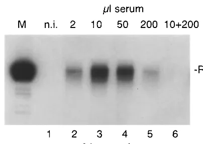
[image:2.612.73.275.72.216.2]Human serum competes with HBV virions for binding to PTH. In the previously published study of infection of PTH FIG. 1. HBV binding to PTH. Cells were incubated for 6 h with
[image:2.612.318.538.78.443.2]increasing volumes of HBV DNA-positive serum (lanes 2 to 5) or a mixture of HBV-positive serum (10 l) and HBV-negative human serum (200l) (lane 6). HBV binding was assessed by Southern blot analysis. Numbers on top of the figure indicate the volume of the respective serum samples. M, 3.2-kbp linear HBV DNA; n.i., nonin-fected control; RC, relaxed circular viral DNA; p.i., postincubation.
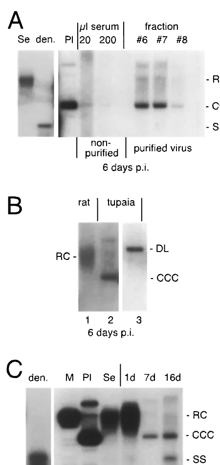
FIG. 2. Effect of virus purification on viral attachment to PTH. (A) Purification of HBV particles by Nycodenz gradient centrifugation. (Top) Coomassie blue staining of fractionated serum proteins. (Mid-dle) Detection of HBsAg by Western blot analysis. (Bottom) Dot blot analysis of viral DNA. Fractions 6, 7, and 8 contain purified virions. (B) Binding assay with purified (right) versus nonpurified (left) viral par-ticles. Viral DNA was detected by Southern blot analysis. For nomen-clature, see the legend to Fig. 1.
on November 9, 2019 by guest
http://jvi.asm.org/
A typical experiment is shown in Fig. 1. At low doses of serum (2 and 10l per ml of culture medium), the amount of particles bound increased with increasing volume of added HBV serum. Surprisingly, however, a higher amount of serum (50 and 200l per ml of culture medium) did not result in an increase of cell-associated viral DNA but rather reduced the number of HBV particles attached to the cells. This effect was possibly due to an inhibitory factor present in the serum sam-ple used. In particular, empty HBsAg particles might compete for HBV binding to the cells. To address this point, 10l of HBV DNA-positive serum was mixed with 200 l of anti-HBsAg-negative, HBsAg-negative normal human serum. Vi-rus attachment to PTH was assessed as described above. As shown in Fig. 1, lane 6, attachment to PTH was dramatically reduced in this experimental setting. The same effect was ob-served with serum samples obtained from seven other healthy and nonvaccinated individuals (data not shown). These results suggest that human serum contains one or more components that compete with HBV for cellular binding sites on PTH and that empty viral particles do not account for the observed effects.
Purification of viral particles enhances binding to PTH.
Based on the above results, it was conceivable that purification of HBV virions from human serum might enhance binding to PTH and increase the maximum multiplicity of infection that can be achieved in this system. To test this hypothesis, HBV DNA-positive serum was subjected to gradient centrifugation, and gradient fractions were analyzed by the virus binding assay. The iodinated bencoic acid derivate Nycodenz was chosen as a gradient medium because it is not toxic to cells in culture and is well suited for virus purification (4). As shown in Fig. 2A, the bulk of serum proteins floated on top of the gradient, while HBV virions readily sedimented into the gradient under our experimental conditions. Compared with nonfractionated in-fectious serum, binding of gradient-purified HBV virions to PTH was greatly enhanced (Fig. 2B). This was not due to a nonspecific biophysical property of Nycodenz, since mixing Nycodenz with the inoculum without sedimentation did not enhance binding (data not shown). These results suggest that serum components which interfere with HBV binding to PTH can be removed by gradient centrifugation.
Gradient purification of viral particles enhances the
effi-ciency of infection.Next, we asked whether enhanced binding
of purified HBV virions to PTH results in enhanced infectivity. Cells were incubated overnight either with gradient-purified viral particles or with nonfractionated HBV-positive serum, followed by 6 days in culture. Cells were harvested and ana-FIG. 3. Infection studies with purified HBV. (A) Infectivity of
pu-rified and nonpupu-rified virus. Viral DNA was detected by Southern blot analysis on day 6 postinfection. Note that cccDNA is clearly detectable only in PTH inoculated with purified virus. (B) Inoculation of primary rat hepatocytes (lane 1) with gradient-purified HBV compared to PTH (lane 2). Note that cccDNA is formed in PTH but not in primary rat hepatocytes. The circular nature of the fast-migrating HBV DNA species was confirmed by digesting the DNA withNcoI, which cuts the HBV genome once (lane 3). (C) Time course of HBV infection in PTH. Cells were incubated with purified HBV virions and harvested on days 1, 7, and 16 postinfection as indicated at the top of the figure. Viral replicative intermediates were analyzed by Southern blot analy-sis. Note that cccDNA was detectable on day 7, and ssDNA was detectable on day 16 post infection. Se, purified HBV DNA from serum; den., denatured serum DNA; Pl, 3.2-kbp circular plasmid DNA; M, 3.2-kbp linear HBV DNA; RC, relaxed circular viral DNA; DL, double-stranded linear DNA.
on November 9, 2019 by guest
http://jvi.asm.org/
[image:3.612.58.283.86.561.2]lyzed for viral replicative intermediates by Southern blotting. Figure 3A shows that cccDNA, a hallmark of hepadnavirus infection (15), is clearly visible in cells infected with purified HBV virions, while it is hardly detectable in cells incubated with nonfractionated HBV DNA-positive serum. DNA diges-tion with the restricdiges-tion enzymeNcoI generated a linear 3.2-kbp fragment, confirming the circular nature of the respective DNA species (Fig. 3B, compare lanes 2 and 3). In a control experiment, primary rat hepatocytes were incubated with gra-dient-purified HBV virions and assayed for cccDNA forma-tion. Figure 3B, lane 1, shows that HBV virions bound to rat hepatocytes but no virus entry occurred, based on the lack of cccDNA formation. Thus, purification of HBV virions by Ny-codenz gradient centrifugation does not mediate nonspecific viral uptake.
We then performed a time course experiment with longer culture times. As illustrated in Fig. 3C, cccDNA but not ssDNA is detectable by Southern blot analysis at day 7 postin-cubation. At day 16, an additional fast-migrating DNA species
is visible which comigrates with denaturated serum HBV DNA and thus most likely represents newly synthesized ssDNA. The total amount of viral DNA only slightly increased from day 7 to day 16 (Fig. 3C, compare lanes 6 and 7), indicating that the rate of HBV replication is low in PTH.
PTH are permissive to WMHBV infection. WMHBV is a
[image:4.612.100.510.519.686.2]recently discovered hepadnavirus that has been isolated from woolly monkeys in captivity (8). We tested whether PTH are susceptible to WMHBV infection. Serum from an infected woolly monkey was processed by gradient centrifugation. PTH were incubated with gradient fractions as described above. In a control experiment, PTH were incubated with purified wood-chuck hepatitis virus (WHV) (1, 14). Cells were harvested at the times indicated and assayed for viral replication by South-ern blotting. While both viruses bound to PTH, cccDNA and ssDNA formation was only observed in cells inoculated with WMHBV. Thus, PTH are permissive to WMHBV infection (Fig. 4A) but not to WHV infection (Fig. 4B). In contrast to HBV (Fig. 3C), WMHBV ssDNA was detectable as early as 7 FIG. 4. Infection of PTH with WMHBV and WHV. (A) WMHBV-positive serum was processed by gradient centrifugation as described for HBV. Cells were harvested on days 1, 7, and 13 postinfection. Viral DNA was analyzed by Southern blotting. Note that cccDNA and ssDNA are generated in WMHBV-infected cells. (B) Control infection with WHV. PTH were incubated with gradient-purified WHV and harvested on day 6 postinfection. Note that WHV relaxed circular DNA but no cccDNA is visible on the Southern blot. For abbreviations, see the legend to Fig. 3.
FIG. 5. Viral replication and transcription in HBV- and WMHBV-infected PTH. (A) Southern blot analysis of infected PTH on day 6 postinfection (top); dot blot analysis of HBV and WMHBV serum (2l each; bottom). Note that only WMHBV produced ssDNA at this time point. For abbreviations, see the legend to Fig. 3. (B) Northern blot analysis of infected PTH. Lysates from cells infected with fractions 6 to 8 were pooled. pgRNA, pregenomic viral RNA. sgRNA, subgenomic viral RNA coding for surface proteins.
on November 9, 2019 by guest
http://jvi.asm.org/
duce different RNA levels in PTH. To test this possibility, HBV- and WMHBV-infected PTH were harvested at day 5 postinfection and analyzed for viral RNA synthesis by North-ern blotting. Figure 5B illustrates that both viruses produced abundant amounts of pre-S/S mRNA (sgRNA), while only low levels of pregenomic RNA (pgRNA) were detected. Notably, however, the ratio of pgRNA to sgRNA was slightly higher for WMHBV than for HBV in this experiment.
DISCUSSION
We have previously reported that primary hepatocytes from
T. belangeri can be infected with HBV (17). However, the
practical use of this system was limited by the low efficiency of infection. Here, we show that human serum interferes with viral attachment, thereby greatly reducing viral infection. On the other hand, purification of virions by gradient centrifuga-tion strongly enhances both HBV binding and infeccentrifuga-tion. Thus, newly synthesized cccDNA and ssDNA and viral RNA were unambiguously detectable by Southern and Northern blot analysis, respectively. Infection of PTH with Nycodenz gradi-ent-purified HBV particles represents an authentic biological process, because the viral host tropism was preserved in cell culture and Nycodenz itself does not affect binding of HBV to PTH. This is in contrast to polyethylene glycol-based protocols (6, 17), because polyethylene glycol may induce nonphysiologi-cal membrane fusion.
The biochemical nature of the inhibitory serum component is unclear at present. Preliminary results indicate that the in-hibitory factor cannot be removed by dialysis and that albumin and immunoglobulins, which are the most abundant serum proteins, do not account for the inhibitory effect (J. Ko¨ck and F. von Weizsa¨cker, unpublished data). Interestingly, attach-ment of duck hepatitis B virus to primary duck hepatocytes is not inhibited by duck serum (J. Ko¨ck and F. von Weizsa¨cker, unpublished). This may explain in part the high efficiency of duck hepatitis B virus infection in vitro (16).
Compared to human hepatocytes, PTH have the principal advantage of being readily available from in-house-bred ani-mals. PTH, therefore, allow the highly reproducible infection with HBV under the experimental conditions described, while the suitability of primary hepatocytes prepared from human liver for infection experiments is very variable (5).
PTH were also infected by WMHBV but not by WHV. This finding substantiates PTH as a useful in vitro model system for studying hepadnavirus infection and rules out the possibility
thesis in PTH revealed an overall low abundance of pgRNA, in HBV- as well as WMHBV-infected cells. The ratio of pgRNA to sgRNA, however, was slightly higher in WMHBV- than in HBV-infected PTH. Since pgRNA is pivotal for replication and capsid formation, subtle changes in pgRNA production might translate into substantial effects on viral replication rates. Compatible with this notion, there are significant differ-ences in the core promoter regions of WMHBV and HBV. We are currently preparing a replication-competent WMHBV construct that would allow us to directly address this question in transfected cells.
ACKNOWLEDGMENTS
This study was supported by grants from the Deutsche Forschungs-gemeinschaft (We 1365/2-2), the Bundesministerium fu¨r Bildung und Forschung (01Kl9951), and the Zentrum fu¨r Klinische Forschung I (3000 0447 01 B2).
We thank H. Schaller for providing cloned HBV DNA, M. Lu for providing WHV serum, and M. Geissler for providing mouse anti-pre-S2/S antibodies.
REFERENCES
1.Aldrich, C. E., L. Coates, T. T. Wu, J. Newbold, B. C. Tennant, J. Summers, C. Seeger, and W. S. Mason.1989. In vitro infection of woodchuck hepato-cytes with woodchuck hepatitis virus and ground squirrel hepatitis virus. Virology172:247–252.
2.Darai, G., A. Schwaiger, D. Komitowski, and K. Munk.1978. Experimental infection ofTupaia belangeri(tree shrews) with herpes simplex virus types 1 and 2. J. Infect. Dis.137:221–226.
3.Dienstag, J. L., E. R. Schiff, T. L. Wright, R. P. Perrillo, H. W. Hann, Z. Goodman, L. Crowther, L. D. Condreay, M. Woessner, M. Rubin, and N. A. Brown.1999. Lamivudine as initial treatment for chronic hepatitis B in the United States. N. Engl. J. Med.341:1256–1263.
4.Fowler, E., N. Raab-Traub, and S. Hester.1985. Purification of biologically active Epstein-Barr virus by affinity chromatography and nonionic density gradient centrifugation. J. Virol. Methods11:59–74.
5.Galle, P. R., J. Hagelstein, B. Kommerell, M. Volkmann, P. Schranz, and H. Zentgraf.1994. In vitro experimental infection of primary human hepato-cytes with hepatitis B virus. Gastroenterology106:664–673.
6.Gripon, P., C. Diot, and C. Guguen-Guillouzo.1993. Reproducible high level infection of cultured adult human hepatocytes by hepatitis B virus: effect of polyethylene glycol on adsorption and penetration. Virology192:534–540. 7.Hoofnagle, J., and A. M. DiBisceglie.1997. The treatment of chronic viral
hepatitis. N. Engl. J. Med.336:347–356.
8.Lanford, R. E., D. Chavez, K. M. Brasky, R. B. Burns III, and R. Rico-Hesse.
1998. Isolation of a hepadnavirus from the woolly monkey, a New World primate. Proc. Natl. Acad. Sci. USA95:5757–5761.
9.Lee, W. M.1997. Hepatitis B virus infection. N. Engl. J. Med.337:1733–1745. 10. Lemon, S. M., and D. L. Thomas.1997. Vaccines to prevent viral hepatitis.
N. Engl. J. Med.336:196–204.
11. Nassal, M., and H. Schaller.1993. Hepatitis B virus replication. Trends Microbiol.1:221–228.
12. Novacek, M. J.1992. Mammalian phylogeny: shaking the tree. Nature356:
121–125.
13. Seeger, C., and W. S. Mason.2000. Hepatitis B virus biology. Microbiol. Mol. Biol. Rev.64:51–68.
on November 9, 2019 by guest
http://jvi.asm.org/
14.Summers, J., J. M. Smolec, and R. Snyder.1978. A virus similar to human hepatitis B virus associated with hepatitis and hepatoma in woodchucks. Proc. Natl. Acad. Sci. USA75:4533–4537.
15. Tuttleman, J. S., C. Pourcel, and J. Summers.1986. Formation of the pool of covalently closed circular viral DNA in hepadnavirus-infected cells. Cell
47:451–460.
16. Tuttleman, J. S., J. C. Pugh, and J. W. Summers.1986. In vitro experimental infection of primary duck hepatocyte cultures with duck hepatitis B virus. J. Virol.58:17–25.
17. Walter, E., R. Keist, B. Niedero¨st, I. Pult, and H. E. Blum.1996. Hepatitis B
virus infection of tupaia hepatocytes in vitro and in vivo. Hepatology24:1–5. 18. Wright, T. L., and J. Y. Lau.1993. Clinical aspects of hepatitis B virus
infection. Lancet342:1340–1344.
19. Xie, Z. C., J. I. Riezu-Boj, J. J. Lasarte, J. Guillen, J. H. Su, M. P. Civeira, and J. Prieto.1998. Transmission of hepatitis C virus infection to tree shrews. Virology244:513–520.
20. Yan, R. Q., J. J. Su, D. R. Huang, Y. C. Gan, C. Yang, and G. H. Huang.1996. Human hepatitis B virus and hepatocellular carcinoma. I. Experimental infection of tree shrews with hepatitis B virus. J. Cancer Res. Clin. Oncol.
122:283–288.