“
CLINICOPATHOLOGICAL STUDY ON
MULTINODULAR GOITRE
”
Dissertation submitted to the
TAMIL NADU DR.MGR MEDICAL UNIVERSITY
CHENNAI, TAMIL NADU
for the degree of
MASTER OF SURGERY IN GENERAL SURGERY
DEPARTMENT OF GENERAL SURGERY TIRUNELVELI MEDICAL COLLEGE
TIRUNELVELI-627011 2014
CHENNAI, TAMIL NADU CERTIFICATE
This is to certify that the dissertation titled “CLINICOPATHOLOGICAL
STUDY ON MULTINODULAR GOITRE” is the original work done by
DR.N.ARUNMOZHI VIJAY , post graduate in the department of GENERAL
SURGERY, TIRUNELVELI MEDICAL COLLEGE, TIRUNELVELI-11
submitted to THE TAMIL NADU DR.MGR MEDICAL UNIVERSITY,
Chennai –32 towards the partial fulfillment of the requirements for the award
of M.S degree in GENERAL SURGERY April 2014 examination.
DR.ALEX ARTHUR EDWARDS M.S.,
DR.S.SOUNDARARAJANM.S.,
UNIT CHIEF PROFESSOR AND HOD
Department Of General Surgery Department of General
Surgery
Tirunelveli Medical College Tirunelveli Medical College
Tirunelveli –11 Tirunelveli -11
THE DEAN
Tirunelveli Medical College
ACKNOWLEDGEMENT
I express my heartfelt thanks to DR.SOUNDARAJAN,M.S., Professor
And Head Of The Department, Department of General Surgery, Tirunelveli
Medical College & Hospital, Tirunelveli, for his timely advice, guidance and
encouragement in all my endeavors and .
I am deeply indebted to so many for guiding and helping me in my
endeavor in making this dissertation a reality. I express my deep sense of
gratitude to my respected teacher and guide, PROF.DR.ALEX ARTHUR
EDWARDS M.S., Professor, Department of General Surgery, Tirunelveli
Medical College & Hospital, Tirunelveli, for their valuable guidance and
constant encouragement throughout the course and the present study.
I express my profound gratitude to Professors DR.R.MAHESWARI
M.S.,DR.V.PANDYM.S.,DR.K.RAJENDRAN,M.S.,DR.M.S.VARADHARAJ
AN.,DR.S.K.SRIDHAR,M.S., DR.G.V.MANOHARAN,M.S., for their constant
guidance throughout my study period.
I express my sincere thanks and gratitude to DR.G.NIRMAL KUMAR
M.S., DR.S.VINOTH KUMAR,M.S., and all my teachers of Department of
General Surgery, Tirunelveli MedicalCollege &Hospital, Tirunelveli for their
constant support of valuable suggestions at every stage of this study.
I thank the DEAN, TIRUNELVELI MEDICAL COLLEGE for
permitting me to use the Hospital facilities for my study.
have been the source and support of companionship throughout this course and I
am indebted to them.
I will be failing in duty, if I do not express my gratitude to all the
patients, who were the subjects of this study. My sincere thanks to them
CONTENTS
S.NO TITLE PAGE NO
1. INTRODUCTION 1
2. AIMS AND OBJECTIVES 2
3. REVIEW OF LITERATURE 5
4. METHODS AND
METHODOLOGY 66
5. RESULTS 67
6. DISCUSSION 77
7. CONCLUSION 80
8. BIBLIOGRAPHY 82
9. ANNEXURES
ANNEXURE-I :
PROFORMA
ANNEXURE-II :
ABSTRACT
CLINICOPATHOLOGICAL STUDY ON MULTINODULAR GOITRE
INTRODUCTION
MULTINODULAR GOITER describes an enlarged, diffusely heterogeneous thyroid gland.Initial presentation may include diffuse enlargement, but the mass often develops asymmetrical nodularity. The cause of this mass is usually iodine deficiency. Initially, the mass is euthyroid; however, with increasing size, elevations in T3 and T4 can occur and progress gradually into clinical hyperthyroidism.
Work-up and diagnosis include evaluation of thyroid function tests. Ultrasound and radioisotopic scanning demonstrate heterogeneous thyroid substance. Nodules with poor uptake can present as lesions suspicious for malignancy.The incidence of carcinoma in multinodular goiter has been reported as 5% to 10%. Therefore, FNAC for diagnosis and resection for suspicious lesions should be considered.
Hyperthyroidism may be adequately controlled by drugs, but surgical management is the preferred treatment. Subtotal or total thyroidectomy may performed depending upon the involvement of the thyroid gland. Radioactive iodine therapy is reserved for elderly individuals who represent poor operative risk. The complications of thyroid surgeries are heamorrhage, respiratory obstruction, vocal cord paralysis, hypoparathyroidism, thyroid insufficiency, thyrotoxic storm and wound infection.
MATERIALS AND METHODS
This is a prospective clinical study of randomly selected patients admitted in the department of surgery, TVMCH diagnosed and treated as a case of multinodular goitre during the study period.
The patients diagnosed as a case of multinodular goitre will undergo detailed history taking, clinical examination, investigations like CBC, thyroid profile, fine needle aspiration cytology, x – ray chest and neck and ultrasonography of neck. After surgery, the patients will be followed up for any immediate post operative complications. The specimen will be sent for histopathological examination and the results will be recorded.
Through this study I intend to find out
Through this study I intend to emphasize the existing data regarding multinodular goitre and find out
- Whether there is a specific age distribution for multinodular goitre in and around tirunelveli.
- To study and discuss the most common presenting clinical features of multinodular goitre.
- The percentage of thyroid malignancies presenting as multinodular goitre and whether FNAC is conclusive in confirming the diagnosis, which will be helpful in planning the surgery necessary ie., whether a subtotal or total thyroidectomy must be performed.
RESULTS
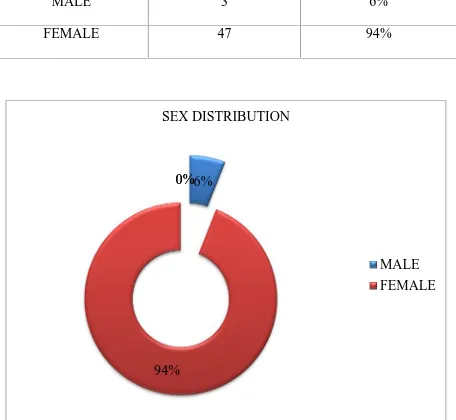
In our study, among the 50 cases three were male which constitutes 6% of the study group. The remaining 47 cases were females (94%). Majority of the cases were in the 30 – 40 years age group (32%), followed by the age group of 40 – 50. The Mean age of incidence was 42.26. The average post operative stay among the 50 cases studied was 5.3 days. 76% of the cases were discharged between 4 to 6 days of post operative stay. The presenting complaint was a swelling in all the cases studied (100%). The swelling was associated with pain in 48% of the cases. Pressure symptoms like dysphagia, dyspnoea and hoarseness of voice were present in 44%, 18% and 20% of cases respectively with an average of 27.3%.Of the 50 cases of Multinodular goiters studied, 7 cases were hyperthyroid on presentation which constitutes 14% of the cases. All the cases were taken up for surgery, 88% of cases underwent total thyroidectomy and 10% of cases underwent subtotal thyroidectomy. The Fine Needle Aspiration Cytology reports of the 50 cases showed Nodular Colloid Goitre (64%) as the most common FNAC finding followed by Hashimoto’s Thyroiditis (11%). The FNAC report was follicular neoplasm for 2 cases, so total thyroidectomy was performed in those cases to rule out malignancy. Post operative complication occurred in three cases. The post operative histopathological examination of the resected specimen showed that 37% of the cases were Colloid nodular goitre and 18% of cases had features suggestive of Hashimoto’s thyroidits. One case of papillary and follicular carcinoma each.
CONCLUSION
5.3 days. Post operative stay in hospital has to be reduced. Thyroidectomy can be done as a day care or short stay procedure in our hospital as is the recent trend in developed countries. However, the applicability of these practices to thyroid surgery remains controversial. Day care surgery can be promoted in selected and educated patients as this will be the future of thyroid surgeries. Hyperthyroidism in multinodular goitre was present in 14% of cases. Hyperthyroidism occurs in cases of multinodular goitre in the natural evolution of the disease and the patient must be treated and brought to euthyroid state before surgery.
Fine Needle Aspiration Cytology is a very useful investigation in the evaluation of Multinodular goitre except for that it cannot differentiate follicular adenoma from follicular carcinoma. Most of the cases had colloid nodular goitre in multinolar goitre. Carcinoma in not uncommon in cases of Multinodular goitre. So, suspicion should always be present. Total thyroidectomy is the preferred surgery for multinodular goitre. But subtotal thyroidectomy may also be performed in cases in whom surgery is done for cosmetic reasons as in Hashimoto’s thyroiditis.
INTRODUCTION
Multinodular goiter describes an enlarged, diffusely heterogeneous
thyroid gland. Initial presentation may include diffuse enlargement, but the
mass often develops asymmetrical nodularity. The cause of this mass is usually
iodine deficiency. Initially, the mass is euthyroid; however, with increasing size,
elevations in T3 and T4 can occur and progress gradually into clinical
hyperthyroidism.
Work-up and diagnosis include evaluation of thyroid function tests.
Ultrasound and radioisotopic scanning demonstrate heterogeneous thyroid
substance. Nodules with poor uptake can present as lesions suspicious for
malignancy.
The incidence of carcinoma in multinodular goiter has been reported as
5% to 10%.Therefore, FNAC for diagnosis and resection for suspicious lesions
should be considered. Hyperthyroidism may be adequately controlled by drugs,
but surgical management is the preferred treatment. Subtotal or total
thyroidectomy may performed depending upon the involvement of the thyroid
gland. Radioactive iodine therapy is reserved for elderly individuals who
represent poor operative risk.
The complications of thyroid surgeries are heamorrhage, respiratory
obstruction, vocal cord paralysis, hypoparathyroidism, thyroid insufficiency,
thyrotoxic storm and wound infection.
AIM OF THE STUDY
- To study the age and sex distribution of multinodular goitre
- To study the presenting clinical features in multinodular goitre
- To study the correlation between FNAC and histopathological
examination
- To study the incidence of malignancy in multinodular goitre
- Discuss the various treatment modalities available in our hospital
- To study the incidence of various post-operative complications
INCLUSION CRITERIA
- All cases admitted in the department of surgery diagnosed as a case of
multinodular goitre
- Above the age of 20 yrs
- Cases presenting with both toxic and non toxic features
EXCLUSION CRITERIA
- Age less than 20 yrs
- Pregnant women
- Cases presenting with solitary nodule
- Cases with diffuse enlargement of thyroid gland
This is a prospective clinical study of randomly selected patients admitted
in the department of surgery, TVMCH diagnosed and treated as a case of
multinodular goitre during the study period.
The patients diagnosed as a case of multinodular goitre will undergo
detailed history taking, clinical examination, investigations like CBC, thyroid
profile, fine needle aspiration cytology, x – ray chest and neck and
ultrasonography of neck.
After surgery, the patients will be followed up for any immediate post
operative complications. The specimen will be sent for histopathological
examination and the results will be recorded.
PURPOSE OF THE STUDY
Through this study I intend to emphasize the existing data regarding
multinodular goitre and find out
- Whether there is a specific age distribution for multinodular goitre in
and around tirunelveli.
- To study and discuss the most common presenting clinical features of
multinodular goitre.
- The percentage of thyroid malignancies presenting as multinodular
goitre and whether FNAC is conclusive in confirming the diagnosis,
which will be helpful in planning the surgery necessary ie., whether a
- The percentage of post operative complications encountered in
REVIEW OFLITERATURE
The name “THYROID” is derived from the Greek word for
“shield-shaped gland in the anterior part of the neck” (Thyroides).
Thyroid gland was referred to as “laryngeal gland” and subsequently
named as “THYROID” by Wharton in 1645.
The relationship between endemic goitre and Cretinism was showed by
Paracelsus (1492-1591).
In 1874, Gull described Cretinoid changes in adults.
Curling in 1950 accounted of Cretinism at London and recorded complete
absence of thyroid gland in both cases.
Thyroxine was isolated by Kendall (1965). Harrington and Banger in
1927 synthesized thyroxine. Triiodothyronine was discovered by Cross,
Pittrivers and Roche in 1953. Radioactive iodine was introduced in 1934
which made clear the understanding of the physiology of thyroid gland.
SURGERY IN THYROID
Roger Frugardi of Salerno in Bamberg manuscripts gave the first credible
account of thyroid gland in 1170.
The first well-documented partial thyroidectomy was performed in Paris
in 1791 by Pierre Joseph Desault. Guillaume Dupuytren performed total
successfully removed six “suffocating goitre” by ligation of all the arteries and
dissection. Most of the thyroid surgeries were disastrous up to the second half of
the 19thcentury due to bleeding and sepsis.
Following the advent of general anaesthesia (1890), antisepsis (1860’s)
and haemostasis (1870’s), surgeons were able to do more thyroid operations and
improvise techniques with great reduction in mortality.
The collar incision was introduced by Tules Boeckial (1848-1927) of
Strasbourg. The first transplantation of thyroid gland was done by Pays in 1906.
In the late 19th century, two surgeon-physiologists Theodor Billroth and Emil Theodor Kocher revolutionized the understanding and management of
thyroid diseases. They established large clinics in Europe and developed skilled
techniques of surgery, provided surgical results that proved the safety and
efficacy of thyroidectomies for both benign and malignant diseases. Kocher
received the Nobel Prize in 1909 for his pioneering developments in the
understanding of thyroid physiology.
The 20th century began with the contribution of Billroth and Kocher. All the advances have allowed the diagnosis and treatment of thyroid diseases to
become rapid, cost-effective and low morbidity procedures.
EMBRYOLOGY
The tissue bud that becomes the thyroid gland arises as a median
tuberculumimpar. It originates from the primitive alimentary tract, which are
cells of endodermal origin. This cellular structure descends into the neck region
to develop into a bilobed solid organ.
The original attachment in the pharynx is at the foraman caecum through
the thyroglossal duct which reabsorbs after 6 weeks of age. The distal end of
this remnant matures as a pyramidal lobe.
Thyroid follicles first appear as the lateral lobes of thyroid gland develop.
Follicles begin to synthesis colloid when the embryo is 6cm in length. In the 3rd month, iodine trapping and thyroid hormone secretion begins.
SURGICAL ANATOMY OF THE THYROID GLAND
Normally adult thyroid gland weighs 20–25 gms. It is a bilobed structure
gland encircles about 75% of the junction of the larynx and the upper part of the
trachea. The gland lies against C5, C6, C7 and T1 vertebrae. The isthmus covers
the 2nd to 4th tracheal rings. The pyramidal lobe is found in about 30% of patients.
The thyroid gland is surrounded by a thin layer of connective tissue
which is called the true capsule. The false capsule is derived from the
pretracheal layer of the deep cervical fascia. These capsules are pierced by
blood vessels of the thyroid gland to form a plexus beneath the true capsule. So
the thyroid gland is removed with its true capsule. The thickening of fascia that
fixes the back of each lobe to cricoid cartilage is called the Ligaments Of Berry.
Relations
Superficial surface is covered by
o Sternohyoid
o Sternothyroid
o Anterior border of sternocleidomastoid
Medial surface is related to
o Trachea and oesophagus
o Inferior constrictor and cricothyroid
o External laryngeal and recurrent laryngeal nerves
Posterior surface is related to the carotid sheath
Anterior border is related to the anterior branch of the superior thyroid
artery
Posterior border is related to
o Inferior thyroid artery
o Thoracic duct on the left side
o Parathyroid glands
Blood supply
Arterial supply consists of four main arteries, two superior and two
inferior thyroid arteries. Superior thyroid artery is the first branch of the external
carotid artery and arises just above the bifurcation of the common carotid artery.
The inferior thyroid artery takes origin from the thyrocervical trunk.
The thyroid gland is drained by three pairs of veins. The superior thyroid
vein lies immediately adjacent to the superior arteries and drains into the
is short and lies horizontally. Usually there are two or three inferior thyroid
veins draining into the innominate and brachiocephalic veins.
The thyroid gland has rich lymphatics draining it in almost every
direction. The lympyhatics are present immediately beneath the capsule and
communicates between the two lobes through the isthmus. They drain into the
regional lymph nodes occupying the pretracheal, paratracheal and
trachea-oesophageal lymph nodes.
Nerve supply
Sympathetic nerve supply is mainly derived from the middle cervical
ganglion and partly from superior and inferior cervical ganglion. They are
vasoconstrictors.
Nerves related to the thyroid gland
Right Recurrent laryngeal nerve :
It branches from the vagus loops around the subclavian artery
crosses behind rt. Common carotid in or near tracheoesophageal groove
Left Recurrent laryngeal nerve :
Arises where the vagus crosses the aortic archloops under ligamentum
arteriosumascend like the rt. Nerve. Both nerves cross the inf. Thyroid artery
near the lower border of middle third of the gland
The nerve can be identified posterior to the inf. Cornu of thyroid
cartilage where it enters the larynx. Lower down, the nerve can be palpated as a
tight strand over the trachea
External laryngeal nerve :
Branch of superior laryngeal nerve. Arises at the level of superior cornu
passes under the sternothyroid along with the superior thyroid vessels, Medial to
them.
PHYSIOLOGY OF THE THYROID GLAND
Thyroid gland is responsible for the production of two families of metabolic
hormones
Thyroxin (T4) and Tri-iodothyronine (T3)
Calcitonin–the hormone regulating calcium
Iodine metabolism
Iodine is essential for the production of thyroid hormones. It is absorbed
from the gastrointestinal tract in the form of inorganic iodide and enters the
body iodine. It is stored in the thyroid as preformed hormone or as an iodinated
amino acid.
Iodine is transported into the follicular cells via an intrinsic
transmembrane protein located in the basolateral membrane. Inside the follicle,
it is rapidly oxidized and bound to thyroglobulin.
Thyroid hormone synthesis
Once organic iodide is oxidized and bound, it couples to thyroglobulin
with tyrosine moieties to form iodotyrosinesmonoiodotyrosine and
diiodotyrosine. It is dependent on thyroid peroxidise, an important intracellular
catalytic agent.
Coupling of monoiodotyrosine and diiodotyrosine which are biologically
circumstances, T4 formation predominates. Both T3 and T4 are bound to
thyroglobulin and stored in the colloid in the centre of the follicular unit, which
allows quicker secretion of the hormones. Most of the thyroid hormones
released from the gland is T4, which is deiodinated in the peripheral tissues and
is converted to T3.
The apical membrane of the follicular cells regulates the release of T3
and T4 via lysosymal hydrolysis of the colloid containing the thyroglobular
bound hormones.
On entering the circulation, the hormones are largely bound to a protein
called thyroxine-binding globulin. The protein has more affinity to T4 than to
T3. The free hormone is physiologically active and the protein-bound fraction
acts as a reserve.
REGULATION OF THYROID HORMONE SYNTHESIS
The hormone secretion from the thyroid is controlled by anterior pituitary
by Thyrotropin (TSH). Thyrotropin is a 210-amino acid, two-chain
glycoprotein. It induces hyperplasia and hypertrophy of the thyroid follicles and
increases blood supply to the gland, promotes trapping of iodine and
organification of iodine.
Synthesis and release of TSH is controlled by the hypothalamus through
secretion is provided by the thyroid hormones. Tri-iodothyronine has been
shown to reduce TRH receptors on thyrotropes.
ACTIONS
I. Growth and development
T3 and T4 are essential for normal growth and development.
Congenital deficiency resulting in cretinism emphasizes their
importance. The developmental milestones are delayed and
practically all the tissues are organs in the body suffer. The nervous
system is the greatest sufferer. Adult hypothyroidism also results in
impairment of intelligence and the movements become slow.
II. Intermediary metabolism
a. Carbohydrate
There is increased utilization of carbohydrates, glycogenolysis
glucose from intestines. There is hyperglycemia, and
diabetes-like state with resistance to insulin occurs in hyperthyroidism.
b. Lipid
Thyroid hormones indirectly enhance lipolysis by potentiating the
action of catecholamines and other lipolytic hormones. The
plasma free fatty acid levels are elevated. Lipogenesis is also
stimulated. Hyperthyroidism is characterized by
hypercholesterolemia. Low-density lipoprotein levels are reduced
in blood.
c. Protein
The overall effect of T3 is catabolic. Increased amount of
protein is being used as energy source. There is negative nitrogen
balance and tissue wasting. Weight loss is a feature of
hyperthyroidism.
III. Calorigenesis
Thyroid hormones increase basal metabolic rate by the stimulation
of celluar metabolism and resetting the energystat.
IV. Cardiovascular system
T3 and T4 cause a hyperdynamic state of circulation. This is
cardiac action. Heart rate, cardiac output and contractility are
increased. Atrial fibrillation and other irregularities are common in
hyperthyroidism due to the direct action of T3 and T4 by direct
action on contractile elements.
V. Nervous system
Thyroid hormones have profound functional effect on the CNS.
Mental retardation is the hallmark of cretinism. Hyperthyroid
individuals are nervous, excitable, anxious and exhibit tremors.
EPIDEMIOLOGY
Multinodulargoitre is world-wide in distribution. It is prevalent among all
races, in all climates and at all altitudes inhabited. It occurs both sporadically
and endemically. Most of the endemic areas are mountainous (Sapaus, 1960).
In India, about 71 million people suffer from iodine-deficiency disorder
and another 200 million people are at risk. A national survey conducted by the
Indian Council of Medical Research (ICMR) in 1989 showed goitre prevalence
of 6.9% (in all age groups). There is a total goitre rate of about 13.5% in
children. In Tirunelveli district, the prevalence is about 7.0% which is less
The Goitre Subcommittee of Medical Research Council of Great Britain
concluded that the mechanism of goitre is due to the failure of thyroid gland to
obtain iodine sufficient to maintain normal function and structure. The failure is
usually due to an absolute environmental iodine deficiency. It may also be
caused by factors interfering with dietary iodine availability, which in turn
imposes an abnormal demand on the thyroid gland.
ETIOLOGY
Marrine a trained pathologist did studies on goitre. One of the classical
experiments in public health shows iodine as a prophylactic supplement. The
recent introduction of tracer methods, urinary radioactive iodine and technique
of chromatography has provided new opportunities for an understanding the
etiology and pathogenesis of nodular goitre. He demonstrated an inverse
relationship between iodine contents of the gland and degree of epithelial
hyperplasia. Vanfallen berg and Mac lendon showed an inverse relationship
between the level of iodine present in food and drinking with the incidence of
goitres.
The most recent phase of study is with the use of radioactive iodine. The
pioneering study by Stanburg in Argentina and followed by similar studies in
India, Holland, Finland and Congo have yielded results are consistent with
iodine deficiency hypothesis.
There is evidence that Thiocynates and Thiouracil like substances exist
in food habitually consumed by man. Thiocynates inhibit the ability to
concentrate iodine and thyroid hormones synthesis. Clement was of opinion that
plant goitrogens are unlikely to be significant etiological factors in thyroid
diseases.
Factors other than iodine deficiency
There are two well-documented epidemics in which the observed features
do not fit with the iodine deficiency hypothesis. Clement observed that school
children in Southern Tasmania developed seasonal goitre even though they have
intake of large iodine supplement. This seasonal increase in goitre incidence
appears to coincide with the spring flush of postures and weeds. Strong
evidence was presented to suggest that a goitrogen in milk from the cows fed on
such postures caused thyroid enlargement. Boys and girls are equally affected
unlike in epidemics where girls predominate.
Coster of Italy showed another exception to iodine deficiency hypothesis.
He observed in several epidemics that iodine levels may be normal or elevated,
PBI were within the normal range and water and urinary iodine values, were not
different from non endemic region. Coster stated that there were some strong
ties between endemic and epidemic goitres in these regions.
The association of lime stone formation with endemic goitre in hard
water was observed by many over the years leading to hypothesis that excessive
ingestion of calcium may be an aetiological factor. Some experimental evidence
have indicate that calcium may enhance the goitrogenic effect on the diet which
is already deficient in iodine. Wilson suggested that excessive fluorine in also a
causative factor.
Thyroid nodule following therapy with I131
Younger the patients, greater is the chance of thyroid nodules developing
following radioiodine therapy for thyrotoxicosis. Nodules usually originate
from focal hyperplastic and regenerated areas of lobules which is apparently due
to prolonged TSH stimulation on the tissues which are still able to react.
Familial Goitres
Defect in organification failure to form organic iodine
Defect in iodine transport
Deficiency of enzymes
Lack of Iodine Peroxidase–complete block
Lack of Iodine Transferase–incomplete block
Abnormal serum iodinated polypeptides
Iodotyrosinedeiodinase defect
Childhood Goitre
The presence of nodules in the thyroid gland in children raises the strong
possibility of malignancy. Possibility of malignancy increases when the nodule
is single, hard and fails to concentrate radioiodine (cold nodule).
Studies performed at Mayo clinic from 1908 to 1955 showed that, of 130
children with nodular enlargement of thyroid, 68 had adenoma of thyroid, 46
had carcinoma, 5 had goitrous cretinism, 8 had lymphocytic thyroiditis and 3
had congenital goitre. Thus carcinoma accounted for 35.4% of patients with
nodular enlargement of thyroid gland. The nature of the diseases producing
nodular enlargement in a child can be determined mostly only by histology.
CLASSIFICATION OF THYROID NODULES
There is no universally accepted classification for thyroid nodules, as the
etiology of the nodular goitre is presumptuous and correlation of pathological
and clinical features is difficult. Any classification must be usable to clinician
and pathologist, simple to avoid confusion and workable, so admittable to
WHO Classification
Stage Clinical findings
A Nogoitre
B Goitre detectable only by palpation and not visible
even when the neck in full extended
1 Goitre palpable, but visible only when the neck is
fully extended
2 Goitre visible with the neck in normal position
3 Very large goitre which cannot be recognized at a
considerable distance
CLASSIFICATION OF TYPES OF GOITRE IN INFANCY AND
CHILDHOOD
I Thyroid enlargement resulting from compensating action of impaired
hormonopoiesis.
1.Familial iodine induced goitre secondary to metabolic inborn errors.
• Iodine transport defect
• Organification defect
Lack of iodine transferase
• Coupling defect
Iodothyrosinedeiodinase defect.
• Abnormal iodinated polypeptides
2.Endemicgoitre resulting from iodine deficiency.
3.Goitre due to drugs or naturally occurring goitrogens
• Inhibitors of hormone synthesis –Thiourea, Thiouracil, Propyl
thiouracil, Methyl xanthine, Methymazole, Sulphonamides, Phenyl
butazone and Para aminosalicylates
• Inhibitors of iodine accumulation –Perchlorates, Thiocynates,
Nitrates
• Natural goitrogens – Cabbages, Turnips, Soyabeans
• others –Cobalt, Resorcinal, Dihyroquinine
II. Thyrotoxicgoitre
• Neonatal
• Childhood
III. Goitre in Thyroiditis
•Acute
•Chronic thyroiditis
•Riedel’s thyroiditis
IV. Nodular goitre
•Cysts
•Benign and malignant neoplasms
CLASSIFICATION OF NODULES BASED ON SCINTI SCAN (Warren’s
classification):
•Hot nodules – hyper functioning
•Warm nodules – Functioning
•Cool nodules –Hypo functioning
•Cold nodules –Non functioning
PATHOGENESIS OF THYROID NODULES
Thyroid nodules whether simple, toxic or malignant seem to occur as a
result of thyroid follicles to lose their dependence on thyroid stimulating
hormone - breakdown in the serve mechanism (Selwyn Taylor,1969). Selwyn
hyperthyroidism. He traced the origin of thyroid nodules back to the stage of
simple hyperplasia of follicular cells. At this stage that the goitre is preventable.
Auto radiographic studies
In this method of study, a slice of the thyroid gland exposed to a
photographic film, which turns black when the tissue contains radioactive
material. Thus it makes possible for compare areas of thyroid gland with its
capacity to concentrate and bind iodine to protein, in other words, ability to
synthesize thyroid hormones. By this study the evolution of the multinodular
goitre is divided into five stages (Selwyn Taylor Hypothesis).
Stage I: Diffuse enlargement of thyroid gland, which shows increase in
vascularity.there is a large uptake of radioiodine and with uniform blackening of
the auto radiograph. This is typical in puberty goitre but may persist for years.
Stage 2: Discrete areas of focal hyperplasia. There are few patients in whom all
functioning thyroid tissue concentrated in a single area. Rawson described this
as a toxic nodule in a nontoxic gland. Patient presents with in a euthyroid state.
Stage 3: increased hyperplasia and vascularity of nodules. This stage is
charecterised by disruption and haemorrhage. The blood supply in the nodules
is by thin walled tortuous dilated vessels, which easily bleed resulting in
haemorrhagic necrosis due to compression of surrounding tissue, fibrous
and formation of Arterio - venous shunt. These perinodular shunts also result in
fall in blood supply of the nodule.
Stage 4: Nodules undergo resolution in two ways. Either a large, lace of colloid
fills the nodule and this is found to be free of iodine or a mass of new follicles
grow to supercede the nodules and their colloid do not take up radioiodine.
Stage 5: The Multinodular goitre is formed by continued repetition of the
above described processes resulting in most of nodules becoming inactive and
incapable to metabolize iodine but among them are few active foci, which
supply normal body requirement.
In Selwyn Taylor hypothesis, the inference that ‘functioning nodule was a
stage in the evolution of all nodular goitre’ is questioned by Scinti-scan studies
to determine the importance of solitary or dominant thyroid nodule. It has been
demonstrated that micro or macro nodules occur in the same thyroid gland
suggesting that small nodules function in this manner from their origin. Auto
radiographic studies have shown that gross autonomous functioning nodules
have similar functioning micronodules.
Transition from functioning to non-functioning nodules may occur
through degenerative changes or possibly by the simple loss of functional
capacity of the follicles. Autonomous micronodules in the gland from the very
functioning micronodule with similar macro nodules strongly suggests that the
large nodules were functioning throughout the period of growth.
HISTOPATHOLOGY OF MULTINODULAR GOITRE
The initial stage of multinodular goitre is a simple goitre. As time
progresses as a result of repeated involution and hyperplasia, multiple nodules
develop gradually. These nodules increase in size and number with advancing
years. The thyroid acini are divided into three types depending upon the stage.
Resting stage: The acini are lined by flattened cells, large and are filled
with dense homogenous colloid.
Secretory Stage: the Acini are lined by cuboidal epithelium and colloid
does not stain intensely.
Response Phase: Acini are lined with columnar cells and are filled with
highly stained vacuolated colloid.
I. Stage of hyperplasia
It is due to increase in TSH, which is secreted as a response to low levels
of circulating thyroid hormones.
Histology : Here the acini are hyperplastic and distorted by invagination of
epithelium. The acini are lined with tall columnar cells and the colloid is less in
II. Colloid Phase
The acini are lined by cuboidal epithelial cells. The concentration of
iodine is less than that of normal gland. When there is increase in supply of
iodine, the thyroid gland undergoes spontaneous involution.
III. Nodular Phase
It is an irreversible phase of goitre and is due to the repeated stimulation
of thyroid gland. The processes hypertrophy, hyperplasia and involution take
place repeatedly. Faulty areas of involution which are associated with colloid
distension will compress the normal glandular tissue. This along with newly
formed fibrous tissue results in the nodule to become encapsulated.
Macroscopically, nodules may be single or multiple. The nodules may be
pale yellow/pink and opaque in appearance. Dissolution of the follicles may
lead to cyst formation.
Histology : Nodular goitre appears like a colloid goitre. It consists of poorly
defined fibrous capsule surrounding colloid filled acini, which are lined by low
cuboidal cells.
RETROSTERNAL GOITRE
Most retrosternal goitres develop from lower pole of nodular goitre. Very
few retrosternal goitre develop from ectopic thyroid tissue. If there is a short
pull the nodules into superior mediastinum. A retrosternalgoitre is often
symptomless. It usually receives its blood supply from the inferior thyroid
vessels.
Retrosternal goitre may be
1.Substernal – commonest type. The lower border of thyroid lies behind
the sternum.
2.Intrathoracic – No thyroid gland is seen in the neck. The Diagnosis is
by radioiodine scan.
3.Plunging type–when intrathoracic pressure raises, the lower border lies
in the neck.
A retrosternal goitre however can cause symptoms which may be life
threatening.
• Dyspnoea
• Dysphagia
•Pressure on the great veins at thoracic inlet
•In severe cases there may be obstruction of the superior vena cava.
A retrosternalgoitre may be malignant or toxic. A chest radiograph in
mediastinum sometimes along with calcification and often causing compression
and deviation of trachea.
COMPLICATIONS OF MNG
1. Secondary Thyrotoxicosis
The toxicity in nodular goitre is due to the hyperactivity of the
internodular thyroid tissue than due to the nodules. There are families of
immunoglobulins which bind to the receptor sites of the thyroid stimulating
hormone on the follicular cells of the membranes. These immunoglobulins have
a prolonged action than TSH.
A few cases of MNG may have PLUMMER’S disease in which some of
the nodules may be overactive as a result of autonomous function of the
nodules.
2.Tracheal compression
It may be due to gross displacement of trachea. It commonly occurs due to
haemorrhage into a nodule, retrosternal goitre and long standing multinodular
goitre.
3.Malignant change
This is one of the most controversial subject which is still not yet settled.
Malignant transformation is an uncommon complication. The incidence of
to 17% of multinodular goitres develop thyroid cancer. The malignancy
occuring is usually of pappilary type.
APPROACH TO MULTINODULAR GOITRE
The thyroid nodules in multinodular goitre may be palpable or hidden and
symptomatic or asymptomatic. Thyroid nodules less than 1-2 cms are usually
not palpable. The gland may be abnormal or normal in structure and function.
Case history
The symptoms are the important evidence to know whether the patient is
in hyper or hypo thyroid state. The patient or the family members may have
noticed alteration in mental activity such as irritability or excitability. The
patient may give history of alteration in temperature tolerance, perspiration and
loss/gain in weight, altered bowel habits and appetite , visual disturbances or
palpitation. Pressure symptoms like dyspnoea, dysphagia and hoarseness of
voice may occur with multinodular goitre.
History regarding diet, ingestion of goitrogenic foods like cabbage,
soyabeans and thyroid inhibiting drugs should be elicited. History of
inhabitation in endemic area and family history of thyroid disorders should be
General examination
In general examination, the signs of hyper or hypothyroidism are to be
noted. The extremities of the patient will be hot and moist in hyperthyroidism
and cold and dry in cases with hypothyroidism. Tremors of hand and tongue
should be noted. Eye signs characteristic of primary thyrotoxicosis are
Stelwag’s sign, Van Graef’s sign, Moebius sign and Joffroy’s sign.
Local examination
Inspection
Inspection is the most important part of examination. It provides lot of
information. Swelling of the thyroid gland is confirmed by movement
ondeglutition. To identify the retrosternal extension of the goitre, one must look
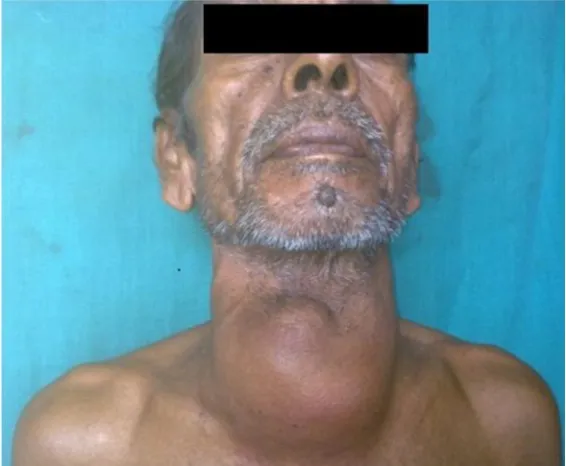
FIG 1 : CLINICICAL PICTURE OF MULTINODULAR GOITRE
[image:43.595.156.439.420.653.2]Palpation
The thyroid gland is palpated both from behind and in front of the patient.
During palpation size, shape, surface, skin over swelling, consistency and
extent of the gland is noted.
The surface is bosselated with gross variation of consistency in MNG,
where as in cases of thyroid carcinoma the consistency is hard, with indistinct
irregular margin and irregular surface. If lower border of the gland is not felt at
the suprasternal notch, it suggests retrosternal goitre. Examination of carotid
pulsation, regional lymph nodes and position of trachea should be done
routinely.
INVESTIGATIONS FOR MNG
I. Tests for thyroid function
1.Thyroid hormones (T4 and T3) assay
2. Pituitary thyroid axis (TSH) assay
3.Free thyroid hormone measurements (FT4 and FT3).
4. Tests of thyroid binding proteins
5. Hypothalamic–pituitary axis (TRH test)
II.Dynamic and Imaging studies of the Thyroid
• T3 Supression test
• TSH stimulation test
• Perchlorate discharge test
2.Thyroidscintigraphy
3.Miscellaneous radioisotope imaging tests
•Gallium/ thallium
•Other radiopharmaceuticals –DMSA
III.Assessment of thyroid anatomy
• Ultrasound scan
• Computerised tomography (CT)
• Nuclear magnetic resonance imaging (NMRI).
IV.Tissue diagnosis
Fine Needle Aspiration Cytology (FNAC)
Core needle biopsy
V.Measurement of thyroid autoimmunity
• Anti-thyroglobulin antibodies
• Long acting thyroid stimulator (LATS)
• Thyroid stimulating immunoglobulins (TSI)
VI. Miscellaneous tests
• X-ray of the neck–AP and lateral vews
• X-ray of the chest - PA view
• Indirectlaryngoscopy
• Thyroglobulin assay
• Calcitonin assay
ULTRASOUND SCAN
It represents the first modality of investigation that has made it possible to
establish the physical state of thyroid lesion before surgery. The thyroid
nodularity evaluation with normal or enlarged volume can be performed with a
much higher sensitivity by high-resolution ultrasonography when compared
with palpation. By ultrasonographic study, solid, cystic and mixed solid cystic
nodules can be differentiated.
Among solid nodules, lesions of high or normal echogenicity can be
distinguished from those with reduced echogenicity by comparison with normal
the nodules to be investigated further by FNAC to exclude malignancy. It also
has to be emphasized that the sonological appearance of a nodule per se does
not exclude malignancy of the gland. Ultrasound also cannot be used to
differentiate between functioning and nonfunctioning nodules.
FINE NEEDLE ASPIRATION CYTOLOGY (FNAC)
For the past 15-20 years, there has been a remarkable change in the
approach to diagnosis and management of thyroid nodules. FNAC has become a
test of utmost importance in the investigation and management of thyroid
nodules. It involves the use of a smaller needle (Gauge 23-25) and 10-20 ml
syringe. This technique provides material for cytological rather than a
histological interpretation of the thyroid. The nodule is fixed between the
fingers and thumb of one hand and the needle is injected into the lesion with the
other hand. Suction is applied in the syringe to create a negative pressure. The
needle is then moved back and forth in the nodule in different directions, by
maintaining the negative pressure. A smear is prepared by using the material in
the syringe. Fixed with 95% alcohol, stained and then studied..
There are four main categories in which the results fall, malignant,
benign, suspicious and inadequate. In case of inadequate sample, the aspirate
should be repeated. Papillary, medullary, anaplastic carcinoma and thyroiditis
can be diagnosed using FNAC. Follicular adenoma cannot be differentiated
form follicular carcinoma by FNAC, as presence of capsular and vascular
FNAC is reported as follicular adenoma or suspicious of malignancy, surgery
has to be performed to confirm the pathology.
RADIOIODINE UPTAKE AND THYROID SCAN
Cassen and his co-workers in 1951 were among the first to implement
radioisotope scanning for diagnosis of diseases. They selected thyroid gland as
the first organ for studying. The remarkable avidity of thyroid gland for
radioiodine permitted visualization of the gland even with primitive scanning
equipment available during that time.
Radioiodine uptake
Radioiodine is tested by administering a known amount of radioactive
iodine to the patient and measuring the percentage accumulated in the thyroid
gland by an external counting device. The radionuclide of choice is I 123. The
radioiodine is given by mouth either in capsule or liquid from and the
measurement is taken at a known time after ingestion.
The measurement depends on the function of the thyroid and the size of
iodine pool or to be more precise the plasma inorganic iodine. Originally the
test was used for the differentiation of normal people from hypothyroid and
Causes of increased radio iodine uptake
1.Hyperthyroidism
•Graves disease
• Toxic MNG
• Toxic adenoma
2. Recovery form thyroiditis
3. Iodine deficiency
4. Dysharmonogenesis except trapping defect
5. After stopping anti-thyroid drugs
Causes of decreased radioiodine uptake
1. Iodine load
• Dietary
• Radiographic contrast agents
• Medicines with inorganic iodine
2.Thyroxine or other thyroid medication–factitious thyroiditis
4. Hypothyroidism
5.Radio nuclide capsules not ingested or digested
6.After severe exercise
THYROID SCINTIGRAPHY
a.RoutineScintigraphy
Thyroid Scintigraphy became possible after the development of the
rectilinear scanner by Casson. Now imaging in thyroid scinigraphy is
performed by using by anger camera with a pinhole collimator. The
radionuclide I123 is administered orally as a capsule and scintigrams are
obtained 3-6 hours later. Recently Technetium 99m (99m TC) which is
normally taken up by thyroid, like iodine, is widely used.
The scan has been used for:
1. Determining the size of the gland
2.Determining whether the nodule concentrates the radioiodine or
(99mTC) or not.
3.Determining if retrosternal shadow on chest x - ray is due to thyroid.
4.Determining whether a lump in the tongue or track of thyroglossal duct
5. For evaluation of toxic multi nodular goitre
b.Whole body Scintigraphy for thyroid metastases
Whole body Scintigraphy study is designed to provide information about
the presence or absence of functioning metastases from a differentiated thyroid
carcinoma. It should be done only after performing a total thyroidectomy
because if it is done when there is a significant amount of normal thyroid tissue,
the radio iodine localizes preferentially in the normal tissue and metastases are
not made out.
Autoradiographic studies have shown that the metastatic thyroid
carcinoma have about 1% of the uptake of normal thyroid gland. 1 or 2 mci of
I131 is used. Whole body Scintigram should be performed about 4 weeks after
surgery to allow endogenously secreted thyroid hormones to be metabolized and
Thyroid stimulating harmone to raise. Blood is examined for TSH and
thyroglobulin before I131 is administered. A whole body scintigraphy scan is
obtained 48-72 hours after the radioiodine dose has been administered.
MISCELLANEOUS RADIOLOGICAL STUDIES
COMPUTERIZED TOMOGRAPHY (CT)
Computerized tomography has no place in routine evaluation of patients
1.Thyroid cancer with spinal metastases.
2. Substernalgoitre not concentrating iodine.
3.Recurrent cancer in neck not taking up iodine
HORMONAL ASSAY
Total thyroid hormones
Total T4 and T3 are measured using specific radioimmunoassay. Since
they are highly protein bound, their values depends upon the levels of binding
proteins present in serum. The normal total plasma T4 is approximately 8 micro
g/dl and plasma total T3 is 0.15micro g/dl.
Causes of high level of thyroid hormones
Hyperthyroidism Increased binding proteins
Hereditary
Pregnancy
Active hepatitis
Oral contraceptive
New born
Antibody to thyroid hormones
Pituitary resistance to thyroid hormones
Acute medical illness
Causes of low levels of thyroid hormones
Hypothyroidism Decreased binding proteins
Hereditary
Androgens
Nephrotic syndrome
Glucocorticoids
Cirrhosis of liver
Low T3 and low T4 (sick euthyroid)
Low T3 syndrome
TESTS OF THYROXINE BINDING PROTEINS
Thyroxine binding capacity (TBC) is measured indirectly by quantitating
the capacity of the binding sites in serum, those which are not carrying
is in the range of 25% to 35%. If there are a lot of unoccupied binding sites over
the proteins, the tracer will bind there and T3RU is low and vice versa.
Knowledge of T3, T4 and T3RU values are usefull in determining if the
problem is due to thyroid diseases, or due to a binding protein abnormality. If
both tests are abnormal towards the same direction, the thyroid is at fault. In
contrast to it if one test is high and the other is low, the defect is due to the
carrier proteins.
PITUITARY THYROID AXIS–TSH
Thyroid stimulating harmone radioimmunoassay became available in
1965. When T4 and T3 are normal and TSH is above normal, it is called as
subclinical hypothyroidism. But older assay could only differentiate TSH levels
of 1 Iu/ml or 2 Iu/ml and since many euthyroid individuals have TSH levels of
0.5 Iu/ml to 2.0 Iu/ml, assay could not differentiate suppressed values from
normal values
With the introduction of immune radiometric assay (IRMA) and
amplified enzyme linked immunoassay (AEIA) techniques, TSH levels even
less than 0.3 micro units/ml can be detected. TSH is an extremely valuable aid
in diagnosis, hypothyroid patients have high levels and hyperthyroid patients
have low levels of TSH, provided pituitary insufficiency is excluded. When T3
HYPOTHALAMIC PITUITARY AXIS (TRH TEST)
TRH was isolated, characterized and synthesized in 1968. This is tested
by injecting TRH intravenously and assessing the ability of pituitary gland to
secrete TSH. A normal response is rise in TSH. The rise in TSH is maximal at
20-30 minutes with return to normal levels by 60-90 minutes. In cases of
hyperthyroidism, the pituitary gland is suppressed by thyroid hormone and there
is no rise in TSH even after injection of TRH. This test was of great value in
early times for understanding the physiology and pathophysiology of
hypothalamic pituitary thyroid interaction and it was clinically valuable in
diagnosis of borderline hyperthyroidism.
PROPHYLAXIS - PLAN OF ACTION IN INDIA
The Government of India launched a NATIONAL GOITRE CONTROL
PROGRAMME. The objectives of the programme are as follows:
1. Initial survey to identify magnitude of problem in the country
2. Production and supply of iodized salt to the endemic regions
3. Health Education & Publicity
4. To undertake monitoring of the quality of iodized salt assessing urinary
5. Re-survey in goiter endemic regions after five years continuous supply
of iodized salt to assess the impact of the control programme. The result of
re-survey in some areas has revealed that the prevalence of goiter has not been
controlled as desired.
TREATMENT OF MULTINODULAR GOITRE
Several treatment modalities are available for patients with Multi Nodular
Goitres. The selection of best therapeutic option depends on several factors,
including size of goitre, location, presence and severity of compressive
symptoms, and the presence or absence of thyrotoxicosis.
THYROID-HORMONE-SUPPRESSIVE THERAPY
As Thyroid stimulating harmone is regarded as a growth factor for the
thyroid epithelial cells,treatment with levothyroxine in doses enough to suppress
TSH has been used for years to reduce or prevent growth of thyroid nodules.
However, the effectiveness of this treatment has remained controversial. A
clinical trial of 78 patients with non-toxic multi nodular goiter who were treated
with levothyroxine or a placebo for a period of nine months and then were
followed up for an additional nine months. Results showed a 58% reduction in
volume of goitre assessed by ultrasonography versus a 4% reduction in placebo
group, but this effect was lost after levothyroxine therapy was stopped. In a
non-toxic goiter, it was found that 60% of patient had decrease in goiter size during
the period of thyroid hormone therapy. The decrease was found to occur within
the first three months of treatment, and a better response was noted in those
patients with diffusethyromegalyrather than nodular goiters.
The efficacy of levothyroxine therapy in preventing the recurrence of
goiter growth after hemithyroidectomy is less clear. Several non randomized
trials show that levothyroxine therapy is effective for these cases, but
randomized controlled trials have failed to demonstrate a significant reduction
in the recurrence of goiter. It has also been showed that in patients with
multinodulargoitre, levothyroxine suppression therapy may prevent the
formation of new nodules by interfering with the process of goitrogenesis,
though it may not cause regression of all the clinically apparent nodules.
Due to known risks associated with subclinical hyperthyroidism as a
result of levothyroxine suppression therapy, caution is essential when
considering this management option in post-menopausal patients, particularly in
those patients with evidence of low bone mass, the elderly patients, and those
patients with cardiac disease, in those whom the risk of this therapy may be
increased.
RADIOIODINE THERAPY
Radioiodine (RAI) has been effectively and widely used for the treatment
of toxic multi nodular goiters. Administered usually as a single dose, orally it is
nodules of the thyroid in period of two to four months. Some patients,
particularly in those with large goiters or severe hyperthyroidism, may need
more than one dose to achieve euthyroidism. Radioiodine administered is
preferentially accumulated in the hyper functioning nodules of thyroid and, so,
subsequent rates of hypothyroidism are lower than in patients who are treated
with radioiodine for Graves’ disease.
Patients with severe thyrotoxicosis, particularly those with cardiac
diseases and the elderly, may be pre-treated with antithyroid drugs
(methimazole or propylthiouracil). There is evidence that propyl thiouracil, but
not methimazole, may reduce the effectiveness of subsequent radioiodine
therapy in these patients. Although RAI has not been traditionally considered as
a treatment option for patients with non-toxic MNGs, several studies have
shown that it is both safe and effective.
This treatment modality has the advantage of resulting in significant
reduction in goitre size (30–60%), along with improvement in obstructive
symptoms in most of the patients. In one study RAI was shown to be more
effective than levothyroxine suppressive therapy in reducing goiter size.
Transient hyperthyroidism may be seen in the first two weeks after treatment,
and permanent hypothyroidism has also been reportedin up to 45% of
patients.Pre-treatment with recombinant human thyroid stimulating harmone
(rhTSH) has been evaluated in recent years as adjuvant to RAI in several studies
as a method to enhance the efficacy of uptake of RAI in non-toxic thyroid tissue
Development of Graves’ hyperthyroidism (with increased levels of
TSH-receptor antibodies) has been described following treatment with RAI in
patients with multinodular goitre and has been found to be more common in
those patients with high thyroid peroxidase (TPO)antibody concentrations
before the initiation of treatment.
SURGERY
Multinodular goitre is mostly an irreversible stage. So, surgery is the
treatment of choice in young patients, for cosmetic reasons, possibility of
malignant transformation, and suspicious malignancies. Patients presenting with
large, obstructive and substernal non-toxic multinodular goitres or those with no
regress in growth are best managed with surgery. Complications of surgery in
cases of large and substernal goiters are more commonly seen than in patients
who undergo thyroidectomy for cervical goiters. Complications include injury
to the trachea, recurrent laryngeal nerves and the parathyroid glands. In a study
which included 34000 patients who underwent thyroidectomy, 1153 (3.4%) had
substernalgoitre showed that this last group of patients were older, men and
more likely to have a comorbid condition. The surgeries usually performed for
multinodulargoitre are subtotal and total thyroidectomy. Hemithyroidectomy
may be performed in special circumstances where there are aremicronodules in
SURGERY FOR NONTOXIC MNG
Most patients of multinodulargoitre are asymptomatic and do not require
surgery. Surgery may be indicated on cosmetic grounds. Retrosternal goitre
with tracheal obstruction is an indication of operation, as is the presence of
dominant area of enlargement among the other nodules, which may be
neoplastic.
Choice of surgery:
a. Total thyroidectomy along with immediate and lifelong replacement of
thyroxine.
b. Any form of partial resection in order to conserve sufficient
functioning thyroid tissue to serve normal function and also eliminating the risk
of hypothyroidism ,which accompanies total thyroidectomy. Partial resection
aims at removing bulk of the gland and leaving upto 8 grams of relatively
normal thyroid tissue i.e., subtotal thyroidectomy. More often the
multinodularity is asymmetrically and distributed with one lobe more
significantly involved than the other lobe, under these circumstances total
lobectomy on the more affected side is the may be performed with either little
or no intervention on the other less affected side.
In many cases, the causative factors persists and recurrence is likely to
occur. Surgery for recurrent nodular goitre is difficult and hazardous. Due to
subtotal thyroidectomy it has been customary to give suppressive doses of
thyroxinein order to suppress TSH secretion and thereby aiming at preventing
recurrence.
SURGERY FOR TOXIC MNG
In toxic MNG the principle behind surgery is by reducing the mass of
overactive tissue. Cure is probable if the remnant thyroid tissue can be
surgically reduced below a critical mass. This may result in a reduction in the
level TSAbs or it may be that the circulating TSAbs however high its level may
be can only produce limited hypertrophy and hyperplasia when the thyroid
tissue mass is meager. In a case of toxic MNG with autonomous toxic nodules,
surgery cures by removing all the overactive nodules. Thereby allows
suppressed normal thyroid tissue to function again.
The extent of resection of thyroid tissue depends on the gland size,
patient’s age, experience of the operating surgeon, the need to minimize
recurrent toxicity and the wish to avoid postoperative thyroid replacement
therapy. Young patients who have a small gland are at greater risk of recurrence
even when there is a very small remnant thyroid tissue.
In recent times, there is an increasing trend towards total thyroidectomy,
as it simplifies the subsequent management and a permanent euthyroid state can
be rapidly achieved on thyroid replacement therapy. In contrast to this a patient
for subtotal thyroidectomy where 4-5 gms of thyroid tissue is left behind in each
lobe.
In toxic Multinodular goitre, surgery is the treatment of choice. Treatment
with radioiodine is highly unreliable in toxic MNG because blood flow through
such goitres is not homogenous or symmetrical as in diffuse goitre. As a result
uptake of radioiodine by the goitre is irregular and many areas escape effective
radiation from radioiodine and hyperthyroidism continues. For these reason
surgery is the treatment of choice for patients with hyperthyroidism due to
Multinodular goitre.
SUBTOTAL THYROIDECTOMY
The patient is made to lie supine on the operating table, with the table
tilted 15 degree at the head end in order to reduce venous engorgement. A sand
bag is placed under the shoulders and the neck is extended to make the thyroid
gland more prominent. General anaesthesia is administered through a flexible
endotracheal tube preferably and good muscle relaxation obtained. A low color
skin crease incision is made two finger breadth above the suprasternal notch.
Upper and lower flaps of skin, subcutaneous tissue and platysma are raised upto
the thyroid notch above and to the suprasternal notch below. The deep cervical
fascia is incised in the midline. The strap muscles are split and not divided as a
routine but may be divided if great exposure is required. In 30% of patients,
middle thyroid veins is present which is short and lies transversely passing
blood supply to the gland is the superior thyroid artery, which must be ligated
and divided with caution and in a secure manner. The superior thyroid artery is
divided close to the gland to prevent the injury to external laryngeal nerve. The
lobe is then freed so it can be rotated out of its bed. The inferior thyroid artery
away from the gland. The recurrent laryngeal nerve should be identified along
its course and injury should be prevented. The parathyroid glands are protected
by identification before resection and avoiding ligatures and sutures close to
hilum of the identified gland. If one of the parathyroid gland is inadvertently
excised or devascularised, it should be autotransplanted within the
sternomastoid muscle in several fragments. Subtotal resection of each lobe is
carried out, leaving behind a remnant of between 3 and 5 grams on each side.
Complete haemostasis is obtained by ligation of individual vessels and by
suturing of thyroid remnants to the tracheal fascia. The pretracheal muscles and
cervical fascia are sutured and the wound is closed with or without suction
drainage to the deep cervical space.
TOTAL THYROIDECTOMY
The technique of operation is same as in subtotal thyroidectomy, except that no
remnant thyroid tissue is left over in total thyroidectomy. Identifying and
sparing recurrent laryngeal nerves and parathyroid glands is very important as
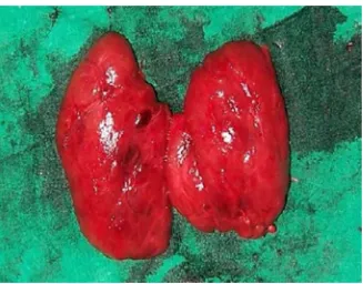
FIG 3 : EXPOSURE OF BOTH THE LOBES OF THYROID
[image:64.595.140.456.409.679.2]FIG 5 : IDENTIFICATION OF RECURRENT LARYNGEAL NERVE
[image:65.595.136.463.402.658.2]POSTOPERATIVE COMPLICATIONS
HAEMATOMA
A tension haematoma deep to thedeep cervical fascia is usually due to
slipping of ligature of the superior thyroid artery; occasionally haemorrhage
from a remnant of thyroid gland or a thyroid vein may be responsible.
immediate surgical exploration is necessary if the patient develops respiratory
distress. On rare occasions, it may be necessary to open the sutures in the
bedside to relieve tension before shifting the patient to the theatre to evacuate
the haematoma and to arrest the site of bleed.
RESPIRATORY OBSTRUCTION
Respiratory distress in most cases is due to laryngeal oedema and very
rarely due to kinking or collapse of trachea. The most important cause of
laryngeal oedema is due to a tension haematoma. Trauma to the larynx by
anaesthetic and surgical manipulation is a main contributory factor particularly
if the goitre is very vascular.
Unilateral or bilateral recurrent nerve paralysis will not usually cause
immediate postoperative respiratory obstruction. Unless laryngeal oedema is
also present but the former two will aggravate the obstruction. If release of the
tension haematoma does not relieve airway obstruction immediate tracheal