Copyright © 2003, American Society for Microbiology. All Rights Reserved.
Molecular and Functional Analyses of Kunjin Virus Infectious cDNA
Clones Demonstrate the Essential Roles for NS2A in Virus
Assembly and for a Nonconservative Residue
in NS3 in RNA Replication†
Wen Jun Liu, Hua Bo Chen, and Alexander A. Khromykh*
Sir Albert Sakzewski Virus Research Centre, Royal Children’s Hospital, and Clinical Medical Virology Centre, University of Queensland, Brisbane, Queensland 4029, Australia
Received 30 December 2002/Accepted 18 April 2003
A number of full-length cDNA clones of Kunjin virus (KUN) were previously prepared; it was shown that two of them, pAKUN and FLSDX, differed in specific infectivities of corresponding in vitro transcribed RNAs by
⬃100,000-fold (A. A. Khromykh et al., J. Virol. 72:7270-7279, 1998). In this study, we analyzed a possible ge-netic determinant(s) of the observed differences in infectivity initially by sequencing the entire cDNAs of both clones and comparing them with the published sequence of the parental KUN strain MRM61C. We found six common amino acid residues in both cDNA clones that were different from those in the published MRM61C sequence but were similar to those in the published sequences of other flaviviruses from the same subgroup. pAKUN clone had four additional codon changes, i.e., Ile59 to Asn and Arg175 to Lys in NS2A and Tyr518 to His and Ser557 to Pro in NS3. Three of these substitutions except the previously shown marker mutation, Arg175 to Lys in NS2A, reverted to the wild-type sequence in the virus eventually recovered from pAKUN RNA-transfected BHK cells, demonstrating the functional importance of these residues in viral replication and/or viral assembly. Exchange of corresponding DNA fragments between pAKUN and FLSDX clones and site-directed mutagenesis revealed that the Tyr518-to-His mutation in NS3 was responsible for an ⬃5-fold decrease in specific infectivity of transcribed RNA, while the Ile59-to-Asn mutation in NS2A completely blocked virus production. Correction of the Asn59 in pAKUN NS2A to the wild-type Ile residue resulted in complete restoration of RNA infectivity. Replication of KUN replicon RNA with an Ile59-to-Asn substitution in NS2A and with a Ser557-to-Pro substitution in NS3 was not affected, while the Tyr518-to-His substitution in NS3 led to severe inhibition of RNA replication. The impaired function of the mutated NS2A in production of infectious virus was complemented in trans by the helper wild-type NS2A produced from the KUN replicon RNA. However, replicon RNA with mutated NS2A could not be packaged intransby the KUN structural proteins. The data demonstrated essential roles for the KUN nonstructural protein NS2A in virus assembly and for NS3 in RNA replication and identified specific single-amino-acid residues involved in these functions.
Kunjin virus (KUN) is an Australian flavivirus closely re-lated to other members of the Japanese encephalitis virus subgroup. The KUN genome consists of single-stranded RNA of positive polarity comprising 11,022 nucleotides (10), with one long open reading frame coding 3,433 amino acids in three structural proteins (C, prM, and E) and seven nonstructural (NS) proteins (NS1, NS2A, NS2B, NS3, NS4A, NS4B, and NS5) (6). The gene order of KUN genome RNA is 5⬘ -C-(pr)M-E-NS1-NS2A-NS2B-NS3-NS4A-NS4B-NS5-3⬘.
Generation of the flavivirus full-length cDNA clones has been hampered by their apparent instability inEscherichia coli, leading to extensive mutagenesis of cDNA sequences during preparation of plasmid DNAs (25). These mutations usually resulted in the complete or partial loss of infectivity of RNAs transcribed in vitro from the cDNA templates. Other research-ers have applied different approaches to overcome this prob-lem, including splitting the full-length cDNA sequence into
two pieces for amplification followed by transcription of RNA from the in vitro-ligated DNA template (24), use of a
partic-ularE. colistrain, and lowering of the temperature of
incuba-tion while growing plasmid DNA inE. coli(7, 18); using very low-copy-number vectors (8); avoiding amplification of cDNA
inE. coliby assembly of full-length cDNA sequence by using
long PCR (7); multiple corrections of mutated sequences (23); and inserting introns to separate toxic regions (30).
Over the years a number of stable infectious full-length cDNA clones of KUN that produced RNAs of different spe-cific infectivities have been generated (10, 12). KUN RNA transcribed from our originally prepared full-length cDNA clone pAKUN had very low specific infectivity (⬃1 PFU per 10
g of RNA), and the recovered virus differed from the paren-tal KUN by a smaller plaque phenotype and a delayed repli-cation in cells and in mice (10, 12). Later reconstruction of KUN cDNA from viral RNA by using reverse transcription and long PCR amplification with high-fidelity DNA polymer-ase resulted in generation of cDNA clones FLSD (with a 7-kb fragment in pAKUN replaced) and FLSDX (with a 9.5-kb fragment in pAKUN replaced), which produced RNAs with dramatically improved specific infectivities (2 ⫻103 and 104
PFU/g of RNA, respectively) (12). Further characterization
* Corresponding author. Mailing address: Sir Albert Sakzewski Vi-rus Research Centre, Royal Children’s Hospital, Brisbane, Queens-land 4029, Australia. Phone: 617 36361568. Fax: 617 36361401. E-mail: [email protected].
† This is publication number 161 from the Clinical Medical Virology Centre and the Sir Albert Sakzewski Virus Research Centre.
7804
on November 8, 2019 by guest
http://jvi.asm.org/
of the virus recovered from FLSD RNAs showed a delayed replication in Vero cells and reduced virulence in mice (9). Selective sequencing of the NS1 gene in the FLSD cDNA clone and in the RNA isolated from virus recovered after transfection of FLSD RNA showed the presence of a proline (Pro)-to-leucine (Leu) substitution of NS1 amino acid codon 250 (Table 1), which was surprisingly stable and was retained in the virus for at least 10 passages (9). Correction of this muta-tion from Leu back to the Pro codon in the FLSD cDNA clone resulted in the recovery of virus indistinguishable from the wild-type KUN strain MRM61C in the kinetics of viral growth in Vero cells and in virulence in mice (9). However, the effect of this correction in NS1 on specific infectivity of transcribed RNA and on the phenotype of viral plaques, as well as the remaining sequence in different cDNA clones leading to such dramatic differences in their infectivity, was not determined. In this study we utilized sequencing analysis of the entire cDNA of pAKUN and FLSDX clones and site-directed mutagenesis for functional analyses of full-length and replicon RNAs to show that the low efficiency of pAKUN RNA in production of infectious KUN was mainly due to the presence of two amino acid substitutions, one in NS3 that severely inhibited RNA replication and another in NS2A that unexpectedly blocked virus assembly. We also employed here previously described complementation and encapsidation assays (12, 13) with full-length and replicon RNAs to show that only wild-type (and not mutated) NS2A when present either in cis or in trans can participate in packaging of RNA into secreted virus particles.
MATERIALS AND METHODS
Cells. BHK21 cells were grown in Dulbecco minimal essential medium (DMEM; Invitrogen, San Diego, Calif.) supplemented with 10% fetal bovine serum (FBS) at 37°C in a CO2incubator. repBHK cells containing stably repli-cating KUN replicon RNA (12) were maintained in the same medium supple-mented with 0.5 to 1 mg of G418 (Geneticin; Invitrogen) per ml.
Construction of plasmids.Plasmids pAKUN/FLSDX2A, pAKUN/FLSDX3, FLSDX/pAKUN2A, and FLSDX/pAKUN3 were obtained by exchanging the fragmentsSacII1482-BssHII5743andBssHII5743-AgeI7897between previously de-scribed KUN full-length cDNA clones pAKUN (10) and FLSDX (12) (Fig. 1). Plasmid FLSDX/pAKUN2A(Ile59) (Fig. 1) was generated by site-directed PCR mutagenesis of the Asn59 codon in NS2A to Ile in FLSDX/pAKUN2A by using high-fidelityPfuDNA polymerase (Stratagene, La Jolla, Calif.). The mutagenesis was performed on the intermediate plasmid pUC-NS2A containing theSphI3628 -BamHI4807fragment from the pAKUN plasmid in the pUC18 vector. After confirmation of the introduced mutation by sequencing analysis, theSphI3628 -BamHI4807fragment including the Asn59-to-Ile mutation in NS2A was trans-ferred into FLSDX/pAKUN2A. The mutation in the resulting FLSDX/pAKUN2A (Ile59) plasmid was confirmed by sequencing analysis. KUN replicon plasmids repPAC-gal/2Amut and repPAC-gal/3mut were prepared by replacing the SphI3628-BstBI5148andBstBI5148-AgeI7897fragments, respectively, in a previous-ly described KUN replicon construct, repPAC-gal (20), with those from the pAKUN plasmid (Fig. 1). Plasmids repPAC-gal/3mut(His518) and repPAC -gal/3mut(Pro557) were constructed by site-directed PCR mutagenesis with high-fidelityPfuDNA polymerase to change Pro557 in NS3 back to the wild-type Ser and His518 in NS3 back to the wild-type Tyr, respectively. The mutagenesis was performed initially on the intermediate plasmid pUC-NS3-5 containing the SalI5384-SalI7884fragment from pAKUN. After confirmation of the introduced mutation by sequencing analysis, theSalI5384-SalI7884fragments including the above mutations were transferred into the repPAC-gal/3mut plasmid. The plasmids FLSDX/pAKUN3(His518) and FLSDX/pAKUN3(Pro557) (Fig. 1) were generated by replacingBssHII5743-AgeI7897fragments in FLSDX/pAKUN3 with those from repPAC-gal/3mut(His518) and repPAC-gal/3mut(Pro557), respectively. All mutations in the resulted replicon and full-length plasmids were confirmed by sequencing.
[image:2.603.44.542.80.271.2]RNA transcription, transfection, and determination of specific infectivity.All RNA transcripts were prepared with SP6 RNA polymerase fromXhoI-linearized plasmids DNAs and were purified by using Bio-spin 30 chromatography columns (Bio-Rad, Hercules, Calif.). Purified RNAs were electroporated into BHK21 cells, essentially as described previously (11). Briefly, 1g of in vitro-transcribed RNAs was electroporated into 2⫻106BHK21 (normal BHK) or repBHK cells in 400l in a 0.2-cm-electrode-gap cuvette (Bio-Rad) with a Bio-Rad Gene Pulser apparatus. Electroporated cells were then used to prepare serial 10-fold dilutions in DMEM–10% FBS, mixed with 106nontransfected BHK21 cells, and seeded on 60-mm-diameter culture dishes for 8 h to allow cells to attach. Then cells were overlaid with DMEM–2% FBS in 0.75% agarose (FMC, Rockland, Maine). After 4 to 6 days of incubation at 37°C, cells were fixed with 20% formaldehyde and were stained with crystal violet.
TABLE 1. Differences between published KUN MRM61C sequences and sequences of infectious KUN cDNA clonesd
Nucleotide position Gene
Amino acid posi-tion in encoded
protein
MRM61C data FLSDX cDNA data pAKUN cDNA data Corresponding residue in related flaviviruses Nucleotide Amino acid Nucleotide Amino acid Nucleotide Amino acid
544 PrM 28 Ca Pro A Thr A Thr Thr (WN, JE, MVE)
1500 E 178 G Leu Tc — — — Leu (WN, JE)/Ala MVE)
1635 E 223 C Leu — — T — Leu (WN, MVE, JE)
1824 E 286 C Leu G — G — Leu (WN, MVE, JE)
2556 NS1 29 Ta Ile G Met G Met Ile (MVE)/Met (WN)/Val (JE)
2924 NS1 152 Aa Asn G Ser G Ser Ser (WN, MVE, JE)
3218 NS1 250 C Pro Tb Leu Tb Leu Pro (WN, MVE, JE)
3701 NS2A 59 T Ile — — Ab Asn Ile (WN, MVE)/Val (JE)
4049 NS2A 175 G Arg — — Ac Lys Arg (WN, MVE, JE)
5937 NS3 442 A Val G — G — Val (WN, MVE, JE)
5996 NS3 462 Ca Thr T Ile T Ile Ile (WN)/Val (JE, MVE)
6017 NS3 469 Ca Ala T Val T Val Val (WN, JE)/Ile (MVE)
6163 NS3 518 T Tyr — — Cb His Tyr (WN, MVE)/Phe (JE)
6280 NS3 557 T Ser — — Cb Pro Ser (WN)/Gln (JE, MVE)
7112 NS4B 66 Ca Thr A Asn A Asn Asn (WN)/Thr (MVE, JE)
7293 NS4B 126 G Val A — A — Val (WN)/Leu (MVE, JE)
7785 NS5 35 C Val T — T — Val (WN, MVE, JE)
10197 NS5 839 C Ser T — T — Ser (WN)/Thr (JE, MVE)
aApparent errors in the published MRM61C. bMutations probably introduced during cloning. cMutation introduced to produce cDNA clones.
dNucleotides in boldface in cDNA clones indicate changes from the original published sequence that lead to changes in corresponding amino acids, also shown in
boldface. —, no differences from wild type were found in the indicated nucleotide or in the corresponding encoded amino acid. WN, West Nile virus; JE, Japanese encephalitis virus; MVE, Murray Valley encephalitis virus.
on November 8, 2019 by guest
http://jvi.asm.org/
IF.Replication and expression of mutated full-length RNAs in transfected cells were monitored by immunofluorescence (IF) analysis with mouse mono-clonal antibodies to KUN E protein (1) as described elsewhere (12).
X-Gal staining and Northern blotting.Detection of replication and expression of mutated replicon RNAs in BHK21 cells were performed by in situ staining with 5-bromo-4-chloro-3-indolyl--D-galactopyopyranoside (X-Gal) or by North-ern blot with32P-labeled probes specific for-galactosidase (-Gal) and-actin nucleotide sequences, as described previously (20).
Nucleotide sequence accession number.The pAKUN and FLSDX cDNA sequences have been deposited in GenBank and have been given the accession numbers AY274505 and AY274504, respectively.
RESULTS
Comparison of plaque morphology and specific infectivities of KUN RNAs transcribed from different cDNA clones and of the recovered viruses.As the first step towards identification of the primary determinates of virus replication in different cDNA clones, we compared specific infectivities of transcribed RNAs and plaque morphologies of the recovered viruses.
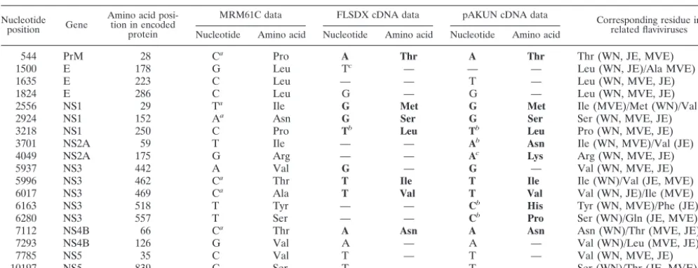
Plaque assays of BHK cells electroporated with RNAs tran-scribed from AKUN, FLSDX, and FLSDX(Pro) (FLSDX cDNA clone with a proline instead of leucine residue at NS1 amino acid 250) cDNA clones (Fig. 1) and of KUN virion RNA (KUN[wt]) in BHK cells showed specific infectivities of ⬍1, 1.3⫻104, 2⫻104, and 2⫻105PFU perg of RNA,
respec-tively (Fig. 2A and Table 2). Incorporation of the antigenomic sequence of hepatitis delta virus ribozyme (HDVr) into the FLSDX(pro) cDNA clone immediately downstream of the last nucleotide of KUN sequence increased specific infectivity of the resulting FLSDX(pro)HDVr RNA by⬃2.6-fold (5.3⫻104
PFU/g [Table 2]) compared to that of FLSDX(pro) RNA. The size of the viral plaques after transfection of FLSDX(pro) and FLSDX(pro)HDVr RNA was similar to that after trans-fection of KUN(wt) virion RNA, while transtrans-fection of FLSDX RNA produced much smaller plaques (Fig. 2A for FLSDX and FLSDX[pro]; results not shown for FLSXD[pro]HDVr). Vi-ruses recovered after transfection of these RNAs into BHK
FIG. 1. Schematic representation of the full-length and replicon KUN cDNA constructs with exchanged fragments. The filled box represents the sequence of the FLSDX clone; the open box represents the sequence of the pAKUN clone.SacII,SphI,BstBI,BssHII,AgeI, andSalI show restriction sites used in construction of plasmids by fragment exchange; numbers under the restriction sites represent corresponding nucleotide positions in the full-length KUN sequence (6, 10). In the designations of the full-length constructs generated by fragment exchange, first letters represent the name of the clone used as the vector backbone and are separated by a slash from the letters representing the name of the clone used as a source of the cloned fragment. Replicon constructs contain the puromycin acetyltransferase gene (PAC) and-Gal gene (-gal) cassette inserted in FLSDX(pro) in place of deleted structural genes (20). Amino acids shown in different recombinant constructs represent corresponding wild-type or mutated residues in NS2A (position 59) and NS3 (positions 518 and 557) genes identified in the original FLSDX and pAKUN constructs. Ile, isoleucine; Asn, asparagine; His, histidine; Pro, proline; Tyr, tyrosine; and Ser, serine.
on November 8, 2019 by guest
http://jvi.asm.org/
cells produced plaques similar in size to those formed in BHK cells transfected with corresponding RNAs (Fig. 2B). The in-crease in size of plaques and specific infectivity of FLSDX(pro) RNA compared to those of FLSDX RNA confirmed previous observations on the importance of amino acid 250 in NS1, shown to be essential for dimerization of this protein, in virus replication in cells and in virulence in mice (9).
Interestingly, in contrast to the extremely low efficiency of plaque formation after transfection of pAKUN RNA, infection by recovered AKUN produced well-defined plaques (Fig. 2B)
and it replicated efficiently (10). The retention of the intro-duced mutation in the NS2A gene (Arg175 to Lys) in the re-covered AKUN has previously been confirmed (10); however, the rest of the sequence of pAKUN cDNA and of the recov-ered AKUN, as well as of FLSDX cDNA (except for the NS1 gene [9]), has not been determined. It was reasonable to as-sume that the low specific infectivity of pAKUN RNA was due to the presence of mutations inhibiting virus replication and/or assembly and that these mutations were corrected in the re-covered AKUN. It was also likely that these inhibiting
[image:4.603.86.501.68.342.2]muta-FIG. 2. Viral plaque morphology in BHK cells transfected with the KUN virion RNA (wild type [wt]) and with the indicated engineered RNAs (A) and infected with the corresponding recovered viruses (B). BHK cells were electroporated with indicated RNAs or infected with indicated recovered viruses and were assayed for plaque morphology as described in Materials and Methods. “ng RNA” under the panels in A shows the amount of transfected RNA in nanograms calculated from appropriate dilution of transfected cells in the corresponding dishes. Viral plaques in panel A were visualized at day 4 after RNA electroporation and in panel B at day 5 after infection with corresponding recovered viruses at 10⫺4 dilution.
TABLE 2. Specific infectivities of KUN RNAs transcribed from different full-length cDNA constructsa
RNA Amino acid in encoded protein (PFU/Specific infectivityg of RNA) NSI-250 NS2A-59 NS2A-175 NS3-518 NS3-557
Wild-type virion RNA Pro Ile Arg Tyr Ser 2⫻105
FLSDX (pro) — — — — — 2.0⫻104
FLSDX (pro)HDVr — — — — — 5.3⫻104
FLSDX Leu — — — — 1.3⫻104
pAKUN Leu Asn Lys His Pro ⬍1
pAKUN/FLSDX2A Leu — Lys His Pro 2⫻103
pAKUN/FLSDX3 Leu Asn Lys — — ⬍1
FLSDX/pAKUN3 Leu — — His Pro 1.8⫻103
FLSDX/pAKUN3 (His518) Leu — — His — 2.5⫻103
FLSDX/pAKUN3 (Pro557) Leu — — — Pro 1⫻104
FLSDX/pAKUN2A Leu Asn Lys — — ⬍1
FLSDX/pAKUN2A (Ile59) Leu — Lys — — 1.4⫻104
a—, no differences from the wild type are shown in the indicated encoded amino acid residue.
on November 8, 2019 by guest
http://jvi.asm.org/
[image:4.603.44.543.570.716.2]tions originally present in pAKUN cDNA were corrected in FLSDX cDNA, leading to dramatic improvement in the spe-cific infectivity of FLSDX RNA.
Sequencing analysis of KUN full-length cDNA clones pAKUN and FLSDX. To confirm the above assumption, we decided to determine the entire genomic sequence of pAKUN and FLSDX cDNA clones and compare them with the previ-ously published sequence of the MRM61C strain of KUN (GenBank accession No. D00246) (6). The differences between pAKUN, FLSDX, and the published KUN sequence are shown in Table 1. FLSDX cDNA had 13 nucleotides different from the published KUN sequence, with seven of them leading to amino acid changes; pAKUN cDNA had 17 nucleotides different from the published KUN sequence, with 11 of them leading to amino acid changes. These amino acid mutations were scattered throughout the entire coding region and were located in the prM, E, NS1, NS2A, NS3, and NS4B genes. No mutations were found in NS2B, NS4A, and NS5 or in the 5⬘
and 3⬘ untranslated regions. pAKUN and FLSDX cDNA clones had six common amino acid substitutions, mainly con-served, that were different from the original MRM61C pub-lished sequence (Table 1). These six common amino acids detected in both FLSDX and pAKUN sequences aligned bet-ter with the sequences of other flaviviruses from the same subgroup than with the published MRM61C sequence, sug-gesting that they represent corrected errors in the originally published MRM61C sequence. The Pro250-to-Leu codon change in the NS1 gene was present in both pAKUN and FLSDX cDNAs and apparently contributed to delayed repli-cation of recovered viruses (9). Thus, with the exclusion of one amino acid difference in NS1 (Pro250 to Leu) that apparently arose during cloning, FLSDX cDNA was identical to the cor-rected wild-type KUN RNA sequence.
One of two mutations in NS2A in pAKUN clone repre-sented a conserved amino acid change (Arg175 to Lys) that was introduced intentionally as a marker mutation during plas-mid construction. It was demonstrated previously that this mu-tation was retained in the recovered AKUN and did not affect viral replication (10). Thus, it left only 3 amino acid codons in pAKUN cDNA different from those in FLSDX cDNAs and/or to those in corrected MRM61C viral RNA sequences (nucle-otide 3701T to A, changing NS2A amino acid Ile59 to Asn; nucleotide 6163T to C, changing NS3 amino acid Tyr518 to His; and nucleotide 6280T to C, changing NS3 amino acid Ser557 to Pro) (Table 1). These changes apparently contrib-uted to the low specific infectivity of transcribed pAKUN RNA. To examine whether these mutations were present in RNA of the recovered AKUN, we performed reverse tran-scriptase PCR and sequence analysis of the corresponding genomic regions in viral RNA. The results showed that all these three mutations but not the marker mutation (Arg175 to Lys in NS2A) reverted to the wild-type sequence in the recov-ered viral RNA (data not shown), clearly demonstrating the importance of these amino acid residues in virus replication.
Mutations in NS3 of pAKUN inhibit virus replication. In order to further identify the specific amino acid substitutions that contributed to the low specific infectivity of pAKUN RNA, we prepared a number of cDNA constructs containing these mutations by exchanging corresponding DNA frag-ments between pAKUN and FLSDX plasmids (Fig. 1).
pAKUN/FLSDX3 and pAKUN/FLSDX2A plasmids con-tained pAKUN backbone and corresponding fragments from FLSDX plasmid, including NS3 and NS2A regions, respec-tively (Fig. 1). FLSDX/pAKUN3 and FLSDX/pAKUN2A con-tained FLSDX backbone and fragments from pAKUN plas-mids, including the NS3 and NS2A regions, respectively (Fig. 1). We first compared replication efficiencies and specific in-fectivities of RNAs containing pAKUN-derived NS3 sequence (pAKUN/FLSDX2A and FLSDX/pAKUN3) with that of the parental pAKUN and FLSDX RNAs by using IF analysis with anti-E antibodies and plaque assays. Transfection of pAKUN/ FLSDX2A, FLSDX/pAKUN3, and FLSDX RNAs into BHK cells showed an increase in the number of IF-positive cells from 24 to 48 h after transfection, demonstrating the spread of infectious virus (Fig. 3). The detection of IF-positive cells in-dicated that all RNAs were replicating, since it was previously clearly demonstrated that nonreplicating KUN RNA degraded quickly in cells and that only replicating KUN RNA could express sufficient amount of proteins for IF detection (11). pAKUN/FLSDX2A and FLSDX/pAKUN3 also produced well-defined small plaques (relative to wild-type virus) charac-teristic for FLSDX RNA (results not shown). As expected from previous experiments, transfection of pAKUN RNA did not lead to the detection of the virus spread within 48 h by IF analysis (Fig. 3) or formation of defined plaques by 4 days in plaque assays (Fig. 2). Specific infectivities of pAKUN/ FLSDX2A and FLSDX/pAKUN3 RNAs were similar (2⫻103
and 1.8⫻103PFU/g, respectively) and were approximately
six- to sevenfold lower than that of FLSDX RNA (Table 2). Site-directed mutagenesis of the His518 and Pro557 mutations to wild-type Tyr and to Ser residues, respectively, showed that the decrease in efficiency of NS3-mutated RNA was largely due to the Tyr518-to-His substitution, while the Ser557-to-Pro substitution had only a marginal effect (Table 2). These data clearly showed that the Tyr518-to-His mutation in NS3 present in pAKUN RNA had an inhibitory effect at least early in virus replication but did not block production and spread of the infectious virus particles.
Mutations in NS2A block production of infectious virus.In contrast to the results with RNAs containing mutations in NS3, transfection of RNAs containing mutations in NS2A, i.e., pAKUN/FLSDX3 and FLSDX/pAKUN2A as well as pAKUN, did not lead to the detection of virus spread from 24 to 48 h by IF analysis (Fig. 3) and did not produce plaques in plaque assays by 4 days (data not shown), demonstrating the inability of these RNAs to produce infectious virus particles at least early in virus replication before the reversion could occur. All these three constructs had only one common amino acid sub-stitution, Ile59 to Asn in NS2A (Table 2), suggesting that residue 59 in NS2A may play a crucial role in ensuring the functioning of NS2A in production of virus particles. To con-firm this observation, we prepared another construct, FLSDX/ pAKUN2A(Ile59), in which we mutated Asn59 in FLSDX/ pAKUN2A back to the wild-type Ile residue (Fig. 1). In con-trast to the parental FLSDX/pAKUN2A RNA, transfection of FLSDX/pAKUN2A(Ile59) RNA into BHK cells resulted in production of well-defined viral plaques (results not shown) and the specific infectivity of this RNA was similar to that of the FLSDX RNA (Table 2). Thus, it was clear from these
on November 8, 2019 by guest
http://jvi.asm.org/
results that a single Ile59-to-Asn substitution in NS2A led to a complete block to the production of infectious virus particles.
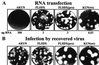
Effects of mutations in NS2A and in NS3 on RNA replica-tion. Although in the previous sections we demonstrated the detrimental effect of mutations in NS2A on production of virus particles and the inhibiting effect of mutations in NS3 on virus replication, it was not clear from these results whether these mutations affected RNA replication, virus assembly, or virus release. To examine the effect of the mutations in NS2A and NS3 on RNA replication, we initially prepared two KUN rep-licon constructs, repPAC-gal/2Amut and repPAC-gal/3mut (Fig. 1), by transferring the fragments with NS2A and NS3 mutations, respectively, from pAKUN into the replicon con-struct repPAC-gal (20). repPAC-gal was prepared previ-ously from FLSDX(Pro) clone and therefore represents the wild-type KUN sequence. It encodes-Gal for easy detection of expression of replicating RNAs. Transfection of BHK cells with repPAC-gal and repPAC-gal/2Amut RNAs resulted in
the detection of similar number of positive cells after X-Gal staining (⬃80 to 90%; panels 1 and 2 in Fig. 4A), while a significantly lower number of positive cells was detected by X-Gal staining after a transfection with repPAC-gal/3mut RNA (⬃5 to 10%; panel 4 in Fig. 4A). Analysis of -Gal expression in transfected cell lysates also showed a significantly smaller amount of -Gal produced from repPAC-gal/3mut RNA than those produced from repPAC-gal and repPAC -gal/2Amut RNAs (compare bar 4 with bars 1 and 2 in Fig. 4B). Northern blot analysis of transfected cells confirmed the re-sults of X-Gal staining and-Gal assay (Fig. 4C). The amount of housekeeping gene (-actin) mRNA in cells transfected with each RNA cell line was similar (Fig. 4C). These results demonstrated that mutations in NS3 but not in NS2A were responsible for the inhibition of RNA replication.
To determine which of the two mutations in NS3 inhibited RNA replication, we prepared two more replicon constructs, repPAC-gal/3mut(His518) and repPAC-gal/3mut(Pro557),
FIG. 3. IF analysis of BHK cells electroporated with mutated RNAs. One microgram of RNAs transcribed from indicated full-length KUN cDNA clones was electroporated into 2⫻106BHK21 cells, and 2⫻105cells were seeded on coverslips in 24-well plates. Twenty-four or 48 h later, cells were fixed with acetone and were stained with KUN anti-E antibodies as described in Materials and Methods.
on November 8, 2019 by guest
http://jvi.asm.org/
each containing corresponding individual amino acid residues different from the wild-type sequence (Fig. 1). X-Gal staining,
-Gal assay, and Northern blotting clearly showed that re-placement of wild-type Tyr518 by His was responsible for se-vere inhibition of RNA replication, similar to that observed with the double mutant, while another substitution, wild-type Ser557 with Pro, produced only a marginal effect (Fig. 4A to C). These results obtained with the mutated replicon RNAs were in agreement with the results obtained with the mutated full-length RNAs (Table 2).
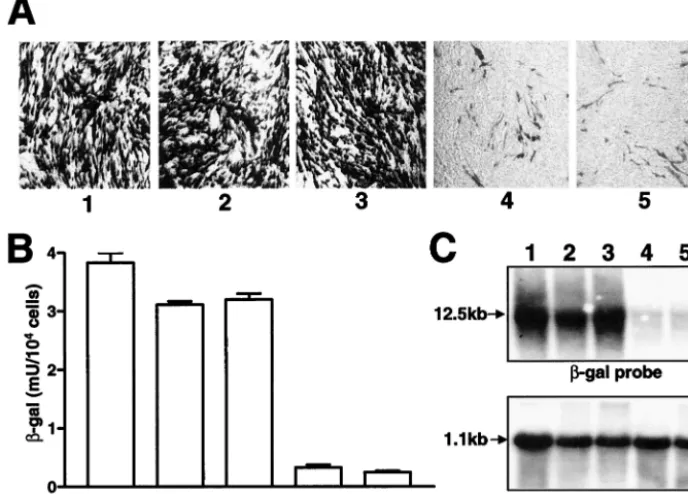
Complementation of mutated NS2A intrans.Recent studies with yellow fever virus (YF) showed that NS2A with a muta-tion abolishing an internal cleavage site, QK2T, at amino acid 190 and leading to an unexpected block in virus production could be rescued intrans by providing helper NS2A protein from another expression vector (17). It was therefore of inter-est whether KUN NS2A with the Ile59-to-Asn mutation block-ing virus production could be complemented intrans. To pro-vide helper NS2A for complementation of NS2A-mutated KUN RNA, we employed a complementation system based on the use of repBHK cells stably expressing NS2A as well as other KUN nonstructural proteins from KUN replicon RNA (15). Transfection of repBHK cells with pAKUN/FLSDX3 and FLSDX/pAKUN2A both containing the Ile59-to-Asn muta-tion in NS2A resulted in detecmuta-tion of viral plaques (Fig. 5), demonstrating successful complementation of their replication
by the wild-type NS2A produced from the helper replicon RNA. It has been demonstrated many times previously that recombination does not occur during complementation of de-fective genomic KUN RNAs in repBHK cells (see, for exam-ple, references 12, 14, and 15) and that therefore, viral plaques shown in pAKUN/FLSDX3 and FLSDX/pAKUN2A panels in Fig. 5 represent the result of true complementation.
[image:7.603.121.465.77.324.2]Replicon RNA with an Ile59-to-Asn substitution in NS2A cannot to be packaged intrans.In order to investigate possible mechanisms by which NS2A may be involved in virus assembly, we decided to examine whether replicon RNA with the Ile59-to-Asn mutation in NS2A could be packaged into virus parti-cles by providing structural proteins intrans. We used an es-tablished trans encapsidation system that employs a Semliki Forest virus (SFV) replicon expression vector for production of KUN structural proteins (SFV-MEC105) (13, 26). BHK cells were first electroporated with KUN replicon RNA rep-PAC-gal or repPAC-gal/2Amut containing the NS2A Asn59 mutation. X-Gal staining of transfected cells showed that transfection efficiencies of both RNAs were similar (data not shown). Thirty-two hours after the electroporation with KUN replicon RNAs, cells were electroporated again with SFV-MEC105 RNA. The culture fluids harvested at 48 h after the second electroporation were tested for the presence of virus-like particles containing encapsidated replicon RNAs by in-fecting Vero cells and staining them with X-Gal 48 h after
FIG. 4. Comparison of replication efficiencies of KUN replicon RNAs with mutations in NS2A and NS3. BHK21 cells were electroporated with the indicated mutated replicon RNAs. Forty-eight hours after electroporation, cells were either fixed by 4% formaldehyde–phosphate-buffered saline and were stained in situ with X-Gal (A) or lysed for a-Gal assay (B). Total RNA was also isolated for analysis of accumulation of replicating RNA by Northern blot with32P-labeled probes specific for-Gal (C, top row) or for-actin (C, bottom row) nucleotide sequences. For panel C, ⬃10g of total RNA was used for hybridization; arrows indicate positions in the gel of RNAs of 12.5 kb (C, top row) and 1.1 kb (C, bottom row) determined relative to migration in the same gel of the ethidium bromide-stained 1-kb ladder (Invitrogen). Numbering of corresponding panels (A), bars (B), and lanes (C) corresponds to cells transfected with repPAC-gal RNA (1), repPAC-gal/2Amut RNA (containing the Asn59 mutation) (2), repPAC-gal/3mut(Pro557) RNA (3), repPAC-gal/3mut RNA (with both His518 and Pro557 mutations) (4), and repPAC-gal/
3mut(His518) RNA (5).
on November 8, 2019 by guest
http://jvi.asm.org/
infection. No -Gal-positive cells were detected in 100 l of undiluted culture fluid collected from BHK cells transfected with repPAC-gal/2Amut and SFV-MEC105 RNAs, while
⬃160 -Gal- positive cells were detected in 100 l of 1:100 dilution of culture fluid collected from BHK cells transfected with repPAC-gal and SFV-MEC105 RNAs (Fig. 6). This re-sult clearly demonstrated that KUN replicon RNA with the Ile59-to-Asn mutation in NS2A could not be packaged intrans.
DISCUSSION
The molecular and functional analyses of two KUN full-length cDNA clones, pAKUN and FLSDX, producing infec-tious RNAs with substantially different specific infectivities have identified two amino acid codon substitutions responsible for such a difference in their efficiency, i.e., Ile59 to Asn in the NS2A gene and Tyr518 to His in the NS3 gene. The Tyr518-to-His substitution in NS3 led to significant inhibition of RNA replication, while the Ile59-to-Asn substitution in NS2A re-sulted in selective blockage of virus assembly and/or secretion. Combination of these two substitutions decreased the specific infectivity of less efficient pAKUN RNA by at least 100,000-fold. Sequence analysis of the virus eventually recovered from the pAKUN RNA after its delayed replication showed that
both codon substitutions reverted to the more efficient wild-type codons, thus further demonstrating the importance of these amino acids in functions of the corresponding proteins during virus replication.
Further detailed comparisons of entire cDNA sequences of pAKUN and FLSDX cDNAs with the published parent KUN MRM61C sequence revealed six additional, mainly conserved amino acid substitutions present in both pAKUN and FLSDX clones, which aligned better with the sequences of other flavi-viruses from the same subgroup than with the published MRM61C sequence (Table 1). It is likely that these mis-matches represent errors in the originally published MRM61C sequence determined from the collection of intermediate cDNA clones (6). It is possible that these errors were intro-duced via mutations during propagation of the intermediate cDNA clones inE. coli. Alternatively, it may represent quasi-species of KUN RNA present in the heterogeneous virus pool and selected during reverse transcription and cDNA cloning. In addition to these six mismatches, both FLSDX and pAKUN cDNAs contained a nonconservative Pro250-to-Leu codon change in the NS1 gene. We previously showed that this mu-tation abolished dimerization of NS1 and contributed to de-layed replication of the recovered virus in vitro and in vivo (9). Correction of this mutation led to restoration of NS1
dimer-FIG. 5. Complementation of infectivity of KUN RNAs with NS2A mutations in repBHK cells. Electroporation of repBHK cells with RNAs and plaque assays were performed as described in Materials and Methods for normal BHK cells. “ng RNA” under the panels is defined as in the Fig. 1A legend. Viral plaques were stained at day 6 after electroporation.
FIG. 6. Detection of packaged KUN replicon RNAs. Panels A and B represent selected fields of Vero cells stained with X-Gal at 48 h after infection with 100l of 1:100 dilution (panel A) or undiluted (panel B) culture fluids collected from BHK21 cells sequentially transfected with either repPAC-gal (A) or repPAC-gal/2Amut (B) RNAs, respectively, followed by transfection with SFV-MEC105 RNA.
on November 8, 2019 by guest
http://jvi.asm.org/
[image:8.603.133.452.560.694.2]ization and recovery of virus indistinguishable from the wild-type KUN in its growth properties in cells and in mice (9). Here we also show that transfection with RNA containing this corrected mutation (FLSDX[pro]) resulted in production of large viral plaques similar in size to those produced by trans-fection of purified KUN virion RNA (Fig. 2).
The effect of substitutions in NS3 on infectivity of full-length RNA and on replication of replicon RNA could be expected in view of the demonstrated functions of flavivirus NS3 in heli-case and RNA-triphosphatase activities (2, 19, 28, 29). How-ever, both replaced residues (amino acids 518 and 557) were located outside the conserved domains proposed to be in-volved in these activities. Particularly surprising was the inhibiting effect on RNA replication of a replacement of the nonconserved Tyr518 (intermediate polarity) by His (also in-termediate polarity), while a more drastic replacement of highly conserved polar residue Ser557 by nonpolar residue Pro did not have such a dramatic effect on RNA replication and virus assembly. It is not clear why the latter mutation, which did not have a significant effect on virus replication, reverted to the wild-type residue in the virus eventually recovered from pAKUN RNA. pAKUN was recovered as a total virus pool after a prolonged (8 or 9 days) incubation of pAKUN RNA-transfected cells (10). It is possible that this mutation could have been retained in some of the individual virus clones if these were analyzed.
Recent sequence analysis studies of the NS3 gene from dif-ferent isolates of hepatitis C virus (HCV) revealed that the region between codons 455 and 485 was conserved and was possibly related to HCV RNA helicase activity (27). The amino acid sequence alignment of KUN and HCV NS3 proteins (not shown) suggests that Tyr518 in KUN NS3 corresponds to Gly484 in HCV NS3, which is situated in the conserved heli-case region, and that therefore Tyr518 may also be involved in the helicase activity of flavivirus NS3. Further comparative studies of in vitro enzymatic activities of the wild-type KUN NS3 and KUN NS3 with the Tyr518-to-His substitution are required to make any definite conclusion on the role of this residue in the NS3 enzymatic functions in RNA replication.
The complete blockage of KUN production caused by a single amino acid substitution, Ile59 to Asn, in the nonstruc-tural protein NS2A despite efficient RNA replication is intrigu-ing. Recent studies with YF showing that a single amino acid substitution in NS2A at a different position (Lys190) blocked virus production despite efficient RNA replication (17) are completely analogous to our results obtained with the Ile59 substitution in KUN NS2A. The functions of NS2A protein in flavivirus replication are not well understood. NS2A is a highly hydrophobic protein that may span the membrane of endo-plasmic reticulum four or five times (6). The mutated residues in NS2A of KUN and of YF are both located in hydrophilic regions. It was shown previously that GST-NS2A bound strongly to 3⬘untranslated region RNA and to other compo-nents of the RNA replication complex, including NS3 and NS5 in labeled infected-cell lysates by glutathione transferase pull-down assays, and was colocalized with the sites of RNA repli-cation by immunoelectron microscopy of cryosections (22). It was also demonstrated that binding of NS4A to NS2A and RNA was sensitive to RNase digestion, indicating the impor-tance of the complex interactions between NS2A, RNA, and
NS4A in formation of the RNA replication complex (22). Based on these results and on earlier data on the composition of the RNA replication complex purified from infected cells (4, 5), members of our group devised the first comprehensive model of formation of the flavivirus replication complex in which NS2A is likely to play an important role by facilitating transport of the partially assembled replicase consisting of RNA, NS2A, NS3, and NS5 to a membrane site of replication via possible hydrophobic interactions with NS4A, which in turn interacts with luminal NS1 (14). Recent immunoelectron mi-croscopy studies with KUN showed that the sites of virus as-sembly in infected cells are adjacent to the endoplasmic retic-ulum located outside the periphery of the virus-induced membranes involved in replication (21). These results, com-bined with other results demonstrating functional coupling be-tween RNA replication and virus assembly (16), indicate that components of the RNA replication complex may be involved in the assembly of progeny RNA into virions possibly via in-direct interactions mediated by the viral RNA.
The impaired function of mutated KUN and YF NS2A pro-teins in virus assembly could be complemented intransby the helper wild-type NS2A, demonstrating that RNAs with these mutations could be packaged into virions by the structural proteins produced incisfrom the same RNA molecule. This implies that RNA sequences in the vicinity of the mutations in NS2A do not represent a part(s) of the packaging signal. It was therefore intriguing to observe that KUN replicon RNA with the Ile59-to-Asn mutation could not be packaged into virus-like particles when the structural proteins were provided in
transfrom the SFV replicon vector (Fig. 6). These results thus
show that the presence of wild-type NS2A produced either in
cisor intransis absolutely essential for packaging RNA into virions or virus-like particles. Although the YF NS2A mutation blocks an internal cleavage site, this cleavage was not required for production of infectious virus. Moreover, such a cleavage does not occur generally among Flavivirus species. Interest-ingly, a suppressor mutation that restored the infectivity of YF NS2A mutant was located in the helicase domain of YF NS3 (at Asn343) (17), suggesting a possible role for interactions between NS2A and NS3 in virus assembly. Although the YF results remain puzzling, we favor a model in which the NS2A-3 region modulates nucleocapsid assembly or budding.
Another possible scenario of NS2A involvement in RNA packaging that we favor is that wild-type NS2A is required for transport of RNA from the site of RNA replication across the induced membranes to the site of virus assembly, whereas the mutated NS2A cannot perform this function. The RNA bind-ing properties of NS2A and its highly hydrophobic nature may facilitate such a transmembrane RNA transport if it indeed occurs in infected cells. In the Flavivirus species, nonpolar amino acids occupy the equivalent site and the flanking sites of the KUN NS2A mutation (Fig. 1 in reference 3); hence, the replacement of Ile by the small polar residues Asn may affect the folding of NS2A such that it is unable to participate in the postulated transport of progeny RNA to the virus assembly site. Further experiments on intracellular colocalization of NS2A, newly synthesized viral RNA, and envelope protein, as well as direct protein-RNA-protein interactions between NS2A, RNA, and envelope protein by using in vitro binding assays and coprecipitation and glutathione transferase
on November 8, 2019 by guest
http://jvi.asm.org/
down assays in infected cells, are required to confirm this hypothesis.
In conclusion, by using nucleotide sequencing and functional analyses of KUN full-length and replicon cDNA clones, we have identified two amino acid substitutions that contributed to severely impaired infectivity of our original full-length cDNA clone pAKUN and demonstrated that one of these mutations, Tyr518 to His in the NS3 protein, was responsible for severe inhibition of RNA replication, and that another, Ile59 to Asn in the NS2A protein, was responsible for a block-age in virus assembly. In addition to providing viral cDNA and RNA that is fully characterized, highly efficient, and identical to the wild-type viral RNA, these results have wider implica-tions for the study of flavivirus replication because they dem-onstrate roles for flavivirus NS3 in RNA replication and for flavivirus NS2A in virus assembly. Previous studies demon-strating an essential role of KUN NS3 in virus assembly (15, 20) and our present results showing the role for KUN NS2A in virus assembly, combined with the recent YF studies demon-strating a role for YF NS2A and NS3 in virus assembly (17), clearly show that the involvement of the nonstructural proteins (i.e., NS2A and NS3) in virus assembly is a common phenom-enon for the Flavivirus genus. It will be of interest to see whether similar findings can be applied to the members of other Flaviviridae genera, the pestiviruses, and the hepacivi-ruses.
ACKNOWLEDGMENTS
We thank Ed Westaway for critical reading of the manuscript and helpful suggestions.
This work was supported by grants 142983 and 142911 to A.A.K. from the National Health and Medical Research Council of Australia.
REFERENCES
1. Adams, S. C., A. K. Broom, L. M. Sammels, A. C. Hartnett, M. J. Howard, R. J. Coelen, J. S. Mackenzie, and R. A. Hall.1995. Glycosylation and antigenic variation among Kunjin virus isolates. Virology206:49–56. 2. Borowski, P., A. Niebuhr, O. Mueller, M. Bretner, K. Felczak, T. Kulikowski,
and H. Schmitz.2001. Purification and characterization of West Nile virus nucleoside triphosphatase (NTPase)/helicase: evidence for dissociation of the NTPase and helicase activities of the enzyme. J. Virol.75:3220–3229. 3. Chang, G.-J.1997. Molecular biology of dengue viruses, p. 175–198.InD. J.
Gubler and G. Kuno (ed.), Dengue and dengue hemorrhagic fever. CAB International, Wallingford, United Kingdom.
4. Chu, P. W., and E. G. Westaway.1985. Replication strategy of Kunjin virus: evidence for recycling role of replicative form RNA as template in semicon-servative and asymmetric replication. Virology140:68–79.
5. Chu, P. W., and E. G. Westaway.1992. Molecular and ultrastructural analysis of heavy membrane fractions associated with the replication of Kunjin virus RNA. Arch. Virol.125:177–191.
6. Coia, G., M. D. Parker, G. Speight, M. E. Byrne, and E. G. Westaway.1988. Nucleotide and complete amino acid sequences of Kunjin virus: definitive gene order and characteristics of the virus-specified proteins. J. Gen. Virol. 69:1–21.
7. Gritsun, T. S., and E. A. Gould. 1998. Development and analysis of a tick-borne encephalitis virus infectious clone using a novel and rapid strat-egy. J. Virol Methods76:109–120.
8. Gualano, R. C., M. J. Pryor, M. R. Cauchi, P. J. Wright, and A. D. Davidson. 1998. Identification of a major determinant of mouse neurovirulence of dengue virus type 2 using stably cloned genomic-length cDNA. J. Gen. Virol. 79:437–446.
9. Hall, R. A., A. A. Khromykh, J. M. Mackenzie, J. H. Scherret, T. I. Khro-mykh, and J. S. Mackenzie.1999. Loss of dimerisation of the nonstructural protein NS1 of Kunjin virus delays viral replication and reduces virulence in mice, but still allows secretion of NS1. Virology264:66–75.
10. Khromykh, A. A., and E. G. Westaway.1994. Completion of Kunjin virus RNA sequence and recovery of an infectious RNA transcribed from stably cloned full-length cDNA. J. Virol.68:4580–4588.
11. Khromykh, A. A., and E. G. Westaway.1997. Subgenomic replicons of the flavivirus Kunjin: construction and applications. J. Virol.71:1497–1505. 12. Khromykh, A. A., M. T. Kenney, and E. G. Westaway.1998.
trans-Comple-mentation of flavivirus RNA polymerase gene NS5 by using Kunjin virus replicon-expressing BHK cells. J. Virol.72:7270–7279.
13. Khromykh, A. A., A. N. Varnavski, and E. G. Westaway.1998. Encapsidation of the flavivirus Kunjin replicon RNA by using a complementation system providing Kunjin virus structural proteins intrans. J. Virol.72:5967–5977. 14. Khromykh, A. A., P. L. Sedlak, and E. G. Westaway.1999.
trans-Comple-mentation analysis of the flavivirus Kunjin ns5 gene reveals an essential role for translation of its N-terminal half in RNA replication. J. Virol.73:9247– 9255.
15. Khromykh, A. A., P. L. Sedlak, and E. G. Westaway.2000.cis- and trans-acting elements in flavivirus RNA replication. J. Virol.74:3253–3263. 16. Khromykh, A. A., A. N. Varnavski, P. L. Sedlak, and E. G. Westaway.2001.
Coupling between replication and packaging of flavivirus RNA: evidence derived from the use of DNA-based full-length cDNA clones of Kunjin virus. J. Virol.75:4633–4640.
17. Ku¨mmerer, B. M., and C. M. Rice.2002. Mutations in the yellow fever virus nonstructural protein NS2A selectively block production of infectious par-ticles. J. Virol.76:4773–4784.
18. Lai, C. J., B. T. Zhao, H. Hori, and M. Bray.1991. Infectious RNA tran-scribed from stably cloned full-length cDNA of dengue type 4 virus. Proc. Natl. Acad. Sci. USA88:5139–5143.
19. Li, H., S. Clum, S. You, K. E. Ebner, and R. Padmanabhan.1999. The serine protease and RNA-stimulated nucleoside triphosphatase and RNA helicase functional domains of dengue virus type 2 NS3 converge within a region of 20 amino acids. J. Virol.73:3108–3116.
20. Liu, W. J., P. L. Sedlak, N. Kondratieva, and A. A. Khromykh. 2002. Complementation analysis of the flavivirus Kunjin NS3 and NS5 proteins defines the minimal regions essential for formation of a replication complex and shows a requirement of NS3 incisfor virus assembly. J. Virol.76:10766– 10775.
21. Mackenzie, J. M., and E. G. Westaway.2001. Assembly and maturation of the flavivirus Kunjin virus appear to occur in the rough endoplasmic retic-ulum and along the secretory pathway, respectively. J. Virol.75:10787– 10799.
22. Mackenzie, J. M., A. A. Khromykh, M. K. Jones, and E. G. Westaway.1998. Subcellular localization and some biochemical properties of the flavivirus Kunjin nonstructural proteins NS2A and NS4A. Virology245:203–215. 23. Mandl, C. W., H. Holzmann, T. Meixner, S. Rauscher, P. F. Stadler, S. L.
Allison, and F. X. Heinz.1998. Spontaneous and engineered deletions in the 3⬘noncoding region of tick-borne encephalitis virus: construction of highly attenuated mutants of a flavivirus. J. Virol.72:2132–2140.
24. Rice, C. M., A. Grakoui, R. Galler, and T. J. Chambers.1989. Transcription of infectious yellow fever RNA from full-length cDNA templates produced by in vitro ligation. New Biol.1:285–296.
25. Ruggli, N., and C. M. Rice.1999. Functional cDNA clones of the Flaviviri-dae: strategies and applications. Adv. Virus Res.53:183–207.
26. Varnavski, A. N., and A. A. Khromykh.1999. Noncytopathic flavivirus rep-licon RNA-based system for expression and delivery of heterologous genes. Virology255:366–375.
27. Wang, H., T. Bian, S. J. Merrill, and D. D. Eckels.2002. Sequence variation in the gene encoding the nonstructural 3 protein of hepatitis C virus: evi-dence for immune selection. J. Mol. Evol.54:465–473.
28. Wengler, G.1991. The carboxy-terminal part of the NS 3 protein of the West Nile flavivirus can be isolated as a soluble protein after proteolytic cleavage and represents an RNA-stimulated NTPase. Virology184:707–715. 29. Wengler, G.1993. The NS 3 nonstructural protein of flaviviruses contains an
RNA triphosphatase activity. Virology197:265–273.
30. Yamshchikov, V., V. Mishin, and F. Cominelli.2001. A new strategy in design of⫹RNA virus infectious clones enabling their stable propagation in E. coli. Virology281:272–280.