CHAPTER 1 INTRODUCTION 1. Inflammation
Inflammation is a protective response intended to abolish the initial cause of cell injury as well as
the necrotic cells and tissues from the original insult[1]. Pathogens,abrasions,chemical irritations,
distortion or disturbances of cells and extreme temperatures causes inflammation. It is an attempt
to dispose of microbes,toxins or foreign materials, at the site of injury, to prevent their spread to
other tissues, and to prepare the site for tissue repair in an attempt to restore tissue homeostasis
.
1.2Signs of inflammation
The four cardinal signs and symptoms of inflammation,described by Roman writer Celsus 1st
century A.D are as follows[2][3]
rubor (redness);
tumor (swelling);
calor (heat); and
dolor (pain).
Inflammation can also cause a loss of function in the injured area depending on the site and
extent of the injury and thus a fifth sign functio laesa (loss of function) was also added by Virchow[4]
1.3Types of inflammation
According to the defence capacity of the host and duration of response , inflammation is
classified as;
Acute inflammation
Chronic inflammation
1.3.1 Acute inflammation
Acute inflammation which has a short duration lasting from few minutes to a few days which is
characterised by fluid and plasma protein exudation and neutrophilic leukocyte accumulation[5].
Acute inflammation has two major components
Changes in vessel calibre resulting in increased blood flow (vasodilation) and structural changes
that permits plasma proteins to leave the circulation (increased vascular permeability)
Cellular events:-
Movement of leukocytes from the microcirculation and accumulation in the focus of the
injury(cellular recruitment and activation)
1.3.1.1 Vascular changes in acute inflammation
Changes in vascular calibre and flow
Changes in blood vessels begin rapidly after infection or injury but may develop at variable
rates,depending on the nature and severity of the inflammatory stimulus.
After transient vasoconstriction, arteriolar vasodilation occurs,which increase the blood
flow locally and engorgement of the downstream capillary beds. This vascular expansion
is the cause of the erythema and warmth characteristically seen in acute inflammation. As the microvasculature becomes more permeable,protein rich fluid moves into the
extravascular tissues.This causes the red blood cells to become more concentrated
thereby increasing blood viscosity and slowing the circulation,known as stasis
As stasis develops, leucocyteswill start to accumulate along the vascular endothelial
surface, a process called margination..
Increased vascular Permeability.
In the early phase of inflammation, arteriolar vasodilation and increased volume of blood flow
lead to a rise in intravascular hydrostatic pressure ,resulting in movement of fluid from
capillaries into thetissues. This fluid, called as transudate is essentially an ultra filtrate of blood
plasma and contains little protein. However, transudation is soon eclipsed by increasing vascular
permeability that allows the movement of protein rich fluid and even cells(called an exudate)
into the interstitium. The loss of protein rich fluid into the perivascularspace reduces the
intravascular osmotic pressure and increases the osmotic pressure of the interstitial fluid. The net
result is the outflow of water and ions into the extravascular tissues. Fluid accumulation in
extravascular spaces is called edema, The fluid may be a transudate or exudates. Whereas
exudates are typical of inflammation, transudates accumulate in various non inflammatory
1.3.1.1.1Mechanisms contributing vascular permeability
a.Endothelial cell contraction leading to intercellular gaps in postcapillary venules
-An immediate transient process which is a reversible process elicited by
histamine,bradykinin,leukotrienes and many other chemical mediators when bound to the
specific receptors due to endothelial contraction.
b.Endothelial injury results in vascular leakage by causing endothelial cell necrosis and detachment.
-Direct injury to endothelial cells causes vascular leakage which begins immediately after the
injury and persist for several hours until the damaged vessels are thrombosed or repaired.
c.Leukocyte-mediated endothelial injury.
-Occur as a consequence of accululation of leukocyte along the vessel wall and the activated
leukocytes release many toxic mediators that cause endothelial injury or detachment
d.Increased transcytosis of proteins via an intracellular vesicular pathway.
-Augments venular permeability especially after exposure to certain mediators such as vascular
endothelial growth factor.(VEGF).
e.Leakage from new blood vessels.
-Tissue repair involves angiogenesis and these vessel sprout remain leaky until proliferating
endothelial cells mature sufficiently to form intracellular junctions.
1.3.1.1.2 Chemistry of vasodilation in acute inflammatory response
The haemodynamics in capillaries and venules are controlled by the precapillary arteriole and the
post capillary venule. The vascular tone is dependant upon the smooth muscle coat in the media
and is under the contol of the control of a number of neuroendocrine influences.some of these
vasoregulatory agents include
Vasoconstrictors:-Leukotrienes, Endothelin, Thromboxane, sympathetic stimuli,
catecholamines
Vasodilators:- PGI2,PGD2, PGE2, Nitric oxide, Histamine, Parasympathetic stimuli,
1.3.1.1.3 Microvascular thrombosis
The contact between the plasma and the collagen of the basement membrane as well as the
platelets and the basement membrane activates the clotting cascade. The result is platelet
aggregation, fibrin deposition causing a small thrombus to form in the capillary. The result of
such thrombi is to further obstruct the blood flow thus causing more capillary distension, more
accumulation of leukocytes and red cells and greater movement of these elements out of the
capillary. Another major role of the fibrin precipitation is that the fibrin threads provide a
scaffolding for the leukocytes to move like “climbers on the rope”.
1.3.1.1.4 Relative tissue hypoxia and lymph nodes
The stagnation of blood quickly drains out the oxygen from the red cells and allow accumulation
of carbon dioxide and with delay in the arrival of more cells,there develops a state of local
hypoxia and anoxia.The change in the milieu promotes increased capillary permeability.
1.3.1.1.5 Involvement of lymphatics and lymph nodes
Normally the passage of fluids into the tissue spaces causes greater drainage of fluid into the
lymphatics to restore the balance. Since the inflammatory fluid is plasma, and contains blood
cells it is an exudates. Not only do the capillaries find it difficult to absorb the larger molecules
but the clotting of the fluids in the tissue further obstructs the lymphatic flow. But in most acute
inflammations, the lymphatics also get dilated and may become red as they transport the cells
and fluids to the regional lymph node. This presents sometimes with very painful thin streaks of
inflamed lymphatics.
1.3.1.2 Cellular events : leukocyte recruitment and activation.
An important function of the inflammatory response is to deliver leukocytes to the site of injury
and to activate them. Leukocytes ingest offending agents,kill bacteria and other microbes and
eliminate necrotic tissue and foreign substances. A price that is paid for the defensive potency of
leukocytes is that, once activated, they may induce tissue damage and prolong inflammation,
since the leukocyte products that destroy microbes can also injure host tissues. Therefore key to
normal function of leukocytes in host defence is to ensure that they are recruited and activated
1.3.1.2.1 Leukocyte recruitment
The sequence of events in the recruitment of leukocytes from the vascular lumen to the
extravascular space consist of
a. Margination, adhesion to endothelium and rolling along the vessel wall.
b. Firm adhesion to the endothelium
c. Transmigration between endothelial cells; and
d. Migration in interstitial tissues towards a chemotactic stimulus.
Margination and rolling
As blood flows from capillary venules, circulating cells are swept by laminar flow against the
vessel wall. In addition,the larger white cells tends to move slower than the smaller red cells. As
a result, leukocytes are pushed out of the central axial column and thus have a better opportunity
to interact with lining endothelial cells, especially as stasis sets in. This process of leukocyte
accumulation at the periphery of the vessels is called margination. Subsequently leukocytes
tumble on the endothelial surface, transiently sticking along the way, a process called rolling.
The weak and transient adhesions which are involved in rolling are mediated by the selectin
family of adhesion molecules. Selectins are receptors expressed on leukocytes and endothelium
which contain an extracellular domain that binds sugars. Selectins binds sialyted
oligosaccharides,that are attached to mucin like glycoproteins on various cells. The three
members of this family are
E- selectin(CD62E) , expressed on endothelial cells;
P-selectins(CD62P), present on endothelium and platelets.
L-selectins(CD62L), present on the surface of leukocytes. Adhesion and transmigration.
The adhesion is mediated by integrins expressed on leukocyte cell surfaces interacting with their
ligands on endothelial cells. Integrins are transmembrane heterodimeric glycoproteins (composed
of different α andβ chains) that also functions as cell receptors for extracellular
matrix.Chemokines are chemoattractant cytokines that are secreted by many cells at site of
inflammation and are displayed bound to proteoglycans on the endothelial surface.When the
adherent leukocytes encounter the displayed chemokines, the cells are activated, and their
integrins undergoes conformational changes and cluster together, thus converting to a high
to increase their expression of ligands for integrins. The result of cytokine stimulated
increasedintegrin affinity and increased expression of integrin ligands is the stable attachment of
leukocytes to endothelial cells at sites of inflammation.
After getting detained on the endothelial surface, leukocytes drift through the vessel wall
primarily by squeezing between cells at intercellular junctions. This movement of leukocytes
called diapedesis, which occurs mainly in the venules of the systemic vasculature. movement of
leukocytes is driven by chemokines produced in extravascular tissues, which stimulate
movement of the leukocytes in the direction of their chemical gradient. After passing through
endothelium, leukocytes cross vascular membranes by focally degrading them with secreted
collagenases.
Chemotaxis
The chemotactic factor-mediated transmigration of leucocytes after passing several barriers
(endothelium, basement membrane, perivascular myofibroblasts and matrix) arrive at the
interstitial tissues is known as chemotaxis. The following agents operate as potent chemotactic
substances or chemokinesfor neutophils:
i) Leukotriene B4 (LT-B4), a product of lipooxygenase pathway of arachidonic acid metabolites
ii) Components of complement system (C5a and C3a in particular)
iii) Cytokines (Interleukins, in particular IL-8)
iv) Soluble bacterial products (such as formylated peptides).
Different types of leukocytes react differently to chemotactic influences. For example:
Most bacteria attract the PMNL
Foreign bodies, whether exogeneous or endogeneous like the infarct attract PMNL Viruses are chemotactic for lymphocytes, usually utilising the MHC antigenon the
surface of the infected cells.
Foreign cells as in a transplant attract lymphocytes and macrophages with the mediation
of the MHC
Mycobacteria like tuberculosis and lepra bacilli attract macrophages and lymphocytes. Allergens attract eosinophils,basophils and mast cells.
Different receptors are there in the surface of leukocytes which can sense the presence of
microbes. The term leukocyte activation stands for a number of responses by microbial products
or various mediators of inflammation with the engagement of receptors..
Leukocyte activation leads to
Phagocytosis
Production of substances which destroys phagoytosed microbes and remove dead tissues.
Production of mediators that enhance inflammatory response.
1.3.1.2.3 Phagocytosis
Phagocytosis is defined as the process of engulfment of solid particulate material by the cells
(cell-eating). The cells performing this function are called phagocytes. There are 2 main types of phagocytic cells:
i) Polymorphonuclear neutrophils (PMNs) which appear early in acute inflammatory response,
sometimes called as microphages.
ii) Circulating monocytes and fixed tissue mononuclear phagocytes, commonly called as
macrophages.
Neutrophils and macrophages on reaching the tissue spaces produce several proteolyitc
enzymes—lysozyme, protease, collagenase, elastase, lipase, proteinase, gelatinase,
and acid hydrolases. These enzymes degrade collagen and extracellular matrix. The microbe
undergoes the process of phagocytosis by polymorphs and macrophages and involves
the following 3 steps
1. Recognition and attachment
2. Engulfment
3. Killing and degradation[6] [7] [8]
Recognition and attachment
Phagocytosis is initiated by the expression of surface receptors on macrophages which recognise
microorganisms: mannose receptorand scavenger receptor. The process of phagocytosis is
further improved when the microorganisms are coated with specific proteins, opsonins, from the
serum or they get opsonised. Opsonins create a bond between bacteria and the cell membrane of
phagocytic cell. The major opsonins present in the serum and their matching receptors on the
i) IgG opsoninis the Fc fragment of immunoglobulin G; it is the naturally occurring antibody in
the serum that coats the bacteria while the PMNs possess receptors for the same.
ii) C3b opsoninis the fragment produce by activation of complement pathway. It is strongly chemotactic for attracting PMNs to bacteria.
iii) Lectins are carbohydrate-binding proteins in the plasma which bind to bacterial cell wall. Engulfment
The opsonised particle bound to the surface of phagocyte is equipped to be engulfed. This is
accomplished by development of cytoplasmic pseudopods around the particle due to activation
of actin filaments under cell wall, enveloping it in a phagocytic vacuole. Eventually, the plasma
membrane enclosing the particle breaks from the cell surface so that membrane lined phagocytic
vacuole or phagosome lies internalised and free in the cell cytoplasm. The phagosome fuses with
one or more lysosomes of the cell and form larger vacuole called phagolysosome.
Killing and degradation
It is the stage of killing and degradation of microorganism to dispose it off justifying the function
of phagocytes as scavenger cells. The microorganisms after being killed by antibacterial
substances are degraded by hydrolytic enzymes. However, this mechanism fails to kill and
Figure 1.1 Phagocytosis
There are intracellular metabolic pathways which generally killmicrobes by oxidative
mechanism and less often nonoxidativepathways. They include
i) Oxidative bactericidal mechanism by oxygen free radicals
a) MPO-dependent
b) MPO-independent
iii) Non oxidative bactericidal mechanism
B) Extracellular mechanisms.
1.3.1.3 Systemic effects of acute inflammation
The account of acute inflammation given up to now above is based on local tissue responses.
However, acute inflammation is associated with systemic effects also. These include fever,
leucocytosis and lymphangitis-lymphadenitis
Fever occurs due to bacteraemia. It ismediated through release of factors like prostaglandins, interleukin-1 and TNF-α in response to infection[9].
Leucocytosis commonly follows the acute inflammatory reactions, usually in the range of 15,000- 20,000/μl. When the counts are higher than this with ‘shift to left’ of myeloid
cells, the blood picture is defined as leukaemoid reaction.
Lymphangitis-lymphadenitis
The lymphatics and lymph nodes that drain the inflamed tissue show reactive inflammatory
changes in the form of lymphangitis and lymphadenitis.
Shock
Enormous release of cytokine TNF-α, a mediator of inflammation, in response to severe tissue
injury or infection results in profuse systemic vasodilatation, amplified vascular permeability and
intravascular volume loss. The net effect of these alterations is hypotension and shock.
1.3.1.4 Morphology of acute inflammation
Pseudomembranous inflammation. It is inflammatory response of mucous surface (oral, respiratory, bowel) to toxins of diphtheria or irritant gases. As a result of denudation of
epithelium, plasma exudes on the surface where it coagulates, and along with necrosed
epithelium, forms false membrane.
Ulcer. In the acute stage, there is infiltration by polymorphs with vasodilatation while long-standing ulcers build up infiltration by lymphocytes, plasma cells and macrophages with
associated fibroblastic proliferation and scarring.
Suppuration (abscess formation).When acute bacterial infection is followed by intense neutrophilic infiltrate in the inflamed tissue, it results in tissue necrosis. A cavity is formed
formation is known as suppuration. Pyogenic bacteria are the bacteria which cause
suppuration.
Cellulitis. It is a diffuse inflammation of soft tissues resulting from distribution effects of substances like hyaluronidase released by some bacteria.
Bacterial infection of the blood. This includes the following 3 conditions:
Bacteraemia
Septicaemia
pyaemia
1.3.1.5 Basic laboratory parameters in acute inflammation.
Acute phase reactants.The increase in C reactive protein level is consistent feature in
inflammation.
Raised erythrocyte sedimentation rate. The raised reactive proteins cause increased clustering of red cells of rouleaux formation that determines ESR. This is prominent in
36-48 hours
Leukocytosis. The total WBC count goes up from the normal of 4500-10000 percmm to an average of about 15000 but may go as high as 30000.[10]
1.3.1.6 Pathological outcomes acute inflammation
Resolution. It means complete return to normal tissue following acute inflammation. This occurs when tissue changes are slight and the cellular changes are reversible e.g.
Resolution in lobar pneumonia.
Healing. Healing by fibrosis takes place when the tissuedamagein acute inflammation is extensive so that there is no tissue regeneration. When tissue loss is superficial, it is
restored by regeneration.
Suppuration. When the pyogenic bacteria causing acute inflammation turn out in severe tissue necrosis, the processleadsto suppuration. Initially, there is intense
neutrophilicinfiltration. Subsequently, mixture of neutrophils, bacteria, fragments of
necrotic tissue, cell debris and fibrin comprise pus which is contained in a cavity to form
an abscess. The abscess, if not drained, may get structured by Dense fibrous tissue, and in
Chronic inflammation. Persisting or recurrent acute inflammation may progress to chronic inflammation in whichthe processes of inflammation and healing proceed side by
Side.
1.3.2 Chronic inflammation
Chronic inflammation is inflammation of prolonged duration(weeks to months to years), in
which active inflammation, tissue injury, and healing proceed simultaneously. In contrast to
acute inflammation,which is characterised by vascular changes, edema and a predominantly
neutrophilic infilterate, chronic inflammation is distinguished by
Infiltration with mononuclear cells, including macrophages,lymohocytes and
plasma cells
Tissue destruction, largely induced by the products of the inflammatory cells.
Repair, involvingnew vessel proliferation (angiogenesis) and fibrosis.
Acute inflammation may progress to chronic inflammation. This change occurs when the acute
response cannot be resolved, either because of the persistence of the inflammatory agent or
because of the intervention with the normal process of healing.
1.3.2.1 Types of chronic inflammation
Non-specific, when the irritant substance give result to a nonspecific chronic
inflammatory reaction with formation of granulation tissue and healing by fibrosis e.g.
chronic osteomyelitis, chronic ulcer.
Specific, when the injurious agent causes a characteristic histologic tissue response e.g.
tuberculosis, leprosy, syphilis.
According to histopathological changes chronic inflammation can be classified as:
Chronic non-specific inflammation. It is characterised by non-specific inflammatory
cell infiltration e.g. chronic osteomyelitis, lung abscess. A alternative of this type of
chronic inflammatory response is chronic suppurative inflammation in which infiltration
by polymorphs and abscess formation are additional features e.g. actinomycosis.
Chronic granulomatous inflammation. It is characterised by formation of granulomas
1.3.2.2 Granulomatous inflammation.
It is a distinctive pattern of chronic inflammation characterised by the aggregates of activated
macrophages that assume an epithelioid appearance. [11]The causes of or conditions in which
granuloma develops are
a. Bacterial infections. (Mycobacteria:leprosy, Tuberculosis; Spirochaete: syphilis)
b. Fungal infections ( Coccidomycosis, Histoplasmosis, Cryptococcosis)
c. Parasites (ova of schistosomiasis)
d. Foreign bodies(minerals, dust,I nsect stings, ruptured cyst in tissues etc)
e. Immune conditions(crohn’s disease, thyroiditiss, Aschoff body of rheumatic heart
disease)
[image:13.612.182.429.330.548.2]1.3.2.2.1 Pathogenesis of granuloma
Figure 1.2 Pathogenesis of granuloma
1.3.2.2.2 Composition of granuloma
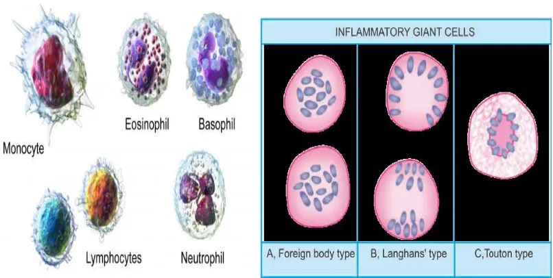
1. Epithelioid cells. It is one of the modified histocyte and are weakly pathogenic.
horseshoe or ring, or are clustered at the two poles (Langhans’ giant cells), or they may
be present centrally (foreign body giant cells).
3. Lymphoid cells 4. Necrosis
5. Fibrosis. It is a feature of healing by proliferatingfibroblasts at the periphery of granuloma.
1.3.2.2.3 Morphological features of foreign body granulomas.
Such granulomas may develop in any organ or skin or mucous membrane where a particulate
foreign body may enter. Visible small nodules may form. It differ from a tubercle in that the
macrophages are smaller not typically epithelioid and the multinucleated giant cells too are not
of the Langhan’s typebut of the foreign body type. The reaction is not organised in compact
tubercles,but more in focal clusters of macrophages and few to many foreign body type giant
cell. There is no caseation, there is considerable vascularisation and much heavier lymphocyte
and plasma cell response around. Depending on the foreign body, neutrophils may also be
associated with it.
1.3.2.3 Systemic effects of chronic inflammation
1. Fever. Invariably there is mild fever, often with loss of weight and weakness. 2. Anaemia. chronic inflammation is accompanied by anaemia of varying degree.
3. Leucocytosis. As in acute inflammation, chronic inflammation also has leucocytosis but generally there isrelative lymphocytosis in these cases.
4. ESR. ESR is elevated in all cases of chronic inflammation.
5. Amyloidosis. Long-term cases of chronic suppurative inflammation may cause secondary systemic (AA) amyloidosis.
1.4 Mediators of inflammation
Biochemical mediators released during inflammation strengthen and broadcast the inflammatory
response. These mediators are soluble, diffusible molecules that can act locally and systemically.
1.4.1.Cell derived mediators
Cell derived mediators are derived from injured tissue cells or leukocytes recruited to the site of
inflammation. Mast cells, platelets, and basophils produce the vasoactive amines serotonin and
histamine.
Histamine causes arteriolar dilation, increased capillary permeability, contraction of nonvascular smooth muscle, and eosinophil chemotaxis and can stimulate nociceptors responsible for the pain
response. Its release is stimulated by the complement components C3a and C5a and by
lysosomal proteins released from neutrophils. Histamine activity is mediated through the
activation of one of four specific histamine receptors, designated H1, H2, H3, or H4, in target
cells. Most histamine-induced vascular effects are mediated by H1 receptors. H2 receptors
mediate some vascular effects but are more important for their role in histamine-induced gastric
secretion. Less is understood about the role of H3 receptors, which may be localized to the CNS.
H4 receptors are located on cells of hematopoietic origin, and H4 antagonists are promising drug
candidates to treat inflammatory conditions involving mast cells and eosinophils (allergic
conditions).
Serotonin (5-hydroxytryptamine) is a vasoactive mediator similar to histamine found in mast cells and platelets in the GI tract and CNS. Serotonin also increases vascular permeability, dilates
capillaries, and causes contraction of nonvascular smooth muscle. In some species, including
rodents and domestic ruminants, serotonin may be the predominant vasoactive amine.
Cytokines, including interleukins 1–10, tumor necrosis factor α (TNF-α), and interferon γ (INF-γ) are produced mainly by macrophages and lymphocytes but can be synthesized by other cell
types also. These polypeptides alter the activity and function of other cells to coordinate and
control the inflammatory response. Two of the more important cytokines, interleukin-1 (IL-1)
and TNF-α, mobilize and activate leukocytes, enhance proliferation of B and T cells and natural
killer cell cytotoxicity, and are involved in the biologic response to endotoxins. IL-1, IL-6, and
TNF-α mediate the acute phase response and pyrexia that may accompany infection and can
induce systemic clinical signs, including sleep and anorexia. In the acute phase response,
components, coagulation factors, protease inhibitors, and metal-binding proteins. By increasing
intracellular Ca2+ concentrations in leukocytes, cytokines are also important in the induction of
PLA2. Colony-stimulating factors (GM-CSF, G-CSF, and M-CSF) are cytokines that promote
expasnsion of neutrophil, eosinophil, and macrophage colonies in bone marrow. In chronic
inflammation, cytokines IL-1, IL-6, and TNF-α contribute to the activation of fibroblasts and
osteoblasts and to the release of enzymes such as collagenase and stromelysin that can cause
cartilage and bone resorption.[12]
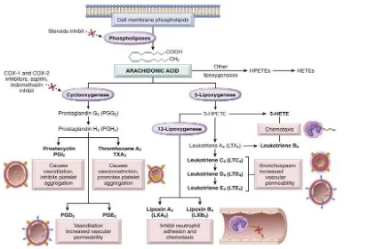
Lipid-derived autacoids play vital roles in the inflammatory response and are a main focus of
research into novel anti-inflammatory drugs. These compounds include the eicosanoids such as
prostaglandins, prostacyclin, leukotrienes, and thromboxane A and the modified phospholipids
such as platelet activating factor (PAF). Eicosanoids are synthesized from 20-carbon
polyunsaturated fatty acids by many cells, including activated leukocytes, mast cells, and
platelets and are therefore widely distributed. Hormones and other inflammatory mediators
(TNF-α, bradykinin) stimulate eicosanoid production either by direct activation of PLA2, or
indirectly by increasing intracellular Ca2+concentrations, which in turn activate the enzyme. Cell
membrane damage can also cause an increase in intracellular Ca2+. Activated PLA2 directly
hydrolyzes AA, which is rapidly metabolized via one of two enzyme pathways—the
cyclooxygenase (COX) pathway leading to the formation of prostaglandin and thromboxanes, or
the 5-lipoxygenase (5-LOX) pathway that produces the leukotrienes.
Cyclooxygenase catalyzes the oxygenation of AA to form the cyclic endoperoxide PGG2, which
is converted to the closely related PGH2. Both PGG2 and PGH2 are inherently unstable and
rapidly converted to various prostaglandins, thromboxane A2 (TXA2), and prostacyclin (PGI1).
PGE1, PGE2, and PGI1 are potent arteriolar dilators and augment the effects of other mediators
by increasing small vein permeability. Other prostaglandins, including PGF2α and thromboxane,
cause smooth muscle contraction and vasoconstriction. Prostaglandins sensitize nociceptors to
pain-provoking mediators such as bradykinin and histamine and, in high concentrations, can
directly stimulate sensory nerve endings. TXA2 is a potent platelet-aggregating agent involved in
Found predominately in platelets, leukocytes, and the lungs, 5-LOX catalyzes the formation of
unstable hydroxyperoxides from AA. These hydroxyperoxides are subsequently converted to the
peptide leukotrienes. Leukotriene B4 (LTB4) and 5-hydroxyeicosatetranoate (5-HETE) are strong chemoattractants stimulating polymorphonuclear leukocyte movement. LTB4 also
stimulates the production of cytokines in neutrophils, monocytes, and eosinophils and enhances
the expression of C3b receptors. Other leukotrienes facilitate the release of histamine and other
autacoids from mast cells and stimulate bronchiolar constriction and mucous secretion. In some
species, leukotrienes C4 and D4 are more potent than histamine in contracting bronchial smooth
[image:17.612.115.538.268.548.2]muscle.[13][14]
Figure 1.3 Generation of arachidonic acid metabolites and their roles in inflammation
Platelet activating factor (PAF) is also derived from cell membrane phospholipids by the action
of PLA2. PAF, synthesized by mast cells, platelets, neutrophils, and eosinophils, induces platelet
aggregation and stimulates platelets to release vasoactive amines and synthesize thromboxanes.
The role of free radical in inflammation is inevitable. A free radical is any species capable of
independent existence containing one or more unpaired electrons. The unpaired electron alter the
chemical reactivity of the molecule/atom, making it more reactive.[15][16] .
The oxygen free radicals include;
superoxide anion radical (O2·-),
singlet oxygen (O2),
hydroxyl radical (·OH) and
perhydroxyl radical (HO2·)
These all are together known as “reactive oxygen species” (ROS). With a single electron
reduction several free radicals and hydrogen peroxide (H2O2) are formed. Reactive oxygen
species are generated by oxidant enzymes, phagocytic cells, ionizing radiation etc.[11]
Superoxide anion is the first radical formed, by the electron transport chain when O2 picks up a
single electron. Radicals such as ·OH, HO
2· and H2O2 are formed from O2·–. O2·– undergoes a dismutation reaction catalysed by the enzyme superoxide dismutase (SOD) to form H2O2, which
by itself is not reactive enough to cause damage to macromolecules.
Among
the reactive oxygen species, ·OH is the most potent destructive radical which can react with all biological macromolecules (lipids, proteins, nucleic acids and carbohydrates). It is extremely
reactive and can lead to formation of DNA-protein cross-links, single and double-strand breaks,
base damage, lipid peroxidation and protein fragmentation . It may also be produced by ionizing
radiation .
(i) Reduction of H2O2 by the Fenton reaction:
Fe2+ + H2O2 → ·OH + OH- + Fe3+ Cu+ + H2O2 → Cu2++·OH + OH
-(ii) Interaction of O2·– with H2O2 by the Haber–Weiss reaction: O2·-+ H2O2 → O2 + H2O +·OH
The oxygen-derived free radicals have the following role in inflammation;
Endothelial cell damage and increased vascular permeability.
Activation of protease and inactivation of antiprotease which caude tissue matrix damage.
Damage to other cells.
Increased generation of free radicals and reactive oxygen species leads to several
pathological conditions like rheumatoid arthritis, myocardial infarction, malignancy, aging,
alzheimer’s disease, cancer etc.[17]
The role of the free radical gas nitric oxide (NO) in inflammation is well established. NO is an
important cell-signaling messenger in a wide range of physiologic and pathophysiologic
processes. Small amounts of NO play a major role in maintaining resting vascular tone,
vasodilation, and antiaggregation of platelets. In response to certain cytokines (TNF-α, IL-1) and
other inflammatory mediators, the production of relatively high quantities of NO is stimulated. In
larger quantity, NO is a potent vasodilator, facilitates macrophage-induced cytotoxicity, and may
contribute to joint destruction in some types of arthritis.[18][19]
1.4.2.Plasma derived mediators.
These include the various products derived from activation and interaction of 4 interlinked
systems: kinin, clotting, fibrinolytic and complement. Hageman factor (factor XII) of clotting
system plays a key role in interactions of the four systems. Activation of factor XII in vivo by
contact with basement membrane and bacterial endotoxins, and in vitro with glass or kaolin,
leads to activation of clotting, fibrinolytic and kinin systems. In inflammation, activation of
factor XII is brought about by contact of the factor leaking through the endothelial gaps. The
end-products of the activated clotting, fibrinolytic and kinin systems activate the complement
system that generate permeability factors. These permeability factors, in turn,further activate
Figure 1.4Interrelationships between the four plasma mediator systems triggered by activation of factor XI
1.4.3 Inflammatory cells
The cells involved in acute and chronic inflammation are circulating leukocytes, plasma cells and
tissue macrophages.
1.4.3.1 Circulating leukocytes
a.Polymorphonuclear neutrophils are acute inflammatory cells, which are involved in initial
phagocytosis of bacteria and foreign bodies.
b. Monocytes are chronic inflammatory cells which are involved in bacterial phagocytosis and
regulates lymphocyte response.
c. Lymphocytes are chronic inflammatory cells which are involved in humoral and cell mediated
immune responses and regulate macrophage responses..
d. Eosinophils are chronic inflammatory cells which are involved during allergic states and
parasitic infestations.
e. Basophils containing electron dense molecules andfunctions as receptor for IgE molecules
These cells are bigger than lymphocytes with more abundant cytoplasm and an eccentric nucleus
which has cart-wheel pattern of chromatin. They develop from Blymphocytes and are rich in
RNA and Ƴ-globulin in theircytoplasm. These cells are active in antibody synthesis.
1.4.3.3 Mononuclear-Phagocyte System (Reticuloendothelial System)
This cell system includes cells derived from 2 sources with common morphology, function and
origin. These are as under:
a.Blood monocytes. These comprise 4-8% of circulating leucocytes.
b. Tissue macrophages. These include the following cells in different tissues:
i) Macrophages in inflammation.
ii) Histiocytes which are macrophages present in connective tissues.
iii) Kupffer cells are macrophages of liver cells.
iv) Alveolar macrophages (type II pneumocytes) in lungs.
v) Macrophages/histiocytes of the bone marrow.
vi) Tingible body cells of germinal centres of lymph nodes.
vii) Littoral cells of splenic sinusoids.
viii) Osteoclasts in the bones.
ix) Microglial cells of the brain.
x) Langerhans’ cells/dendritic histiocytes of the skin.
xi) Hoffbauer cells of the placenta.
xii) Mesangial cells of glomerulus.
The mononuclear phagocytes are the scavenger cells of the body as well as participate in
immune system of the body; their functions in inflammation are as under:
i) Phagocytosis (cell eating) and pinocytosis (cell drinking).
ii) Macrophages on activation by lymphokines released by T lymphocytes or by non
immunologic stimuli elaborate a variety of biologically active substances like proteases,
plasminogen activator,products of complement,coagulation factors etc.
1.4.3.4 Giant Cells
A few examples of multinucleate giant cells exist in normal tissues (e.g. osteoclasts in the bones,
inflammation when the macrophages fail to deal with particles to be removed, they fuse together
and form multinucleated giant cells. Besides, morphologically distinct giant cells appear in some
tumours also. Some of the common types of giant cells are
i) Foreign body giant cells.
ii) Langhans’ giant cells..
iii) Touton giant cells..
[image:22.612.108.511.220.423.2]iv) Aschoff giant cells.[21][22]
Figure 1.5 Inflammatory cells
1.5 Pain
Pain is a multidimensional occurrence that is essential for the maintenance and preservation of an
individual. It warns of the danger of bodily harm and alerts to trauma and injury. Pain is a
specific enteroceptive sensation; it can be perceived as arising from a particular portion of the
body, its temporal properties can be detailed, it can be differentiated qualitatively (for example,
as stinging, pricking, burning, throbbing, dull or aching), and it involves dedicated subsets of
peripheral and central neurons. The experience of pain has a distinctly unpleasant character, that
is, an affective or motivational aspect that can be distinguished from its discriminative sensory
aspects and from the long-term emotional experience of ‘suffering’. The unpleasantness ranges
in intensity from the discomfort of a cold room, fatigued muscles or colonic tension to the
Under normal circumstances, primary afferent pain fibres activate particular central
pathways that engage protective mechanisms at many functional levels: autonomic, homeostatic,
motor, behavioural and mnemonic. However, injury or disease can change the balance of this
system and result in persistent, pathological pain. Analgesic substances, such as aspirin and
morphine, which interact with the transmitters and modulators of the pain system are helpful for
many people with pain, but there is a great need for the development of better methods for the
lessening and control of both acute (immediate) and chronic (long-term, pathological) pain.[23]
During the process of inflammation, a mixture of inflammatory mediators such as
peptides (bradykinin), lipids (prostaglandins), neurotransmitters (ATP), protons and
neurotrophins (NGF) are released.The release of neuropeptides from peripheral neurones
supports plasma extravasation of further mediators and chemoattraction of inflammatory cells, a
process called as “neurogenic inflammation”. The sensitisation of the primary afferent neurones
by local inflammatory mediators can occur by a direct action on the sensory neurones, or it can
involve an indirect action on non-neuronal cells, especially but not exclusively on immune cells,
from which further inflammatory mediators can then be released.[24]
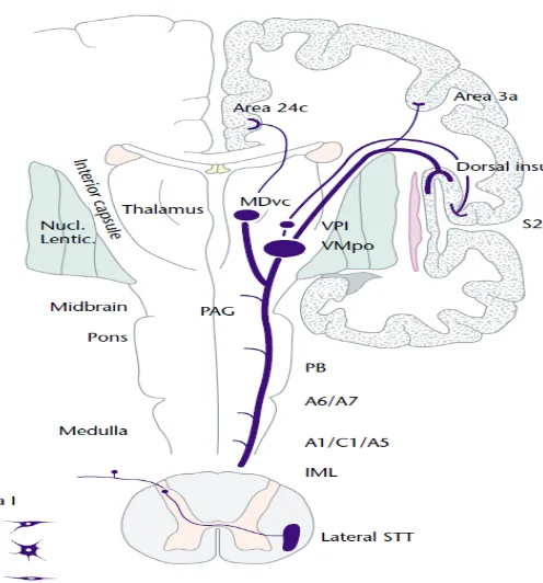
1.5.1 Pain pathways
Nociceptors, or pain receptors, are free nerve endings that respond to painful stimuli which
transmit information to the brain. Pain perception occurs when electrical, thermal, mechanical,
biological and chemical stimuli are transmitted to the spinal cord and then to the central areas of
the brain. Pain impulses travel to the dorsal horn of the spine, where they synapse with dorsal
horn neurons in the substantia gelatinosa and then ascend to the brain. The basic sensation of
pain occurs at the thalamus. It continues to the limbic system and the cerebral cortex, where pain
Figure 1.6 Pain pathway 1.5.2 Pain theories:
Specificity theory.
Pattern theory.
Gate control theory.
Anodal blocking theory and,
Central inhibition theory.
1) Specificity Theory:
It proposes that a mosaic of specific pain receptors in body tissue projects to a main center in the
brain. It maintain that free nerve endings are pain receptors which generate pain impulses that are
carried by A-delta and C-fibers in peripheral nerves and by the lateral spinothalamic tract in the
spinal cord to a pain center in the thalamus."Pain receptor" implies that stimulation of one type
of receptor elicits a single psychological or physiological response.
According to this theory, pain is the result of the stimulation of certain nerve impulses
that form a pattern and are then brought together and deserted into the spinal cord as a lump sum
of pain, a process called “central summation.”
3) Gate Control Theory:
Melzack and Wall published this theory in 1965. According to this theory, a mechanism
in the brain acts as a gate to increase or decrease the flow of nerve impulses from the peripheral
fibers to the Central nervous system. An “open” gate allows the flow of nerve impulses, and the brain can perceive pain. A “closed” gate does not allow flow of nerve impulses, decreasing the
perception of pain.
4) Anodal Blocking Theory:
This theory proposes maintaining the absolute refractory period in the nerve by rapidly
and repeatedly stimulating the nerve fiber, thus preventing pain impulses from being transmitted.
5) Central Inhibition Theory:
It suggests that large-fiber and small-fiber activity do not result in presynaptic effects of
opposite polarity as suggested by the gate control theory. The concept of inhibitory balance
depends on minor inhibitory feedback from small fiber input that activates the nociceptive
marginal neuron and major inhibitory feedback to the marginal neuron from large
non-nociceptive fibers. Modulation of pain producing input thus depends on the balance between
large fiber and small fiber activity via a postsynaptic inhibitory mechanism acting on the
nociceptive relay neurons.[26]
1.6 Anti-inflammatory drugs and analgesics
Anti-inflammatory drugs are the drugs which are used for the treatment of
inflammation. They are mainly used in infections like rheumatoid arthritis and gout . Analgesic
is a drug that selectively relieves pain by acting in the CNS or on peripheral pain mechanisms,
without significantly altering consciousness.
Analgesics are divided into two groups;
Nonopioid / Non-narcotic /Antipyretic or anti-inflammatory analgesics.
1.6.1 Narcotic analgesics: 1.6.1.1 Classification:
I) Narcotic Agonist Analgesics
i) Natural opium alkaloids: Morphine, codeine
ii) Semi synthetic opiates: Diacetylmorphine, Pholcodeine, Ethylmorphine
iii) Synthetic opioids: Pethidine, Fentanyl, Methadone, Tramadol
II) Narcotic Agonist-Antagonist Analgesics
i) Phenanthrenes: Buprenorphine, Nalbuphine
ii) Morphinan: Butorphanol
iii) Benzomorphans: Phenazocine , Pentazocine
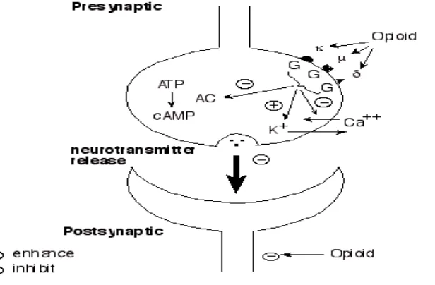
1.6.1.2 General mechanism Of Action Of Narcotic Analgesics:
It exerts its effects by interacting with opioid receptors (μ, κ and δ ) located on
prejunctional neurons. Activation of opioid receptor reduces intracellular cAMP formation and
opens potasium channels or suppresses voltage gated N type Calcium channels. These actions
leads to neuronal hyperpolarisation and reduced availability Of intracellular calcium, decreased
[image:26.612.158.466.449.651.2]neurotransmitter release by CNS and myenteric neurons.[27]
1.6.1.3Adverse Effects associated with opiods.
The various adverse effects associated with opioids include respiratory depression, drowsiness,
nausea, vomiting, endocrine disturbances, tolerance to analgesic effect, physical dependence,
abuse potential and interaction with CNS depressant drugs. [28]
1.6.2 Nonsteroidal Anti-inflammatory Drugs and Antipyretic-Analgesics : 1.6.2.1 Classification:
A) Nonselective COX inhibitor
1. Salicylates: Aspirin.
2. Propionic acid derivatives: Ibuprofen , Ketoprofen.
3. Fenamates: Mephenamic acid.
4. Oxicams: Piroxicam, Tenoxicam.
5. Pyrazolones: Phenylbutazone, Oxyphenbutazone.
6. Indole derivatives: Indomethacin, Sulindac.
7. Aryl-acetic acid derivatives: Diclofenac.
8. Pyyrrolo-pyrrole derivatives: Ketorolac.
B) Preferential COX-2 Inhibitors: Nimesulide.
C) Selective COX-2 Inhibitors: Celecoxib, Etoricoxib.
D) Analgesic-antipyretics with poor anti-inflammatory action.
1. Paraaminophenol derivative: Paracetamol.
2. Pyrazolone derivatives: Metamizol, Propiphenazone.
3. Benzoxazocine derivative: Nefopam.
1.6.2.2 General mechanism Of Action Of NSAIDs:
Nonsteroidal anti-inflammatory drugs exert their anti-inflammatory effect through
inhibition of cyclooxygenase, which is the enzyme catalyzing the transformation of arachidonic
acid to prostaglandins and thromboxanes.NSAIDs also inhibit expression of cell adhesion
Figure 1.8 : Mechanism of action of NSAIDs.
1.6.2.3 Adverse effects of NSAID Therapy
NSAIDs produces various adverse effects as it interfere with various pathways that maintain
[image:28.612.111.512.63.303.2]homeostasis.The manifestations are as follows[30]
Table 1.1 Adverse drug reactions of NSAIDs
SYSTEM MANIFESTATIONS
GI Abdominal pain, nausea, anorexia, gastric erosions, anemia, GI hemorrhage, perforation, diarrhea
Renal Salt and water retention, hyperkalemia, Decreased effectiveness of diuretic and antihypertensive medications
CNS Vertigo, dizziness, lowering of seizure threshold ,hyperventilation, depression.
Platelets Inhibited platelet activation
Uterus prolongation of gestation and possible prolongation of labor Hypersensitivity Vasomotor rhinitis, angioedema, asthma, urticaria, flushing,
hypotension, shock
1.7 Cyclo oxygenase as a drug target
Three isoforms of COXs have been identified. Cyclooxygenase-1 (COX-1) is a glycoprotein of
71kDa, which is constitutively expressed in different tissues. COX-1 is encoded by a gene on
chromosome 9 and plays a role in tissue homeostasis by modulating several cellular processes
ranging from cell proliferation to angiogenesis or platelet aggregation due to thromboxane
production . Cyclooxygenase-2 (COX-2) is the inducible isoform, which is regulated by growth
factors and different cytokines such as IL1β, IL6, or TNFα , therefore overexpressed during inflammation. The COX-2 gene is located on chromosome 1 and its promoter displays an NFκB response element as well as other cytokine-dependent (i.e., IL6) response elements . The protein
shows a 60% homology with COX-1 [31]; in addition, COX-2 presents a C-terminal extension
and a different binding site for NSAIDs, which makes COX-2 a preferential target compared to
COX-1, thus being specifically inhibited at lower doses . Finally, COX-3 has been identified as a
splice variant of COX-1, and it is present mainly in brain and spinal cord . Currently, the role of
COX-3 is not known. Some pieces of evidence suggest a possible role in pain sensitivity, based
on studies focused on the mechanism of action of acetaminophen (paracetamol), recently evoked
as a selective inhibitor of COX-3[32].
The COX molecule consists of three independent folding units: an epidermal growth
factor-like domain, a membrane binding site, and an enzymatic domain . The active COX site is
a hydrophobic channel with a series of amino acids. Aspirin binds irreversibly to serine 580 by
acetylation, whereas most other NSAIDs bind sterically and reversibly to Tyrosine 385 or
Arginine 120, blocking the COX action resulting in the alleviation of pro-inflammatory ARA
metabolites, and in particular, prostaglandin E2(PGE2)[33]. The use of NSAIDs is often limited
by side effects emanating from disrupting the levels of these protective COX metabolites.
Following the discovery of a second form of COX enzyme (COX-2) ,COX-2 selective inhibitors
(coxibs) such as celecoxib and rofecoxib were subsequently developed in an effort to
circumvent these problems. Coxibs are effective against inflammation and pain, with
comparatively less risk of severe GI toxicity associated with conventional NSAIDs. However,
there are safety concerns with coxib use due to an increase in the risk of cardiovascular events
associated with the imbalance of the PGI2 and thromboxane (TXA2) metabolite levels .
Inhibition of COX-1 decreases platelet-derivedTXA2, an eicosanoid which functions as a
PGI2/TXA2 ratio to favor TXA2, increasing the risk of mortality from ischemic heart
disease.Recently, a metabolomic approach to study the relationship between adverse
cardiovascular eventsand the use of rofecoxib suggested that this drug acts, in part,through
accumulation of 20-hydroxyeicosatetraenoic acid (20-HETE) which is a potent vasoconstrictor
among ARA metabolites. Treatment of inflammation and pain constitutes significant medical
needs because more people are prone to these conditions than any other disease state. Thus, there
is a growing demand for safer but efficacious NSAIDs or coxibs . Upcoming concepts and
approaches forthe treatment of inflammation and pain have moved towards simultaneously
targeting multiple enzymes in the ARA cascade through combination therapy and multi target
inhibitors such as dual inhibitors with the aim of overcoming the risks in single enzyme or
pathway inhibition[34]
1.8 Inflammation and cancer
Inflammatory conditions in selected organs augment the risk of cancer. An inflammatory
component is present also in the microenvironment of tumors which are not epidemiologically
related to inflammation[35]. Over expression of COX-2 has been detected in a number of tumors,
such as colorectal breast as well as pancreatic and lung cancers which can be correlated with a
poor prognosis. Moreover, over expression of COX-2 has been reported in hematological cancer
models like RAJI (Burkitt’s lymphoma) and U937 (acute promonocytic leukemia) as well as in patient’s blast cells. Data suggested that COX-2 may play a role in different steps of cancer
progression, by increasing proliferation of mutated cells, thus favoring tumor promotion as well
as by affecting programmed cell death and affecting the efficacy of anticancer therapies to be,
finally, concerned in metastasis formation, for example, by affecting apoptosis induced by loss of
cell anchorage (anoikis). Combination of preferential orselective COX-2 inhibitors with
cancer agents already used in clinics were tested with the goal to improve the efficiency of
anti-cancer protocols.
1.9 In silico drug design
Pharmaceutical research has successfully incorporated a wealth of molecular modelling methods,
within a variety of drug discovery programs, to study complex biological and chemical systems.
identification and development of novel promising compounds. Broadly used in modern drug
design, molecular docking methods explore the ligand conformations adopted within the binding
sites of macromolecular targets. This approach also estimates the ligand-receptor binding free
energy by evaluating critical phenomena involved in theintermolecular recognition process.
Two approaches are used in drug design
1.Structure based drug design
2.Ligand based drug design
1.9.1 Structure Based drug design
If reliable information about the 3-D structure and active sites of the target protein can be
obtained from X-ray crystallography, nuclear magnetic resonance, or 3-D structure databases,
and integrated into a computer model, compounds binding to the target can be designed. This
approach is called “structure-based drug design”. Commonly used techniques in this approach
are docking and molecular dynamics simulation. Potent ligands can be found by screening a
molecule database with docking software. Molecular dynamics simulation can be useful to
determine not only how a molecule interacts with the target protein, but also to determine some
other properties of the molecule itself, such as membrane permeability.[36]
1.9.2 Ligand Based drug Design
When the receptor structure is unknown but the ligand structures are known, a ligand based
approach is used. An extension of the QSAR approach is used to study the active ligands, also
known as pharmacophore based drug design. The pharmacophore refers to an ensemble of steric
and electronic features that enables it to exhibit specific biological activity.generally, this method
depend on the application of descriptors of molecular structure and properties, including
Figure1.9 Workflow of various approaches in in silico drug design
Heterocycles always remain as a potential scaffold with variety of pharmacological significance.
Numerous research reports have indicated the coumarin nucleus as a potential candidate for
development of anti-inflammatory drugs.[37]As an important class of compounds,
2-ones are isomeric to 4-quinol2-ones and isosteric to coumarins. The compounds that have
quinolin-2-one moiety are associated with biologic activities such as antibacterial, anticancer, antiviral,
cardiotonic, and N-methyl-D-aspartate receptor inhibitor functions, among others. Thiazole,
oxazoles ,N-substituted piperazines and aminopyridine containing scaffolds are proven to be
promising entities with good anti inflammatory, analgesic and antioxidant potential. In the
current study, based on the rational approach, various analogs of 2-quinolones were designed for
CHAPTER 2 LITERATURE REVIEW
Quinolinone derivatives
1. Lan et al.,(2014)[39]; synthesised and evaluated the analgesic potential of 3,4-dihydroxy-2(1H)-quinolinone derivatives as novel sigma-1 receptor antagonists. The compounds were
evaluated in vitro in sigma-1 and sigma-2 receptor-binding assays in guinea pig brain membranes.7-(3-(piperidin-1-yl)propoxy)-1-(4-fluorobenzyl)-3,4-dihydro-2(1H) quinolinone (1) was profiled with highest affinity and greatest selectivity which possess a high binding constant
for sigma-1 receptor and high sigma-1/2 selectivity (1066-fold). In formalin test, compound (1)
produced dose-dependent anti-nociception in both phases with ED50 values 49.4 ± 4.1 and 50.5
±2.5 mg/kg for phases I and II, respectively.
N O
F
O N
(1)
2.Kumar et al.,(2014)[40]; synthesised and evaluated the antimicrobial and anti-inflammatory activity of isoxazolineincorporared 2 quinolones.Among the synthesised compounds compound
(2) and (3) showed maximum percentage inhibition of edema volume at 4th hour when compared to the standard drug diclofenac sodium in the carrageenan induced paw edema model at dose of
200mg/kg
N O
O N
H3C
NH2
N
H NH O
CH3
Cl O
(2) (3)
synthesised compound (4) was found to possess high free radical scavenging activity in DPPH (EC50 = 12.2 µM) and superoxide anion assays (EC50 =138.8µM)
N
H NH O
CH3
Cl O
(4)
4.Nitesh et al.,(2012)[42]; elucidated the anticancer activity of synthesized 2-quinolone derivatives without N-methyl or 3-aryl substitution. Significant cytotoxicity was observed in
MCF-7 cells treated with (5) and (6).Both the derivatives’ treatment showed damage to the DNA. In vivo studies for (5) and (6)were performed at two doses 100 and 200 mg/kg using Ehrlich ascites carcinoma (liquid) and Dalton lymphoma ascites (solid) models. Both derivatives
showed a significant reduction in the tumor progression by increasing the mean life span and by
improving the haematological profile and antioxidant status of the liver in a liquid tumor model.
More prominent effect was observed in a solid tumor model by reduction in solid tumor weight
and tumour volume. The CTC50 of (5) and (6) were found to be, 83.04µM and 22.04 µM
respectively
N
H O
CH3
HN
O
Cl
N
H O
CH3
HN
O
(5) (6)
5.Jayashree et al.,(2010)[43]; designed and synthesised a series of 2-quinolone derivatives as anti oxidants and antimicrobials. Based on the docking stimulation and QSAR studies, the
7-amino-4-methylquinolin-2(1H)-one parent compound and its carboxamides) were synthesized
N
H O
CH3
N H CH3
O
(7)
6.Sankaran et al., (2010)[44]; synthesised and conducted antioxidant and toxicological study of novel pyrimido quinolone derivatives from 4-hydroxy-3-acyl quinolin-2-one. They were
screened for their in vitro antioxidant activities against radical scavenging capacity using DPPH, Trolox equivalent antioxidant capacity (TEAC), total antioxidant activity by FRAP, superoxide
radical scavenging activity, metal chelating activity and nitric oxide scavenging activity. Among
the compounds screened, (8) and (9) exhibited significant antioxidant activities.
N
H O
N HN
O
N
H O
N NH HN
S
(8) (9)
7.Detsi et al.,(2007)[45]; designed and synthesised a series of N-substituted-quinolinone-3-aminoamides and their hybrids containing the R-lipoic acid functionality as potential
bifunctional agents combining antioxidant and anti-inflammatory activity. The compounds were
evaluated for their antioxidant activity and for their ability to inhibit in vitro lipoxygenase as well as for their anti-inflammatory activity in vivo. Among the compounds synthesised compound (10) was found to be most potent anti-inflammatory agent with 85.3% inhibition of paw edema volume in carrageenan induced paw edema method.
N O
OH
CH3
N H O
NH2
(10) Piperazine derivatives
to be the most potent antioxidant giving an IC50 of 20.30 µg/mL and 5.62 µg/mL in DPPH and
ABTS assay method respectively.
O
O O N
N
OH
(11)
9.Silva et al.,(2015)[47]; investigated the anti-nociceptive and anti-inflammatory effects of piperazine derivative 4-[(1-phenyl-1H-pyrazol-4-yl) methyl]1-piperazine carboxylic acid ethyl
ester (12) and the involvement of the serotonergic pathway. In the formalin test, treatments with
this compound (15 and 30 mg/kg p.o.) reduced the licking time in both neurogenic and
inflammatory phases. In the tail flick and hot plate tests, (12) treatment increased latency to thermal stimulus and in the carrageenan-induced paw edema test, at the doses of 15 and 30
mg/kg reduced the edema at all tested time points, while the dose of 7.5 mg/kg reduced the
edema only for the first hour.Compound (12) (30 mg/kg p.o.) reduced both cell migration and protein exudation in the carrageenan-induced pleurisy test.
N
N O
O
N N
(12)
10.Ghorbani et al., (2015)[48]; synthesised a series of novel piperazine analogues bearing quinolin-8yloxy-butan-1-ones/pyridine-2-yloxy-Ethanone. The analogues were evaluated for in vitro antioxidant activity against DPPH and ferrous ion radical scavenging activities and anti-inflammatory activity by inhibition of Vipera russelli venom (PLA2) and gastric K+/H+ ATPase activities.It was found that pyridine ring with phenyl and nitro-phenyl group of (13) and (14)
showed potent inhibition against all the assays
O
O N
N N
O N
N N N+
O
-O O
11.Mistry et al., (2015)[49] synthesised new Mannich base series of piperazine linked berberine analogues and were evaluated for antioxidant and anti-cancer activities. The radical scavenging
potential of the final derivatives was found excellent with IC50s, <13 lg/mL and<8 lg/mL in
DPPH and ABTS assay, respectively, whereas some analogues showed significant Fe+3
reducing power with absorption at around 2 nmin the FRAP assay.compound(15) was found to
be more potent. Anticancer effects of titled compounds were inspected against cervical cancer
cell line Hela and Caski adapting SRB assay, in which the analogues presented <6 lg/mL of
IC50s, and>30 of therapeutic indices, thus exerting low cytotoxic values against Malin–Darby
canine kidney (MDCK) cell lines at CC50s >125 lg/mL.
N
N N
O O
HO O
F
Cl-(15)
12.Andonova et al., (2014)[50]; synthesised and evaluated the antioxidant activity of some 1-aryl/aralkylpiperazine derivatives with xanthine moiety at N4. All compounds were in vitro
screened for their activity as antioxidants using DPPH, and FRAP methods. The antioxidant
activity of the studied compounds against lipid peroxidation was also measured. The highest
antioxidant activity was demonstrated by compound (16) with IC50 values . 189.42 µmol/L2,3.45
µmol/L2and173.99 µmol/L2 for DPPH, ABTS and FRAP assays respectively.
N N
N
N O
O
N N OH
(16)
Compounds (17)and (18) were proven to be good anti-inflammatory agents at adose 100mg/kg in
carrageenan induced paw edema method.
O
N N S
O O N N CF3 Cl O
N N S
O O N N CF3 Cl
(17) (18)
14.Girish et al.,(2012)[52]; synthesised a series of novel 6-methoxy-2-(piperazin-1-yl)-4H -chromen-4-one and 5,7-dimethoxy-2-(piperazin-1-ylmethyl)-4H-chromen-4-one derivatives of biological interest and screened for their pro-inflammatory cytokines (TNF-α and IL-6) and
antimicrobial activity (antibacterial and antifungal). Among all the compound screened, the
compounds (19)(82%TNF-α and 87 %IL-6 inhibitory activities ) and (20) (85%TNF-α and 91 %IL-6 inhibitory activities) were found to have promising anti-inflammatory activity at
concentration of 10 μM with reference to standard dexamethasone (71% TNF-a and 84% IL-6
inhibitory activities at 1 μM)
O N N N O O N N N O N N N O O O O
(19) (20)
15.Kimura et al., (2004)[53]; synthesised a new series of diphenylalkylpiperazine derivatives with high affinities for the dopamine transporter (DAT), which were modified at both the
diphenylalkyl moiety and the phenyl ring in the phenylamino moiety of
1-[4,4-bis(4-fluorophenyl)butyl]-4-[2- hydroxy-3-(phenylamino)propyl]piperazine and was evaluated for
their inhibitory activities against auto-oxidative lipid peroxidation in canine brain
homogenates.The 4-hydroxyphenyl derivative (21) showed the most potent anti-oxidative activity with an IC50 value of 0.32 lM, exhibiting approximately 5-fold more potent activity than