Rochester Institute of Technology
RIT Scholar Works
Theses
Thesis/Dissertation Collections
1984
Determination of the neighboring molecule to the
FC receptor on human macrophages
Lorri Jean Malinski
Follow this and additional works at:
http://scholarworks.rit.edu/theses
This Thesis is brought to you for free and open access by the Thesis/Dissertation Collections at RIT Scholar Works. It has been accepted for inclusion in Theses by an authorized administrator of RIT Scholar Works. For more information, please contactritscholarworks@rit.edu.
Recommended Citation
DETERMINATION OF THE NEIGHBORING MOLECULE
TO THE FC RECEPTOR
ON HUMAN MACROPHAGES
.
By
Lorri Jean Malinski
A RESEARCH PROJECT
Submitted to
Rochester Institute of Technology
in partial fulfillment of the requirements
for the degree of
ABSTRACT
DETERMINATION OF THE NEIGHBORING MOLECULE TO THE FC RECEPTOR
ON HUMAN MACROPHAGES
By
Lorri Jean Malinski
Chemical cross-linking studies have been carried out to investigate near neighbors to
the receptor for immunoglobulin G[lgGJ on U937 cells both before and after solubilization
of the receptor. A cross-linked product with a molecular weight of 350.000 daltons
was achieved with. OMS on intact and lysed U937 cells. OTBP on intact cells and OSS
on cell Iysates. with anti-FcRIl as the immunoprecipitation reagent. Following
cross-I ink ing of the U937 cells with OTBP and analysis by two-dimensional gel
electrophoresis the p350 molecule appeared to be a p 170 dimer. The p 170 molecular
has been related to some nonspecificity of the ant i-Fc Rj]. These procedures were thus
unsuccessful in identifying a neighboring molecule to the U937 cell FcR.
OSS cross-linking of Fc fragments of IgG and Fab fragments of anti-FcRIl to the FcR
on intact U937 cells was unsuccessful. Lack of success in this regard is more likely
attributed to difficulties in OSS usage then to inability to cross-link close-lying molecules
to the FcR.
The investigations presented clarify the problems that must be overcome before
successful cross-I inking can be achieved between the U937 cell FcR and its neighboring
TO TOM
Your love, understanding and encouragement
TABLE OF CONTENTS
SECTION PAGE
INTRODUCTION 1
I. BACKGROUND INFORMATION 2
1 Fc Receptors 2
2 Endocytosis-FcR Mediated 2
3 IgE/FcR System 4
4 IgG/FcR System 7
5 Cross-Linking Reagents 9
6 U937 Cells 15
7 Preliminary Investigations 16
B Proposed Investigation 19
II. MATERIALS AND METHODS 20
2.1 U937 Cells 20
2.2 Iodi nation 20
2.3 SDS-PAGE and Autoradiography 20
2.4 Reagents 21
2.5 Cross-linking of Intact U937 Cells 22
2.6 Cross-linking of Lysed U937 Cells 24
2.7 FcR Precipitation 24
2.B Cross-linking of Fragment Coated U937 Cells 25
2.9 TCA Precipitation 25
III. RESULTS 26
3.1 Cross-linking of intact U937 Cells 26
3.2 Cross-linking of lysed U937 Cells 28
3.3 Cross-linking of fragment coated U937 cells 29
IV. DISCUSSION AND SUGGESTED FURTHER STUDIES 30
REFERENCES 32
INTRODUCTION
The cell surface receptor for immunoglobulin G [IgG] on human macrophages and
monocytes mediates the triggering of endocytosis. The endocytic process begins with
the binding of IgG immune complexes through their Fc units to these receptor sites
on macrophages [See Figure 1 ]. The nature of the triggering signal as well as the entire
sequence of events of this process are largely unknown. A proper starting point to
achieve an understanding of these events is the characterization of the entire Fc binding
site which is known as the Fc receptor [FcR ] [ 1]. What is known is that the FcR
is an integral membrane protein which travels as a single major heterodisperse band
on polyacrylamide gel electrophoresis [PAGE] in sodium dodecyl sulfate [SDS] with
a molecular weight of approximately 72.000 daltons ( 2]. It is believed that this p72
molecule does not make up the FcR in its entirety and that the FcR is a complex of
subunits. The objective of this study has been to determine the neighboring molecule
to the FcR using cross-linking reagents with the consideration that the close lying
molecule may be a subunit of the receptor site. Cross-linking reagents in use were
two imidates. dimethyl suberimidate [DMS] and dimethyl-3,3'-dithiobispropionimidate
[DTBP], an n-hydroxysuccinimide ester, disuccinimidyl suberate [DSS], as well as a
Fc FRAGMENT Sites lor:
ComplementFixation Reactivitywith RheumatoidFactors Membrane Transmission SkinFixation
MacrophageFixation
Regulationof'.aubolism
epsin Cleavage
LIGHT CHAIN (212 residues)
PAPAINCLEAVAGE
INVARIABLE \ ? \\ REGION
VARIABLE REGION
FabFRAGMENT (Antigenbindingsite)
Figure 1. The four chain structure of an IgG molecule showing both interchain and
intrachain disulfide bridges. Indicated are the Fc and Fab fragments. Pepsin digests
SECTION
BACKGROUND MATERIAL
1.1 Fc RECEPTORS
Fc receptors can be found on the plasma membrane of many different cell types and
tissues, including cells of the immune system, platelets and placental tissue. These
cells and tissues may contain single or multiple types of receptors as there are specific
Fc receptors for each immunoglobulin class and in some cases, subclass. In other words.
the FcR for IgG is a separate entity from the IgE FcR and IgGj has a different receptor
site than lgG2- The logic of this arises from the distinct function each immunoglobulin
fulfills. For example, the binding of IgE to its FcR on mast cells and basophils is
responsible for the explosive degranulation of these cells which leads to the physiological
consequences of the allergic response.
1.2 ENDOCYTOSIS
Endocytosis, a collective term for phagocytosis and pinocytosis. is a process by which
cells internalize nutrients, toxins, effector molecules [growth factors, hormones.
antibodies], enzymes and pathogens. These substances bind specifically or
nonspecifically to the cellular plasma membrane [PM] prior to internalization. Ingested
material is then enclosed in membrane vacuoles or vesicles. Fusion of these vacuoles
with lysosomes or other organelles allows modification of the endocytosed material
within a closed compartment.
the more highly differentiated responses of mammalian polymorphonuclear leukocytes
and macrophages are designed to locate and remove antibody coated material by the
process of FcR-mediated endocytosis. Bacteria represent one example of material
that is removed in this manner. Antigenic material from bacteria is recognized and
ingested following interaction with IgG and complement components. IgG and
complement facilitate adherence and subsequent phagocytosis by macrophages. The
bacteria are then killed and degraded by enzymes contained in the lysosomes and specific
granules.
The process by which foreign material is ingested results from an interaction that
occurs between membrane receptor protein and components of the cytoskeleton.
Filamentous structures within the cytoskeleton are involved in cellular motility.
exocytosis, membrane ruffling and subsequently the "zipper phenomena"
as illustrated
in Figure 2.
During endocytosis a macrophage can rapidly interiorize a large portion of its plasma
membrane. Studies on the Fc receptor would not be complete without giving
consideration to its fate during this internalization process. Mellman e_t. aL utilized
a method for labelling integral membrane proteins of pinocytic vesicles of the murine
macrophage and found the presence of the FcR comparable in the pinocytic vesicle
and plasma membrane [3 ]. This indicated to Mellman that the FcR is cycled from
the plasma membrane to the pinocytic vesicle and back again. It is not understood
what happens to the FcR once a vesicle fuses with a lysosome.
A second concern when studying the FcR is that of macrophage FcR turnover. Synthesis
and insertion times for the FcR have been found to be 36 to 55 minutes from pulse
chase experiments. Under normal physiological conditions, turnover of the protein
was biphasic since 60% was lost with a half time of 13 hours and the remainder with
Figure 2 . The formation of a phagosome in a macrophage interacting with IgG-coated
4
The FcR mediated endocytic process can be likened to the process by which drugs,
hormones and neurotransmitters exert their effect. That is. receptor proteins must
transduce information from the external environment into meaningful intracellular
signals. To do so. these receptors must perform three distinct functions: 1] recognition
of a specific stimulus, 2] transduction of the signal across the plasma membrane and
3] initiation of a response in the cell. Because of this, receptor proteins are assumed
to span the entire membrane. The following model for receptor function has been
suggested from these assumptions:
1. Specific interaction of the binding domain of the receptor with an Fc region
of an immunoglobulin involves the usual molecular complementarity observed in
enzyme/substrate and antigen/antibody reactions.
2. Transduction involves conformational changes in the transducer domain. The
binding of ligand could cause changes in the individual receptor molecules; or aggregation of identical membrane receptors with subsequent interaction could
induce conformational changes.
3. The initiator domain may act through a variety of pathways. There may be
direct interaction between the initiator and the cytoskeletal system which initiates
or terminates movement, opens or closes specific ion gates, or activates nucleotide
cyclases to produce second messengers cyclic AMP [cAMP] or cyclic GMP [cGMP].
These second messengers then modulate the activity of soluble or secretory proteins
[5].
1.3 IgE/FcR SYSTEM
An example of a receptor dependent process which follows the model just suggested
is the IgE mediated allergic response. Symptoms of hypersensitivity are the result
of the interaction of immunoglobulin E with mast cells and/or basophils. Upon primary
exposure to an allergen, antigen specific IgE is produced and becomes fixed to mast
cells via the FcR. Upon subsequent exposure to the same antigen, an antigen/antibody
reaction occurs on the mast cell plasma membrane. This triggers a series of events
histamine, from mast cells [6].
1.3A Binding Characteristics
Investigations into the structure of the IgE FcR and the mechanism of histamine release
have been performed primarily on rat mast cells and the related tumor line, rat
basophilic leukemia cells [RBL]. The general properties as well as the binding
characteristics of the IgE FcR's found on these cells are listed in Table 1. [IB]
1.3B FcR Dimerization
The IgE isotype was discovered in the late I960's by Ishizaka and Ishizaka [7]. Their
early work with this immunoglobulin focused on identifying the transducer signal for
histamine release. Using a ragweed antigen-human antibody system they showed that
an antigen bound to two or more IgE antibody molecules could produce a biological
reaction such as skin reactivity: whereas a single antibody could not [B]. They later
observed that responses could also be initiated with aggregated IgE [9]. Ishizaka also
developed human anti-lgE antibodies and found that they were capable of inducing
mediator release from human basophils. Enzymatic digests were made of the anti-lgE
antibodies to form
[Fab']2
fragments and monomeric Fab [See Figure 1]. The[Fab'Jp
fragments were capable of producing a response while the monomer bound yet could
not generate a response [10].
The above findings suggested that a dimeric signal was sufficient to produce a biological
reaction. With this theory in mind, Isersky et. aj_. [11] developed rabbit IgG antibodies
directed against the IgE FcR on rat mast cells. These anti-receptor antibodies and
their [Fab1]? fragments caused histamine release from the mast cells while monomeric
Fab fragments bound but were not active. These studies implicated receptor aggregation
Table 1 . General Properties of receptor for IgE
Characteristic
studied
Number/cell
Distribution
Mobility
Binding of IgE
Valence
Finding
3 x 10⁵-1 x 10⁶
Diffuse
D = 2 x 10⁻¹⁰ cm2/s
Readily patched, capped
Reversible
k] 1 x 10⁵ M"1 s_1 k_] 1 x 10~5 s_1
KA = 1 x 10⁹-1 x 10¹² M_1
Univalent
Experimental approach
Binding studies
Fluorescence and electron
microscopy; radioautography
Photobleaching studies Fluorescence and electron
microscopy
Binding studies
Binding studies
Binding studies
Binding studies
Comigration, coimmobilization,
coprecipitation studies; mol
6
to interact for the response. IgE antibody was covalently crosslinked into dimers.
trimers and higher forms and their effect on normal rat mast cells was investigated [12].
The mast cells were found to release equally well with all the IgE forms. Similiar
studies on human basophils using covalently
cross-linked human IgE antibodies revealed
that IgE dimers. trimers and higher oligomers caused histamine release [13]. In this
particular study. IgE trimers were found to be more potent than dimers. However.
a comparison of the histamine release curves for dimers and trimers showed parallelism
as well as similiar maxima. Other studies indicate that the human IgE trimers may
be more active due to a greater probability of binding [14]. A logical conclusion from
the above findings is that the transduction signal in IgE-mediated reactions is FcR
dimerization.
1.3.C Biochemical Studies
In order to evaluate the relationship between plasma membrane receptor proteins
and the cytoskeleton, biochemical analyses were performed on various cell types
following the stimulation of their IgE Fc receptors. One particular area under
investigation was the methylation of phospholipids in cell membranes. Hirata and
Axelrod [15] discovered that the conversion of phosphatidylserine to
phosphatidylethanolamine and then to phosphatidylcholine is facilitated by
methyltransferases in the plasma membrane of rat erythrocytes. These same
investigators found that rat mast cells stimulated by Concanavalin A [Con A] in the
presence of ^^C-phosphatidylserine undergo the above sequence of phospholipid
methylations [16], Since Con A is also responsible for histamine release a correlation
7
As a result of the above findings Ishizaka studied phospholipid methylation in rat mast
cells stimulated by anti-rat basophilic leukemia antibody or anti-RBL. Ishizaka found
that [methyl- H] L-methionine uptake was maximal 15 seconds after cell stimulation
with a rapid decline thereafter. Phospholipid methylation was then followed by
histamine release and 45Ca uptake. Fab'
monomers of anti-RBL did not induce these
reactions [17]. Further studies by Ishizaka suggested that phospholipid methylation
was intrinsic to the opening of Ca+2 channels for Ca influx and histamine release.
Ishizaka also measured intracellular cAMP and found the levels to be increased after
receptor bridging. The exact relationship between adenylate cyclase, methyltransferases
and IgE receptors could not be clarified although Ishizaka believes it is crucial to this
cellular process.
1.3.D IgE FcR Structure
The structure of the high-affinity mast cell receptor for IgE has been clarified. Metzger
describes this receptor as having two subunits. alpha [C] and beta (^][1BL Theocchain
has a molecular weight of approximately 50.000 daltons [19] while the^chain has a
molecular weight of 30,000 daltons [20], Cleavage of these chains with proteases has
delineated two domains in each [cC^eC2 \P-^ [21].
TheC2
domain appears to be thesite of IgE binding since it can not be labelled when IgE is present. TheeCand^chains
are associated noncovalently in a 1:1 ratio. Metzger has actually proposed a
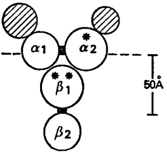
topographical model of the subunits of the IgE FcR and this can be seen in Figure 3.
[18] This model has not. as yet. been supported by sufficient data. Further support
of receptor subunit structure is provided by other receptor systems such as insulin
and acetylcholine [22, 23].
The above studies and findings on the IgE FcR serve as a prototype for the research
being conducted on the IgG FcR.
1.4 IgG/FcR SYSTEM
T.
BOA
1
BOA
Figure 3. Schematic representation of receptor for IgE. The horizontal line represents
the surface of the outer leaflet of the plasma membrane bi-layer. The hatched areas
represent carbohydrate, the black areas sites of proteolytic cleavage. [*] Principal site
of surface labeling. [**] The circles represent spheres whose volumes are proportional
[image:16.543.206.375.245.407.2]B
The first step of the endocytic process, the binding of monomeric IgG to macrophage
FcR sites, has been characterized by radioligand studies [24-26). This characterization
was made possible by the development of a method to separate cell bound radioligand
from free radioligand. In this manner monomeric IgG was found to bind only for a
matter of minutes and with affinity constants [ka) of 10B to 108 . These ka's are
significantly lower than those for IgE.
1.4B FcR dimerization
As with the IgE System, IgGFcR dimerization appears to trigger the respective cell
function of endocytosis. The fate of monomeric IgG upon binding to its receptor at
0C is subsequent dissociation from the cell membrane [27). Anderson has determined
that the kinetics and reversibility of monomer binding is the same at 37C [27]. This
finding seems appropriate since it would be impossible for macrophages to maintain
endocytosis and degradation of the monomeric IgG normally found in serum. However,
immune complexes of dimeric IgG or larger, once bound to the macrophage membrane,
are rapidly endocytosed and catabolized [2B, 29). More importantly, the endocytic
rate does not increase with an increase in complex size (28, 29]. Thus only two Fc
receptors need to interact for irreversible endocytosis to occur. An important
assumption here is that the IgG FcR has a valency of a single Fc unit and therefore
binds a single immunoglobulin. This assumption is based on the fact that the IgE FcR
hassuch a valency [30, 31, 32].
1.4 C Biochemical Studies
Young et. al. have studied the effects of specific ligand binding, to the IgG FcR. on
the membrane potential of a macrophage cell line. Rapid membrane depolarization
was observed within 20 seconds of ligand binding. The extent of membrane
9
cross-linking of ligand bound to those receptors. The depolarization was also determined
to be due to Na+ influx into the cell. This determination was made by eliminating
Na+
from the cell incubation media and again observing the membrane potential change
[*]. In this same manner extracellular Ca2+
was found not to play a significant role.
These same investigators also studied the effect
on membrane potential changes of
three other ligands which recognize other major membrane surface antigens on the
same monocyte line. A similiar >Jr was not observed. This phenomena appears to
be specific for the FcR. From these studies Young has suggested that the FcR is a
ligand-dependent ion channel and that the
^p
that occurs activates subsequent cellularresponses (33).
1.4D IgG FcR Structural Investigations
It is strongly believed that the IgG FcR has a subunit structure although there have
been no findings to support the hypothesis thus far. The investigations of Young and
Unkeless 04] actually suggest that the FcR functions independently. Young and Unkeless
purified the IgG FcR from a monocyte cell line using a monoclonal antibody. The
FcR material was then reconstituted into proteoliposomes and planar bilayer membranes.
In the presence of specific ligands the purified receptor increased the cation
permeability of the proteoliposomes. The FcR was thus capable of initiating a response
without the presence of any other associated proteins.
1.5 CROSS LINKING REAGENTS
Chemical cross-linking reagents are short linear molecules that contain reactive groups
at both ends. The reactive groups may be specifically directed toward a particular
class of functional groups on macromolecules. or may have no specificity at all. The
primary use of these compounds is in structural studies, particularly on cell membranes.
10
by these reagents and are then analyzed. The subunit stoichiometry of several protein
systems have been determined in this manner (35, 36).
The use of cross-linking reagents is particularly popular for structural investigations
in the 5-20 Angstrom (A) range. At 5 A or less, various types of spectroscopy and
diffraction procedures provide definitive information. Electron microscopy is useful
above 20-30 A. The information obtained from cross-linking studies is not as precise
as the physical techniques yet has been very valuable. Quaternary structure of
oligomeric proteins and even larger molecular aggregates can be unambiguously
established. A comparison of results obtained from cross-linking experiments and
crystallography on proteins of known structure found the two methods to be in
agreement.
The utilization of cross-linking reagents in structural studies of biological systems
provides other advantages. The information is obtained from such studies without
altering the biological function of the molecule. In addition, straightforward results
are achieved since these reagents do not contribute to molecular weight.
The cross-linking reagents are not without their disadvantages. First of all. it is often
difficult to establish optimal conditions for cross-linking reactions. Also, in protein
solutions that are sufficiently concentrated, there is increased formation of cross-linked
collision complexes which can provide misleading information. Another problem arises
when many different macromolecules are present in the system. Analysis of such
mixtures can be very difficult. Finally, when cross-linking intact cells, the reagents
can be so effective that the cells cannot be lysed for subsequent analysis.
1.5B Cross-Linking Reagent Categories
11
readily be synthesized. General categories of these reagents include homobifunctional,
heterobifunctional and photosensitive. The homobifunctional class of reagents contains
the same reactive group at each end while the heterobifunctional contains two dissimiliar
groups. The photosensitive reagents can be either homobifunctional or
heterobifunctional and contain reactive groups [one or both] which cross-link only
upon exposure to light.
These three types of reagents are available in various lengths and cleavable forms.
The shorter the reagent, especially at 5 A or less, the fewer the cross-links formed.
Cleavable cross-linking analogs contain either disulfide or glycol bridges within their
structure. The disulfide linkages can be cleaved by reducing agents such as
2-mercaptoethanol [2-ME] while the glycol bridges are cleaved by sodium periodate.
The cleavable cross-linkers are especially invaluable in structural studies. Individual
components of a cross-linked complex can be identified following cleavage of the
cross-linking compound.
1.5C Photosensitive Cross-linking Reagents
When appropriate functional groups of a specific macromolecule are not available
for cross-linking, reagents of high reactivity and low specificity can be used. The
photochemical cross-linking reagents are one such group which is gaining popularity.
These reagents remain totally inert until a photon of light is absorbed. Upon light
absorption an intermediate reactive species is generated which can then become
attached onto biological molecules. The concentration of the reactive species formed
will depend directly on the number of light quanta absorbed and indirectly on the
concentration of the precursor. Shortwave ultraviolet lamps are most commonly used
to provide the necessary light for photolysis. If flash photolysis is desired, ordinary
camera flash units can be used. For this particular investigation a xenon lamp, which
12
The photosensitive reagents have gained increasing popularity since they overcome
inherent problems of conventional reagents such as random collision cross-links, long
reaction times and difficulty in
controlling reactions. Photolysis in the millisecond
range can initiate cross-linking reactions that provide a picture image of interacting
systems at the desired moment. Random collision cross-links still occur, but at a much
lower frequency. By cross-linking with heterobifunctional photosensitive reagents.
cross-linking is not only rapid but can also be controlled sequentially. Cross-linking
with these reagents involves two steps: the first step being a dark incubation for the
reaction of the conventional end. The photoreactive precursor can then be manipulated
into a desired position prior to photolysis.
Reagents that have proven useful for photolabelling/photochemical cross-linking include
precursors to carbenes. nitrenes and free radicals (37). Discussion will be limited to
the nitrenes.
Nitrenes are generated photochemically from suitable aryl or alkyl azides. The general
reaction mechanism of nitrene formation from an aryl azide is shown in Fig. 8. The
half life of the intermediate reactive species is of the order of 10~4 to 10~2
seconds.
Once a nitrene is generated it may undergo any of the following reactions: 1] hydrogen
abstraction 2] insertion at C-H bonds 3] addition 4) condensation 5] insertion at
N-H bonds and 6] rearrangement B6). These reactions are illustrated in Figure 9.
The most widely used class of photolabel precursors is the aryl azides. Their present
popularity stems from the ease with which they can be synthesized, their stability
in storage and their lack of reactivity under physiological conditions in the absence
of light. In addition, the aryl nitrenes are less susceptible to internal rearrangement
than are the alkyl nitrenes. Furthermore, the absorption maxima for the alkyl nitrenes
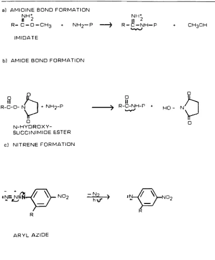
Figure 8. General reaction mechanisms of a] imidates and b) N-hydroxysuccinimide
esters with primary amino groups, c] Illustrates nitrene generation from aryl azides.
a) AMIDINE BOND FORMATION
NHt II 2
R-C-O-CH3 + NH2-P
IMIDATE
NH+
n 2
R-C-NH-P CH3OH
b] AMIDE BOND FORMATION
+ NH9-P
N-HYDROXY
SUCCINIMIDE ESTER
O
II
R-C-NH-P +
HO - N
/S
o
c) NITRENE FORMATION
;n=n/n Z'
Vno2
R
- N
hi/
v->
:"\
/^: [image:22.543.43.475.98.625.2]Figure 9 . Some possible fates of nitrenes
1) R
2] R
3) R
4)
R-5] R-N
N
N
N
N
6) R-N
+ H-C!
+ H-C^
+ R-N
i >
-* RNH +
-C; > other products
- R-NH-CJ
-* R-N=N-R
[R']?NH 9 R-NH-N/
^ rearrangement products
Nr-1] hydrogen abstraction, 2] insertion at C-H, 3] addition, 4] condensation,
13
Substituted aryl azides have been developed that can be photolyzed
in the near visible
light region; wavelengths at which such protein damage cannot occur.
1.5D Experimental Design
The use of cross-linking reagents is not
technically difficult, however the challenge
lies in finding optimal conditions for the reagent within the restrictions of the
experimental system. Attention must be given to the reaction time period, temperature,
pH, buffer composition and cross-linker concentration. These factors will vary with
the reagent in use and conditions for the more widely used reagents have been well
established. Of note here is reagent concentration since many of these compounds
are extremely unstable and will undergo hydrolysis upon solubilization. For this reason
reagent excess is required for quantitative cross-linking to occur. To avoid adverse
side reactions several successive additions of reagent is preferred.
When performing cross-linking on cell membrane proteins, further consideration must
be given to experimental design. From the Fluid Mosaic Model B8)> membrane proteins
can be peripheral or integral. Integral proteins, as are receptor molecules, are strongly
associated with the membrane phospholipid. These proteins can either face the cell
cytoplasm or the exterior of the cell, or can be transmembraneous. and span the entire
membrane. For those proteins that face inward, suitable reactive groups may be
difficult to reach with cross-linking reagents. For this reason cross-linking of cell
lysate material should also be performed.
1.5E Cross-linking Reagents Utilized
Imidates are the most common cross-linking reagents used for cell membrane structural
,NH
studies. Imidates are imido esters and have the general formula
R-C'qr
. Those14
soluble and most are permeable to cell membranes. Depending on the reagent's rate
of hydrolysis, half-lives range from several minutes to 30 minutes. Because this
hydrolysis occurs, up to 100 fold reagent excess may be required. For a more complete
review of these compounds the investigator is referred to Peters and Richards [36].
The primary reaction of imidates involves formation of an amidine bond with primary
amino groups on protein: a reaction referred to as amidination. The actual mechanism
of amidination has been elucidated by Hand and Jencks [39] and is illustrated in Figure
8.
Temperature and pH significantly effect the imidate reaction. For maximum formation
of amidine bonds with a minimum of side products, the reaction should be carried out
at pH 10 or slightly above [40]. In many cases this is not feasible since proteins, and
in particular cells, do not tolerate such harsh alkaline conditions. On the other hand,
if the pH of the reaction system is at a pH of B or lower, side reactions are more
predominate. As regards temperature, the reaction rate decreases severalfold as the
temperature is lowered from 39C to 25C. and then again from 25C to 0C. From
previous cross-linking experiments, optimal conditions for reaction with imidates are
pH 9.3. 30-60 minute incubation at room temperature and a total reagent concentration
of 1-5 mM with incremental additions every 10-30 minutes [41.42). These conditions
are idealistic and serve only as a guideline as actual conditions should be optimized
for the particular experimental system under consideration.
As mentioned earlier. DMS and DTBP were to be the two imidates utilized. Both of
these reagents are homobifunctional. DTBP represents the cleavable analog of DMS
as it contains a disulfide bridge in its structure. The molecular weights of DMS and
DTBP are 273 and 281 respectively. The chain length of DMS is 1 1 A and that of DTBP
is 1 1.9
A.
Their structures can be seen in Figure 4.a]
NH^
CI II[S-[CH2)2-C-0-CH3]2
11.9 A
DTBP
c)
ANB NOS
b) CH3-0-C-(CH2)-C-OCH3
NHt cr6 NH+cr
2 2
11 A
DMS
d]
\
Kj-O-C
-tCH2M^-0-N,12 A
DSS
Figure 4. Structures of the cross-linking reagents, (a] and (b) show homobifunctional bisimidates. (a) represents cleavable dimethyl-3. 3'-dithiobispropionimidate [DTBP]
and (b) dimethyl suberimidate (DMS). (c) represents the
heterobifunctional photosensitive
N-5-azido-2-nitrobenzoyloxysuccinimide (ANB-NOS) with its n-hydroxysuccinimide ester
end and (d) disuccinimidyl suberate (DSS) also containing
[image:26.543.33.497.129.536.2]15
order to evaluate whether different cross-linked products could be formed using reagents
with different reactive groups. These reagents are known to react primarily with
amino groups to form an amide bond [43] as can be seen in Figure 8. These reagents
are unstable in aqueous media and have a half life on the order of 10 minutes at pH
B.6 and 4C [44] and several hours at pH 7.0 and 0C [45]. Reactions are complete
within 10 minutes at 0-4C (44.45). DSS was to be the N-hydroxysuccinimide ester
in use and its structure can be seen in Figure 4. DSS has a molecular weight of 368
O
and a chain length of 12 A. Since DSS is more highly reactive, extensive cross-linking
can occur so that much lower reagent concentrations must be used.
The photosensitive reagent in use, ANB-NOS, contains an N-hydroxysuccinimide ester
as its conventional group and a substituted aryl azide as its photosensitive group.
ANB-NOS is called a substituted aryl azide since it contains a nitro group on its aromatic
ring. This substitution enhances the reactivity of the photogenerated aryl nitrene
and allows photoactivation at 320-350 nm. ANB-NOS has a molecular weight of 305
and its structure can also be seen in Figure 4. The distance between the epsilon amino
and azido group, upon protein modification, is 6.5 A (46). Because of the nonspecific
reactions of nitrenes. an interpretation of chain length and optimal cross-linking is
still rather difficult at this time.
1.6 U937 CELLS
The cells for this study were a human monocyte line designated U937. U937 is a tumor
cell line derived from a patient with histiocytic lymphoma. These cells are more easily
maintained in large numbers and pure form, than are normal monocytes. Cell numbers
can be doubled in 24 hours and densities of 1.5 x 10⁶ cells/ml are possible [Anderson
unpublished results). In addition to providing a ready supply of cells, the U937 FcR
should provide a reliable model for the FcR structure and function in normal human
16
radiolabeled human myeloma proteins have shown strong similiarities to normal
monocytes 127]. As with the normal monocytes.
IgGj
andlgG3
were found to bind moreefficiently to the U937 FcR. while
lgG2
andlgG4
bound less readily. U937 cells alsoshare such features with normal macrophages as strong esterase activity, lysozyme
and endogenous pyrogen production, phagocytic capabilities and complement receptors
(47.4B). U937 cells are lacking in such characteristics as myeloid colony-stimulating
activity [49), lymphocyte activating factor [50) and prostaglandin E (49). Kurland et.
al. suggest that these missing features indicate a lack of differentiation (5(3.
Purified normal human monocytes were to be utilized in key procedures so that a true
picture of normal endocytosis could be obtained. These cells would have been obtained
from peripheral blood using a counter flow centrifugation-elutriation technique 61).
1.7 PRELIMINARY STUDIES
1.7A IgG FcR Characterization
The IgG FcR on U937 cells and normal peripheral monocytes has been identified as
a 72.000 dalton glycoprotein using classical affinity column chromatography methodology
(52). These investigations also revealed a possible second minor component of 40.000
daltons. In brief, detergent lysates of chloroglycouril iodinated U937 cells were made
in 1 % NP-40. The lysates were then passed through a Sepharose-IgG affinity column
and the column eluted with acetic acid. The acid eluates were then analyzed by
SDS-PAGE and autoradiography.
Anderson was able to show that the p72 molecule, isolated from the above procedure.
is all or part of the IgG FcR on the basis of the following findings: a) radiolabelling
of the molecule using chloroglycouril was blocked by occupation of the FcR with IgG
17
receptor c] the molecule was not recovered from affinity adsorbents bearing proteins
that do not bind to the Fc receptors, nor d] from a human T cell line that does not
bear Fc receptors 62).
The significance of the p40 molecule has not been fully clarified. Actin appears to
make up at least part of this band since the p40 was found to migrate comparably
with the actin marker on two-dimensional electrophoresis. The remaining portion
of the p40 band seems to have a molecular weight that is slightly higher and has
isoelectric points much more basic, than actin [52). Since actin is a contractile protein
of the cell membrane cytoskeleton, one can speculate that the p40 may be a transducer
domain of the FcR.
1.7B Cross-linking Studies
The ability to achieve cross-linking with DMS. of molecules situated on the surface
of U937 cells, has also been tested. U937 cells were incubated with radiolabeled IgG
to allow binding of the radioligand to the cell prior to cross-linking. As a matter of
comparison, samples that were not to be cross-linked received comparable amounts
of the DMS solvent. The cross-linking reaction was quenched with glycine and the
cells then lysed. Lysed material was then prepared in Laemmli's Sample Buffer
containing 2-ME for the SDS-PAGE procedure. Following processing of the gel. a
prefogged autoradiographic film was apposed to it. The autoradiogram (AR) was allowed
to develop for a length of time which varied with the amount of radioactivity loaded
on to the gel. The developed AR revealed radioactive bands at molecular weights
corresponding to various combinations of linked
immunoglobulin heavy chain, light
chain and receptor when DMS was present [Anderson unpublished results). When DMS
was not present bands were visualized at molecular weights of 50.000 and 22.000 daltons
only, the approximate molecular weights of immunoglobulin heavy chain and light
IB
Once it was ascertained that cross-linking could be achieved with DMS. Anderson then
attempted to cross-link the U937 FcR p72 molecule to a neighboring molecule while
the cells were intact. The protocol used was similiar to the aforementioned procedure
except that after cell lysis the FcR was immunoprecipitated from the lysates using
either goat anti-FcR|| (a goat anti-FcR antibody developed by Anderson] or aggregated
IgG (AgglgG). Heat fixed Staphyloccus aureus Cowan I [SAC I] was then added to all
samples. This particular Staph strain contains a membrane protein called Protein
A which is capable of binding the Fc portion of an IgG. These SAC "beads" add weight
to the entire immune complex so that they can be removed by centrifugation. [With
regards the AgglgG. one assumes that not all Fc regions are occupied with receptor.]
The radioactive material attached to the SAC beads was then separated by SDS-PAGE
and visualized by AR. The results are reflected in the Table below. Besides the FcR
being present, two new molecules were immunoprecipitated with the goat anti-FcR||,
a 170,000 and a 350,000 dalton protein. AgglgG did not precipitate these bands. The
pi70 molecule was present with or without the presence of DMS and has been attributed
to nonspecificity of the goat anti-FcR||. Thus, the only product of interest is the
molecule having an approximate molecular weight of 350.000 daltons. The individual
components of the p350 were to be the subject of analysis undertaken within this project.
hDMS -DMS
AgglgG 72.000 72.000
Anti-FcR 72.000 72.000
170.000 170.000
350.000
19
l.B PROPOSED INVESTIGATIONS
The primary objective of this study is to identify a near-neighbor molecule to the FcR
on human macrophages with the consideration that the neighboring molecule may also
be involved in FcR mediated endocytosis. The investigations will be performed on
a cell line designated U937.
FcR near neighbor analysis will be attempted using several cross-linking reagents. The
two midates. DMS and DTBP will be the primary reagents utilized. Depending on the
success of these reagents, other cross-linking reagents with different reactive groups
and chain lengths will also be considered.
Preliminary cross-linking attempts by Anderson using DMS on intact U937 cells have
yielded a cross-linked product with a molecular weight of 350,000 daltons. My
experimentation will begin with attempting to reproduce these same results. If it
is ascertained that the p350 molecule can be reproduced, a second cross-linking
experiment will be performed using DTBP. Two-dimensional gel electrophoresis
following DTBP cross-linking should then characterize the molecular weights of the
individual components of the p350 molecule.
Cross-linking of Fc fragments of IgG and Fab fragments at anti-FcR|| to the
radiolabeled U937 cell FcR will be attempted. By using specific Fc and Fab fragments
of known molecular weights (approximately 50.000 daltons], it can be ascertained
whether close lying molecules are capable of being cross-linked to the FcR. The efficacy
of cross-linking can also be analyzed in this manner.
Depending on the time frame, cross-linking will be attempted on plasma membrane
vesicles. These vesicles are formed by homogenizing cells in the absence of detergent.
Vesicles formed can be inside or right side out. Inside out vesicles will allow access
20
SECTION II
MATERIALS AND METHODS
2.1 U937 Cells
Cells. Cultures of U937 cells were obtained from Dr. Peter Ralph. Sloan-Kettering
Institute for Cancer Research. Rye. NY. These cells were maintained in spinner culture
in RPMI 1640 [Gibco Laboratories, Grand Island, NY) containing 10% fetal calf serum
[FCS] [Sterile Systems, Logan, UT), penicillin [lOOU/ml], and streptomycin (100 g/ml],
both from Gibco Laboratories.
2.2 lodination
The U937 cells were radioiodinated by the chloroglycouril method 64]. 0.7 ml of cells
[14.3 x 10B cells/ml of PBS] and 1 mCi
125l
(IMS.300, Amersham. Arlington Heights.ID in 10aI were placed in a vial coated with 5 uq chloroglycouril and incubated for
30 minutes at 0C. The reaction was quenched and the cells were washed three times
in 5 mM Kl in PBS. The cells were then either resuspended in bicine pH 9.5. a Good
Buffer.(55] to 3.0 x 10Bc/ml. or lysed in 1% Nonidet P-40 [NP-40], 2 mM
phenylmethylsulfonylfluoride [PMSF], and aprotinin (1 Trypsin inhibitor unit [TIU/ml]]
in PBS for 30 minutes at 0C. depending on whether lysate or whole cells were used
for cross-linking. The cell nuclei and unlysed material were sedimented at lO.OOOg
for 30 minutes at 4C.
2.3 SDS-PAGE and Autoradiography
21
66). The acid eluate samples containing 2% SDS. 10% glycerol. 5% 2-ME. and 0.001%
bromphenol blue, were immersed in boiling water for 2 minutes and applied to the
stacking gels. Molecular weight markers were myosin [200.000). B-galactosidase
[130.000], phosphorylase B [94,000], Bovine Serum Albumin [BSA] [68,000], ovalbumin
[OA] (43,000), soybean trypsin inhibitor [21,000], and lysozyme (14,300), from Bio-Rad
Laboratories and chloramine T-radioiodinated human
IgG]
(27). After being stained,destained and dried, the gels were apposed to pre-fogged Xomat AR X-ray film [Eastman
Kodak Co.. Rochester. NY] and placed in Kodak film cassettes at -70C. Two
dimensional gel electrophoresis was performed on 5
-10% gradient slab gels in both
dimensions. Sample buffer was the same as that noted previously except that 2-ME
was not present in the buffer used in the first dimension. The lane of interest was
outlined using fluorescent markers, dansylated BSA and dansylated ovalbumin [57],
[dansyl chloride from Bio-Rad Laboratories] in adjacent lanes. The markers were
visualized with a UV lamp while the lane was cut out. The entire lane was then soaked
in LSB+ for a minimum of 2 hours. Electrophoresis was performed in the second
dimension by placing the lane at the top of a second gradient slab gel and anchoring
it with agarose (Marine Colloids Inc.. Rockland, ME].
2.4 Reagents
The following reagents were obtained from Sigma Chemical Co. (St. Louis. MO]: BSA.
OA. cytochrome c, aprotinin, PMSF, TRIS, L-methionine, glycine, bicine, tricine. DMSO.
NP-40 and trichloracetic acid (TCA). EDTA. potassium iodide [Kl], sodium carbonate
[NaoCO-a). and ammonium persulfate were obtained from Fisher Scientific (Fair Lawn,
NJ). Sodium azide [NaN3L bromphenol blue and acetone was obtained from Eastman
Kodak. Sodium chloride (NaCI). sodium hydroxide [NaOHL and dioxane were obtained
from Mallinckrodt (St. Louis. MO). Sodium dodecyl sulfate (SDS). N, N'
methylene-bis-acrylamide, acrylamide and temed were obtained from Bio-Rad
22
human IgG was purchased as Pentex human gamma globulin and further purified by DEAE
cellulose ion exchange chromatography. A human
IgG]
myeloma protein [Woo] andFc fragments from the same protein, as well as a human IgG myeloma (Allendorf]
and Fab fragments of goat anti-FcR|(. were purified as described (27). Aggregated
IgG
(Agg
IgG) was prepared by heating human IgG at 10 mg/ml to 63C for 12 minutes.The medium in which U937 cells were incubated with
IgG]
to saturate Fc receptorswas balanced salt solution (BSS) [5B] containing BSA (1 mg/ml] and
NaN3
[0.2%] (BBAL2.5 Cross-linking of intact U937 Cells
Dimethylsuberimidate (DMS). dimethyl-3,3'-dithiobispropionirnidate [DTBP] and
disuccinimidyl suberate [DSS] were all purchased from Pierce Chemical Company
[Rockford. ID. N-5-azido-2-nitrobenzoyloxysuccinimide (ANB-NOS) was synthesized
by Sheldon Isaacson in the summer of I9B0 in Dr. Clark Anderson's laboratory. University
?f Rochester (Rochester, NY]. The ANB-NOS was stored refrigerated in a brown bottle
covered with aluminum foil. All cross-linking reagents were prepared fresh just prior
to addition and all reactions were performed on ice. Cross-linking was performed
on 2.5 ml aliquots of radiolabeled U937 cells resuspended to 3.0 x 10B cells/ml in the
buffer indicated for the particular cross-linker.
Surface-labelled U937 cells were chemically cross-linked with DMS after resuspension
in a bicine pH 9.5 buffer. Bicine buffer pH 9.5 was used to maintain a pH of B.5 during
cross-linking (20). DMS was prepared at 90 mg/ml of 0.1 M Na2C03. DMS was added
to [3] 2.5 ml aliquoits of the cell suspension; the initial concentrations of the DMS
being 1, 3 and 6 mM. After the second addition, final DMS concentrations were 3.
6 and 10 mM. Incubation after the first DMS addition was for 1 hr. mixing every 15
minutes. The second incubation was for three hours with mixing. Subsequent
experiments revealed sufficient cross-linking with 2 incubations of 30 minutes.
23
5 mM KI/50mM glycine in PBS was used to wash the cells two times prior to lysis.
The cells were lysed at 50 x 10B cells/ml as described in 2.2. FcR imunoprecipitation
then followed.
A positive control consisted of cross-linking IgG-Woo coated U937 cells with DMS
after two additions at 6 and then 10 mM. For the negative control, bicine buffer pH
9.5 was added in comparable amounts to the cross-linking reagent. The cells were
then lysed with 1% NP-40/PMSF/TRAS*ee note below.] and the TCA or acetone
precipitates of the lysates were analyzed by SDS-PAGE.
DTBP was prepared and used in the exact same manner as DMS.
Washed '2^l labelled U937 cells were resuspended in PBS prior to cross-linking with
DSS. DSS was prepared at 50 mM in DMSO. Two DSS additions were made for
concentration ranges of 0.5 to 1.0. 1.0 to 2.0. and 2.0 to 4.0 mM. DSS addition was
followed by 10 minute incubations. Quenching and further processing was also the
same as for DMS.
Labelled U937 cells were prepared in PBS for cross-linking with ANB-NOS. ANB-NOS
was prepared at 100 mM in dioxane and protected from light at all times. Dioxane
was kept at 10% of the protein. (59) Final ANB-NOS concentrations achieved for the
single 15 minute
"dark"
incubation were 1, 5. 10 and 20 mM. Higher concentrations
of ANB-NOS resulted in a precipitate due to excessive derivitization of cellular proteins
by the NOS end. All sample tubes were foil covered and capped. For comparison,
one sample with 20 mM ANB-NOS was kept in the dark for the entire procedure. A
negative tube with dioxane addition alone was also run. Following the dark incubation.
the samples were photolyzed with a xenon lamp at 6000 volts. 14 pulses/second for
5 minutes. Quenching and further processing was then as before.
24
2.6
Cross-linking
of Lysed U937 cellslzal labelled U937 cells were washed in 5 mM KI/PBS twice, once with 5 mM Kl/bicine
pH 9.5. then solubilized with 1% NP-40 in bicine pH 9.5. No protease inhibitors were
added during solubilization so all procedures were maintained at 4C and performed
without delay. High speed centrifugation of the extracts was as before. 100^1 of
the lysates were cross-linked by two additions of DMS in the following range: 1 to
2. 2 to 4. 3 to 6, 4 to 8 and 6 to 12 mM. The latter concentration represents the final
DMS concentration. Incubations were for 30 minutes following each addition. 1%
NP-40 in PBS was used to prepare cell extracts for DSS cross-linking. 100j<U of lysate
was used and DSS concentrations and incubation times were the same as for intact
cells. Quenching was again with 0.5 M glycine/PBS. Sample volumes were kept as
small as possible for FcR immunoprecipitation.
2.7 FcR precipitation
The FcR was immunoprecipitated from lysed radiolabeled U937 cells in two ways;
using goat antibody to the FcR (>FcR(|] at 13.9 mg/ml. and aggregated IgG [AgglgG]
at 10 mg/ml. To one tube of 50yul of lysate was added 2^l of AgglgG and to a second
tube was added 7 u\ of >FcR||. After 30 minutes 200^1 of a 10% suspension of
Staphylococcus aureus Cowan I (IgGSorb or SAC bacteria) was then added to all samples
to adsorb IgG immune complexes. After another 30 minutes the samples were
centrifuged at 3000 rpm for 15 minutes at 4C to yield a firmly packed pellet of immune
complexes. A series of washes using SAC buffers (60) was then performed. After the
final wash. 80 mI of Laemmli's Sample Buffer was added to the pellets to dissociate
the immune complexes upon boiling, in preparation for SDS-Page. All samples were
counted on a gamma counter prior to loading to the gels to determine the time exposure
25
2.B Cross-linking of fragment coated U937 cells.
,25l
labelled U937 cells were washed in BBA once and resuspended in the same to30 x 10 cells/ml. [3] 0.5 ml cell samples were prepared for separate incubations with
IgGpWoo Fc fragments at IB.7 mg/ml. >FcR|, Fab fragments at 9.34 mg/ml and with
BBA alone. 54ytl of each were added to the cell suspensions and binding was allowed
to occur for 1 hour at 4C. Following this incubation the cells were washed once in
BBA. twice in BSS and resuspended in PBS to 3 x 10⁶ cells/ml for cross-linking. 2.5
mis of each suspension were reacted with DSS at initial concentrations of 0.5 and 1
mM and with second additions of DSS for final concentrations of 1 and 2 mM. Incubation
times after each addition were 10 minutes on ice. Quenching, cell solubilization,
immunoprecipitation and analysis was as before.
2.9 TCA precipitation
TCA precipitations performed on lysate material were used to monitor the recovery
of radioactivity and success of the labelling procedure. 10kI of cell lysate was used
to make a 1:100 dilution in 1% NP-40. 10u.l of the diluted sample was then added to
0.5 ml BSA/PBS at 5 mg/ml and 0.5 ml of 25% TCA and incubated on ice for 20 minutes.
The sample was spun at 3000 rpm for 10 minutes at 4C. The supernatant was carefully
26
Section III
RESULTS
3.1 Cross-linkingof intact U937 cells
3.1 A DMS Utilization
When
125l-surface
labelled U937 cells are cross-linked with DMS for 4 hours andsolubilized with 1% NP-40/PMSF/TRAS, goat anti-FcR|| immunoprecipitation yields
a slight band with a molecular weight of 72,000 and denser bands at 170,000 and 350,000
on autoradiogram [AR). When no DMS is present bands can be seen at 170,000 only.
Immunoprecipitation with AgglgG did not yield any bands. These last two observations
suggest that a loss of FcR occurred during the procedure. The positive control, which
consisted of cross-linking U937 cells coated with *lgG-Woo. indicated that cross-linking
was successful since radioactive bands were seen at molecular weights which correspond
to combinations of heavy and light chains.
3.1 B Analysis of FcR loss
Before any further cross-linking studies were done an analysis of pH effects, bicine
toxicity and long cross-linking incubationson FcR recovery was performed. This analysis
was performed in the following manner. Surface radiolabeled U937 cells were
resuspended in 5 mM Kl in PBS. divided into four aliquots and centrifuged at 930 rpm.
4C for 7 minutes. The cells of the first aliquot were lysed as before. The other three
aliquots were resuspended to 3 x 10B cells/ml in BSS. bicine pH 7.4 and bicine pH B.5
and put on ice for 4 hours with mixing every 15 minutes. Lysisof the three cell samples
was then performed with 1% NP-40/PMSF/TRAS followed by FcR immunoprecipitation
27
recovery. Labelled U937 cells maintained in bicine pH 7.4 or BSS yielded bands on
AR after FcR immunoprecipitation that were somewhat more dense than those cells
maintained in bicine pH 8.5. These results suggest that pH may have a small effect
on FcR recovery and that bicine is only minimally toxic to the FcR. However, the
cells in BSS that were not processed through an incubation period, yielded bands
significantly denser than the other samples above. Thus it was concluded that the
FcR was not clearly visualized following DMS cross-linking due to loss of radioactivity.
Incubation times of 30 minutes were used for subsequent procedures.
3.1C DTBP Utilization
Cross-linking with the cleavable analog DTBP for 30 minutes yielded results that were
comparable to those from the preliminary studies done by Anderson when DMS was
used for 30 minutes.
Anti-FcR||
precipitation yielded bands on AR at 72.000. 170,000and 350.000. A single disperse band centered at 72,000 as well as dense bands stacked
at the interface of the stacking and running gel were seen with AgglgG as the
precipitating agent. (See Figure 5) The dense bands at the top of the gel probably
represent larger cross-linked products incapable of entering the gel.
In order to identify the individual components of the 350.000 cross-linked molecules
achieved with DMS and DTBP. two-dimensional gel electrophoresis was performed
as described in Methods following cross-linking with DTBP in the 6 to 10 mM range.
Of the original bands, the 350.000 molecule alone was cleaved into smaller products
as seen by an AR of the 2-D gel. [Figure 6] The cleaved product had a molecular weight
of 170.000 indicating that the p350 is a dimer of the original p170 and thus is unrelated
to the FcR. The 350.000 band did not entirely disappear after reduction indicating
that there was incomplete DTBP cleavage or that there may be impurities in the
cross-linking reagent. Also of interest on the 2-D gel AR was the appearance of the
p72 molecule. Although it still migrated as a disperse band in the molecular
weight
region between 60.000 and 80,000, it appeared heterogenous
as several distinct spots
** -200
-D
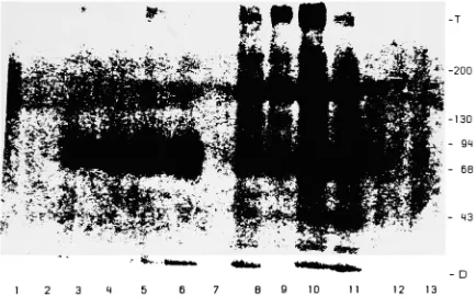
[image:40.543.60.494.169.449.2]B 9 10 1 1 12 13
Figure5 . Autoradiogram of SDS-PAGE gradient slab gel (5-10%)
analyzing the
immunoprecipitation products following DTBP cross-linking of intact U937 cells.
The cells were lysed in 1 % NP-10/PMSF/TRAS prior to SDS-PAGE. SDS-PAGE was
performed under nonreduced conditions. Products in Lanes 3 through 6 are from AgglgG immunoprecipitation while anti-FcR|| was used in the samples in Lanes B
through 1 1. DTBP was highest in Lanes 5 and 10. Samples from Lanes 6 and 1 1
received no DTBP. Bands can be seen stacked at the top of the gel. Numbers to
the right of the autoradiograph indicate apparent molecular weight x 10~3. T -top
of 5
Figure 6. Autoradiogram of the SDS-PAGE two-dimensional gel analyzing the p350
cross-linked molecule produced from DTBP cross-linking of intact surface-labelled U937 and immunoprecipitated with anti-FcR||. The second dimension was run in the
presence of 2ME. Due to incomplete cleavage some of the p350 remained [spot I). Spot 2 is a cleavage product of the p350 and has a molecular weight of 170. Spots
3 and 4 did not cleave and represent the original pi70 and p72 (FcR) molecules
[image:41.543.72.483.66.334.2]Lanes
B
Figure 7. Autoradiograms of SDS-PAGE gradient slab gels [5-10%) analyzing the immunoprecipitation products following cross-linking of soluble extracts of surface
labelled U937 cells using (A) DMS and (B) DSS. In (A) Lanes 1 through 6 are products
of anti-FcR|| precipitation. DMS concentrations decrease from Lanes 1 to 5 and B
to 12. Lanes 6 and 13 contained no DMS. In (B) Lane 1 is missing. AgglgG was used for Lanes 2 to 5 and anti-FcR|| was used for Lanes 7 to 10. Lane 11 is a radiolabeled
IgG-Woo marker. DSS was also used in decreasing concentrations from Lane 2 to 4
and 7 to 9. No DSS was used in samples in Lane 5 and 10. Numbers to right of
auto-radiographs indicate apparent molecular weight x 10"^. T
-top of 5-10% gradient gel. D - bromphenyl blue dye front. * indicates slight band
[image:42.543.30.508.65.536.2]2B
3. ID ANB-NOS cross-linking
A U937 cell suspension at 40 x 10B cells/ml of BBA was incubated with
125l
labelledIgG-Allendorf to allow binding to occur as was done for the control cells from previous
procedures. Following cross-linking of the intact U937 cells with ANB-NOS the AR
showed bands at the following molecular weights: 22.000 (light chain). 50,000 [heavy
chain], 95-100,000 (2 heavy chains] and 140,000 [an intact immunoglobulin IgG). No
bands were seen that would indicate cross-linking to the FcR had occurred.
3.2 Cross-linking of
125l
lysed U937 cells,25l-surface
labelled U937 cells were solubilized in 1% NP-40/bicine pH 9.5. withoutthe presence of enzyme inhibitors for cross-linking with DMS. AgglgG
immunoprecipitation of the soluble extract following cross-linking yielded a very slight
band on the autoradiogram at 140.000 when DMS was at its highest concentration range
of 6 to 12 mM. The usual disperse band at 72.000 was also seen. This was the first
time that AgglgG yielded anything other than the p72. Immunoprecipitation with
anti-FcR|| yielded a disperse 72.000 dalton band as well as bands at 130.000. 170.000
and 350.000. The 130.000 band was also very slight. [See Figure 7) The gel was put
back to film for a two week period of time so that the p140 from AgglgG precipitation
could be visualized more clearly. The two week AR did show slight bands at 140.000
in all lanes precipitated with AgglgG when DMS was present while this p140 band was
not present in the absence of DMS. These bands were so slight that it would have
been difficult to analyze the molecule any further.
Cross-linking of soluble extracts of U937 cells with DSS yielded no bands on AR that
had not been seen with the other reagents. A p350 cross-linked product was also
achieved with DSS. Denser bands were seen with decreasing DSS concentrations
suggesting that over cross-linking may have
been a problem despite the low
concentration of DSS used. (See Figure 7) At higher DSS
29
not be lysed properly such that the FcR is recovered in smaller amounts or not at all.
Centrifugation following lysis of the cells did indeed yield NP-40 pellets larger than
normal and which did not have the usual stringy appearance.
3.3 Cross-linking of fragment coated U937 cells
Fab fragments of goat anti-FcR|| and Fc fragments of IgG-Woo were coated onto
separate samples of radiolabeled U937 cells and cross-linking then attempted on the
cells with DSS as described in Methods. Incomplete lysis of the cells again posed a
problem. The bands seen on AR were similiar to those from all the other cross-linking
30
SECTION IV
DISCUSSION
With regards to the objective of this research, a neighboring molecule to the FcR has
not been identified. It cannot be concluded however, that the FcR does not have
molecular associations with neighboring molecules or that the FcR does not have a
subunit structure. It is clear that the conditions used were adequate for cross-linking
with the reagents used since the control cells coated with radiolabeled IgG and
cross-linked gave positive results and 170 + 170350. Thus a suitable conclusion
is that there are no reactive amino groups on adjacent molecules to the receptor which
approach the FcR at the necessary distance [1 1-12 A). A second possibility is that
the FcR may need to be activated by an IgG dimer before an association exists with
surrounding membrane proteins. Dimeric IgG is being developed now. as well as trimers.
tetramers and so on. so that such a study can be performed. Such a procedure would
be technically difficult but would be the next possibility to investigate. There is also
a third possibility that the FcR near neighbor is not susceptible to probes of the intact
membrane and upon solubilization for cross-linking of the cell extract, the neighboring
molecule may lose its association with the FcR.
As mentioned in the introduction it is possible that the FcR functions alone in the
process of endocytosis. In order to substantiate the negative findings from the studies
done, cross-linking of Fc fragments of IgG-Woo and Fab fragments of goat anti-FcRM
to the FcR was attempted. By successfully cross-linking protein fragments of known
molecular weight to the FcR and at the same time not being able to cross-link a
neighboring molecule to the FcR. one could
more appropriately conclude that the FcR
is not associated with other molecules. Successful cross-linking of such fragments
using DSS was not achieved however, but this may have been partially due to the
The 350.000 molecule isolated following cross-linking of intact cells and cell lysates
with all of the cross-linking reagents, was conclusively identified as a dimer of the
pl70 molecule by this study. The p170 molecule is immunoprecipitated from U937
cells using goat anti-FcR|| as a result of some nonspecificity of the antibody. The
p350 molecule is thus not associated with the FcR.
Cross-linking reagents are more often than not difficult to work with but with continued
perseverance and further experimentation these reagents may help to identify a subunit
structure of the IgG FcR and thus aid in a more complete understanding of the endocytic
32
REFERENCES
1. Unkeless. J.C.. H. Fleit. and LB. Mellman. 1981. Structural aspects and heterogeneity of immunoglobulin Fc receptors. Adv. Immunol. 31:247.
2. Mellman. I.S.. and J.C. Unkeless. I9B0. Purification of a functional mouse Fc receptor
through use of a monoclonal antibody. J. Exp. Med. 152:1048-1069.
3. Mellman. I.S.. R.M. Steinman. J.C. Unkeless. and
Z.A. Cohn. 1979. Selective iodination
and polypeptide composition of pinocytic vesicles. J. Cell. Biol. 86:712-722.
4. Kaplan. G.. J.C. Unkeless. and Z.A. Cohn. 1979. Insertion and turnover of macrophage plasma membrane proteins. Proc. Natl. Acad. Sci. 76:3824-3828.
5. Hood. L.E.. I.L. Weissman, and W.B. Wocd. I97B. Immunology. Forkner. M.. M.
Moore, and J. Hall, editors. Benjamin/Cummings Publishing Co. pp. 322-330.
6. Hyde. R.M., and R.A. Patnode. I97B. jn Immunology. Reston Publishing Company. Inc. pp. 141.
7. Ishizaka. K.. and
![Figure 8.General reaction mechanisms of a] imidates and b) N-hydroxysuccinimideesters with primary amino groups, c] Illustrates nitrene generation from aryl azides.](https://thumb-us.123doks.com/thumbv2/123dok_us/101102.9460/22.543.43.475.98.625/figure-general-reaction-mechanisms-imidates-hydroxysuccinimideesters-illustrates-generation.webp)