Development of RNA-based Genetic Control Elements
for Predictable Tuning of Protein Expression in Yeast
Thesis by
Andrew Harris Babiskin
In Partial Fulfillment of the Requirements
For the Degree of
Doctor of Philosophy
California Institute of Technology
Pasadena, California
2011
© 2011
Andrew Harris Babiskin
Acknowledgements
First, I would like to express my appreciation to my research advisor, Christina
Smolke. Her enthusiasm for her students, her hands-on approach, and her accessibility
were major factors in my development as an independent researcher. Her guidance has
been immeasurable in the development and the progress of my research projects.
Over the years, I developed close relationships with several colleagues in lab and
in the community at the California Institute of Technology. My fellow Smolke lab
members have provided a great atmosphere for the facilitation of the sharing of
information (and vectors) and for social interaction. Maung Win, Joe Liang, Leo
d’Espaux and I spent numerous enjoyable hours together -- whether lunch at Chandler or
coffee break at Red Door -- discussing research, politics, pop culture, and whatever else
came to mind. I would like to thank Drew Kennedy for spending many half-price burger
and chicken sandwich dinners with me during my time at Stanford University. Outside of
lab, I developed close friendships with Armin Sorooshian (my roommate of 4 years),
Marc Woodka, Ubaldo Córdova-Figueroa, Edgardo García-Berríos, and Muang Win. We
enjoyed many visits to all-you-can-eat buffets, our sometimes successful seasons of
intramural basketball, our unsuccessful attempt at intramural football, and assembling the
greatest Gradiators team of all time. I am lucky to have great colleagues who are also
such great friends. Outside of Caltech, I would like to thank Justin Aefsky, Mark
Gaylord, and Peter Schmidt. My many trips to San Diego to visit them always gave me a
nice, quick weekend away from graduate school. I express my appreciation to my friends
For their constant support and love, I would like to thank my parents, Carol and
Robert Babiskin, and my sisters, Rachel and Jenny. They have made me everything I am
today and my success can be attributed to their involvement. To my deceased
grandfather, Julius Babiskin, I would like to acknowledge his doctorate in physics as my
source of inspiration for achieving my own doctorate. I would also like to thank my
grandparents, Melvin and Lee Rosen, for their constant love and support. Being so far
away was tough, but I enjoyed visiting home and going out to lunches with them. Even
though they may not understand what I am actually doing, I was always glad to provide
them with additional bragging material.
Last but not least, I would like to thank my wife, Rosario Babiskin, whom I love
deeply. She has been with me for almost my entire graduate career. Because of her, I had
a family away from home with whom I could spend numerous holidays with and eat
ridiculous amounts of amazing food. We share together the cutest dog in the world, Cleo.
When I moved away to Stanford, it was tough on the both of us being apart and not
knowing when we would live together again, but she stayed strong. Though that period
was difficult, we now have memories that we will cherish forever, including our
wedding. Having her back living with me after our marriage has been amazing and her
support and understanding has been invaluable in these final months as I finished my
Development of RNA-based Genetic Control Elements
for Predictable Tuning of Protein Expression in Yeast
Andrew Harris Babiskin
B.S., University of Maryland
M.S., California Institute of Technology
Ph.D., California Institute of Technology
Abstract
The proper functioning of many biological processes and synthetic genetic
networks depends on the precise tuning of expression levels of key protein components.
With growing interests in eukaryotic hosts and the increasing complexity of networks in
synthetic biology, there is a need for the expansion of the genetic toolbox, particularly for
the bioprocessing and biosynthesis applications in the yeast Saccharomyces cerevisiae. The available control elements in yeast generally focus on the regulation of transcription
through alternative promoter systems. Synthetic RNA-based control elements placed in
the untranslated regions (UTRs) of transcripts have the ability to regulate the
posttranscriptional mechanisms of translation initiation and transcript stability. Such
posttranscriptional elements have the added advantage of being coupled to any promoter
for enhanced control strategies.
Two types of posttranscriptional elements were examined in this thesis. The first
transcript, the endonucleolytic cleavage due to Rnt1p activity resulted in the rapid
degradation of the transcript. We developed two libraries of RNA hairpins based on the
randomization of critical regions in Rnt1p substrates that affect the enzyme’s ability to
associate and/or cleave the hairpin. The modulation of the strength of binding and
cleavage by Rnt1p resulted in changes in the steady-state transcript levels and thus
protein levels. Through integration of an aptamer into the stem of an Rnt1p hairpin, we
were able to develop a riboswitch based upon the direction inhibition of Rnt1p cleavage
through association of the ligand in the sites of cleavage. The second type of
posttranscriptional elements examined is the placement of internal ribosome entry sites
(IRESes) in the 5’ UTR that initiate translation independent of the 5’ cap through direct
interaction with the ribosomal machinery. We propose that the activity of small
sequential IRESes can be tuned through varying the complementarity with the 18S
ribosomal RNA (rRNA) to advance the creation of yeast multicistronic vectors. The
application of Rnt1p hairpins and IRESes provide a key tool in synthetic biology for the
construction of complex genetic networks in yeast where the predictable tuning of gene
Table of Contents
Acknowledgements ... iii
Abstract ... v
Table of Contents ... vii
List of Tables ... xii
List of Figures ... xiii
Chapter I: Introduction ... 1
1.1. Synthetic biology and metabolic engineering ...1
1.2. Common pathways of transcriptional decay and translation in yeast ....4
1.2.1. Deadenylation-dependent decapping pathway of transcript degradation ...6
1.2.2. Cap-dependent translation initiation ...7
1.3. Posttranscriptional regulation through transcript stability and translation ...8
1.3.1. Control of transcript decay ...8
1.3.2. Control of the initiation of translation ...11
1.3.3. RNA processing by the RNase III enzyme Rnt1p ...13
1.3.4. Translation initiation mediated through internal ribosome entry sites ...14
1.4. Interrelationship among the thesis projects ...15
References ...17
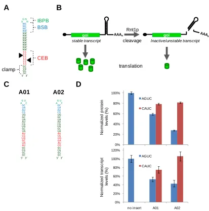
Chapter II: Synthetic RNA control modules that tune gene expression in yeast through directed processing by Rnt1p ... 33
Abstract ...33
2.1. Introduction ...34
2.2.1. Implementing Rnt1p hairpins as RNA-based gene regulatory
components ...37
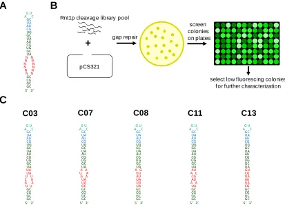
2.2.2. Design and selection of an Rnt1p cleavage library to achieve tunable gene regulatory control...41
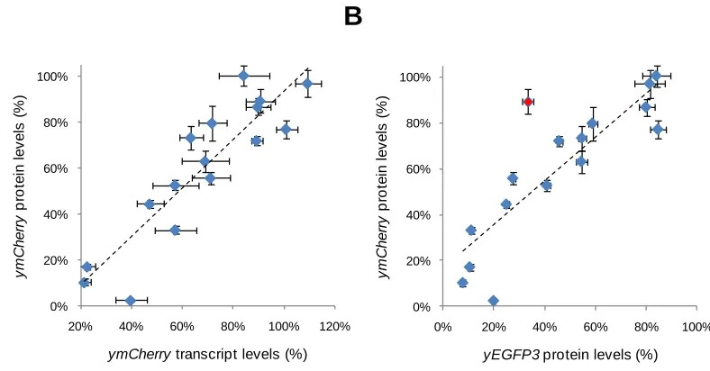
2.2.3. A synthetic Rnt1p hairpin library exhibits a range of gene regulatory activities in vivo ...43
2.2.4. Rnt1p library hairpins maintain regulatory activity in a different genetic context ...45
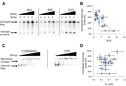
2.2.5. In vitro characterization demonstrates that Rnt1p library members achieve differential activity through alterations in Rnt1p cleavage rates ...47
2.2.6. Control of endogenous ERG9 expression by 3’ UTR replacement with Rnt1p library members ...52
2.3. Discussion...58
2.4. Materials and Methods ...63
2.4.1. Plasmid construction ...63
2.4.2. 3’ UTR replacement cassette and integration ...67
2.4.3. Rnt1p substrate characterization assays ...67
2.4.4. Fluorescence quantification ...68
2.4.5. Quantification of cellular transcript levels ...69
2.4.6. Cell growth rate determination ...70
2.4.7. Cellular ergosterol quantification ...70
2.4.8. In vitro transcription of Rnt1p substrates ...71
2.4.9. Rnt1p expression and purification ...72
2.4.10. In vitro Rnt1p substrate cleavage assay ...73
2.4.11. In vitro Rnt1p substrate mobility shift assay ...73
2.5. Supplementary Information ...74
Supplementary Figures and Tables ...74
Acknowledgements ...79
Chapter III: Synthetic RNA modules for precise control of expression
levels in yeast by tuning RNase III activity ... 87
Abstract ...87
3.1. Introduction ...88
3.2. Results ...91
3.2.1. Design and selection of an Rnt1p binding library to achieve tunable gene regulatory control...91
3.2.2. A synthetic Rnt1p binding library exhibits a range of gene regulatory activities in vivo ...97
3.2.3. Synthetic BSBs exhibit modular activity with different CEBs in vivo ...100
3.2.4. In vitro characterization demonstrates that Rnt1p binding library members achieve differential activity through alterations in Rnt1p cleavage rates and affinity ...102
3.3. Discussion...107
3.4. Materials and Methods ...110
3.4.1. Plasmid construction ...110
3.4.2. Library-scale yeast transformation...111
3.4.3. FACS and sorted library retransformation ...113
3.4.4. Rnt1p substrate characterization assays ...114
3.4.5. Fluorescence quantification ...114
3.4.6. Quantification of cellular transcript levels ...115
3.4.7. In vitro transcription of Rnt1p substrates ...116
3.4.8. Rnt1p expression and purification ...117
3.4.9. In vitro Rnt1p substrate cleavage assay ...118
3.4.10. In vitro Rnt1p substrate mobility shift assay ...118
3.5. Supplementary Information ...120
Supplementary Figures and Tables ...120
Acknowledgements ...124
Chapter IV: Engineering ligand-responsive RNA controllers in yeast
through the assembly of RNase III tuning modules ... 131
Abstract ...131
4.1. Introduction ...132
4.2. Results ...134
4.2.1. Design of a ligand-responsive RNA switch based on Rnt1p processing ...134
4.2.2. Replacement of the aptamer sequence modulates ligand responsiveness and Rnt1p processing ...141
4.2.3. Incorporation of synthetic BSBs modulates ligand responsiveness and processing of the Rnt1p switch ...143
4.2.4. The application of multiple switch modules decreases theophylline responsiveness and increases fold-change ...145
4.2.5. Combined tuning strategies support the rational design of Rnt1p switch control systems with enhanced regulatory properties ...147
4.3. Discussion...150
4.4. Materials and Methods ...155
4.4.1. Plasmid construction ...155
4.4.2. Rnt1p substrate characterization assays ...157
4.4.3. Fluorescence quantification ...158
4.4.4. Quantification of cellular transcript levels ...158
4.4.5. In vitro transcription of Rnt1p substrates ...159
4.4.6. Rnt1p expression and purification ...160
4.4.7. In vitro Rnt1p substrate cleavage assay ...161
4.5. Supplementary Information ...162
Supplementary Figures and Tables ...162
Acknowledgements ...165
Chapter V: Synthetic IRESes promoting translation under normal
physiological conditions in S. cerevisiae ... 170
Abstract ...170
5.1. Introduction ...171
5.2. Results ...174
5.2.1. Implementing internal ribosome sites as RNA-based gene regulatory elements in dicistronic vectors ...174
5.2.2. Development of a plate-based screen for IRES activity ...176
5.2.3. Implementation of short sequential IRESes in tandem drives translation initiation of MEL1 ...177
5.2.4. Design of an IRES library to achieve tunable gene regulatory control ...180
5.3. Discussion and Future Work ...182
5.4. Materials and Methods ...186
5.4.1. Plasmid and strain construction ...186
5.4.2. MEL1 quantification ...191
5.4.3. CyPET and YPET fluorescence distribution ...192
5.5. Supplementary Information ...193
Supplementary Figures and Tables ...193
Acknowledgements ...194
References ...195
Chapter VI: Conclusions ...199
References ...206
List of Tables
Table 2.1 In vivo characterization data for the Rnt1p cleavage library ...40 Table 2.2. In vitro characterization data for Rnt1p cleavage library ...50 Table 2.3. Gene regulatory and phenotypic measures of the impact of Rnt1p
hairpins on ERG9 expression ...56 Table 3.1. In vivo characterization data for the binding library ...99 Table 3.2. In vitro characterization data for the binding library ...105 Table 4.1. Relevant parameters for all RS-based switches and the Rnt1p
and ligand binding controls, RSN and RSnt ...140
Supplementary Table 2.1. Sequence and in vivo characterization data for all
screened Rnt1p hairpins. ...77
Supplementary Table 2.2. In vivo characterization data for the Rnt1p cleavage library in the context of ymCherry (pCS1749). ...79 Supplementary Table 3.1. Sequence and in vivo characterization data of all
tested Rnt1p hairpins ...123
Supplementary Table 4.1. The theoretical fold-change and dynamic range of all
Rnt1p switches examined in this study as determined
from experimentally measured baseline expression at
0 mM theophylline (b) and the theoretical maximal output (M) calculated by fitting the dose response
data to the binding model ...163
Supplementary Table 4.2. The previously reported gene-regulatory activity of
the synthetic BSB modules selected for use in this
study in the context of the Rnt1p hairpin genetic
control element (A02) ...164
Supplementary Table 4.3. Oligonucleotide template sequences for all switches
built in this study ...164
Supplementary Table 5.1. The oligonucleotide template sequences of all
List of Figures
Figure 1.1. Maturation of eukaryotic transcripts after transcription ...5
Figure 1.2. The deadenylation-dependent decapping pathway of transcript degradation in eukaryotes ...6
Figure 1.3. The mechanism of eukaryotic cap-dependent translation ...8
Figure 1.4. The processing of eukaryotic transcripts following endonucleolytic cleavage is independent of the 5’ cap and the 3’ poly(A) tail ...11
Figure 1.5. Genetic elements that affect the initation of translation in eukaryotes ...12
Figure 1.6. Simplified schematic of the interactions with structural and sequential IRESes with the translational machinery ...15
Figure 2.1. Genetic control elements based on Rnt1p hairpins ...38
Figure 2.2. Design and in vivo screening of an Rnt1p cleavage library ...42
Figure 2.3. In vivo characterization of the selected Rnt1p cleavage library ...44
Figure 2.4. Demonstration of functional modularity of the hairpin library in the context of a different genetic construct ...47
Figure 2.5. In vitro characterization of the Rnt1p library supports the tuning of gene regulatory activity through modulation of cleavage rates ...48
Figure 2.6. Synthetic Rnt1p hairpins enable posttranscriptional control over endogenous ERG9 expression levels ...53
Figure 3.1. Implementation of Rnt1p hairpins as posttranscriptional genetic control elements and binding library design ...92
Figure 3.2. In vivo screening of an Rnt1p binding library ...95
Figure 3.3. In vivo characterization of the selected Rnt1p binding library and demonstration of the modularity of the BSB sequences ...98
Figure 4.1. Design and implementation of Rnt1p switches as
posttranscriptional genetic control elements ...135
Figure 4.2. Tuning the response curve of the Rnt1p switch through the
integration of different theophylline aptamers ...142
Figure 4.3. Tuning the response curve of the Rnt1p switch through the
integration of different synthetic BSB modules ...143
Figure 4.4. Predictive tuning of the Rnt1p switch response curve through the
integration of multiple copies of the switch module ...146
Figure 4.5. Combinatorial implementation of multiple tuning modules results in
predictive tuning of the Rnt1p switch regulatory response curve ...149
Figure 5.1. A yeast dicistronic vector based on insertion of an internal
ribosome entry site (IRES) into the intercistronic region (IR)
between the two genes of interest (goi1 and goi2) ...175
Figure 5.2. Visualization of MEL1 activity on X--gal plates ...177
Figure 5.3. Quantification of MEL1 activity of constructs baring IRES
modules ...178
Figure 5.4. Proposed design for selection of a 10-nt IRES library in a
dicistronic vector ...180
Figure 5.5. Expression profiles for the CyPET- and YPET-based constructs ...182
Supplementary Figure 2.1. Flow cytometry histograms of pCS321-based
constructs bearing control and library Rnt1p hairpins ..74
Supplementary Figure 2.2. Sequences and structures of the final Rnt1p cleavage
library and the positive control Rnt1p hairpins ...75
Supplementary Figure 2.3. Flow cytometry histograms of pCS1749-based
constructs bearing control and library Rnt1p hairpins ..75
Supplementary Figure 2.4. Correlation analysis of protein and transcript levels
for all hairpins identified from the fluorescence-based
Supplementary Figure 3.1. FACS analysis and gating procedure for pCS1585
system on FACSAria ...112
Supplementary Figure 3.2. FACS analysis and gating procedure for pCS1748
system on FACSAria II ...113
Supplementary Figure 3.3. Sequences and structures of the selected Rnt1p
binding library and control hairpins containing the
‘parent’ BSB ...114
Supplementary Figure 3.4. Plasmid map of pCS321-based vectors ...115
Supplementary Figure 4.1. Sequences illustrating the placement of the TCT-4
aptamer within R31L-3B4Inv at multiple locations ...162
Supplementary Figure 4.2. The dose response curves of RS, RS-B03, RS-B05,
and RS-B06 indicate that these synthetic BSB
modules increase baseline expression relative to the
original Rnt1p switch (RS) ...162
Supplementary Figure 4.3. Plasmid map of pCS321, the Rnt1p hairpin
characterization plasmid ...163
Supplementary Figure 5.1. Plasmid map of pRM, the dicistronic IRES
Chapter I: Introduction
1.1. Synthetic biology and metabolic engineering in Saccharomyces
cerevisiae
Synthetic biology is an emerging field that joins biology and engineering to
design and build new biological systems exhibiting desired functions, such as the
biosynthesis of drugs and biofuels in microorganisms and genetic therapies that can target
diseased cells in humans1-4. Synthetic biologists have focused on the development and
application of genetic tools and engineering principles to design and implement synthetic
gene networks and the rewiring or reprogramming of endogenous cellular networks5.
Developed genetic regulatory tools function in the cellular environment to control
transcriptional, posttranscriptional, and posttranslational processes. Precise levels of gene
expression are critical for the proper functioning of genetic networks6-8. As complexity
increases with the size of engineered networks, there is a growing need for control
elements that allow for the fine-tuning of the levels of protein components in the
network9. There is a particular need for the development of genetic regulatory tools that
function in eukaryotes, as the majority of devices to date have been built in prokaryotes.
Metabolic engineering is defined as the redirection of cellular metabolism for the
production of valuable chemicals and the removal of harmful or toxic compounds from
the environment10. Research in this area often involves the implementation of gene
expression tools to precisely control enzyme levels and thus regulate flux through natural
or heterologous pathways11-13. While synthetic chemistry has traditionally been the main
particularly ones with multiple chiral carbon centers, have proven extremely difficult to
synthesize through these traditional methods14. Metabolic engineering addresses these
challenges by utilizing enzymes to perform chemical conversions, which generally
exhibit stereospecificity, thereby resulting in the efficient production of chiral products.
Enzymatic reactions performed inside cells offer several advantages over in vitro based systems in that cells can be used to generate and replenish the desired enzymes and
necessary cofactors from inexpensive starting materials and provide appropriate
precursor chemicals15. However, the redirection and construction of cellular metabolic
networks is not as straightforward as cloning the genes that encode the appropriate
enzymes into the cell. Cellular productivity can be negatively impacted by metabolic
burden associated with enzyme overexpression16-17, the accumulation of cytotoxic
intermediates12, 18-19, and the redirection of cellular resources from central metabolism
20-22
. The tuning of enzymes levels has been found to be crucial for optimizing metabolic
flux to alleviate these detrimental issues and achieve the desired function, namely
increased product yield12, 16, 23-24.
There are many examples of plants and other higher-level organisms that
naturally produce chemicals that are of interest to various industries25-27. In particular,
many plant species produce compounds with diverse pharmacological activities that are
of interest as drug molecules25. These compounds have been traditionally extracted from
their natural hosts. However, higher-order eukaryotic cells have very long doubling times
and, due to differentiation, not every cell necessarily produces the product of interest. For
example, natural products of interest in plants have been found to amass at low quantities
similar compounds and the use of toxic solvents28-29. In addition, there are additional
costs associated with the land and resources (including manpower) required to grow
plants. It is desirable to transfer the ability to make these chemicals into organisms that
grow more rapidly on inexpensive energy sources to lower the cost of these compounds.
The construction of a biosynthetic network begins with the selection of the appropriate
organism that naturally produces required intermediates or demonstrates similar
chemistries30. A common tactic is to reconstitute the system in common host organisms
used in industrial fermentation applications: a bacterium, Escherichia coli; and a eukaryotic microorganism, Saccharomyces cerevisiae.
Although E. coli is robust and fast growing, there are limitations in its ability to effectively express enzymes from eukaryotic host organisms. Many of these problems
arise from differences in the protein expression pathways between bacteria and
eukaryotic organisms. For instance, posttranslational processes such as glycosylation and
the localization of enzymes to intracellular membranes are present in eukaryotes but not
in prokaryotes. Therefore, S. cerevisiae can overcome these deficiencies present in E. coli, while having advantages over other eukaryotic cell lines due to its small fully-sequenced genome, fast doubling time, ability to grow in inexpensive chemically defined
medium, and ease of scale-up to fermentation vessels similar to those used to grow E.
coli31-32. Additional advantages of S. cerevisiae are associated with the accumulated
knowledge of the organism‟s genetics, physiology, and biochemistry, its classification as
GRAS (generally regarded as safe) by the U.S. Food and Drug Administration (FDA),
Enzyme levels are typically regulated by controlling the copy number of
heterologous genes, transcription efficiency, translation efficiency, transcript abundance,
and protein abundance33. In S. cerevisiae, very few genetic tools exist to control transcript levels and the translation of transcripts. The majority of genetic tools developed to date
have focused on the incorporation of different endogenous promoter systems or the
re-engineering of promoters to modulate the transcriptional output or the response to factors
of transcriptional activation34-38. Posttranscriptional elements have the advantage of being
coupled to any promoter of choice, providing for enhanced control strategies. Internal
ribosome entry sites (IRESes) and AU-rich elements (AREs) have demonstrated the
ability to modulate gene expression in yeast, while more recently, antisense- and
ribozyme-based riboswitches have shown the ability to enhance or repress gene
expression due to presence of a small molecule effector39-43.
In the following sections of the Introduction, a detailed explanation of eukaryotic
posttranscriptional mechanisms will be provided. Numerous RNA-based elements that
regulate or bypass these mechanisms will be described. Finally, two specific regulatory
elements, Rnt1p hairpin substrates and IRESes, will be described, including their function
in S. cerevisiaie.
1.2. Common pathways of transcriptional decay and translation in yeast
The cellular processes of transcription, translation, and transcript turnover are
common across all eukaryotes and prokaryotes. However, the eukaryotic gene expression
pathway is more complex and contains intermediate steps between transcription and
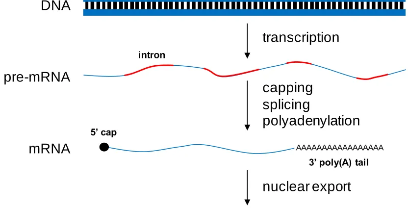
and nuclear export. In eukaryotes, a mature transcript is formed through a series of
coupled processing events (Figure 1.1). Initially, a pre-messenger RNA (pre-mRNA) is
transcribed from a gene by RNA polymerase II. The first processing step in the
production of a translationally-competent transcript is the addition of a 5‟ cap, which
contains a methylated guanine nucleotide44. The purpose of the cap is to protect the
transcript from degradation by 5‟ to 3‟ exonucleases and to stimulate the initiation of
translation45. The next processing step is the removal of introns, intervening noncoding
sequences found within the coding region, to form the mature transcript through a process
called splicing44. The final step before nuclear export to the cytoplasm is a 3‟ end
modification in which a poly(A) tail is added. The transcript is then exported to the
cytoplasm where it undergoes cytoplasmic decay or translation to produce protein
molecules. Cells control the level of proteins by regulating each one of these steps, from
the chromatin remodeling necessary for transcription of many genes to posttranslational
[image:20.612.119.535.472.684.2]protein stability.
Figure 1.1. Maturation of eukaryotic transcripts after transcription.
transcription
capping
splicing
polyadenylation
mRNA
DNA
pre-mRNA
AAAAAAAAAAAAAAAAA
intron
5’ cap
3’ poly(A) tail
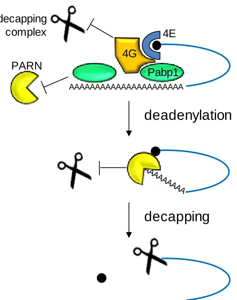
1.2.1. Deadenylation-dependent decapping pathway of transcript degradation
There are several mechanisms by which transcripts are degraded, including the
deadenylation-dependent decapping pathway, the deadenylation-independent decapping
pathway, and the endoribonucleolytic cleavage pathway. In S. cerevisiae, the most common degradation pathway is deadenylation-dependent decapping.
Figure 1.2. The deadenylation-dependent decapping pathway of transcript degradation in eukaryotes. 4E denotes eIF4E and 4G denotes eIF4G. Adapted from Wilusz et al. (2001)46.
Transcripts are present in a circular conformation due to the interaction of the
cap-binding protein, eukaryotic transcription factor (eIF)4E, on the 5‟ cap and the
poly(A) binding protein (Pab1p) on the 3‟ poly(A) tail mediated through binding to
eIF4G, a scaffolding protein (Figure 1.2). This circularization promotes translation and
prevents the activity of decapping and deadenylation enzymes46. The interaction of Pab1p
Pabp1
AAAAAAAAAAAAAAAAAAAAAA
4E
4G decapping
complex
PARN
deadenylation
[image:21.612.208.448.216.519.2]with the poly(A) tail inhibits deadenylation47. When the poly(A) ribonuclease (PARN)
binds to the 5‟ cap, it disrupts the cap‟s interaction with eIF4E causing a displacement of
Pab1p, which allows deadenylation to occur46, 48. Deadenylation is the rate-limiting step
in transcript decay49. While there are several different deadenylases that could be
functioning50, the predominant form in yeast is Ccr4p51. Once deadenylation is
completed, PARN no longer stays associated with the cap and the decapping complex of
Dcp1 and Dcp2 cleaves off the cap allowing a 5‟ to 3‟ exonuclease (Xrn1p) to rapidly
degrade the rest of the transcript46-47, 52. 3‟ to 5‟ exonucleolytic activity does occur after
deadenylation, but it tends to be slower than the activity of Xrn1p49, 53.
1.2.2. Cap-dependent translation initiation
Translation initiation begins when the 40S small ribosomal subunit associates
with two eIFs, eIF2 and eIF3, and the initiator methionine tRNA to form the 43S
preinitiation complex (Figure 1.3)54. eIF2 must also be bound by guanosine
5‟-triphosphate (GTP) in order for it to associate with the 40S ribosome. On the transcript,
the 5‟ cap is bound by a cap-binding protein complex, eIF4F, which consists of three
subunits: eIF4A, an RNA helicase; eIF4E, the actual cap-binding protein; and eIF4G, a
scaffolding protein55-56. The transcript is initially in a closed, circular form due to
eIF4G‟s interactions with both eIF4E at the 5‟ cap and Pab1p at the 3‟ poly(A) tail. The
43S complex binds eIF4F to form the 48S complex and scanning of the transcript begins
for the initiating AUG start codon57. The scanning by the complex for AUG is caused by
more initiation factors powered by ATP. Once the start codon is located, eIF1 and eIF1A
the hydrolysis of GTP on eIF2 followed by the association of the large 60S ribosomal
subunit with the 48S complex to form the complete 80S ribosome44, 56. The complex is
[image:23.612.114.541.161.380.2]then in a form where translation can initiate and proceeds to the elongation stage.
Figure 1.3. The mechanism of eukaryotic cap-dependent translation. 4A denotes eIF4A; 4E denotes eIF4E; and 4G denotes eIF4G. Adapted from Klann and Dever (2004)58.
1.3. Posttranscriptional regulation through transcript stability and
translation
1.3.1. Control of transcript decay
As discussed in Section 1.2.1, the deadenylation-dependent decapping pathway is
the primary mechanism by which eukaryotic transcripts are degraded. Briefly, transcripts
contain a 3‟ poly(A) tail whose interactions with proteins inhibit decapping. Once the tail
is removed, decapping proceeds and the transcript is degraded 5‟ to 3‟ by an exonuclease.
Eukaryotes have evolved additional non-coding elements in their genetic untranslated
regions (UTRs) and sometimes in the actual coding regions by which this pathway can be
43S association Pabp1
4E 4A
AAAAAAAAAAAAAAAAAAAAAA AUG
4G
AAAAAAAAAAAAAAAAAAAAAA AUG
scanning
AAAAAAAAAAAAAAAAAAAAAA AUG
60S association
AAAAAAAAAAAAAAAAAAAAAA
attenuated or bypassed52. These elements include deadenylation-independent decapping
elements, transcript stability elements, and elements conveying endonucleolytic activity.
In the deadenylation-independent decapping pathway, transcript degradation
proceeds without the removal of the 3‟ poly(A) tail. This pathway proceeds through the
recruitment of elements that enhance decapping. The RPS28B transcript in S. cerevisiae
contains a stem-loop structure within its 3‟ UTR and encodes for a protein, Rps28B, that
binds directly to that stem-loop59. The Rps28B protein product recruits a
decapping-enhancing protein, Edc3, and Edc3, in turn, recruits several other factors that lead to the
decapping of the transcript. The EDC1 transcript in S. cerevisiae encodes for a decapping-enhancing protein, Edc1, though it is unknown if Edc1 plays a role in EDC1
degradation52. EDC1 contains a stretch of uridine nucleotides that interacts with the poly(A) tail inhibiting deadenylation60. The decapping of the transcript is caused by
several protein factors including those associated with deadenylation.
Transcript stability elements are located at multiple positions on the transcript, but
primarily in the 3‟ UTR52
. The largest class of elements that has been examined is the
ARE in the 3‟ UTR. AREs are identified by a consensus AUUUA pentamer, but its
activity is dependent on the context and number of those pentamers52. AREs can
destabilize the transcript through the interaction of the sequence itself or ARE-binding
proteins with the transcript decay protein complex61-62. AREs can also stabilize
transcripts, where proposed mechanisms are based on competition with destabilizing
factors or inhibition of deadenylation-dependent decapping decay, such as through
strengthening the interaction between PABP and the poly(A) tail52. In addition, the PUF
Destabilization by PUF binding is due to recruitment of deadenylases63. As another
example, the proteins CP1 and CP2 are responsible for the stabilization of several
genes through interactions with pyrimidine-rich elements in the 3‟ UTR. It is believed
that the observed transcript stabilization is due to interactions with PABP that protect the
poly(A) tail from deadenylases64.
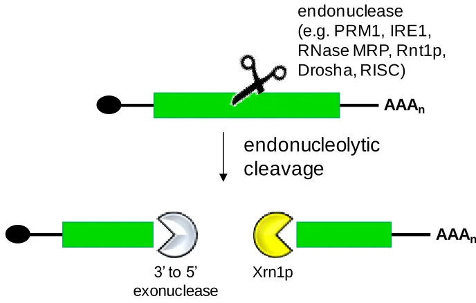
Endoribonucleolytic decay can also be described as deadenylation-independent
and decapping-independent decay. Internal cleavage of the transcript results in the
generation of two RNA fragments with unprotected ends (Figure 1.4)52. The 3‟ fragment
is susceptible to exonucleolytic decay by Xrn1p in the 5‟ to 3‟ direction, while the 5‟
fragment is degraded in the same manner once the cap is removed or by 3‟ to 5‟
exonucleases. In eukaryotes containing the RNA interference (RNAi) pathway, gene
expression is modulated through directed endonucleolytic cleavage, referred to as
“Slicer” activity, of the target transcript65
. Cleavage is mediated through components of
the RNA-induced silencing complex (RISC), which contains the RNase III enzyme
variant Dicer66-68. MicroRNAs (miRNAs) or small interfering RNAs (siRNAs) are loaded
onto RISC and direct the complex to the transcript through perfect or nearly perfect
(some mismatches allowed) base-pairing between the transcript and the miRNA/siRNA
69-71
. There are numerous endoribonucleases that regulate expression levels in eukaryotes,
although for many of the enzymes, such as PRM1, IRE1, and RNase MRP, cis-acting consensus binding regions have not yet been determined72-74. As an alternative to the
RNase III variant Rnt1p has been shown to cleave transcripts containing stem loop
structures76-77. Rnt1p specifically processes transcripts containing hairpins with AGNN
[image:26.612.145.489.164.383.2]tetraloops78 and is explained in further detail in Section 1.3.3.
Figure 1.4. The processing of eukaryotic transcripts following endonucleolytic cleavage is independent of the 5‟ cap and the 3‟ poly(A) tail. Scissors denote the endonuclease. Adapted from Garneau et al. (2007)52.
1.3.2. Control of the initiation of translation
As discussed in Section 1.2.2, the initiation of translation in eukaryotes is
mediated through protein interactions at the 5‟ cap between eIFs and the small ribosomal
subunit. The preinitiation complex scans the transcripts for the AUG start codon, where
the large ribosomal subunit binds and translation begins. The majority of translational
control is due to interference with the normal processes of the ribosome and the eIFs
(Figure 1.5)79. Cap-independent translation by IRESes is described in its own section
(1.3.4). The majority of these elements are located in the 5‟ UTR.
endonuclease (e.g. PRM1, IRE1, RNase MRP, Rnt1p, Drosha, RISC)
AAAn
endonucleolytic
cleavage
AAAn
Xrn1p 3’ to 5’
Figure 1.5. Genetic elements that affect the initation of translation in eukaryotes. The blue ovals in the 5‟ and 3‟ UTRs represent binding sites for protein factors that typically inhibit translation. Adapted from Gebauer and Hentze (2004)79.
Translational repression for the ferritin transcript is mediated through a stem-loop
structure called an iron-responsive element (IRE) located 40 nucleotides from the cap in
the 5‟ UTR80-81
. Iron regulatory proteins (IRPs) bind the IRE blocking the recruitment of
the preinitiation complex to the 5‟ cap due to steric hinderance82. Steric hinderance is also
observed with secondary and tertiary structures in the 5‟ UTR, such as RNA hairpins and
pseudoknots79. Transcripts containing a U-rich sequence known as a cytoplasmic
polyadenylational element (CPE) in the 3‟ UTR interact with the CPE-binding protein
(CPEB)83. CPEB represses translation by associating with another protein, Maskin, that
contains an eIF4E-binding domain, which inhibits eIF4E‟s interaction with eIF4G84.
Translational repression can also interfere with the ribosome after it has been bound to
the cap. The sex-lethal protein (Sxl) binds to U-rich sites on the msl-2 transcript located in both the 5‟ and 3‟ UTR85
. The binding of Sxl interferes with ribosomal scanning.
Another cap-independent method interferes with the association of the large ribosomal
submit in LOX3 transcripts86. Here, two proteins, hnRNP K and hnRNP E, bind a differentiation-control element (DICE), which is a repeated CU-rich element, in the 3‟
UTR and block formation of the 80S ribosome. In the previous section, we discussed
endonucleolytic cleavage mediated by the interaction of siRNA and miRNA to RISC.
AAAn
AUG hairpins, pseudoknots
uORFs
The miRNA silencing pathway is also known to repress translation through direct or
indirect interference with eIFs65, 87.
1.3.3. RNA processing by the RNase III enzyme Rnt1p
The RNase III family is a class of enzymes that cleaves double-stranded RNA
(dsRNA)88. Dicer is an RNase III enzyme in humans and other eukaryotes that cleaves
dsRNA into 21–23 nt fragments referred to as siRNAs that go on to induce gene silencing
though the RNAi pathway89. Drosha, another eukaryotic RNase III involved in the RNAi
pathway, is involved in the processing of miRNA from long dsRNA transcripts referred
to as primary (pri-)miRNA90. Rnt1p was discovered in S. cerevisiae due to similarities to the E. coli RNase III91 and has been shown to cleave cellular ribosomal RNA (rRNA) precursors, small nuclear RNAs (snRNAs), small nucleolar RNAs (snoRNAs), and
messenger RNA (mRNA)91-94. This protein is localized to the nucleus95 and contains two
domains: an RNase III domain and a dsRNA-binding domain (dsRBD)96.
The RNA hairpin substrates of Rnt1p contain a consensus AGNN tetraloop with a
cleavage site 14–16 base-pairs (bp) from the tetraloop78. The AGNN tetraloop forms a
predetermined fold that is recognized by the dsRBD88, 97. The dsRNA region of Rnt1p
substrates has an effect on the binding affinity and cleavage rate with this enzyme. The
base-pairs immediately below the tetraloop can impact Rnt1p binding, while sequences
near the cleavage site influence the cleavage rate98. These observations led to the
definition of three regions on Rnt1p substrates: the initial binding and positioning box
(IBPB) which consists of the tetraloop; the binding stability box (BSB) which is the
(CEB) which is the region containing and surrounding the cleavage site98. An unique
feature of Rnt1p is that it uses the tetraloop as its primary binding site, whereas for other
RNase III enzymes it is the RNA helix99.
Rnt1p is involved in the natural regulation of several genes in S. cerevisiae. Previously, transcripts had been discovered that undergo endonucleolytic cleavage but by
factors other than Rnt1p100-101. The RPS22B and RPL18A transcripts contain intronic Rnt1p substrates that deplete unspliced transcripts as well as reducing levels of the
mature transcripts94. The MIG2 transcript contains an Rnt1p substrate in its coding region that increases the transcript‟s sensitivity to glucose-dependent degradation77. Several
transcripts involved in iron uptake or iron mobilization contain Rnt1p substrates in the
coding region that help avoid cytotoxicity to the cellular iron starvation reponse102. The
diversity of structure and sequences in natural Rnt1p substrates, as well as the
identification of critical regions, support that a set of engineered Rnt1p hairpins can be
generated with differential activity.
1.3.4. Translation initiation mediated through internal ribosome entry sites
IRESes were initially discovered during the analysis of the 5‟ UTRs of
picornaviral transcripts where it was determined that the transcripts lacked a 5‟ cap and
translation continued in the absence of the cap-binding protein, eIF4F103. IRESes are
critical elements for the translation of the genome of several viruses, including the
Hepatitis A virus (HAV)104, the Hepatitis C virus105, the foot-and-mouth-disease virus
(FMDV)106, and the human immunodeficiency virus (HIV)107. Viral IRESes contain a
ribosomal machinery or mimic the interaction of such components with the 5‟ cap and
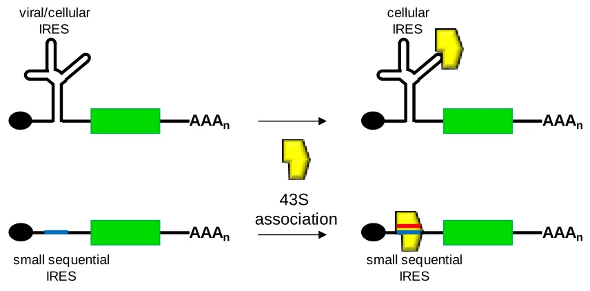
other protein factors (Figure 1.6)108-113. IRESes illustrate the scaffolding power of RNA
structures and the creative mechanisms by which viruses have evolved to essentially
hijack the host-based expression machinery.
Figure 1.6. Simplified schematic of the interactions with structural and sequential IRESes with the translational machinery. Red line denotes the sequences on the 18S rRNA complementary to the IRES sequence (blue line).
Cellular IRESes were first discovered when researchers observed that
immunoglobulin heavy chain binding protein (BiP) continued being expressed after
cap-dependent translation had been shut down due to poliovirus infection109. Many cellular
IRESes characterized thus far contain a Y-shaped stem-loop structure upstream of the
initiation codon; however, the activity of these IRESes may not necessarily depend on
that secondary structure114-115. In a mouse cell line, deletional studies of the structured 5‟
UTR of the Gtx protein, which demonstrated IRES activity, identified a 9 nucleotide (nt)
module that retained the ability to internally initiate translation114. When multiple
modules of the 9-nt module were placed in tandem, a synergistic effect was observed as
AAAn
viral/cellular IRES
AAAn
small sequential IRES
AAAn
cellular IRES
AAAn
small sequential IRES
[image:30.612.107.535.189.400.2]overall IRES activity increased. This 9-nt segment was determined to be completely
complementary to a segment of the 18S ribosomal RNA, a critical component of the
ribosomal machinery (Figure 1.6)116. In S. cerevisiae, two IRES-containing 5‟ UTRs for the YAP1 and p150 genes were also found to contain several regions of complementarity to 18S rRNA39. These studies have demonstrated that cellular IRESes contain regions
that directly base-pair to the 18S rRNA. This mechanism of translation initiation by
cellular IRESes in eukaryotes suggest that their function is analogous to Shine-Dalgarno
sequences in prokaryotes, which initiate translation through direct base-pairing with the
prokaryotic analogue of the 18S rRNA, the 16S rRNA117. Based on this observation, a
short segment of nucleotides in the intercistronic region (IR) of a yeast and mammalian
dicistronic vector were randomized and screened for IRES activity by expression of the
second cistron118-119. The resultant IRESes demonstrated complementarity to the 18S
rRNA.
Prokaryotic genes are typically expressed from operons, where multiple coding
regions are located on one transcript under the control of a single promoter. Each coding
region contains a Shine-Dalgarno sequence upstream of its start codon in order to initiate
translation of each gene. Viruses are also known to produce multicistronic transcripts or
genomes. For example, the entire positive-strand genome of HCV is contained on a
single piece of RNA120. The entire genome is translated through an IRES at the 5‟ end.
The resultant polyprotein is then processed by a series of proteases and peptidases to
create each individual protein product. HIV translation is similar to HCV except that its
IRES can also cause translation initation at multiple start codons resulting in different
can be generated through the introduction of an IRES element before each gene.
Retroviral mulitcistronic vectors had been developed in mammalian systems where
multiple viral IRESes were incorporated122-123. Recently, a dicistronic reporter construct
had been characterized in S. cerevisiae where the YAP1 and p150 IRES were placed in the IR to alter the ratio of expression between two genes124. Since only the p150 IRES worked in this system, the work highlights the need for additional IRESes to be
discovered or engineered in yeast to increase the ability to tune gene expression through
this method.
1.4. Interrelationship among the thesis projects
Chapter I provides an overview of the field of synthetic biology and metabolic
engineering and gives an in-depth examination of the cellular processes of transcript
translation initiation and decay and the RNA elements that control these processes.
Chapter II describes the development of a library of RNA hairpins that regulate
posttranscriptional decay to attenuate gene expression due to the endonucleolytic
processing of the hairpins by the S. cerevisiae RNase III Rnt1p. The library is based on the randomization of nucleotides associated with controlling the cleavage rate by the
enzyme. Chapter III describes a second library of Rnt1p-cleaved hairpins based on the
randomization of nucleotides associated with the binding of Rnt1p to the hairpin. In
addition, the two library elements are integrated combinatorially to extend the accessible
levels of gene expression. Chapter IV describes the integration of the small
molecule-responsive aptamers into Rnt1p substrates to achieve ligand-controlled cleavage. The
binding in proximity of the cleavage sites and switching dynamics are altered through
incorporation of additional aptamers and Rnt1p-based modules, as well as the
construction of multiple switch devices in tandem. Chapter V describes a strategy to
select for a library of small sequential IRES elements with various strengths to initiate
translation at physiological conditions. These elements will aid in the development of
yeast „operons‟ or multicistronic vectors where relative gene expression levels can be
controlled. These research projects collectively demonstrate the capacity of utilizing
RNA-based control elements to predictably tune gene expression levels in S. cerevisiae.
References
1. Chen, Y.Y., Jensen, M.C. & Smolke, C.D. Genetic control of mammalian T-cell
proliferation with synthetic RNA regulatory systems. Proc Natl Acad Sci U S A
107, 8531-8536 (2010).
2. Culler, S.J., Hoff, K.G. & Smolke, C.D. Reprogramming cellular behavior with
RNA controllers responsive to endogenous proteins. Science 330, 1251-1255 (2010).
3. Lee, S.K., Chou, H., Ham, T.S., Lee, T.S. & Keasling, J.D. Metabolic engineering
of microorganisms for biofuels production: from bugs to synthetic biology to
fuels. Curr Opin Biotechnol19, 556-563 (2008).
4. Lee, S.Y., Kim, H.U., Park, J.H., Park, J.M. & Kim, T.Y. Metabolic engineering
of microorganisms: general strategies and drug production. Drug Discov Today
5. Andrianantoandro, E., Basu, S., Karig, D.K. & Weiss, R. Synthetic biology: new
engineering rules for an emerging discipline. Mol Syst Biol2, 2006 0028 (2006). 6. Elowitz, M.B. & Leibler, S. A synthetic oscillatory network of transcriptional
regulators. Nature403, 335-338 (2000).
7. Gardner, T.S., Cantor, C.R. & Collins, J.J. Construction of a genetic toggle switch
in Escherichia coli. Nature403, 339-342 (2000).
8. Basu, S., Mehreja, R., Thiberge, S., Chen, M.T. & Weiss, R. Spatiotemporal
control of gene expression with pulse-generating networks. Proc Natl Acad Sci U S A101, 6355-6360 (2004).
9. Khalil, A.S. & Collins, J.J. Synthetic biology: applications come of age. Nat Rev Genet11, 367-379 (2010).
10. Smolke, C., Martin, V. & Keasling, J. in Protein expression technologies: current
status and future trends. (ed. F. Baneyx) (Horizon Bioscience, Wymondham,
Norfolk; 2004).
11. Pitera, D.J., Paddon, C.J., Newman, J.D. & Keasling, J.D. Balancing a
heterologous mevalonate pathway for improved isoprenoid production in
Escherichia coli. Metab Eng9, 193-207 (2007).
12. Pfleger, B.F., Pitera, D.J., Smolke, C.D. & Keasling, J.D. Combinatorial
engineering of intergenic regions in operons tunes expression of multiple genes.
Nat Biotechnol24, 1027-1032 (2006).
13. Peralta-Yahya, P.P. & Keasling, J.D. Advanced biofuel production in microbes.
14. Hughes, E.H. & Shanks, J.V. Metabolic engineering of plants for alkaloid
production. Metab Eng4, 41-48 (2002).
15. Smolke, C.D., Martin, V.J. & Keasling, J.D. in Protein Expression Technologies:
Current Status and Future Trends. (ed. F. Baneyx) (Horizon Scientific Press,
U.K.; 2004).
16. Jin, Y.S., Ni, H., Laplaza, J.M. & Jeffries, T.W. Optimal growth and ethanol
production from xylose by recombinant Saccharomyces cerevisiae require
moderate D-xylulokinase activity. Appl Environ Microbiol69, 495-503 (2003). 17. Jones, K.L., Kim, S.W. & Keasling, J.D. Low-copy plasmids can perform as well
as or better than high-copy plasmids for metabolic engineering of bacteria. Metab Eng2, 328-338 (2000).
18. Zhu, M.M., Lawman, P.D. & Cameron, D.C. Improving 1,3-propanediol
production from glycerol in a metabolically engineered Escherichia coli by
reducing accumulation of sn-glycerol-3-phosphate. Biotechnol Prog 18, 694-699 (2002).
19. Zhu, M.M., Skraly, F.A. & Cameron, D.C. Accumulation of methylglyoxal in
anaerobically grown Escherichia coli and its detoxification by expression of the
Pseudomonas putida glyoxalase I gene. Metab Eng3, 218-225 (2001).
20. Alper, H., Jin, Y.S., Moxley, J.F. & Stephanopoulos, G. Identifying gene targets
21. Alper, H., Miyaoku, K. & Stephanopoulos, G. Construction of
lycopene-overproducing E. coli strains by combining systematic and combinatorial gene
knockout targets. Nat Biotechnol23, 612-616 (2005).
22. Paradise, E.M., Kirby, J., Chan, R. & Keasling, J.D. Redirection of flux through
the FPP branch-point in Saccharomyces cerevisiae by down-regulating squalene
synthase. Biotechnol Bioeng100, 371-378 (2008).
23. Dueber, J.E. et al. Synthetic protein scaffolds provide modular control over
metabolic flux. Nat Biotechnol27, 753-759 (2009).
24. Alper, H., Fischer, C., Nevoigt, E. & Stephanopoulos, G. Tuning genetic control
through promoter engineering. Proc Natl Acad Sci U S A 102, 12678-12683 (2005).
25. Morant, M., Bak, S., Moller, B.L. & Werck-Reichhart, D. Plant cytochromes
P450: tools for pharmacology, plant protection and phytoremediation. Curr Opin Biotechnol14, 151-162 (2003).
26. Veen, M. & Lang, C. Production of lipid compounds in the yeast Saccharomyces
cerevisiae. Appl Microbiol Biotechnol63, 635-646 (2004).
27. Hughes, E.H., Hong, S.B., Gibson, S.I., Shanks, J.V. & San, K.Y. Metabolic
engineering of the indole pathway in Catharanthus roseus hairy roots and
increased accumulation of tryptamine and serpentine. Metab Eng 6, 268-276 (2004).
28. Chemler, J.A. & Koffas, M.A. Metabolic engineering for plant natural product
29. Silva, G.L., Lee, I. & Kinghorn, A.D. in Natural Products Isolation. (ed. R.J.P.
Cannell) (Humana Press, Totowa, New Jersey; 1998).
30. Alper, H. & Stephanopoulos, G. Engineering for biofuels: exploiting innate
microbial capacity or importing biosynthetic potential? Nat Rev Microbiol7, 715-723 (2009).
31. Cereghino, G.P. & Cregg, J.M. Applications of yeast in biotechnology: protein
production and genetic analysis. Curr Opin Biotechnol10, 422-427 (1999).
32. Gerngross, T.U. Advances in the production of human therapeutic proteins in
yeasts and filamentous fungi. Nat Biotechnol22, 1409-1414 (2004).
33. Nevoigt, E. Progress in metabolic engineering of Saccharomyces cerevisiae.
Microbiol Mol Biol Rev72, 379-412 (2008).
34. Hawkins, K.M. & Smolke, C.D. The regulatory roles of the galactose permease
and kinase in the induction response of the GAL network in Saccharomyces
cerevisiae. J Biol Chem281, 13485-13492 (2006).
35. Nevoigt, E. et al. Engineering promoter regulation. Biotechnol Bioeng 96, 550-558 (2007).
36. Nevoigt, E. et al. Engineering of promoter replacement cassettes for fine-tuning of
gene expression in Saccharomyces cerevisiae. Appl Environ Microbiol 72, 5266-5273 (2006).
37. Mumberg, D., Muller, R. & Funk, M. Yeast vectors for the controlled expression
38. Jeppsson, M., Johansson, B., Jensen, P.R., Hahn-Hagerdal, B. &
Gorwa-Grauslund, M.F. The level of glucose-6-phosphate dehydrogenase activity
strongly influences xylose fermentation and inhibitor sensitivity in recombinant
Saccharomyces cerevisiae strains. Yeast20, 1263-1272 (2003).
39. Zhou, W., Edelman, G.M. & Mauro, V.P. Transcript leader regions of two
Saccharomyces cerevisiae mRNAs contain internal ribosome entry sites that
function in living cells. Proc Natl Acad Sci U S A98, 1531-1536 (2001).
40. Vasudevan, S. & Peltz, S.W. Regulated ARE-mediated mRNA decay in
Saccharomyces cerevisiae. Mol Cell7, 1191-1200 (2001).
41. Lautz, T., Stahl, U. & Lang, C. The human c-fos and TNFalpha AU-rich elements
show different effects on mRNA abundance and protein expression depending on
the reporter in the yeast Pichia pastoris. Yeast27, 1-9 (2010).
42. Bayer, T.S. & Smolke, C.D. Programmable ligand-controlled riboregulators of
eukaryotic gene expression. Nat Biotechnol23, 337-343 (2005).
43. Win, M.N. & Smolke, C.D. A modular and extensible RNA-based
gene-regulatory platform for engineering cellular function. Proc Natl Acad Sci U S A
104, 14283-14288 (2007).
44. Alberts, B. Molecular biology of the cell, Edn. 4th. (Garland Science, New York;
2002).
45. Day, D.A. & Tuite, M.F. Post-transcriptional gene regulatory mechanisms in
eukaryotes: an overview. J Endocrinol157, 361-371 (1998).
46. Wilusz, C.J., Wormington, M. & Peltz, S.W. The cap-to-tail guide to mRNA
47. Tucker, M. & Parker, R. Mechanisms and control of mRNA decapping in
Saccharomyces cerevisiae. Annu Rev Biochem69, 571-595 (2000).
48. Dehlin, E., Wormington, M., Korner, C.G. & Wahle, E. Cap-dependent
deadenylation of mRNA. Embo J19, 1079-1086 (2000).
49. Jacobson, A. & Peltz, S.W. Interrelationships of the pathways of mRNA decay
and translation in eukaryotic cells. Annu Rev Biochem65, 693-739 (1996).
50. Mitchell, P. & Tollervey, D. mRNA stability in eukaryotes. Curr Opin Genet Dev
10, 193-198 (2000).
51. Tucker, M. et al. The transcription factor associated Ccr4 and Caf1 proteins are
components of the major cytoplasmic mRNA deadenylase in Saccharomyces
cerevisiae. Cell104, 377-386 (2001).
52. Garneau, N.L., Wilusz, J. & Wilusz, C.J. The highways and byways of mRNA
decay. Nat Rev Mol Cell Biol8, 113-126 (2007).
53. Caponigro, G. & Parker, R. Mechanisms and control of mRNA turnover in
Saccharomyces cerevisiae. Microbiol Rev60, 233-249 (1996).
54. Huang, H. & Donahue, T.F. in mRNA Metabolism and Post-transcriptional Gene
Regulation. (eds. J.B. Harford & D.R. Morris) (Wiley-Liss, New York; 1997).
55. Richter, J.D. & Sonenberg, N. Regulation of cap-dependent translation by eIF4E
inhibitory proteins. Nature433, 477-480 (2005).
56. Weaver, R. Molecular biology, Edn. 2nd. (McGraw-Hill, New York; 2005).
58. Klann, E. & Dever, T.E. Biochemical mechanisms for translational regulation in
synaptic plasticity. Nat Rev Neurosci5, 931-942 (2004).
59. Badis, G., Saveanu, C., Fromont-Racine, M. & Jacquier, A. Targeted mRNA
degradation by deadenylation-independent decapping. Mol Cell15, 5-15 (2004). 60. Muhlrad, D. & Parker, R. The yeast EDC1 mRNA undergoes
deadenylation-independent decapping stimulated by Not2p, Not4p, and Not5p. EMBO J 24, 1033-1045 (2005).
61. Anderson, J.R. et al. Sequence-specific RNA binding mediated by the RNase PH
domain of components of the exosome. RNA12, 1810-1816 (2006).
62. Chen, C.Y. et al. AU binding proteins recruit the exosome to degrade
ARE-containing mRNAs. Cell107, 451-464 (2001).
63. Goldstrohm, A.C., Hook, B.A., Seay, D.J. & Wickens, M. PUF proteins bind
Pop2p to regulate messenger RNAs. Nat Struct Mol Biol13, 533-539 (2006). 64. Wang, Z., Day, N., Trifillis, P. & Kiledjian, M. An mRNA stability complex
functions with poly(A)-binding protein to stabilize mRNA in vitro. Mol Cell Biol
19, 4552-4560 (1999).
65. Valencia-Sanchez, M.A., Liu, J., Hannon, G.J. & Parker, R. Control of translation
and mRNA degradation by miRNAs and siRNAs. Genes Dev20, 515-524 (2006). 66. Liu, J. et al. Argonaute2 is the catalytic engine of mammalian RNAi. Science305,
1437-1441 (2004).
67. Okamura, K., Ishizuka, A., Siomi, H. & Siomi, M.C. Distinct roles for Argonaute
68. Song, J.J., Smith, S.K., Hannon, G.J. & Joshua-Tor, L. Crystal structure of
Argonaute and its implications for RISC slicer activity. Science 305, 1434-1437 (2004).
69. Guo, H.S., Xie, Q., Fei, J.F. & Chua, N.H. MicroRNA directs mRNA cleavage of
the transcription factor NAC1 to downregulate auxin signals for arabidopsis
lateral root development. Plant Cell17, 1376-1386 (2005).
70. Mallory, A.C. et al. MicroRNA control of PHABULOSA in leaf development:
importance of pairing to the microRNA 5' region. EMBO J23, 3356-3364 (2004). 71. Yekta, S., Shih, I.H. & Bartel, D.P. MicroRNA-directed cleavage of HOXB8
mRNA. Science304, 594-596 (2004).
72. Gill, T., Cai, T., Aulds, J., Wierzbicki, S. & Schmitt, M.E. RNase MRP cleaves
the CLB2 mRNA to promote cell cycle progression: novel method of mRNA
degradation. Mol Cell Biol24, 945-953 (2004).
73. Hollien, J. & Weissman, J.S. Decay of endoplasmic reticulum-localized mRNAs
during the unfolded protein response. Science313, 104-107 (2006).
74. Yang, F. & Schoenberg, D.R. Endonuclease-mediated mRNA decay involves the
selective targeting of PMR1 to polyribosome-bound substrate mRNA. Mol Cell
14, 435-445 (2004).
75. Serganov, A. & Patel, D.J. Ribozymes, riboswitches and beyond: regulation of
gene expression without proteins. Nat Rev Genet8, 776-790 (2007).
76. Gregory, R.I., Chendrimada, T.P. & Shiekhattar, R. MicroRNA biogenesis:
isolation and characterization of the microprocessor complex. Methods Mol Biol
77. Ge, D., Lamontagne, B. & Elela, S.A. RNase III-mediated silencing of a
glucose-dependent repressor in yeast. Curr Biol15, 140-145 (2005).
78. Chanfreau, G., Buckle, M. & Jacquier, A. Recognition of a conserved class of
RNA tetraloops by Saccharomyces cerevisiae RNase III. Proc Natl Acad Sci U S A97, 3142-3147 (2000).
79. Gebauer, F. & Hentze, M.W. Molecular mechanisms of translational control. Nat Rev Mol Cell Biol5, 827-835 (2004).
80. Gray, N.K. & Hentze, M.W. Iron regulatory protein prevents binding of the 43S
translation pre-initiation complex to ferritin and eALAS mRNAs. EMBO J 13, 3882-3891 (1994).
81. Muckenthaler, M., Gray, N.K. & Hentze, M.W. IRP-1 binding to ferritin mRNA
prevents the recruitment of the small ribosomal subunit by the cap-binding
complex eIF4F. Mol Cell2, 383-388 (1998).
82. Stripecke, R., Oliveira, C.C., McCarthy, J.E. & Hentze, M.W. Proteins binding to
5' untranslated region sites: a general mechanism for translational regulation of
mRNAs in human and yeast cells. Mol Cell Biol14, 5898-5909 (1994).
83. Mendez, R. & Richter, J.D. Translational control by CPEB: a means to the end.
Nat Rev Mol Cell Biol2, 521-529 (2001).
84. Stebbins-Boaz, B., Cao, Q., de Moor, C.H., Mendez, R. & Richter, J.D. Maskin is
85. Gebauer, F., Grskovic, M. & Hentze, M.W. Drosophila sex-lethal inhibits the
stable association of the 40S ribosomal subunit with msl-2 mRNA. Mol Cell 11, 1397-1404 (2003).
86. Ostareck, D.H., Ostareck-Lederer, A., Shatsky, I.N. & Hentze, M.W.
Lipoxygenase mRNA silencing in erythroid differentiation: The 3'UTR regulatory
complex controls 60S ribosomal subunit joining. Cell104, 281-290 (2001).
87. Humphreys, D.T., Westman, B.J., Martin, D.I. & Preiss, T. MicroRNAs control
translation initiation by inhibiting eukaryotic initiation factor 4E/cap and poly(A)
tail function. Proc Natl Acad Sci U S A102, 16961-16966 (2005).
88. Wu, H. et al. A novel family of RNA tetraloop structure forms the recognition site
for Saccharomyces cerevisiae RNase III. Embo J20, 7240-7249 (2001).
89. Lingel, A. & Izaurralde, E. RNAi: finding the elusive endonuclease. Rna 10, 1675-1679 (2004).
90. Lee, Y., Jeon, K., Lee, J.T., Kim, S. & Kim, V.N. MicroRNA maturation:
stepwise processing and subcellular localization. EMBO J21, 4663-4670 (2002). 91. Elela, S.A., Igel, H. & Ares, M., Jr. RNase III cleaves eukaryotic preribosomal
RNA at a U3 snoRNP-dependent site. Cell85, 115-124 (1996).
92. Chanfreau, G., Elela, S.A., Ares, M., Jr. & Guthrie, C. Alternative 3'-end
processing of U5 snRNA by RNase III. Genes Dev11, 2741-2751 (1997).
93. Chanfreau, G., Rotondo, G., Legrain, P. & Jacquier, A. Processing of a dicistronic