T E C H N I C A L N O T E
Open Access
Comparative analysis of four methods to extract
DNA from paraffin-embedded tissues: effect on
downstream molecular applications
Cornelis JJ Huijsmans
1*, Jan Damen
2, Johannes C van der Linden
2, Paul HM Savelkoul
3, Mirjam HA Hermans
1Abstract
Background:A large portion of tissues stored worldwide for diagnostic purposes is formalin-fixed and paraffin-embedded (FFPE). These FFPE-archived tissues are an extremely valuable source for retrospective (genetic) studies. These include mutation screening in cancer-critical genes as well as pathogen detection. In this study we
evaluated the impact of several widely used DNA extraction methods on the quality of molecular diagnostics on FFPE tissues.
Findings:We compared 4 DNA extraction methods from 4 identically processed FFPE mammary-, prostate-,
colon-and lung tissues with regard to PCR inhibition, real time SNP detection colon-and amplifiable fragment size. The
extraction methods, with and without proteinase K pre-treatment, tested were: 1) heat-treatment, 2) QIAamp DNA-blood-mini-kit, 3) EasyMAG NucliSens and 4) Gentra Capture-Column-kit.
Amplifiable DNA fragment size was assessed by multiplexed 200-400-600 bp PCR and appeared highly influenced by the extraction method used. Proteinase K pre-treatment was a prerequisite for proper purification of DNA from FFPE. Extractions with QIAamp, EasyMAG and heat-treatment were found suitable for amplification of fragments up to 400 bp from all tissues, 600 bp amplification was marginally successful (best was QIAamp). QIAamp and Easy-MAG extracts were found suitable for downstream real time SNP detection. Gentra extraction was unsuitable. Hands-on time was lowest for heat-treatment, followed by EasyMAG.
Conclusions:We conclude that the extraction method plays an important role with regard to performance in downstream molecular applications.
Findings
Due to the tremendous progress in molecular pathology during the last decade, molecular techniques are moving rapidly from research to routine use in diagnostic pathology. At present, routine tests include for example the detection of bacteria and viruses [1-7], neoplasm-associated mutations [8-10], microsatellite instability [11,12] and up- and down-regulation of mRNA’s [13]. Archives of formalin-fixed paraffin-embedded (FFPE) tissues -that are often maintained over decades- repre-sent an extraordinary source of morphologically well defined tissues that now allow retrospective studies to
correlate molecular findings with therapy and clinical outcome [14].
However, the application of molecular DNA-based techniques to FFPE tissues suffers from challenges. For-malin fixation, the most widely used fixative in histo-pathology, has many advantages such as the ease of tissue handling, the possibility of long-term storage, an optimal histological quality and its availability in large quantities at low price [15,16]. Unfortunately formalin fixation induces DNA-tissue protein cross-links, which can prevent amplification. In addition, nucleic acid frag-mentation may occur in formalin fixed tissue due to aging of the specimen or the pH of the fixative [17]. Previous studies have shown that DNA extraction and subsequent downstream processes such as PCR from FFPE tissues is difficult, especially when longer stretches of DNA templates are targeted [18]. It has been * Correspondence: [email protected]
1
Laboratory of Molecular Diagnostics, Jeroen Bosch Hospital,
‘s-Hertogenbosch, The Netherlands
Full list of author information is available at the end of the article
reported that DNA fragments of up to only 100-300 bp are obtained from FFPE tissues [19].
To recover nucleic acids from non-fixed tissues we routinely combine proteinase K (prot. K) digestion with commercial extraction methods. The aim of this study was to test whether DNA isolation techniques routinely available in molecular diagnostic laboratories can be applied to FFPE tissues. In addition, we think it is handy to employ DNA extraction kits that are suitable for the extraction of DNA from a wide variety of patient mate-rials, e.g. blood, buccal swabs and FFPE tissues. We therefore compared four different extraction protocols with and without prot. K digestion, and evaluated the impact of these DNA isolation methods on downstream molecular techniques. The extractions tested were: 1) heat-treatment, 2) QIAamp DNA-blood-mini-kit extrac-tion, 3) EasyMAG NucliSens extraction and 4) Gentra Capture-Column-kit extraction.
Experiments were carried out regarding i) the inhibi-tion of PCR by monitoring amplificainhibi-tion of an internal control DNA virus, ii) the performance of the isolated DNA in SNP analysis by real time PCR and iii) perfor-mance in a conventional multiplex PCR amplifying 200, 400 and 600 bp human DNA fragments. Studies com-paring the suitability of different DNA extraction meth-ods such as (modified) phenol-chloroform extraction, boiling, microwave and QIAamp DNA-blood-mini-kit extraction have been published [20-24]. However these studies did not include a comparison of the in this study described commercial methods, which are routi-nely used by hospital laboratories when performing molecular diagnostics.
The data presented here indicate that proteinase K digestion is required for obtaining DNA of sufficient quality by all 4 extraction methods. The size of the amplifiable DNA fragments highly depended on the extraction method. QIAamp extraction and heat-treat-ment in combination with proteinase K digestion resulted in amplification of the longest DNA fragments - up to 600 bp. Amplification inhibitors were found in all Gentra extracts and in one colon tissue extract after prot. K digestion and heat-treatment. EasyMAG Nucli-Sens extraction and the QIAamp method seemed to be equally effective in extracting 200 bp fragments, and therefore suitable for real time SNP detection. An advantage of heat-treatment and the EasyMAG Nucli-Sens extraction was their lower hands-on time.
Methods
Experimental set-up
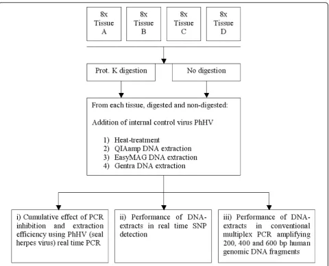
The workflow of the presented study is described in figure 1. Four tissues (A-D) were subjected to proteinase K digestion and no proteinase K digestion. Subse-quently, 4 different extraction methods were performed:
1) heat-treatment, 2) QIAamp DNA-blood-mini-kit extraction, 3) EasyMAG NucliSens extraction and 4) Gentra Capture-Column-kit extraction. All extracts were tested regarding: i) cumulative effect of PCR inhibition and extraction efficiency, ii) performance in real time SNP detection and iii) Performance in conventional multiplex PCR amplifying 200, 400 and 600 bp human genomic DNA fragments.
Tissue processing
Four randomly chosen biopsies, taken for diverse clinical purposes, from different patients and organs were used for this study: A. mammary, B. colon, C. prostate and D. lung. The tissues were rendered anonymous before use in this study and were formalin-fixed and paraffin-embedded 1-4 weeks ago. Tissues were fixed in 0.01 mol/L buffered (0.005 mol/L disodium hydrogen phos-phate anhydrous and 0.005 mol/L sodium dihydrogen phosphate dihydrate, pH 7.0) 10% formalin, and pro-cessed for paraffin embedding using a Tissue-Tek VIP 5 (Sakura, Torrance, USA). The dehydration program con-sisted of 14 steps of 1 hour under continuous agitation, pressure, vacuum, and heating. At 40°C, two 10% forma-lin steps were followed by one 70% (v/v) ethanol step, two 96% ethanol steps, three 100% ethanol steps, and two 100% xylene steps. Paraffin embedding was done at 60°C in four 100% paraffin steps.
DNA extractions
The four DNA isolation procedures were: heat-treat-ment, QIAamp DNA-blood-mini-kit extraction (silica membrane-based column extraction; Qiagen, Hilden, Germany), EasyMAG NucliSens extraction (magnetic silica beads-based extraction; Biomerieux, Boxtel, The Netherlands) and Gentra Capture-Column-kit extraction (silica membrane-based column extraction; Gentra Sys-tems Inc, Minneapolis, USA).
Paraffin-embedded tissues were trimmed of paraffin excess and cut into 3-μM-thick sections. Approximately 1 to 1.5 cm2 of sectioned tissue (a single section or short ribbons depending on the surface per section) was put in 250μL of digestion solution (digestion solution with proteinase K was prepared by adding 100 μL of proteinase K solution (20 mg/mL; Roche Diagnostics GmbH, Mannheim, Germany) and 10μL of Tween 20 (Merck BV, Amsterdam, The Netherlands) to 2 mL of TE buffer (1 mmol/L ethylenediaminetetraacetic acid, and 10 mmol/L Tris-HCl buffer, pH 8.0)) and incubated overnight at 45°C. Proteinase K was inactivated the next day by incubation at 100°C for 15-30 minutes. After-wards, samples were centrifuged for 2 minutes at 14,000 rpm.
From each material eight paraffin sections were cut, 4 were digested in digestion solution with proteinase K Huijsmanset al.BMC Research Notes2010,3:239
http://www.biomedcentral.com/1756-0500/3/239
(according to the method described above) and 4 were submerged in digestion solution without proteinase K. All 32 samples were further processed as described above. To assure an equal quantity of DNA in each pro-cedure, the supernatants -located beneath the paraffin cap- from the 4 proteinase K digested samples as well as the 4 non-digested samples were pooled for each mate-rial. These pools were homogenized and processed as detailed below.
Heat treatment
Two-hundredμL of pool was mixed with 10μL of Pho-cine herpes virus (PhHV, seal herpes virus, kindly pro-vided by the Erasmus Medical Centre in Rotterdam, The Netherlands), which served as an internal extraction control, and was used directly in the downstream applications.
QIAamp DNA-blood-mini-kit extraction
Two-hundredμL pool and 10 μL of PhHV were added to 200μL AL buffer, homogenized and incubated for
10 min. at room temperature. Two-hundredμL of 96% ethanol (Merck KgaG, Darmstadt, Germany) was added. The mixture was transferred to a QIAamp column and centrifuged for 1 min. at 8,000 rcf. The column was put in a new collection tube, 500μL AW1 buffer was added and centrifuged for 1 min. at 8,000 rcf. This procedure was repeated with 500 μL AW2 buffer and the column was centrifuged for 1 min. at 14,000 rcf. To remove all ethanol from the column it was put in a new collection tube and then subjected to a dry spin for 1 min. at 14,000 rcf. Elution was performed by adding 200μL EL buffer, incubating for 5 min. at room temperature followed by centrifugation for 1 min. at 8,000 rcf. EasyMAG NucliSens extraction
[image:3.595.59.540.86.475.2]water) was subsequently added. The DNA was extracted on the EasyMAG machine using the “Generic 2.0.1” program. Elution was performed in 200μL NucliSens Extraction buffer 3.
Gentra Capture-Column-kit extraction
Two-hundredμL pool and 10μL of PhHV were directly added to the column and incubated for 5 min. at room temperature. Four-hundredμL of Purification Solution 1 was added followed by 5 min. incubation at room tem-perature and a 15 sec. centrifugation step at 8,000 rcf. This step was performed twice. Subsequently 200μL of Elution Solution 2 was added followed by a 15 sec. cen-trifugation step at 8,000 rcf. To elute the purified DNA, 200 μL of Elution Solution 2 was added to the column and incubated for 10 min. at 100°C. Collection of the DNA was performed by centrifugation for 25 sec. at 8,000 rcf.
Internal control amplification
A PhHV specific real time PCR was performed. Twenty-fiveμL of PCR, using a homebrew“JBZ” 4× mastermix, contained 20 mmol/L Tris-HCl, pH 8.4, 50 mmol/L KCl, 3 mmol/L MgCl2 (prepared from 10× PCR buffer and 50 mmol/L MgCl2solution delivered with Platinum Taq polymerase), 0.75 U of Platinum Taq polymerase (Invitrogen BV, Breda, The Netherlands), 4% glycerol (molecular biology grade; Calbiochem, VWR Interna-tional BV, Amsterdam, The Netherlands), 200 μmol/L of each dNTP (Invitrogen BV), 0.5 μL of Rox reference dye (Invitrogen BV), 300 nM of PhHV forward primer 5′-GGG CGA ATC ACA GAT TGA ATC-3′, 300 nM of PhHV reverse primer 5′-GCG GTT CCA AAC GTA CCA A-3′, 100 nM PhHV TaqMan probe 5′-FAM-TTT TTA TGT GTC CGC CAC CAT CTG GAT C-TAMRA-3′ and 10 μL extracted DNA [25]. Real time PCR was performed in an ABI Prism 7000 SDS (Applied Biosystems (ABI), Foster City CA, USA) for 2 minutes at 50°C, 10 minutes at 95°C, followed by 45 cycles of 15 seconds at 95°C and 1 minute at 60°C.
SNP analysis using real time PCR
Predesigned TaqMan Assays-on-Demand SNP genotyping products rs2043731 and rs1350138 (ABI) were used according to the manufacturer’s instructions. Two master-mixes were tested: the homebrew JBZ 4× mastermix and the commercial ABI 2× TaqMan Universal PCR master-mix. Twenty-fiveμL of PCR, using the JBZ 4× mastermix contained, 20 mmol/L Tris-HCl, pH 8.4, 50 mmol/L KCl, 3 mmol/L MgCl2(prepared from 10× PCR buffer and 50 mmol/L MgCl2 solution delivered with Platinum Taq polymerase), 0.75 U of Platinum Taq polymerase (Invitro-gen BV, Breda, The Netherlands), 4% glycerol (molecular biology grade; Calbiochem, VWR International BV, Amsterdam, The Netherlands), 200μmol/L of each dNTP
(Invitrogen BV), 0.5μL of Rox reference dye (Invitrogen BV), 1.25 μL of predeveloped assay reagent from the Assays-on-Demand SNP genotyping products (ABI) con-taining two primers and two MGB TaqMan probes (5′ VIC for allele 1, 5′FAM for allele 2 and a 3′black hole quencher for both alleles), and 11.25μL of target DNA. Twenty-fiveμL of PCR, using the commercial mastermix, contained 12.5μL of 2× TaqMan Universal PCR Master-mix (ABI), 1.25μL of predeveloped assay reagent from the Assays-on-Demand SNP genotyping products and 11.25 μL of target DNA. Real time PCR was performed in an ABI Prism 7000 SDS (ABI) for 2 minutes at 50°C, 10 min-utes at 95°C, followed by 45 cycles of 15 seconds at 95°C and 1 minute at 60°C.
Assessment of maximum amplicon length
Five μL of eluate was added to 12.5 μL of 2× Qiagen Multiplex Mastermix® (Qiagen), 2.5 μL of primer pool (containing 2μM of each primer) and 5 μL of ultrapure water (Gibco BRL division of Invitrogen, Gaithersburg, USA). Amplification was performed in a Veriti (ABI): 5 min. at 95°C, 35 cycles of 30 sec. at 94°C, 1:30 min. at 57°C and 1:30 min. at 72°C, followed by 10 min. at 72°C, and finally∞at 10°C.
Separation on gel, visualization and quantification of the PCR products was performed in a 2100 BioAnalyzer (Agilent Technologies, Santa Clara CA, USA) using the DNA 1000 series II kit according to the manufacturer’s protocol.
Results
Internal control amplification
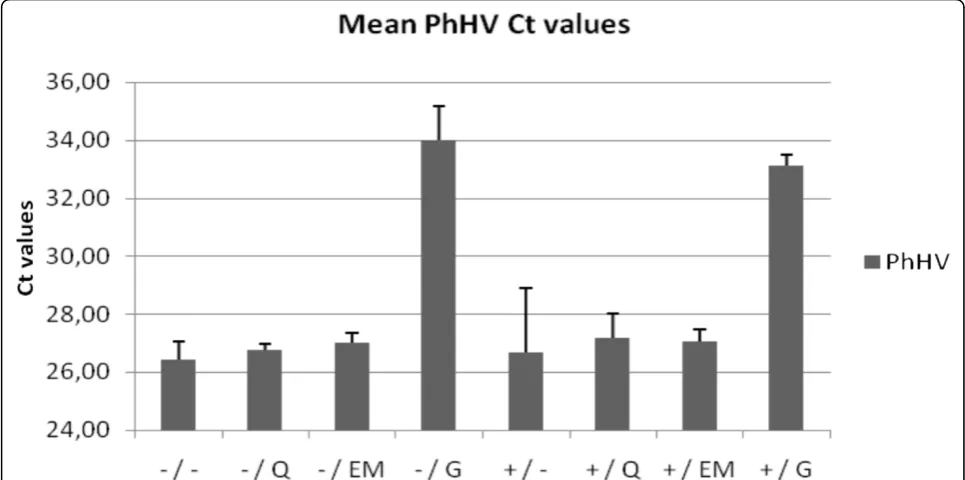
To monitor the presence of inhibiting substances in the PCR, 10 μL of seal herpes virus (PhHV) was added to each material before DNA isolation and a PhHV specific real time PCR was performed after DNA isolation. The fixed quantity of PhHV added to each extraction method in this study is exactly in accordance with the quantity used in our routine diagnostics. Based on 340 measurements of DNA isolations for diagnostic pur-poses, the expected Ct value is the mean Ct value = 25.8; coefficient of variation, 2.5% (data not shown); the threshold for inhibition was set at Ct 27.7 being the mean + 3 standard deviations. DNA isolates with PhHV values that exceed 27.7 are considered to be inhibited.
Figure 2 shows the mean PhHV PCR Ct values of the 4 tissues (A, B, C and D) for the different extraction methods. PhHV amplification after Gentra extraction from all four tissues (with and without prot. K pre-treat-ment; Ct 33.1 ± 0.4 and Ct 34.0 ± 1.2, respectively) and prot. K plus heat-treatment for colon tissue B (Ct 30.0) generated Ct values > 27.7 indicating PCR inhibition. PhHV amplification of DNA-extracts obtained using all other methods showed no inhibition (Ct values < 27.7). Huijsmanset al.BMC Research Notes2010,3:239
http://www.biomedcentral.com/1756-0500/3/239
SNP analysis using real time PCR
To determine the suitability of the different DNA extracts for SNP analysis, real time amplification using Assays-On-Demand SNP genotyping products was per-formed. Different PCR buffer systems and/or different Taq polymerases may yield different real time PCR results [26,27]. We therefore evaluated 2 different mastermixes.
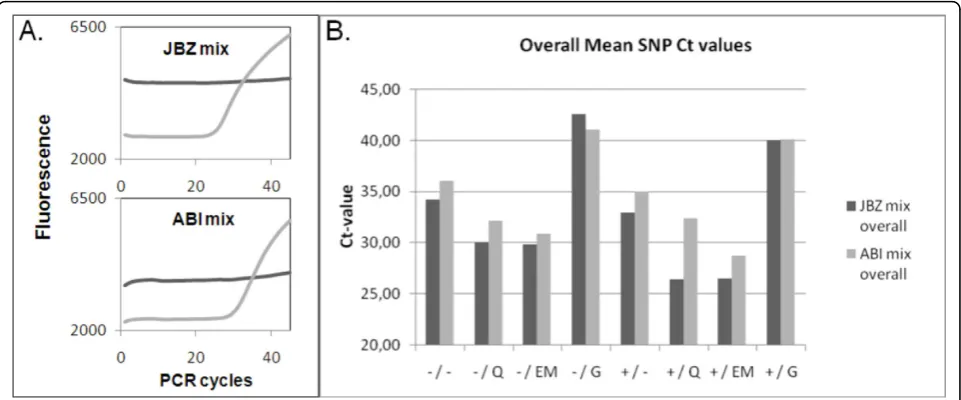
Figure 3A shows a representative example (colon tis-sue) of real time amplification component plots using SNP assay rs1350138 in JBZ 4× (home made) mastermix and ABI 2× mastermix. Figure 3B shows the mean Ct values for the different DNA extraction procedures after real time amplification using SNP Assays-On-Demand Genotyping products rs2043731 and rs1350138 in JBZ 4× mastermix and ABI 2× mastermix. Use of the JBZ 4× mastermix resulted in slightly higher fluorescence and lower mean Ct values than the ABI 2× mastermix. Mean Ct’s were lowest after prot. K digestion followed by QIAamp and EasyMAG extraction (mean Ct value in JBZ mix = 26.5 and 26.6, respectively).
Assessment of maximum amplicon length
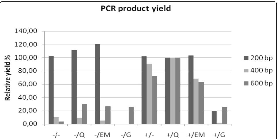
A multiplex PCR was performed to assess the ability of 200, 400 and 600 bp human DNA fragments to be amplified using the DNA yielded by the different extrac-tion methods. Visualizaextrac-tion of the PCR products on gel of representative tissue B (colon) is shown in figure 4. The mean yields of the multiplexed 200 bp, 400 bp and
600 bp PCR products of 4 different tissues (A, B, C and D) for the different extraction methods are shown in fig-ure 5. To compare the methods’ yields the QIAamp DNA extraction in combination with proteinase K diges-tion was set at 100%. The multiplex amplificadiges-tion of DNA extracted by prot. K digestion in combination with QIAamp, EasyMAG or heat-treatment extracts was suc-cessful for fragments up to 400 bp from all tissues (400 bp amplicon yields of 11.0 ± 1.2 ng/μL, 8.2 ± 5.8 ng/μL and 9.02 ± 4.56 ng/μL, respectively), 600 bp amplifica-tion was marginally successful in 3/4 tissues for QIAamp and EasyMAG and in 4/4 tissues for heat-treatment with low yields (6.5 ± 8.2 ng/μL, 3.5 ± 4.6 ng/ μL and 3.42 ± 4.96 ng/μL, respectively).
Discussion
Currently routine molecular techniques are increasingly used on FFPE tissues. An important basic requirement is optimal DNA preparation. We tested human DNA extracts from 4 commonly used DNA extraction meth-ods for the presence of inhibiting substances, and in two downstream applications: real time SNP amplification and multiplexed 200-400-600 bp PCR.
[image:5.595.56.539.90.330.2]influence nucleic acid fragmentation [17]. In addition, the DNA extraction methods were studied using a lim-ited number of samples. However the consistency of results justify several conclusions.
Gentra DNA extraction can be successfully employed on blood samples [28-32]. However, the reduced ampli-fication of PhHV, the internal control virus, after Gentra extraction showed that the Gentra method was not able to sufficiently remove the inhibitory substances (figure 2). In addition, Ct values < 27.7 generated by amplifying
PhHV DNA extracted by the other methods imply that proteinase K digestion is not necessary for the removal of possibly present inhibitory substances.
[image:6.595.58.539.89.289.2]QIAamp as well as EasyMAG are both methods that are currently widely used in routine molecular diagnos-tics regarding the detection of pathogens, e.g. HPV detection [33,34], and mutation screening in cancer-cri-tical genes, e.g. K-ras mutation detection [10]. With regard to real time SNP detection both methods per-formed well after proteinase K digestion. In line with
[image:6.595.57.540.511.682.2]Figure 4 Assessment of maximum amplicon length: visualisation on gel. Gel image of 200-400-600 bp multiplex PCR-products of representative tissue B. The +/ and -/ indicate the use of proteinase K digestion or no digestion, respectively. The different extraction methods are indicated by: heat-treatment =/-, QIAamp DNA extraction =/Q, EasyMAG DNA extraction =/EM, Gentra DNA extraction =/G. Neg = ultrapure water in PCR, Pos = QIAamp extracted DNA from EDTA-blood.
Figure 3SNP analysis using real time PCR.A. Two representative component plots generated after proteinase K treatment followed by EasyMAG extraction from colon tissue B and real time amplification using SNP assay rs1350138. The light grey line indicates allele 1 (VIC label), the dark grey line indicates allele 2 (FAM label).B. Mean Ct value of SNP rs2043731 and rs1350138 of the 4 materials (A, B, C and D) in JBZ 4× mastermix or ABI 2× mastermix for the different extraction methods. The +/ and -/ indicate the use of proteinase K digestion or no digestion, respectively. The different extraction methods are indicated by: heat-treatment =/-, QIAamp DNA extraction =/Q, EasyMAG DNA extraction =/EM, Gentra DNA extraction =/G.
Huijsmanset al.BMC Research Notes2010,3:239 http://www.biomedcentral.com/1756-0500/3/239
previous findings (e.g. [35,36]) we observed that protei-nase K digestion is required for optimal purification of paraffin-embedded DNA. The homebrew JBZ 4× mas-termix yielded better results than the commercial ABI 2× mastermix: fluorescent signals were slightly higher and Ct values lower, suggesting a better real time PCR environment for SNP amplification (figure 3A and 3B). The absence and presence of > 200 bp PCR products after multiplex PCR (figure 4 and 5) of non-digested and digested samples (resp.) indicate that proteinase K treatment plays an important role in proper purification of fragments > 200 bp. Also for RNA it has been shown that small molecules are recovered more easily from FFPE tissues than larger RNA molecules [27,37]. The relatively high 200 bp PCR product yield for the extrac-tion methods without proteinase K digesextrac-tion is probably due to the lack of competition for PCR ingredients by the absence of amplification of the higher molecular DNA targets, which are known to be extracted better when proteinase K digestion is used in contrast to no digestion [35,36]. This observation is important with regard to applications that target stretches of DNA > 200 bp, e.g. STR testing, P53 sequencing and APO-E genotyping [38-40]. Overall multiplex PCR results after Gentra extraction were very poor.
During the processes of paraffin embedding, section-ing and further analysis by (real time) PCR, small traces of foreign DNA, e.g. introduced by floater tissue or a
contaminated microtome blade, may contaminate the material under investigation thereby possibly influencing interpretation of results [41,42]. Thus, caution is advised when using FFPE tissues in combination with molecular techniques. In addition, we routinely process paraffin blocks without tissue, which we use as negative controls. DNA extracts from these blocks may generate real time PCR signals above Ct 35. To be sure that the signal under investigation is not due to background, we set the cut-off Ct value at 33 when using a SNP-profiling assay for identity confirmation [43], implicating that test results with Ct values > 33 were rejected, whereas test results with Ct values < 33 were accepted.
[image:7.595.57.541.87.330.2]suitable, followed by heat-treatment and EasyMAG extraction. An advantage of the heat-treatment and EasyMAG was the reduced hands-on time (when extracting 24 samples: approximately 60 min. for QIAamp versus 5 min. for heat-treatment and 25 min. for EasyMAG).
Conclusions
We conclude that the extraction method significantly influences downstream molecular analysis, which is in line with the findings of previous studies [17,20-24]. The Gentra Capture-Column-kit is not suitable for DNA recovery from FFPE tissues. Of the four methods tested QIAamp DNA-blood-mini-kit extraction and EasyMAG NucliSens extraction performed best for real time SNP detection. Amplification of 400-600 bp fragments appeared most successful after QIAamp isolation fol-lowed by the heat-treatment and EasyMAG.
Thus the method used for DNA isolation from FFPE tissues should be matched with the intended application.
Abbreviations
FFPE: formalin-fixed paraffin-embedded; SNP: single nucleotide polymorphism; Ct value: cycle threshold value; the number of cycles required for the fluorescent signal to cross the threshold (i.e. exceeds background level); PCR: polymerase chain reaction; PhHV: phocine herpes virus; SDS: sequence detection system;
Acknowledgements
We thank Kathelijn Geraats-Peters for critical reading of this manuscript.
Author details
1
Laboratory of Molecular Diagnostics, Jeroen Bosch Hospital,
‘s-Hertogenbosch, The Netherlands.2Laboratory of Pathology, Jeroen Bosch Hospital,‘s-Hertogenbosch, The Netherlands.3Medical Microbiology and Infection Control, VU University Medical Center, Amsterdam, The Netherlands.
Authors’contributions
CJJH participated in the design of the study, carried out all molecular experiments and drafted the manuscript. JD performed the tissue processing. JCvdL participated in drafting the manuscript and supplied the tissues. PHMS and MHAH participated in the design of the study and in drafting the manuscript. All authors read and approved the final manuscript.
Competing interests
The authors declare that they have no competing interests.
Received: 30 April 2010 Accepted: 14 September 2010 Published: 14 September 2010
References
1. Shibata DK, Arnheim N, Martin WJ:Detection of human papillomavirus in paraffin-embedded tissue using the polymerase chain reaction.J Exp Med1988,167:225-230.
2. Brandsma J, Lewis AJ, Abramson AL, Manos MM:Detection and typing of papillomavirus DNA in formalin-fixed, paraffin-embedded tissue.Arch Otolaryngal1990,116:844-848.
3. Cao M, Xiao X, Egbert B, Darragh TM, Yen TSB:Rapid detection of cutaneous herpes simplex virus infection with the polymerase chain reaction.J Invest Dermatol1989,82:391-392.
4. Unger ER, Vernon SD, Lee DR, Miller DL, Reeves WC:Detection of Human Papillomavirus in Archival Tissues: Comparison of In Situ Hybridization and Polymerase Chain Reaction.J Histochem Cytochem1998,46:535-540. 5. Staudach EA, Dietze O, Hauser-Kronberger C:Comparison of real-time PCR
signal-amplified in situ hybridization and conventional PCR for detection and quantification of human papillomavirus in archival cervical cancer tissue.J Clin Microbiol2004,42:3758-3765.
6. Johansen IS, Thomsen VØ Forsgren A, Hansen BF, Lundgren B:Detection of Mycobacterium tuberculosis complex in formalin-fixed, paraffin-embedded tissue specimens with necrotizing granulomatous inflammation by strand displacement amplification.J Mol Diagn2004, 6:231-236.
7. Beqaj SH, Flesher R, Walker GR, Smith SA:Use of the real-time PCR assay in conjunction with MagNA Pure for the detection of mycobacterial DNA from fixed specimens.Diagn Mol Pathol2007,16:169-173. 8. Burmer GC, Rabinovitch PS, Loeb LA:Analysis of c-Ki-rasmutations in
human colon carcinoma by cell sorting, polymerase chain reaction, and DNA sequencing.Cancer Res1989,49:2141-2146.
9. Lyons J, Landis CA, Harsh G, Vallar L, Grunewald K, Feichtinger H, Duh QY, Clark OH, Kawasaki E, Bourne H, McCormick F:Two G protein oncogenes in human endocrine tumors.Science1990,249:655-659.
10. Kramer D, Thunnissen FB, Gallegos-Ruiz MI, Smit EF, Postmus PE, Meijer CJ, Snijders PJ, Heideman DA:A fast, sensitive and accurate high resolution melting (HRM) technology-based assay to screen for common K-ras mutations.Cell Oncol2009,31:161-167.
11. Thibodeau SN, Bren G, Schaid D:Microsatellite instability in cancer of the proximal colon.Science1993,260:816-819.
12. Riet van der P, Karp D, Framar A, Wei Q, Grossman L, Tokino K, Ruppert JM, Sidransky D:Progression of basal cell carcinoma through loss of chromosome 9q and inactivation of a single p53 allele.Cancer Res1994, 54:25-27.
13. Iverson AA, Gillett C, Cane P, Santini CD, Vess TM, Kam-Morgan L, Wang A, Eisenberg M, Rowland CM, Hessling JJ, Broder SE, Sninsky JJ, Tutt A, Anderson S, Chang SY:A single-tube quantitative assay for mRNA levels of hormonal and growth factor receptors in breast cancer specimens.J Mol Diagn2009,11:117-130.
14. Lehmann U, Kreipe H:Real-time PCR analysis of DNA and RNA extracted from formalin-fixed and paraffin-embedded biopsies.Methods2001, 25:409-418.
15. Kayser K, Stute H, Lübcke J, Wazinski U:Rapid microwave fixation - a comparative morphometric study.Histochem J1988,20:347-352. 16. Perlmutter MA, Best CJ, Gillespie JW, Gathright Y, González S, Velasco A,
Linehan WM, Emmert-Buck MR, Chuaqui RF:Comparison of snap freezing versus ethanol fixation for gene expression profiling of tissue specimens.J Mol Diagn2004,6:371-377.
17. Gilbert MTP, Haselkorn T, Bunce M, Sanchez JJ, Lucas SB, Jewell LD, Van Marck E, Worobey M:The Isolation of Nucleic Acids from Fixed, Paraffin -Embedded Tissues- Which Methods Are Useful When?PLoS One2007,2: e537.
18. Quach N, Goodman M, Shibata D:In vitro artifacts after formalin fixation and error prone translesion synthesis during PCR.BMC Biotechnology
2004,4:1.
19. Bonin S, Petrera F, Niccolini B, Stanta G:PCR analysis in archival postmortem tissues.Mol Pathol2003,56:184-186.
20. Dedhia P, Tarale S, Dhongde G, Khadapkar R, Das B:Evaluation of DNA Extraction Methods and Real Time PCR Optimization on Formalin-fixed Paraffin-embedded Tissues.Asian Pacific J Cancer Prev2007,8:55-59. 21. Cao W, Hashibe M, Rao JY, Morgenstern H, Zhang ZF:Comparison of
methods for DNA extraction from paraffin-embedded tissues and buccal cells.Cancer Detect Prev2003,27:397-404.
22. Chan PK, Chan DP, To KF, Yu MY, Cheung JL, Cheng AF:Evaluation of extraction methods from paraffin wax embedded tissues for PCR amplification of human and viral DNA.J Clin Pathol2001,54:401-403. 23. Rivero ER, Neves AC, Silva-Valenzuela MG, Sousa SO, Nunes FD:Simple salting-out method for DNA extraction from formalin-fixed, paraffin-embedded tissues.Pathol Res Pract2006,202:523-529.
24. Wu L, Patten N, Yamashiro CT, Chui B:Extraction and Amplification of DNA From Formalin-Fixed, Paraffin-Embedded Tissues.Appl Immunohistochem Mol Morphol2002,10:269-274.
Huijsmanset al.BMC Research Notes2010,3:239 http://www.biomedcentral.com/1756-0500/3/239
25. van Doornum GJ, Guldemeester J, Osterhaus AD, Niesters HG:Diagnosing herpesvirus infections by real-time amplification and rapid culture.J Clin Microbiol2003,41:576-580.
26. Wolffs P, Grage H, Hagberg O, Rådström P:Impact of DNA Polymerases and Their Buffer Systems on Quantitative Real-Time PCR.J Clin Microbiol
2004,42:408-411.
27. Li J, Smyth P, Cahill S, Denning K, Flavin R, Aherne S, Pirotta M,
Guenther SM, O’Leary JJ, Sheils O:Improved RNA quality and TaqMan Pre-amplification method (PreAmp) to enhance expression analysis from formalin fixed paraffin embedded (FFPE) materials.BMC Biotechnol2008, 8:10.
28. Kowalski A, Radu D, Gold B:Colorimetric microwell plate detection of the factor V Leidenmutation.Clin Chem2000,46:1195-1198.
29. Fahle GA, Fischer SH:Comparison of six commercial DNA extraction kits for recovery of cytomegalovirus DNA from spiked human specimens.J Clin Microbiol2000,38:3860-3863.
30. Esteve M, Rosinach M, Fernández-Banãres F, Farré C, Salas A, Alsina M, Vilar P, Abad-Lacruz A, Forné M, Mariné M, Santaolalla R, Espinós JC, Viver JM:Spectrum of gluten-sensitive enteropathy in firstdegree relatives of patients with coeliac disease: clinical relevance of lymphocytic enteritis.Gut2006,55:1739-1745.
31. Pruthi RK, Rodriguez V, Allen C, Slaby JA, Schmidt KA, Plumhoff EA: Molecular analysis in a patient with severe factor VII deficiency and an inhibitor: report of a novel mutation (S103G).European Eur J Haematol
2007,79:354-359.
32. Hillman MA, Wilke RA, Yale SH, Vidaillet HJ, Caldwell MD, Glurich I, Berg RL, Schmelzer J, Burmester JK:A prospective, randomized pilot trial of model-based warfarin dose initiation using CYP2C9 genotype and clinical data. Clin Med Res2005,3:137-145.
33. Koidl C, Bozic M, Hadzisejdic I, Grahovac M, Grahovac B, Kranewitter W, Marth E, Kessler HH:Comparison of molecular assays for detection and typing of human papillomavirus.Am J Obstet Gynecol2008,199(144):e1-6. 34. Klaassen CH, Prinsen CF, de Valk HA, Horrevorts AM, Jeunink MA,
Thunnissen FB:DNA microarray format for detection and subtyping of human papillomavirus.J Clin Microbiol2004,42:2152-2160.
35. Isola J, DeVries S, Chu L, Ghazvini S, Waldman F:Analysis of changes in DNA sequence copy number by comparative genomic hybridization in archival paraffin-embedded tumor samples.Am J Pathol1994, 145:1301-1308.
36. Banerjee SK, Makdisi WF, Weston AP, Mitchell SM, Campbell DR: Microwave-based DNA extraction from paraffin-embedded tissue for PCR amplification.BioTechniques1995,18:768-773.
37. Li J, Smyth P, Flavin R, Cahill S, Denning K, Aherne S, Guenther SM, O’Leary JJ, Sheils O:Comparison of miRNA expression patterns using total RNA extracted from matched samples of formalin-fixed paraffin-embedded (FFPE) cells and snapfrozen cells.BMC Biotechnol2007,7:36. 38. Moretti TR, Baumstark AL, Defenbaugh DA, Keys KM, Smerick JB, Budowle B:
Validation of short tandem repeats (STRs) for forensic usage: performance testing of fluorescent multiplex STR systems and analysis of authentic and simulated forensic samples.J Forensic Sci2001, 46:647-660.
39. Ahrendt SA, Halachmi S, Chow JT, Wu L, Halachmi N, Yang SC, Wehage S, Jen J, Sidransky D:Rapid p53 sequence analysis in primary lung cancer using an oligonucleotide probe array.Proc Natl Acad Sci USA1999, 22:7382-7387.
40. Gioia L, Vogt LJ, Freeman WM, Flood A, Vogt BA, Vrana KE:PCR-based apolipoprotein E genotype analysis from archival fixed brain.J Neurosci Methods1998,80:209-214.39.
41. Fox EA:Preparation of DNA from fixed, paraffin-embedded tissue.Curr Protoc Hum Genet2001.
42. Mosse CA, Stumph JR, Best DH, Vnencak-Jones CL:A B-cell lymphoma diagnosed in“floater”tissue: implications of the diagnosis and resolution of a laboratory error.Am J Med Sci2009,338:248-251. 43. Huijsmans R, Damen J, van der Linden H, Hermans M:Single nucleotide
polymorphism profiling assay to confirm the identity of human tissues.J Mol Diagn2007,9:205-213.
doi:10.1186/1756-0500-3-239
Cite this article as:Huijsmanset al.:Comparative analysis of four
methods to extract DNA from paraffin-embedded tissues: effect on downstream molecular applications.BMC Research Notes20103:239.
Submit your next manuscript to BioMed Central and take full advantage of:
• Convenient online submission
• Thorough peer review
• No space constraints or color figure charges
• Immediate publication on acceptance
• Inclusion in PubMed, CAS, Scopus and Google Scholar
• Research which is freely available for redistribution