R E S E A R C H A R T I C L E
Open Access
Mapping the cellular and molecular
heterogeneity of normal and malignant breast
tissues and cultured cell lines
Patricia J Keller
1,2†, Amy F Lin
1,2†, Lisa M Arendt
1,2, Ina Klebba
1,2, Ainsley D Jones
1,2, Jenny A Rudnick
1,2,
Theresa A DiMeo
1,2, Hannah Gilmore
3, Douglas M Jefferson
4, Roger A Graham
5, Stephen P Naber
6, Stuart Schnitt
3, Charlotte Kuperwasser
1,2*Abstract
Introduction:Normal and neoplastic breast tissues are comprised of heterogeneous populations of epithelial cells exhibiting various degrees of maturation and differentiation. While cultured cell lines have been derived from both normal and malignant tissues, it remains unclear to what extent they retain similar levels of differentiation and heterogeneity as that found within breast tissues.
Methods:We used 12 reduction mammoplasty tissues, 15 primary breast cancer tissues, and 20 human breast epithelial cell lines (16 cancer lines, 4 normal lines) to perform flow cytometry for CD44, CD24, epithelial cell adhesion molecule (EpCAM), and CD49f expression, as well as immunohistochemistry, andin vivotumor xenograft formation studies to extensively analyze the molecular and cellular characteristics of breast epithelial cell lineages. Results:Human breast tissues contain four distinguishable epithelial differentiation states (two luminal phenotypes and two basal phenotypes) that differ on the basis of CD24, EpCAM and CD49f expression. Primary human breast cancer tissues also contain these four cellular states, but in altered proportions compared to normal tissues. In contrast, cultured cancer cell lines are enriched for rare basal and mesenchymal epithelial phenotypes, which are normally present in small numbers within human tissues. Similarly, cultured normal human mammary epithelial cell lines are enriched for rare basal and mesenchymal phenotypes that represent a minor fraction of cells within reduction mammoplasty tissues. Furthermore, although normal human mammary epithelial cell lines exhibit features of bi-potent progenitor cells they are unable to differentiate into mature luminal breast epithelial cells under standard culture conditions.
Conclusions:As a group breast cancer cell lines represent the heterogeneity of human breast tumors, but individually they exhibit increased lineage-restricted profiles that fall short of truly representing the intratumoral heterogeneity of individual breast tumors. Additionally, normal human mammary epithelial cell lines fail to retain much of the cellular diversity found in human breast tissues and are enriched for differentiation states that are a minority in breast tissues, although they do exhibit features of bi-potent basal progenitor cells. These findings suggest that collections of cell lines representing multiple cell types can be used to model the cellular heterogeneity of tissues.
* Correspondence: charlotte.kuperwasser@tufts.edu
†Contributed equally 1
Department of Anatomy & Cellular Biology, Sackler School, Tufts University School of Medicine, 136 Harrison Ave, Boston, MA 02111, USA
Full list of author information is available at the end of the article
Introduction
Human breast cell lines have long served as models for a wide array of applications including the study of molecular, cellular, and biochemical mechanisms that regulate breast epithelial biology. Breast cancer cell lines are also commonly used in xenograft models for drug discovery and in the assessment of pre-clinical experi-mental therapeutic efficacy. Despite their crucial role for rational drug discovery and development and in under-standing molecular pathophysiology of cancer, their ability to accurately reflect phenotypes of tumors remains controversial. Several studies have suggested that cell lines exhibit a narrow range of genetic profiles, harbor genetic alterations due to adaptation of tissue culture environment, and are poor predictors of in vivo
sensitivity to drug efficacy [1-3]. Cell line-derived xeno-graft models also fail to recapitulate the heterogeneous histopathology characteristic of the parent tumor histol-ogy. However, other studies have indicated that cell lines, as a system, actually mirror many of the biological and genomic properties found within primary human tumors [4,5]. Genomic approaches have revealed that like primary tumors, the gene expression signatures of breast cancer cell lines can distinguish luminal from basal subtypes of breast cancer [6-9]. Moreover, cell line-derived gene signatures can correctly classify human tumor samples [6,7,10], suggesting that despite their acquired ability to grow in vitro, and acquired mutations following adaptation to culture conditions, cell lines continue to share many of the molecular and genetic features of the primary breast cancers from which they were derived.
The use of primary human breast tissues for experi-mental studies and breast cancer research has been fueled by the notion that cell lines are not accurate models of the heterogeneity found in vivo. As such, reduction mammoplasty and cancer tissues have been used to identify and characterize epithelial differentia-tion states and lineages since it is presumed that not all cell types are maintained or mirrored in vitro. Expres-sion of epithelial cell adheExpres-sion molecule (EpCAM) and CD49f+(a6 integrin) have been used to identify luminal and basal/myoepithelial cells from breast tissues [11-14]. Mature luminal cells are reported to express an EpCAM+/CD49f- phenotype while luminal progenitors express an EpCAM+/CD49f+marker profile. Myoepithe-lial cells and basal progenitor cells are defined by an EpCAM-/CD49f+ phenotype [11,13,15]. In addition to EpCAM and CD49f, surface expression of CD44 and CD24 have also been used to identify luminal epithelial cells that express genes involved in hormone responses (CD24+) and cells resembling progenitor cells that express genes involved in motility (CD44+) [16].
Reflecting the normal cell types within the breast, tumors are broadly classified histopathologically by expression of either luminal cytokeratins (CK8/18) or stratified epithelial cytokeratins (CK5/6/14, basal-type) [17,18]. Similarly, tumor subclasses identified by micro-array were named to reflect the gene expression patterns of the normal breast luminal and myoepithelial/basal cells [19-23]. Luminal-type breast cancers (Luminal A and Luminal B) express estrogen receptor (ER). Her2-type breast cancers typically overexpress or amplify Her2, are generally negative for ER expression and tend to express the genes associated with the Her2-amplicon. Lastly, Basal-like breast cancers are also often referred to as triple-negative tumors since they do not express ER, progesterone receptor (PR), or Her2 [19-22].
To determine if cell lines mirror or maintain the cellu-lar differentiation states found in primary tissues, we examined the molecular and cellular profiles of normal and malignant human breast epithelial cell lines and compared them to normal and cancerous tissues. In doing so, we found four distinguishable cell states across a collection of cell lines that mirrored the four differen-tiation states present within normal and malignant breast tissues. However, we also found that the cellular heterogeneity within cell lines was remarkably restricted in culture and was enriched for cellular phenotypes that were normally present as a minor componentin vivo.
Materials and methods
Cell lines and tissue culture
SUM cell lines were obtained from Dr. Stephen Ethier (Kramanos Institute, Detroit, MI, USA) and are com-mercially available (Asterand, Detroit, MI, USA). The MCF7, T47 D, BT20, MCF10A, MCF10F, MDA.MB.231, MDA.MB.361 and HCC cell lines were obtained directly from the American Type Culture Collection (ATCC; Manassas, VA, USA). The MCF10A and MCF10F cell lines are non-tumorigenic mammary epithelial cell lines that were produced by long-term culture in serum-free medium with low calcium; the MCF10A cells were derived from an the adherent population in these cul-tures, while the MCF10F line was derived from floating cells within the MCF10 cultures [24]. All of the ATCC cell lines used in this study were low passage (< 10). SUM225CWR, SUM149PT, and SUM159PT cells were cultured in F12 with 5% calf serum (CS), insulin (5μg/ ml), and hydrocortisone (1 μg/ml), while SUM1315 MO2 cells were cultured in F12 with 5% CS, insulin (5
Hyclone, Logan, UT, USA) with 10% FBS. The TUM177 breast cancer cell line was established from a primary invasive ER-positive adenocarcinoma. An ER-negative cancer cell line spontaneously emerged after two months of cultivations. TUM177 cells were cultured in DMEM with 10% fetal bovine serum (FBS; Invitrogen, Carlsbad, CA, USA).
HME I and HME II cells were derived from reduction mammoplasty tissues from two different patients grown in Mammary Epithelial Growth Medium (MEGM) until the generation of variant cells [25] and then immorta-lized through the ectopic expression of the catalytic sub-unit of human telomerase (hTERT) [26].
MCF10F cells were cultured in Dulbecco’s modified Eagle’s medium-Ham’s F12 (DMEM/F12; 1:1) with 5% horse serum, insulin (5 μg/ml), hydrocortisone (1 μg/ ml), and human epidermal growth factor (hEGF; 10 ng/ ml), and cholera toxin (100 ng/ml) (all, Sigma, St. Louis, MO, USA). MCF10A and immortalized human mam-mary epithelial (HME) cell lines were cultured in MEGM supplemented with bovine pituitary extract (52
μg/ml), hydrocortisone (0.5 μg/ml), hEGF (10 ng/ml) and insulin (5 μg/ml) (MEGM Bullet Kit, Lonza Cor-poration, Walkersville, MD, USA). MCF10A cells were further supplemented with cholera toxin (100 ng/ml). For serum differentiation experiments, HME or MCF10A cells were switched to growth in the MCF10F medium with substitution of 5% CS for the horse serum and omission of the cholera toxin, or 5% CS was added to MEGM and cells were allowed to differentiate for six days before use in experiments. For mammosphere cul-ture, cells were plated at 20,000 cells/ml and grown on ultra-low adherence six-well plates for one week (Corn-ing Life Sciences, Lowell, MA, USA). Quantification of mammospheres was accomplished using a Multisizer 3 COULTER COUNTER (Beckman-Coulter, Brea, CA USA) that provides number, and size distributions with an overall sizing range of 14μm to 336 μm.
Reduction mammoplasty and tumor tissue specimens All human breast tissue procurement for these experi-ments was obtained in compliance with the laws and institutional guidelines, as approved by the Institutional Review Board committee from Beth Israel Deaconess Hospital and Tufts Medical Center. Fresh disease-free reduction mammoplasty tissues (n = 12) and tumor tis-sues (n = 15; 8 fresh, 15 formalin-fixed paraffin embedded) were obtained from discarded material from patients undergoing elective reduction mammoplasty surgeries or from patients undergoing partial or com-plete mastectomy for excision of tumor tissue from the Pathology departments at BIDMC or Tufts Medical Center. All samples were obtained from de-identified discarded material and therefore, informed consent was
not required for these studies. All samples were evalu-ated histologcially and confirmed to be invasive ductal carcinomas. The following histopathologic variables, determined for all tumor tissue specimens, were done on full sections, and cases with 10% or more positive for ER, p53 or EGFR staining were grouped as positive. The scoring of Her2 was performed using the ASCO/CAP guidelines, as follows: Cases with 30% or more strongly positive cells with strong complete membrane staining were defined as Her2+ tumors. Cases with 10% or more positive cells with weak to moderate complete mem-brane staining were considered Her2+ but were not defined as Her2+ tumors solely on this basis. IHC analy-sis for estrogen receptor (ER), progesterone receptor (PR), Her2, p53 and EGFR were independently reviewed by expert breast pathologists (HG and SN). Breast tumor subtypes were defined as follows: Luminal A (ER+ and/or PR+, Her2-), Luminal B (ER+ and/or PR+, Her2+), Her2+ (ER-, PR-, Her2+), and Basal-like (ER-, PR-, Her2-, and epidermal growth factor receptor (EGFR)+/-) and p53+.
Uncultured cells from reduction mammoplasty or human breast tumor organoid preps [27] were disso-ciated to a single-cell suspension by trypsinization and filtered through a 20 μm nylon mesh (Millipore, Dan-vers, MA, USA). Human breast tumors were plated in DMEM supplemented with 10% CS for one to two hours to deplete stromal cells.
Immunohistochemical analysis and scoring
Immunohistochemistry was performed by the Histology Special Procedures Laboratory at Tufts Medical Center on paraffin-embedded tissue sections on a Ventana (Tucson, Arizona, USA) automated slide stainer with the iVIEW DAB detection kit for visualization. Antibo-dies used were CK14 (1:500, clone LL002, Vector (Bur-lingame, CA, USA)), CK8/18 1:500, clone DC-10, Vector), Vimentin (1:500, clone V9, Vector), S100A4 (1:200, clone 1F12-1G7, Sigma), S100A6 (1:200, clone CACY-100, Sigma), p53 (Ventana Medical Systems), ER (Ventana Medical Systems), Her2 (Ventana Medical Sys-tems), EGFR (1:20, clone 31G7, Zymed), and PR (Ven-tana Medical Systems). All Ven(Ven-tana antibodies are prediluted.
added to obtain a total stain score for each field. An average total stain score was calculated for the staining for a particular sample. Statistical analysis was per-formed using the student’s t-test across the different patient samples.
Flow cytometry and FACS
Uncultured cells from reduction mammoplasty tissues (n = 12) or primary breast tumor tissues (n = 8) from organoid preparations were dissociated to single-cell suspensions, as described above. For reduction mammo-plasty tissues, endothelial, lymphocytic, monocytic, and fibroblastic lineages were depleted with antibodies to CD31, CD34 and CD45 (all Thermo/LabVision, Fre-mont, CA, USA) and Fibroblast Specific Protein/IB10 (Sigma) using a cocktail of Pan-mouse IgG and IgM Dynabeads (Dynal, Invitrogen) according to the manu-facturers instructions and as described previously [28]. Depleted single cells suspensions were resuspended at 1 × 10⁶ cells/ml in phosphate-buffered saline containing 1% calf serum (FACS buffer, FB) and bound with fluor-escently-conjugated antibodies to human EpCAM (APC), CD49f (PE), and CD24 (FITC) (all, BD Bios-ciences, San Jose, CA, USA) for 20 minutes at 4°C. Anti-body-bound cells were washed and resuspended at 1 × 10⁶ cells/ml in FB and run on a FACSCalibur flow cyt-ometer. Flow cytometry data was analyzed with the Flowjo software package (TreeStar, Ashland, OR, USA).
For fluorescence-activated cell sorting (FACS), cells from reduction mammoplasty tissue were prepared as above for flow cytometry and resuspended at 5 x10⁶ cells/ml in FB and sorted on a BD Influx Cell sorter (BD Biosciences) into culture medium (MEGM) con-taining 50% CS.
For cell lines, non-confluent cultures of cells were trypsinized into single cell suspension, counted, washed with PBS, and stained with antibodies specific for human cell CD24 (PE) and CD44 (APC) (BD Bios-ciences). The cells were stained with antibodies specific for human cell surface markers: EpCAM-fluorescein iso-thiocyanate (FITC), CD24-phycoerythrin (PE), and CD49f-PE-Cy5 or CD44-allophycocyanin (APC) (BD Biosciences). Additional cells were stained with isotype controls for each antibody: Ms IgG1-FITC, Ms IgG2a-PE, and Rat IgG2a-PE-Cy5 or Ms IgG2b-APC (BD Bio-sciences). A total of 200,000 to 800,000 cells were incu-bated with antibodies or isotype controls for 20 minutes on ice. The cells were washed with PBS to remove any unbound antibody and analyzed no later than one hour post-staining on a FACSCalibur flow cytometer (BD Biosciences). Antibody-bound cells were resuspended at 1 × 10⁶ cells/ml in FB and run on a FACSCalibur flow cytometer (BD Biosciences) or sorted on an BD Influx FACS sorter (BD Biosciences). Flow cytometry data was
analyzed with the Flowjo software package (TreeStar). Each cell line was analyzed in three to five different bio-logical replicates.
Immunofluorescence
Collected cell fractions from FACS were counted and cytospun onto glass slides at 10,000 cells per spot with a Cytospin 4 cytospinner (Thermo Scientific, Waltham, MA, USA). Cultured cell lines were plated at 10 to 20,000 cells per well in eight-well chamber slides (BD Biosciences) and grown two to three days. Cytospins and cells in chamber slides were fixed in 100% methanol and stained overnight at 4°C with primary antibodies directed to EpCAM (VU-ID9, 1:100, Stem Cell Technol-ogies, Vancouver, BC, Canada), CK8/18 (5D3, 1:500, Vector Labs, Burlingame, CA, USA), ERa (1D5, 1:100, Santa Cruz Biotechnology, Santa Cruz, CA, USA) CK14 (ASM-1, 1:500, Thermo Scientific/LabVision),a-smooth muscle actin (SMA; 1:250, Vector Labs) and vimentin (V9, 1:500, Vector Labs) followed by secondary antibo-dies (1:500 Alexa488 or Alexa555 conjugated anti-mouse and anti-rabbit H+L IgG, Invitrogen) for one hour at room temperature. Nuclei were counterstained with 4’, 6-diamidino-2-phenylindole (DAPI) and images were captured with the Spot imaging software (Diagnos-tic Instruments, Inc., Sterling Heights, MI, USA); stain-ing was analyzed by countstain-ing the total number of cells positive stain compared to the total number of cells in multiple fields with at least 50 cells analyzed per condi-tion. Negative staining represents no cells staining posi-tive, Mixed staining is > 1% but < 50% of the cells staining positive, while positive staning is > 80% of the cells staining positive.
An average total stain score of a cell line was calcu-lated using three to five different regions of the plate. Statistical analysis was performed using the student’s T-test across the different patient samples.
Animals and surgery
Statistical analysis
Fisher exact tests were used when comparing the binary categories of expression of proteins between groups. All
P-values reported are two-sided.
Results
CD44 and CD24 expression in human breast cancer cell lines
Studies have suggested that the pre-existing differentia-tion state of normal precursor cell types is so strongly encoded it survives the neoplastic transformation and accounts in part for tumor phenotype [29,30]. Based on this notion, we reasoned that it might be possible to map different tumor subtypes to their normal cellular precursors within human breast tissues based on the expression of cell surface markers. Recently, the cell sur-face markers CD24 and CD44 have been used to define normal human breast epithelial differentiation states: CD44 is expressed in basal cells while CD24 is expressed in luminal cells [16].
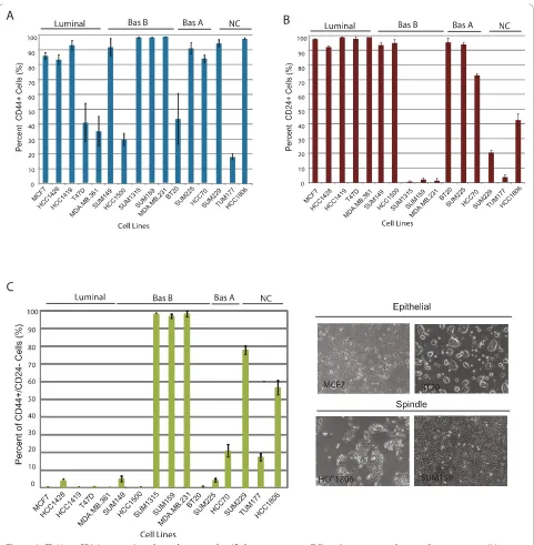
We wanted to determine whether these markers could be used to classify luminal and basal breast cancer cell lines, many of which have been previously classified on the basis of gene expression profiling [7,31]. Using a panel of 16 cancer lines we found that all breast cancer cell lines contained a population of CD44+cells regardless of tumor subtype. Most of the lines (11/16) contained a majority (> 80%) of CD44+cells, while the remaining cell lines (5/16) contained a minority (< 40%) of CD44+cells (Figure 1a, Additional files 1 and 2). There was no correlation (P= 0.14,P= 0.44,P= 1) between the proportion of CD44+ (greater than or 80% or less than 40%) cells within the cell line with breast cancer subtype.
In contrast to CD44 expression, not all breast cancer cell lines contained CD24+cells. Rather, 10/16 lines contained a large proportion (> 70%) of CD24+cells, while 6/16 lines contained very few (< 5 to 45%) CD24+cells (Figure 1b, Additional files 1 and 2). As with CD44 expression, there was no correlation between the proportion of CD24+cells in cell lines and tumor subtype. Since CD44 and CD24 expression alone could not be used to classify cell lines based on tumor subtype, we examined whether together these markers might be able to categorize cell lines. While, the proportion of CD44+/CD24-cells did not correlate with gene expression-based classifiers of breast cancer subtype, consistent with previous reports, there was a striking relationship between the proportion of CD44+/ CD24- cells in the line and spindle-cell morphology (Figure 1c), [32,33] (Additional files 1, 2, 3 and 4).
EpCAM, CD24 and CD49 expression reduction mammoplasty tissues
Since CD44 and CD24 were not useful markers to clas-sify tumor cells, we wanted to determine whether
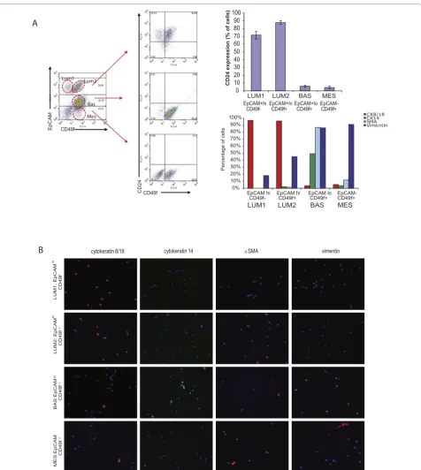
additional lineage markers might be able to refine cellu-lar differentiation states. Accordingly, we used flow cyto-metry to characterize breast epithelial cells from reduction mammoplasty tissues (n = 12) using EpCAM, and CD49f expression. EpCAM and CD49f have been used previously to define cells within the luminal and basal lineages from normal human breast tissue [11,14,15].
We identified four epithelial cell populations (two populations of luminal cells and two populations of basal cells) from freshly dissociated, lineage-depleted breast epithelial cells from reduction mammoplasty tis-sues on the basis of EpCAM/CD24/CD49f expression (Figure 2). There were three populations of cells identi-fied on the basis of EpCAM expression; EpCAMhi cells, which expressed CD24 but were either CD49f+ or CD49f-, EpCAMlow cells that lacked CD24 expression but expressed CD49f, and EpCAM-negative cells that also lacked CD24 expression but were CD49f-positive.
To confirm the nature of these cell types, we sorted lineage-depleted cells from reduction mammoplasty tis-sues by FACS, and cytospun freshly sorted cells to examine the expression of established markers of lumi-nal and myoepithelial/basal cells (Figure 2b, c). Highly expressing EpCAM+luminal cells were either CD49f+ or CD49f-, consistent with the definition of mature luminal cells and luminal progenitor cells, respectively [14,15]. EpCAM+/CD49f- and EpCAM+/CD49f+ cells were pre-dominantly CK8/18 positive, lacked CK14 and SMA expression thus were termed Luminal 1 and Luminal 2 cells, respectively. EpCAM+/CD49f- and EpCAM +
/CD49f+ cells also both expressed CD24. However, unlike previous reports, we observed a second EpCAM +
/CD49f+population of cells that expressed lower levels of EpCAM. Unlike EpCAM+/CD49f+luminal progenitor cells, this population of EpCAM+/CD49f+ cells lacked CD24 expression. In addition, EpCAM+/CD24-/CD49f+ cells were predominantly CK14-positive, while EpCAM +
/CD24+/CD49f+ cells were predominantly CK18-postive (Figure 2). Furthermore, EpCAM+/CD24-/CD49f+cells expressed SMA and vimentin; and thus were termed Basal. Finally, an EpCAM-negative population which lacked CD24 expression was also identified. This popu-lation expressed CD49f, expressed lower levels of CK14, and strong levels of vimentin (Figure 2b, c). Although EpCAM-/CD49f+ cells expressed basal epithelial mar-kers, they were termed mesenchymal, due to the lack of luminal epithelial markers (CD24 and EpCAM), and the higher levels of vimentin expression.
Cellular and molecular heterogeneity in breast cancer tissues
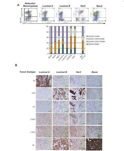
analyzed freshly dissociated breast epithelial cells from primary human breast cancers (n = 8) by flow cytome-try. Primary tumor tissues, in general, showed a different spectrum of cellular heterogeneity compared to breast reduction mammoplasty tissue by flow cytometry when stained for EpCAM, CD49f, and CD24 (Figure 3a).
[image:6.595.58.539.86.577.2]Although the four major cell types were still present regardless of the tumor classification (Luminal (A or B), Her2, Basal), several tumor tissues contained a larger proportion of EpCAM-/CD49f+Mesenchymal cells com-pared to reduction mammoplasty tissues. Although the number of tumors analyzed was too small to make any
Figure 1CD44 or CD24 expression alone does not classify breast cancer cell lines into tumor subytpes. Breast cancer cell lines are grouped based on tumor subtype classification defined by [7] as Luminal, Basal A (Bas A), Basal B (Bas B), or those that have not been
statistically significant conclusions, it was interesting to note that basal tumors, which have been considered to express mesenchymal markers, contained the fewest numbers of EpCAM-/CD49f+Mesenchymal cells, while Her2-positive tumors, which are traditionally viewed as a subset of luminal tumors, retained the fewest numbers of EpCAM+/CD49f-Luminal 1 cells. It will be interest-ing to determine if these observations can be expanded across a wider spectrum of tumor specimens.
We also analyzed breast cancer tissues (n = 15) by immunohistochemistry for markers of Luminal 1, Lumi-nal 2, Basal and Mesenchymal cells. Consistent with the flow cytometry data, human breast cancers exhibited heterogeneous and variable expression of markers of Luminal 1, Luminal 2, Basal and Mesenchymal cells, regardless of tumor subtype (Figure 3b). Future prospec-tive studies are needed to determine whether the differ-ences in cell state proportions within tumors are associated with clinical and prognostic information.
EpCAM, CD24 and CD49 epithelial subtypes in breast cancer cell lines
All 16 breast cancer cell lines were analyzed for the expression of EpCAM, CD24, and CD49f to determine whether the same four differentiation states present within human breast tissues were retained in cultured lines. While we indeed observed the presence of all four of these differentiation states within the panel of human breast cancer cell lines, the majority of cell lines failed to retain Luminal 1 EpCAM+/CD49f- cells. Rather only one class of cell lines could be readily distinguished from all other lines by retaining this population of EpCAM+/CD24+/CD49f- cells (Figure 4a, Additional files 4, 5 and 6); these cell lines are thereafter referred to as Luminal 1-type lines. Luminal 1 cell lines were derived from pleural effusions, and are strongly ER-posi-tive, thus of the luminal subtype. A second class of cell lines were distinguished by a prominent population (> 90%) of EpCAM+/CD24+/CD49f+ luminal cells and are thus referred to as Luminal 2 lines (Figure 4a, Addi-tional files 4 and 5). Luminal 2 cell lines (6/16) included cell lines that were derived from either pleural effusions or primary tumor tissues and express ER, Her2 or both ER and Her2 (Figure 4a, Additional files 4 and 6). The third class of cell lines could be distinguished by two prominent populations (> 15%) of EpCAM+/CD49f+ cells: EpCAM+/CD24+/CD49f+ luminal cells and EpCAM+/CD24-/CD49f+ basal cells, the latter of which were rare or absent in other cell lines. Thus, these can-cer lines were referred to as Basal lines (Figure 4a, Addi-tional files 4 and 6). All Basal cell lines (4/16) in this category were derived from primary breast tumors and are ER-, PR-, and Her2-negative. Finally, cell lines that exhibited a spindle-like morphology in culture, were
derived from either pleural effusions or primary tumor tissues and were largely comprised of EpCAM-/CD24-/ CD49f+ Mesenchymal cells (> 90%) (Figure 4a, Addi-tional files 4 and 6); thus, referred to as Mesenchymal lines. Notably, all Mesenchymal cell lines lack ER, PR and Her2 expression.
Consistent with previous reports, we observed a strong association between the cell surface-based categories, morphology and molecular markers. Luminal cells (Luminal 1 and 2) grew as epithelial-differentiated monolayers with tight cell-cell junctions. They all expressed CK8/18 and EpCAM, and all lacked expres-sion of the basal cytokeratin CK14 and mesenchymal vimentin (Figure 4b, Additional files 4 and 6). In con-trast, Mesenchymal cells appeared less differentiated and exhibited a spindle-like appearance. They lacked expres-sion of both of CK8/18 and CK14 expresexpres-sion and were all strongly positive for vimentin expression (Figure 4b, Additional files 4 and 6). Interestingly, Basal cell lines generally exhibited a more scattered morphology com-pared to Luminal cell lines but were more epithelial compared to Mesenchymal cell lines. Consistent with their luminal-like morphology, Basal cell lines all expressed CK8/18 and EpCAM, but they all also expressed the basal maker CK14 (Figure 4b, Additional files 4 and 6), which was absent in both Luminal and Mesenchymal cell lines. Moreover, vimentin expression was rarely detected in Basal lines and when it was, it was focal and restricted to rare cells within the popula-tion (Addipopula-tional files 4 and 6). These findings indicate that breast cancer cell lines retain the four cell differen-tiation states that map to normal precursors found in reduction mammoplasty tissues.
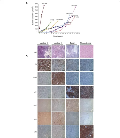
In vivotumorigenicity and growth characteristics of human breast cancer cell lines
ER-positive and negative for p53, vimentin and Her2. Luminal 2 cell lines also formed tumors that expressed either ER and/or Her2, but failed to express p53 or vimentin (Figure 5b, Additional files 6 and 7). Basal cell lines formed tumors that expressed robust p53 but lacked ER and Her2 expression (Figure 5b, Additional files 6 and 7). Basal tumors also lacked vimentin expres-sion with the exception of the tumor-stromal interface (data not shown). Unlike Luminal and Basal cell lines, Mesenchymal cancer cell lines formed almost exclusively spindle-cell metaplastic tumors that lacked obvious epithelial features (Figure 5b, Additional files 6 and 7). In addition, tumors derived from Mesenchymal lines were strongly and uniformly positive for vimentin and p53, consistent with clinical basal-like tumors (Figure 4b, Additional files 6 and 7). However, unlike primary human basal-like breast cancers that have been reported to express EGFR protein, EGFR expression in cell-line derived xenograft tumors was only weakly expressed in HCC1806 and TUM177 xenografts and not expressed preferentially in tumors derived from other Basal or Mesenchymal cell lines despite its expression in these cultured cell lines (Additional files 6 and 7, and [10]).
Enrichment for basal phenotypes in normal breast cell lines
Since the majority of breast cancer cell lines failed to maintain EpCAM+/CD24+/CD49f-Luminal 1 cells
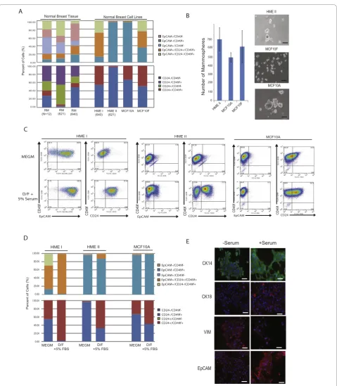
in vitro, we wanted to determine whether this was a general feature ofin vitrocell cultivation or was a con-sequence of malignancy. We therefore compared non-transformed human breast epithelial cell lines (HMECs (HME I, HME II), MCF10A and MCF10F) with reduc-tion mammoplasty tissues for cell surface and molecular features. Surprisingly, we found that under serum-free conditions none of the normal human mammary epithe-lial cell lines contained Luminal 1 cells in culture, nor could they be classified as Luminal 2 cells. Rather nor-mal human breast epithelial cell lines were classified into two categories: Basal lines (HME I and MCF10F cell lines) that contained a prominent Basal population, and Mesenchymal lines (HME II and MCF10A cell lines) that were comprised of a majority (> 90%) Mesenchymal EpCAM-/CD24-/CD49f+ cells (Figure 6a). These data indicate that the selection for basal and mesenchymal cell states in cultured breast epithelial cells is not a consequence of genetic mutation or malig-nant transformation, but is likely the result of adherent
in vitroselection.
We used immunofluorescence to determine whether non-transformed Basal and Mesenchymal cell lines expressed similar markers of normal reduction mammo-plasty counterparts (Figure 6e). In contrast to Mesench-ymal cancer cell lines, which failed to express CK8/18
or CK14 and grew as spindle cells, normal Mesenchymal epithelial cell lines expressed both CK14 and vimentin, and grew as cobblestone islands of cells, suggesting they retained some of the molecular features of normal Mesenchymal epithelial cells found in reduction mam-moplasty tissues. In addition, Basal mammary cell lines expressed CK8/18 and CK14 but also expressed vimen-tin, reminiscent of Basal cells in breast tissues. These data suggest that normal Basal and Mesenchymal cell lines may retain more features that mirror differentia-tion in reducdifferentia-tion mammoplasty tissues than Basal and Mesenchymal cells in cancer cell lines.
The expression of CK14, CK8/18, and vimentin com-bined with the high CD44 expression in HMEC cultures (data not shown) suggested that Basal and Mesenchymal cells may retain characteristics of bi-potent progenitor cells. Mammosphere formation is associated with the ability to generate cells of both breast lineages in culture [34]. Therefore, we performed mammosphere assays to gauge progenitor activity in normal mammary epithelial cell lines. Indeed, HME I, HME II, MCF10A and MCF10F cells all formed mammospheres at similar rates, although MCF10A cells formed much larger spheres compared to the other lines (Figure 6b, data not shown). The potential progenitor activity of HMEC cultures combined with the obvious absence of EpCAM +
/CD24+/CD49f-Luminal 1 cells prompted us to deter-mine whether Basal or Mesenchymal lines could differ-entiate and give rise to Luminal 1 cells in vitro. It has been reported that luminal-type cells are growth-promoted in the presence of serum while basal/ mesenchymal cells are selected for in the presence of serum-free media, which is the typical growth medium for HMECs [35]. Therefore, we treated HME I/II and MCF10A cell lines with serum and assessed whether this might affect the differentiation of cells into Luminal 1 cells. The addition of serum to Basal HME I cells indeed led to the development of a Luminal 2 cell line due to an increase in the proportion of EpCAM+/CD24 +
/CD49f+cells (> 90%) and the loss of EpCAM+/CD24-/ CD49f+ cells (Figure 6c, d). However, the addition of serum failed to induce differentiation of Luminal 1 cells. In contrast to Basal lines, the addition of serum to Mesenchymal lines only resulted in a modest increase in Luminal 2 cells. However, a significant increase in the proportion of CD24+ luminal cells lacking EpCAM expression was observed in Mesenchymal cell lines. Since this cell type does not exist in any significant pro-portion in reduction mammoplasty tissues, it is unclear what type of luminal cell this is.
Figure 6Human breast cell lines are enriched for basal and mesenchymal phenotypes.(a)Normal breast cell lines demonstrate loss of EpCAM+/CD49f- and EpCAM+/CD24+/CD49f+populations compared to primary breast epithelial cells isolated from reduction mammoplasty. Reduction mammoplasty tissues (RM) and normal breast cells lines as well as matched RM with HME cell lines were stained with EpCAM, CD24 and CD49f and quantified by flow cytometry as described in Materials and methods.(b)Quantification of mammospheres formed in non-adherent culture by HME II, MCF10A and MCF10F cell lines (left) and representative images (right). Bar = 100μm.(c, d)Addition of 5% serum to the culture conditions of HME cells increases differentiation to a more luminal state as assessed by flow cytometry for EpCAM, CD49f and CD24. Representative dot plots are shown in C and quantification in D.(e)Changes in Basal/Luminal differentiation were assessed by
cultivation of human breast epithelial cells selects for the Mesenchymal and Basal cells which retain the capa-city to differentiate into EpCAM+/CD24+/CD49f+ Lumi-nal 2 cells or CD24+cells.
Discussion
We have used flow cytometry and immunostaining for lineage markers to identify four epithelial cell states pre-sent within normal human breast epithelial tissues and have shown that these cell states can be used to stratify a panel of human breast cancer cell lines. Through use of a three-marker strategy, we have subdivided human breast tissue into Luminal 1 cells, characterized by the majority of cells having an EpCAMhiCD24+CD49f- pro-file; Luminal 2 cells, characterized by a majority of EpCAMhiCD24+CD49f+ cells; Basal cells, characterized by EpCAM+/loCD24-CD49f+ cells, and Mesenchymal cells, characterized by EpCAM-CD24-CD49f+ cells. Our description of four major cell types within breast tissue is similar to previously published reports describing epithelial populations through the use of EpCAM and CD49f staining [11-15]. Notably, Villadsen et al. described two luminal populations representing lobular and ductal-oriented luminal cells characterized as EpCAMhiCD49f-and EpCAMhiCD49f+, respectively, and lobular and ductal myoepithelial/basal populations with EpCAMlo/-CD49f+phenotypes [11].
Recently, several groups have identified breast bi-po-tent progenitor/stem-like activity in EpCAM+/hiCD49f+ populations but also in EpCAM-/loCD49f+ populations [11-15]. These conflicting differences may arise from use of different fluorescently conjugated antibodies for flow cytometry and gating strategies. Alternatively, it could be that human breast tissue may contain two dis-tinct populations of bi-potent stem/progenitor cells. Consistent with this notion, ductal (CD24loCD49fhi) and lobular/alveolar (CD24hiCD49flo) progenitors that both give rise to luminal and myoepithelial cells have been described in the mouse mammary gland [36,37]. By using CD24 to further define luminal populations in human breast tissues, it may be that EpCAMhi/+/CD24-/ CD49f+ and EpCAMlo/+/CD24-/CD49f+ represent the lobule and ductal progenitors in the human breast. CD24+ cells have been previously described to be asso-ciated with the EpCAM+CD49f+ luminal progenitors [14]. However, we have observed that CD24+ cells are found in both the EpCAMhiCD49f-and EpCAMhi -CD49f+populations. It is worth speculating that the use of CD24 as an additional marker might reveal different bi-potent potentials of progenitor cells. Indeed, we found that HMEC lines with bi-potent and differentia-tion potential contained EpCAM+/CD24-/CD49f+ cells, while those that were nearly all EpCAM-/CD49f+ cells were only able to differentiate into an EpCAM-/CD24+
phenotype which does not exists in human breast tissue. Therefore, future studies that further define the normal breast epithelial cell hierarchy using additional markers will be necessary to fully understand the complex cell types and differentiation states in human tissues.
In this small study, we surprisingly found that the majority of human breast cancer tissues exhibited a EpCAM+/CD49f+ luminal epithelial differentiation phe-notype regardless of their molecular subtype. This is consistent with immunohistochemistry studies that have reported that breast cancers largely express luminal makers despite being of the basal molecular subtype [38]. We found that in tissues and cell lines, the EpCAM+/CD49f+ phenotype contains both CD24+ and CD24-cells. In reduction mammoplasty tissues, EpCAM +
/CD24-/CD49f+cells exhibited a basal cytokeratin phe-notype while breast cancer cell lines with a basal-like phenotype also contained a unique population of EpCAM+/CD24-/CD49f+ cells. Gene expression profiling of cell lines that exhibit a large EpCAM+/CD49f+ popu-lation most closely corresponded with the expression profile of Basal-like breast tumors [14] suggesting that EpCAM+/CD49f+cells may be the cellular precursors to both luminal and basal-like tumors. Future studies will need to be performed to determine if this is indeed the case.
We found that adherent cultures of normal human breast epithelial cells and to a lesser extent, cancer cell lines lead to enrichment of cells that exhibited basal and mesenchymal differentiation states with limited capacity to differentiate into fully-committed luminal cells. This suggests that standard adherent culture may select pre-ferentially for cells of basal-orientation, or may result in epigentic loss of luminal differentiation programs.
Data from studies in mouse mammary glands and human tissues suggest that bi-potent progenitor/stem-like activity is correlated with the formation of colonies that contain cells of both luminal and basal lineages, defined by keratin CK8/18/19 or CK14/5 expression, respectively. However, since luminal cells are lost fol-lowing in vitrocultivation, this suggests that bi-potent progenitor/stem-like activity from luminal cells has not been well studied. This does not discount the evidence that mammary stem-like cells have basal characteristics but it does suggest that in vitromethods need to be improved to allow for maintenance or cultivation of cells of the luminal lineage to better model cells that are likely of great importance for human breast tumor development.
characterized at low passage (less than 10 passages) and were grown in specified medium. Under these condi-tions, we found a strong association between epithelial or spindle-cell morphology, marker expression (CK14, CK18, vimentin, and EpCAM), and the proportion of CD44+/CD24-cells. It is well established that cancer cell lines evolve over time in culture and may be influenced by a variety of factors including confluency, media com-positions as well as passage number. Thus, it is highly likely that as certain cell lines have evolved in culture when grown under differing conditions and in turn have acquired different morphological features. However, it is likely the case that such cell lines could still be classified on the basis of cell surface phenotypes and be grouped into one of the four breast epithelial differentiation states. Future studies will be needed to determine whether the plasticity of the cell state dynamics within cancer cell lines is due tode novoacquired mutations or due to epigenetic changes associated with extracellular environment.
Conclusions
Our data indicate that, while cell lines as a group indeed represent the heterogeneity of human breast tumors, individually, they exhibit a notable increase in lineage-restricted profiles that falls short of truly representing the intratumoral heterogeneity of individual breast tumors, regardless of their molecular classification. This is in large part due to the loss of Luminal 1 cells in cul-ture, which represents a major cell phenotype of normal and malignant breast tissues. Additionally, we found that normal human breast epithelial cell lines, like can-cer cell lines, have a Basal/Mesenchymal-restricted line-age phenotype under normal serum-free culture conditions but that they can be induced to partially dif-ferentiate under serum-containing conditions. However, the four normal breast cell lines tested, representing some of the most commonly used cell lines for studying the behavior of mammary epithelial cells in culture, have a phenotype that does not represent the major cell types within breast tissue, namely, differentiated luminal epithelial cells and luminally-oriented progenitors. These results serve as a resource for further understanding the behavior and origins of breast cell lines, which are cru-cial and widely used research models. However, they also demonstrate that additional models and cell lines are needed to more accurately depict and study human breast epithelial cell types and tumors in a manner that is more efficient for developing effective therapies. These findings also indicate that further studies are needed to identify culture conditions that can allow for the growth and expansion of Luminal 1 cells, which seem to be unable to survive or expandin vitro.
Additional material
Additional file 1: Morphology and surface markers EpCAM, CD24, and CD49f classify breast cancer cell lines into distinct
differentiation states. Human Luminal breast cancer cell lines can be classified into Luminal 1 or Luminal 2 cell lines based on morphology in tissue culture (left panels, original magnification: 100×) and by expression of EpCAM, CD24 and CD49f cell surface markers (dot plots, right panels). Cell lines were stained for EpCAM, CD24, and CD49f and quantified by flow cytometry as described in Materials and methods.
Additional file 2: Morphology and surface markers EpCAM, CD24, and CD49f classify breast cancer cell lines into distinct
differentiation states. Human Basal breast cancer cell lines can be classified into Basal or Mesenchymal cell lines based on morphology in tissue culture (left panels, original magnification: 100×) and by expression of EpCAM, CD24 and CD49f cell surface markers (dot plots, right panels). Cell lines were stained for EpCAM, CD24, and CD49f and quantified by flow cytometry as described in Materials and Methods.
Additional file 3: CD44+/CD24-/EpCAM+cells are variable across a panel of cultured human breast cell lines. Human breast cancer cell lines were stained for EpCAM, CD24, and CD44 and quantified by flow cytometry as described in Materials and Methods. Cell staining CD44 +/CD24-(upper left quadrant, dot plots) were analyzed for the
percentage of EpCAM+cells, which is shown in the histogram to the right of the dot plots. The percentage of CD44+/CD24-/EpCAM+cells is calculated by multiplying the percentage of EpCAM+cells by the percentage of CD44+/CD24+cells.
Additional file 4: Luminal 1, Luminal 2, Basal, and Mesenchymal cell lines identified by EpCAM, CD24, and CD49f expression were classified on the basis of CK14, CK8/18, ERa, EpCAM, and vimentin expression. Representative immunofluorescent images are from the panel of Luminal 1, Luminal 2, Basal, and Mesenchymal cell lines. Nuclei were counterstained with DAPI (blue). Original magnification: 200×.
Additional file 5: Table 1. Molecular and cellular characterization of human breast cell lines.
Additional file 6: Table 2. Histopathological characteristics of breast cancer cell line xenografts.
Additional file 7: Table 3.In vitrovs.in vivocomparative molecular marker expression of breast cancer cell lines.
Abbreviations
APC: allophycocyanin; CK: cytokeratin; CS: calf serum; DAPI: 4’ ,6-diamidino-2-phenylindole; DCIS: ductal carcinomain situ; DMEM: Dulbecco’s modified Eagle’s medium; EMT: epithelial to mesenchymal transition; EpCAM: epithelial cell adhesion molecule; ER: estrogen receptor; F12: Ham’s F12 medium; FACS: fluorescence activated cell sorting; FB: flow buffer; FBS: fetal bovine serum; FITC: fluorescein isothiocyanate; hEGF: human epidermal growth factor; HME: human mammary epithelial; HMEC: human breast epithelial cell; hTERT: human telomerase; MEBM: mammary epithelial basal medium; MEGM: mammary epithelial growth medium; PBS: phosphate buffered saline; PE: phycoerythrin; PR: progesterone receptor; RPMI: Roswell Park Memorial Institute-1640 medium; SMA: smooth muscle actin.
Acknowledgements
Author details
1Department of Anatomy & Cellular Biology, Sackler School, Tufts University
School of Medicine, 136 Harrison Ave, Boston, MA 02111, USA.2Molecular Oncology Research Institute, Tufts Medical Center, 75 Kneeland St, Boston, MA 02111, USA.3Department of Pathology, Beth Israel Deaconess Medical Center, Harvard Medical School, 330 Brookline Avenue, Boston, MA 02215, USA.4Department of Physiology, Sackler School, Tufts University School of Medicine, 136 Harrison Ave, Boston, MA 02111, USA.5Department of Surgery, Tufts Medical Center, 750 Washington St., Boston, MA 02111, USA. 6
Department of Pathology, Tufts Medical Center, 750 Washington St., Boston, MA 02111, USA.
Authors’contributions
PJK, AL, LMA and ADJ took part in the conception and design of the study, the collection of data, data analysis and interpretation, and manuscript writing. IK collected and/or assembled data. CF and CMP took part in the collection of data, and data analysis and interpretation. JAR and TAD collected data. HG, SS, RAG, DJ and SN dealt with the provision of study materials, including the procurement of resources and samples (cell lines or reduction mammoplasty and tumor tissues). CK took part in the conception and design of the study, the collection and/or assembly of data, data analysis and interpretation, manuscript writing and financial support. All authors approved of the final manuscript.
Competing interests
The authors declare that they have no competing interests.
Received: 12 May 2010 Revised: 25 August 2010 Accepted: 21 October 2010 Published: 21 October 2010
References
1. Rubin H:Cell agingin vivoandin vitro.Mech Ageing Dev1997,98:1-35. 2. Truong K, Guilly MN, Gerbault-Seureau M, Malfoy B, Vielh P, Dutrillaux B:
Evidence forin vitroselection during cell culturing of breast cancer: detection by flow and image cytometry.Cancer Genet Cytogenet1999,
114:154-155.
3. Weisenthal LM, Kern DH:Prediction of drug resistance in cancer chemotherapy: the Kern and DiSC assays.Oncology (Williston Park)1991,
5:93-103.
4. Wistuba II, Behrens C, Milchgrub S, Syed S, Ahmadian M, Virmani AK, Kurvari V, Cunningham TH, Ashfaq R, Minna JD, Gazdar AF:Comparison of features of human breast cancer cell lines and their corresponding tumors.Clin Cancer Res1998,4:2931-2938.
5. Lacroix M, Leclercq G:Relevance of breast cancer cell lines as models for breast tumours: an update.Breast Cancer Res Treat2004,83:249-289. 6. Charafe-Jauffret E, Ginestier C, Monville F, Finetti P, Adelaide J, Cervera N,
Fekairi S, Xerri L, Jacquemier J, Birnbaum D, Bertucci F:Gene expression profiling of breast cell lines identifies potential new basal markers.
Oncogene2006,25:2273-2284.
7. Neve RM, Chin K, Fridlyand J, Yeh J, Baehner FL, Fevr T, Clark L, Bayani N, Coppe JP, Tong F, Speed T, Spellman PT, DeVries S, Lapuk A, Wang NJ, Kuo WL, Stilwell JL, Pinkel D, Albertson DG, Waldman FM, McCormick F, Dickson RB, Johnson MD, Lippman M, Ethier S, Gazdar A, Gray JW:A collection of breast cancer cell lines for the study of functionally distinct cancer subtypes.Cancer Cell2006,10:515-527.
8. Kao J, Salari K, Bocanegra M, Choi YL, Girard L, Gandhi J, Kwei KA, Hernandez-Boussard T, Wang P, Gazdar AF, Minna JD, Pollack JR:Molecular profiling of breast cancer cell lines defines relevant tumor models and provides a resource for cancer gene discovery.PLoS One2009,4:e6146. 9. Greshock J, Nathanson K, Martin AM, Zhang L, Coukos G, Weber BL,
Zaks TZ:Cancer cell lines as genetic models of their parent histology: analyses based on array comparative genomic hybridization.Cancer Res 2007,67:3594-3600.
10. Hollestelle A, Nagel JH, Smid M, Lam S, Elstrodt F, Wasielewski M, Ng SS, French PJ, Peeters JK, Rozendaal MJ, Riaz M, Koopman DG, Ten Hagen TL, de Leeuw BH, Zwarthoff EC, Teunisse A, van der Spek PJ, Klijn JG, Dinjens WN, Ethier SP, Clevers H, Jochemsen AG, den Bakker MA, Foekens JA, Martens JW, Schutte M:Distinct gene mutation profiles among luminal-type and basal-type breast cancer cell lines.Breast Cancer Res Treat2010,121:53-64.
11. Villadsen R, Fridriksdottir AJ, Ronnov-Jessen L, Gudjonsson T, Rank F, Labarge MA, Bissell MJ, Petersen OW:Evidence for a stem cell hierarchy in the adult human breast.J Cell Biol2007,177:87-101.
12. Stingl J, Eaves CJ, Zandieh I, Emerman JT:Characterization of bipotent mammary epithelial progenitor cells in normal adult human breast tissue.Breast Cancer Res Treat2001,67:93-109.
13. Raouf A, Zhao Y, To K, Stingl J, Delaney A, Barbara M, Iscove N, Jones S, McKinney S, Emerman J, Aparicio S, Marra M, Eaves C:Transcriptome analysis of the normal human mammary cell commitment and differentiation process.Cell Stem Cell2008,3:109-118.
14. Lim E, Vaillant F, Wu D, Forrest NC, Pal B, Hart AH, Asselin-Labat ML, Gyorki DE, Ward T, Partanen A, Feleppa F, Huschtscha LI, Thorne HJ, kConFab, Fox SB, Yan M, French JD, Brown MA, Smyth GK, Visvader JE, Lindeman GJ:Aberrant luminal progenitors as the candidate target population for basal tumor development in BRCA1 mutation carriers.
Nat Med2009,15:907-913.
15. Eirew P, Stingl J, Raouf A, Turashvili G, Aparicio S, Emerman JT, Eaves CJ:A method for quantifying normal human mammary epithelial stem cells within vivoregenerative ability.Nat Med2008,14:1384-1389. 16. Shipitsin M, Campbell LL, Argani P, Weremowicz S, Bloushtain-Qimron N,
Yao J, Nikolskaya T, Serebryiskaya T, Beroukhim R, Hu M, Halushka MK, Sukumar S, Parker LM, Anderson KS, Harris LN, Garber JE, Richardson AL, Schnitt SJ, Nikolsky Y, Gelman RS, Polyak K:Molecular definition of breast tumor heterogeneity.Cancer Cell2007,11:259-273.
17. Dairkee S, Puett L, Hackett A:Expression of basal and luminal epithelium-specific keratins in normal, benign, and malignant breast tissue.J Natl Cancer Inst1988,80:691-695.
18. Nielsen TO, Hsu FD, Jensen K, Cheang M, Karaca G, Hu Z, Hernandez-Boussard T, Livasy C, Cowan D, Dressler L, Akslen LA, Ragaz J, Gown AM, Gilks CB, van de Rijn M, Perou CM:Immunohistochemical and clinical characterization of the basal-like subtype of invasive breast carcinoma.
Clin Cancer Res2004,10:5367-5374.
19. Sorlie T, Perou CM, Tibshirani R, Aas T, Geisler S, Johnsen H, Hastie T, Eisen MB, van de RM, Jeffrey SS, Thorsen T, Quist H, Matese JC, Brown PO, Botstein D, Eystein LP, Borresen-Dale AL:Gene expression patterns of breast carcinomas distinguish tumor subclasses with clinical implications.Proc Natl Acad Sci USA2001,98:10869-10874.
20. Perou CM, Sorlie T, Eisen MB, van de RM, Jeffrey SS, Rees CA, Pollack JR, Ross DT, Johnsen H, Akslen LA, Fluge O, Pergamenschikov A, Williams C, Zhu SX, Lonning PE, Borresen-Dale AL, Brown PO, Botstein D:Molecular portraits of human breast tumours.Nature2000,406:747-752.
21. van‘t Veer LJ, Dai H, van de Vijver MJ, He YD, Hart AA, Mao M, Peterse HL, van der Kooy K, Marton MJ, Witteveen AT, Schreiber GJ, Kerkhoven RM, Roberts C, Linsley PS, Bernards R, Friend SH:Gene expression profiling predicts clinical outcome of breast cancer.Nature2002,415:530-536. 22. Weigelt B, Glas A, Wessels L, Witteveen A, Peterse J, van’t Veer L:Gene expression profiles of primary breast tumors maintained in distant metastases.Proc Natl Acad Sci USA2003,100:15901-15905. 23. Sorlie T, Tibshirani R, Parker J, Hastie T, Marron JS, Nobel A, Deng S,
Johnsen H, Pesich R, Geisler S, Demeter J, Perou CM, Lonning PE, Brown PO, Borresen-Dale AL, Botstein D:Repeated observation of breast tumor subtypes in independent gene expression data sets.Proc Natl Acad Sci USA2003,100:8418-8423.
24. Soule HD, Maloney TM, Wolman SR, Peterson WD Jr, Brenz R, McGrath CM, Russo J, Pauley RJ, Jones RF, Brooks SC:Isolation and characterization of a spontaneously immortalized human breast epithelial cell line, MCF-10.
Cancer Res1990,50:6075-6086.
25. Holst CR, Nuovo GJ, Esteller M, Chew K, Herman JG, Tlsty TD:Methylation of p16(INK4a) promoters occursin vivoin histologically normal human mammary epithelia.Cancer Res2003,63:1596-1601.
26. Elenbaas B, Spirio L, Koerner F, Fleming MD, Zimonjic DB, Donaher JL, Popescu NC, Hahn WC, Weinberg RA:Human breast cancer cells generated by oncogenic transformation of primary mammary epithelial cells.Genes Dev2001,15:50-65.
27. Proia DA, Kuperwasser C:Reconstruction of human mammary tissues in a mouse model.Nat Protoc2006,1:206-214.
29. Gupta P, Kuperwasser C:Disease models of cancer: Breast cancer.Drug Discovery Today2004,1:9-16.
30. Ince TA, Richardson AL, Bell GW, Saitoh M, Godar S, Karnoub AE, Iglehart JD, Weinberg RA:Transformation of different human breast epithelial cell types leads to distinct tumor phenotypes.Cancer Cell2007,12:160-170. 31. Hollestelle A, Wasielewski M, Martens JW, Schutte M:Discovering
moderate-risk breast cancer susceptibility genes.Curr Opin Genet Dev 2010,20:268-276.
32. Sheridan C, Kishimoto H, Fuchs RK, Mehrotra S, Bhat-Nakshatri P, Turner CH, Goulet R Jr, Badve S, Nakshatri H:CD44+/CD24- breast cancer cells exhibit enhanced invasive properties: an early step necessary for metastasis.
Breast Cancer Res2006,8:R59.
33. Fillmore CM, Kuperwasser C:Human breast cancer cell lines contain stem-like cells that self-renew, give rise to phenotypically diverse progeny and survive chemotherapy.Breast Cancer Res2008,10:R25.
34. Dontu G, Abdallah WM, Foley JM, Jackson KW, Clarke MF, Kawamura MJ, Wicha MS:In vitropropagation and transcriptional profiling of human mammary stem/progenitor cells.Genes Dev2003,17:1253-1270. 35. Kao C, Nomata K, Oakley CS, Welsch CW, Chang C:Two types of normal
human breast epithelial cells derived from reducuction mammoplasty: phenotypic characterization and response to SV40 transfection.
Carcinogenesis1995,16:531-538.
36. Jeselsohn R, Brown NE, Arendt L, Klebba I, Hu MG, Kuperwasser C, Hinds PW:Cyclin D1 kinase activity is required for the self-renewal of mammary stem and progenitor cells that are targets of MMTV-ErbB2 tumorigenesis.Cancer Cell2010,17:65-76.
37. Smith GH, Medina D:Re-evaluation of mammary stem cell biology based onin vivotransplantation.Breast Cancer Res2008,10:203.
38. Park SY, Lee HE, Li H, Shipitsin M, Gelman R, Polyak K:Heterogeneity for stem cell-related markers according to tumor subtype and histologic stage in breast cancer.Clin Cancer Res2010,16:876-887.
39. Asselin-Labat ML, Sutherland KD, Barker H, Thomas R, Shackleton M, Forrest NC, Hartley L, Robb L, Grosveld FG, van der Wees J, Lindeman GJ, Visvader JE:Gata-3 is an essential regulator of mammary-gland morphogenesis and luminal-cell differentiation.Nat Cell Biol2007,
9:201-209.
40. ATCC-LGC Standards Partnership.[http://www.atcc.org]. 41. LBL Breast Cancer Cell Collection.[http://icbp.lbl.gov/breastcancer].
doi:10.1186/bcr2755
Cite this article as:Kelleret al.:Mapping the cellular and molecular heterogeneity of normal and malignant breast tissues and cultured cell lines.Breast Cancer Research201012:R87.
Submit your next manuscript to BioMed Central and take full advantage of:
• Convenient online submission
• Thorough peer review
• No space constraints or color figure charges
• Immediate publication on acceptance
• Inclusion in PubMed, CAS, Scopus and Google Scholar • Research which is freely available for redistribution