R E S E A R C H A R T I C L E
Open Access
miR-629-3p may serve as a novel biomarker
and potential therapeutic target for lung
metastases of triple-negative breast cancer
Jin Wang
1*†, Cailu Song
1†, Hailin Tang
1†, Chao Zhang
2, Jun Tang
1, Xing Li
1, Bo Chen
1and Xiaoming Xie
1*Abstract
Background:Different breast cancer subtypes show distinct tropisms for sites of metastasis. Notably, the lung is the most common site for the first distant recurrence in triple-negative breast cancer (TNBC). The identification of novel biomarkers for lung metastasis is of great importance to improving the outcome of TNBC. In this study, we sought to identify a microRNA (miRNA)-based biomarker and therapeutic target for lung metastasis of TNBC. Methods:A total of 669 patients without de novo stage IV TNBC were recruited for this study. miRNA profiling was conducted in the discovery cohort. Diagnostic accuracy and prognostic values of candidate miRNAs were evaluated in the training and validation cohorts, respectively. The biological functions of candidate miRNAs, as well as potential targets, were further evaluated through bioinformatic analysis as well as by performing in vitro and in vivo assays. Results:In the discovery set, we found that miR-629-3p was specifically upregulated in both metastatic foci (fold change 144.16,P< 0.0001) and primary tumors (fold change 74.37,P= 0.004) in patients with lung metastases. In the training set, the ROC curve showed that miR-629-3p yielded high diagnostic accuracy in discriminating patients with lung metastasis from patients without recurrence (AUC 0.865, 95% CI 0.800–0.930,P< 0.0001). Although miR-629-3p predicted poor overall survival and disease-free survival in the validation set, it failed to show significance after
multivariate analysis. Notably, logistic regression analyses confirmed that miR-629-3p was an independent risk factor for lung metastasis (OR 4.1, 95% CI 2.5–6.6,P< 0.001). Inhibition of miR-629-3p drastically attenuated the viability and migration of TNBC cells, and it markedly suppressed lung metastasis in vivo. Furthermore, we identified the leukemia inhibitory factor receptor (LIFR), a well-known metastatic suppressive gene, to be a direct target of miR-629-3p.
Conclusions:miR-629-3p may serve as a novel biomarker and potential therapeutic target for lung metastases of TNBC mediated via LIFR.
Keywords:miR-629-3p, LIFR, Biomarker, Lung metastasis, Triple-negative breast cancer
Background
Breast cancer is the most common malignancy in females [1]. It is a heterogeneous disease that is classified into five genetically distinct subtypes [2]. Triple-negative breast can-cer (TNBC), characterized by an absence of estrogen recep-tors (ERs), progesterone receprecep-tors (PRs), and human epidermal growth factor receptor 2 (HER2) [3], displays the
poorest clinical outcomes, owing to early recurrence and a propensity for distant visceral metastases [4]. Importantly, different breast cancer subtypes show distinct tropisms for sites of metastasis [5, 6]. In particular, the lung is the most common site for first distant recurrence in TNBC, which accounts for 40% of metastatic cases [3]. As such, a better understanding of the molecular mechanisms of lung metas-tasis and the development of new targeted therapies are of great importance to improving the clinical outcome of TNBC.
So far, there has been minimal research on identifying predictive metastatic biomarkers at specific sites in TNBC [5, 7]. Among many proposed mechanisms underlying * Correspondence:wangjin1@sysucc.org.cn;xiexm@sysucc.org.cn
†Equal contributors
1
Department of Breast Oncology, Sun Yat-sen University Cancer Center, State Key Laboratory of Oncology in South China, Collaborative Innovation Center for Cancer Medicine, No.651 Dongfeng East Road, Yuexiu District,
Guangzhou, Guangdong 510060, People’s Republic of China Full list of author information is available at the end of the article
metastasis [8, 9], microRNA (miRNA)-regulated transcrip-tional dynamics has emerged as a critical step [10–13], par-tially owing to the ability to concurrently target multiple effectors of pathways [14, 15]. miRNA-based anticancer therapies have recently been explored, either alone or in combination with other therapies [16].
In this study, miRNA expression profiles of surgical specimens revealed that miR-629-3p is a specific miRNA associated with lung metastasis in TNBC and is validated as a poor prognostic marker. Although miR-629-3p has been reported to play important roles in cell invasion and metastasis in multiple types of clinically aggressive cancers [17–20], the oncogenic role of miR-629-3p in breast can-cer remains unclear. Notably, we identified that the leukemia inhibitory factor receptor (LIFR), which has been proven to be a critical metastasis suppressor [21–26], is a direct target of miR-629-3p.
Methods
Clinical specimens and study design
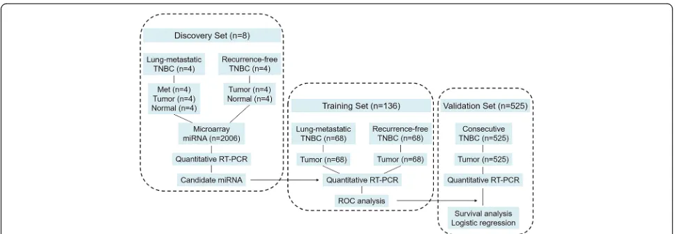
Consecutive female patients with infiltrating TNBC who underwent curative surgical treatment (mastectomy or breast-conserving surgery with axillary evaluation) at the Sun Yat-sen University Cancer Center between January 1999 and December 2013 were recruited for this study. Pa-tients with inflammatory breast carcinomas, synchronic bi-lateral carcinomas, history of other malignant tumors, or incomplete archives of pathological samples were excluded. The subjects’ ER, PR, and HER2 status was evaluated by the department of pathology using immunohistochemistry (IHC) or fluorescence in situ hybridization at the time of diagnosis. TNBC was defined according to the St. Gallen Expert Consensus [27]. Two pathologists independently reevaluated all slides, and any disagreements were resolved by consensus. The pathological tumor stage was assessed according to the criteria described in the seventh edition of the American Joint Committee on Cancer’sAJCC Cancer Staging Manual. The tumors were classified into histo-logical grades I–III according to the Nottingham combined histological grading system. Adjuvant chemotherapy and radiation therapy were administered according to the breast cancer guidelines of the National Comprehensive Cancer Network. Follow-up for these patients was completed by 31 December 2015. In brief, after total RNA isolation and quality control, a final cohort of 669 patients with TNBC with formalin-fixed, paraffin-embedded (FFPE) surgical specimens was allocated to either the discovery set, the training set, or the validation set. Figure 1 depicts the differ-ent phases of the clinical study design.
Discovery set
Four patients with TNBC with lung metastasis underwent pulmonary lobectomy or segmental resection. Paired sur-gical specimens of lung metastasis (Met), primary breast
carcinoma (Tumor), and normal adjacent breast tissue (Normal) were subjected to miRNA profiling. After com-parison with Normal, the miRNAs with altered expression levels in both Met and Tumor were deemed to be associ-ated with lung metastasis. Furthermore, primary tumors and corresponding normal breast tissue from another four patients with TNBC with 10-year disease-free survival (DFS) were also prepared for miRNA profiling; the differ-entially expressed miRNAs in this group were not consid-ered to be involved in promoting lung metastasis.
Training set
Primary tumors collected from a larger cohort of patients with lung metastasis (n= 68) and patients without recur-rence (n= 68) were subjected to quantitative reverse tran-scription polymerase chain reaction (qRT-PCR) analysis of the candidate miRNA. The efficacy of miRNA in diagnos-ing lung metastasis was determined usdiagnos-ing the ROC curve.
Validation set
The prognostic effects of the candidate miRNA for over-all survival (OS), DFS, distant metastasis-free survival (DMFS), and locoregional recurrence-free survival (LRRFS) were evaluated in the validation cohort, which was composed of 525 TNBC samples. Correlations be-tween the candidate miRNA and clinicopathologic char-acteristics were also assessed. Furthermore, associations between the candidate miRNA and sites of distant re-lapse were evaluated using univariate and multivariate logistic regression models.
miRNA profiling
miRNA expression profiling was conducted using the Human miRNA V19.0 Microarray (Platform GPL19730, G4872A; Agilent Technologies, Santa Clara, CA, USA), which consists of probes for 2006 human miRNAs based on Sanger miRBase release 19.0. Total RNA was extracted and purified from FFPE tissue using Recover-AllTMTotal Nucleic Acid Isolation Kit (Ambion, Aus-tin, TX, USA) according to the manufacturer’s instructions. Microarray image information was con-verted to spot intensity values using Feature Extraction version 10.7 software (Agilent Technologies). Raw data were normalized by quantile algorithm using Gene-Spring software version 12.6 (Agilent Technologies). Logarithmic transformation, using log base 2, was then analyzed. A paired-sample t test was used to identify miRNAs with significantly altered expression (fold change >1.5, P< 0.05). All microarray data were deposited in the National Center for Biotechnology
Information Gene Expression Omnibus (GEO)
Quantitative real-time polymerase chain reaction analysis
Total RNA of tissue samples or cells was extracted with TRIzol reagent (Life Technologies, Carlsbad, CA, USA). Reverse transcription of miRNA was done using the TaqMan MicroRNA Reverse Transcription Kit (Applied Biosystems, Foster City, CA, USA). qPCR reactions were performed in triplicate using TaqMan Universal PCR Master Mix (Applied Biosystems) according to the man-ufacturer’s instructions. Results were quantified using ei-ther a 7900 HT sequence detection system (Applied Biosystems) or the IQTM5 Multicolor Real-Time PCR Detection System (Bio-Rad Laboratories, Hercules, CA, USA). All primers were synthesized by Invitrogen (Carlsbad, CA, USA). The expression of miRNA was normalized to U6 small nuclear RNA (snRNA), and fold change was calculated using the comparative cycle threshold (2−TΔΔC) method [28].
Prediction of target genes and enrichment analysis
Putative target genes of miRNAs were predicted using the miRWalk 2.0 database (http://zmf.umm.uni-heidel-berg.de/apps/zmf/mirwalk2/miRretsys-self.html) [29], which integrates eight prediction programs, including DIANA-microT, miRanda, miRDB, miRWalk, RNAhy-brid, PICTAR2, RNA22, and TargetScan. To increase the accuracy of the prediction, only target genes predicted by at least five programs were retained for further ana-lysis. To annotate the biological functions of candidate miRNAs, the list of target genes was submitted to DA-VID Bioinformatics Resources 6.8 (https://david.ncifcrf.-gov/tools.jsp), whereby Gene Ontology (GO) function and Kyoto Encyclopedia of Genes and Genomes (KEGG) pathway enrichment analysis were conducted [30]. Path-ways with fold enrichment >1.5 and P< 0.05 were con-sidered to be of interest.
Cell lines and animals
Nine human breast cancer cell lines (MDA-MB-453, MDA-MB-468, MDA-MB-231, BT-549, MCF-7, T47D, BT-474, SKBR-3, and HCC-38) and one normal mam-mary epithelial cell line (MCF-10A) were obtained from the American Type Culture Collection (Manas-sas, VA, USA). These cells were cultured according to the supplier’s instructions and passaged within 6 months of purchase. Six-week-old female BALB/c nude mice (Animal Experimental Center, Guangdong Academy of Medical Sciences, Guangzhou, China) were maintained under specific pathogen-free condi-tions at the Laboratory Animal Center of Sun Yat-sen University.
Transfection and stably engineered cell lines
Cells cultured in six-well plates were transfected with miRNA mimic, antagomiRNA, RNA, short hairpin RNA, and their corresponding negative controls using Lipofectamine™ 2000 reagent (Invitrogen) according to the manufacturer’s instructions. All miRNA oligonu-cleotides, RNA-expressing lentiviral plasmids, or RNA-interfering lentiviral plasmids were synthesized by GeneCopoeia (Rockville, MD, USA) and are de-scribed in Additional file 1: Figure S1. For later in vivo studies, stable inhibition of miR-629-3p in MDA-MB-231 cells and ectopic expression of miR-629-3p in MCF-7 cells were achieved using the pEZX-AM03 and pEZX-MR03 lentiviral delivery systems, respect-ively. Scrambled nonsilencing vectors were used as negative controls. The stable selection markers were Hygromycin (for anti-miR-629 vectors) and puro-mycin (for pre-miR-629 vectors). All the vectors were constructed by GeneCopoeia and are described in Additional file 2: Figure S2.
[image:3.595.56.540.86.254.2]Cell proliferation assay
Transfected MDA-MB-231 and MCF-7 cells were seeded at a density of 5 × 10³cells per well into 96-well plates and incubated at 37 °C for 24 h. Cell viability was assessed at 24, 48, 72 and 96 h using a 3-(4,5-dimethylthiazol-2-yl)-2,5-diphenyltetrazolium bromide (MTT) assay [31]. Ab-sorbance values were determined at 570 nm (SpectraMax 250 spectrophotometer; Molecular Devices, Sunnyvale, CA, USA).
Cell migration and invasion assays
Cell migration was examined using wound-healing assays. At 24 h after cell seeding, confluent monolayers were wounded linearly by scraping with sterile 1-ml pipette tips. Progression of migration was observed and photographed using an inverted microscope at 48 h after wounding. The extent of wound closure was assessed using cellSens Dimen-sion software (Olympus Life Science, Waltham, MA, USA).
Cell invasion was analyzed using transwell chamber assays as described previously [31]. Briefly, cells were seeded onto the basement membrane matrix present in the insert of a 24-well extracellular matrix (ECM) cul-ture plate (BD Biosciences, San Jose, CA, USA). Medium containing 10% FBS was added to the lower chamber as a chemoattractant. The noninvading cells and ECM were gently removed with a cotton swab after an additional 48 h. Invasive cells located on the lower side of the chamber were stained with crystal violet, imaged, and counted in five randomly chosen fields.
Luciferase assays
To determine whether miR-629-3p regulates LIFRdirectly through the interaction with its predicted 3′-untranslated region (UTR) binding sites, luciferase reporter assays were performed using MDA-MB-231 cells. The full length of the LIFR3′-UTR was synthesized by GeneCopoeia and cloned into the pMIR-REPORT luciferase vector (Ambion) using PCR-generated fragments, which served as the wild-type LIFR 3′-UTR luciferase vector (wt-LIFR). The first seven complementary nucleotides ofLIFR bound to the seed re-gion of miR-629-3p were mutated by site-directed muta-genesis (Stratagene, San Diego, CA, USA) to generate the mutantLIFR3′-UTR luciferase vector (mut-LIFR). The wt-LIFR and mut-wt-LIFR were cotransfected with miR-629-3p mimics or scrambled oligonucleotides into MDA-MB-231 cells. Luciferase activity was measured in cell lysates 48 h after transfection using a dual-light luminescent reporter gene assay kit (Applied Biosystems) according to the manu-facturer’s instructions.
Western blotting
Western blotting was performed as described previously [31]. Briefly, proteins extracted from cell lines were transferred to polyvinylidene difluoride membranes
(EMD Millipore, Billerica, MA, USA), which were blocked with 5% skim milk powder and incubated with primary anti-LIFR antibody (1:200 dilution, ab101228; Abcam, Cambridge, UK). A peroxidase-conjugated sec-ondary antibody (1:2000 dilution) and enhanced chemi-luminescence Western blot detection reagents were used to visualize the target proteins (New England BioLabs, Ipswich, MA, USA), which were quantified with a Bio Image Intelligent Quantifier 1-D (version 2.2.1; Nihon Bio Image Ltd., Tokyo, Japan). An anti-β-actin antibody (Boster Biological Technology, Pleasanton, CA, USA) was used as a protein-loading control.
Animal studies
For tumor growth analysis, the previously mentioned sta-bly transfected cells (5 × 10⁶cells/mouse) were orthotopi-cally injected into the mammary fat pads of mice (n= 5 in each group). Estradiol pellets (0.72 mg, 60-day release; In-novative Research of America, Sarasota, FL, USA) were implanted subcutaneously into mice with MCF7 cells to induce tumor growth according to previous studies [32]. The size of the tumors was measured every 4 days and calculated following the ellipsoid volume formula: π/6 × length × width2(mm3). Primary tumors were excised with the mice under anesthesia 4 weeks after inoculation.
For pulmonary metastasis studies, cells (1 × 10⁵ cells/ mouse) were resuspended in 0.1 ml of PBS and injected into the lateral tail veins of mice (n= 5 in each group). Progression of lung metastasis was monitored using the Xenogen IVIS Spectrum Imaging System (PerkinElmer, Waltham, MA, USA). After a further 8 weeks, lungs were isolated with the mice under anesthesia, and the number of macroscopically visible pulmonary metastases nodules per mouse was counted by three professional pathology experts, with subsequent validation by hematoxylin and eosin (H&E) staining under a microscope [22].
For histological analysis, slides were stained with H&E for microscopic observations. IHC was performed using the standard streptavidin/peroxidase staining method as previously described [33]. Briefly, after deparaffinization and rehydration, slides were incubated with primary antibody against LIFR (1:50 dilution, ab101228; Abcam) at 4 °C overnight. The immunoreactivity assay was performed using the EnVisionTM Detection System (K500711; Dako, Glostrup, Denmark). Results were assessed and captured using an Eclipse 80i microscope (Nikon Instruments, Otawara, Japan).
Statistical analysis
test, respectively. ROC analysis was conducted to test the specificity and sensitivity of miR-629-3p in predicting lung metastasis. The optimal threshold of miR-629-3p was deter-mined by Youden’s index (by maximizing the sum of sensi-tivity and specificity) [34], which was used to dichotomize miR-629-3p expression to high or low levels in the valid-ation cohort. Correlvalid-ations between miR-629-3p and clinico-pathologic characteristics were evaluated using Spearman’s rank correlation coefficient presented withrvalues. Survival analysis was conducted using the Kaplan-Meier method with the log-rank test. Significant prognostic factors in the univariate analysis were included in the Cox proportional hazards regression model for multivariate analyses. HRs from the final models were presented with 95% CIs. Risk factors for sites of distant relapse were assessed in multivari-ate models using logistic regression presented with ORs and 95% CIs.P< 0.05 was considered statistically significant.
Results
miRNAs associated with lung metastasis
All samples in the discovery phase met the quality con-trol parameters recommended by the manufacturer. As shown in Fig. 2a, supervised hierarchical clustering of miRNAs clearly discriminated normal breast tissues from tumors. In addition, the miRNAs (Tumor and Met) in the lung metastasis group clustered separately from miRNAs (Tumor) in the recurrence-free group.
Compared with corresponding normal breast tissues, ex-pression of 45 miRNAs and 17 miRNAs in the lung me-tastasis group was significantly altered in Met and Tumor, respectively, of which 11 miRNAs overlapped (Fig. 2b, c). To distinguish the specific miRNAs associ-ated with lung metastasis, three miRNAs (hsa-miR-21-3p, hsa-miR-21-5p, and hsa-miR-211-3p) were excluded because they were found to be upregulated in primary tumors in the recurrence-free group. The remaining eight dysregulated miRNAs (Fig. 2b) were identified to be putatively involved in the process of lung metastasis, especially miR-629-3p, which was upregulated in both Met and Tumor by more than 70-fold (P< 0.001) (Fig. 2c). Subsequent qRT-PCR to reevaluate the expres-sion levels of the eight miRNAs in the discovery set was consistent with microarray results.
Diagnostic efficacy of miR-629-3p in lung metastasis of TNBC
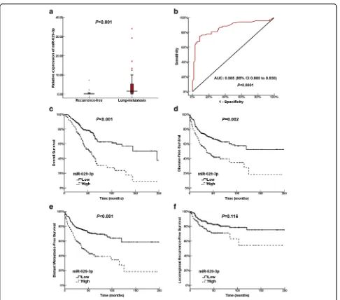
[image:5.595.57.539.436.662.2]To further evaluate the findings of the discovery set, the expression levels of miR-629-3p in primary tu-mors were examined in two independent TNBC co-horts. Characteristics of patients in the training and validation sets are summarized in Additional file 3: Table S1, which were well matched. As shown in Fig. 3a, the expression of miR-629-3p in the lung me-tastasis group was significantly higher than that in
a
b
c
Fig. 2Specific microRNAs (miRNAs) altered in patients with triple-negative breast cancer (TNBC) with lung metastases.aHierarchical clustering represented in the heat map clearly discriminated normal breast tissues from tumors. Samples are presented in columns, and differently expressed miRNAs are presented in rows.Colored barsshow the range of normalized log2signals.bandcVenn diagram and the corresponding table
the recurrence-free group (P< 0.001). ROC curve ana-lysis demonstrated that miR-629-3p yielded high diag-nostic accuracy in discriminating patients with lung metastasis and patients without recurrence (AUC 0.865, 95% CI 0.800–0.930, P< 0.0001) (Fig. 3b). When the sensitivity and specificity of miR-629-3p were 75% and 91.3% respectively (maximum of You-den’s index), the corresponding relative expression value of miR-629-3p was 0.6 (normalized against U6
snRNA expression), which was regarded as the opti-mal cutoff value for further analysis.
Relationships between miR-629-3p and clinicopathologic parameters
[image:6.595.57.543.83.511.2]3p were more likely to develop lymphatic metastasis (r= 0.139,P= 0.001) as well as advanced stage (r= 0.135, P= 0.002). Notably, a strong positive association between the expression of miR-629-3p and lymphovascular inva-sion (LVI) was identified (r= 0.241,P< 0.0001).
Prognostic effects of miR-629-3p on TNBC
The prognostic effects of miR-629-3p were evaluated in the validation set. With a median follow-up period of 43 months (range 3–199 months), the 5-year OS and DFS of this cohort were 65.6% and 57.9%, respectively.
Univariate and multivariate analyses of OS, DFS, DMFS, and LRRFS are summarized in Table 2. Clinicopathologic variables, including lymphatic metastasis, advanced stage, high grade, and LVI, were established as independent poor prognostic factors for OS, DFS, and DMFS. On one hand, it is noteworthy that high expression of miR-629-3p was associated with a decrease in OS (P< 0.001), DFS (P= 0.002), and DMFS (P< 0.001) in univariate analysis (Fig. 3c–e). On the other hand, there was no difference in LRRFS with respect to miR-629-3p expression (P= 0.116) (Fig. 3f). Multivariate analysis demonstrated that miR-629-Table 1Relationships between miR-629-3p and clinicopathologic factors
Variables miR-629-3p Correlation
coefficient
Pvalue
Low High
Number of patients (n= 367) % Number of patients (n= 158) %
Age, years −0.016 0.717
< 35 48 13.1 24 15.2
35–65 291 79.3 121 76.6
> 65 28 7.6 13 8.2
Menopause 0.077 0.077
No 230 62.7 86 54.4
Yes 137 37.3 72 45.6
T 0.071 0.106
T1 138 37.6 49 31.0
T2 180 49.0 82 51.9
T3 32 8.6 17 10.8
T4 17 4.6 10 6.3
N 0.139 0.001
N0 184 50.1 54 34.1
N1 94 25.6 52 32.9
N2 44 12.0 26 16.5
N3 45 12.3 26 16.5
TNM stage 0.135 0.002
I 97 26.4 26 16.5
II 165 45.0 68 43.0
III 105 28.6 64 40.5
Grade 0.054 0.218
I 67 18.3 18 11.4
II 141 38.4 67 42.4
III 159 43.3 73 46.2
Ki-67 0.045 0.298
≤14% 102 27.8 37 23.4
> 14% 265 72.2 121 76.6
LVI 0.241 <0.001
Negative 326 88.8 109 69.0
Positive 41 11.2 49 31.0
[image:7.595.56.538.100.574.2]3p failed to maintain a significant association after adjust-ing for confoundadjust-ing factors. Regardadjust-ing the strong associ-ation of miR-629-3p with lung metastasis, correlassoci-ations between miR-629-3p and other specific metastatic sites were further explored.
Correlations between miR-629-3p and sites of distant metastasis
Of the validation cohort, distant metastasis occurred in 192 (36.6%) patients. Univariate and multivariate ana-lyses of metastatic sites are summarized in Table 3. Multivariate analysis revealed that lymphatic metastasis was an independent risk factor for both lung (P= 0.046) and liver (P= 0.014) metastases. Meanwhile, advanced stage (II/III) independently increased the risk of bone metastasis (P= 0.005). Interestingly, positive LVI was as-sociated with a higher prevalence of relapses in the brain
(P< 0.001). Notably, univariate analysis showed that high miR-629-3p expression not only correlated with lung metastasis (P< 0.001) but also increased the risk of brain metastasis (P= 0.002). However, multivariate logistic re-gression analyses demonstrated that miR-629-3p was an independent risk factor for lung metastasis (OR 4.1, 95% CI 2.5–6.6, P< 0.001) but not for brain metastasis (OR 1.6, 95% CI 0.8–3.1,P= 0.147).
GO annotation and KEGG pathway analysis of miR-629-3p
[image:8.595.58.539.109.267.2]Target genes of miR-629-3p predicted by eight programs are listed in Additional file 4: Table S2. A total of 2267 tar-get genes were predicted by at least five programs. GO an-notation analysis (Additional file 5: Table S3) indicated that the biological process of the candidate targets was significantly (fold enrichment >1.5,P< 0.05) related to cell proliferation, cell migration, cell-matrix adhesion, blood Table 2Univariate and multivariate analyses of prognostic factors for overall survival, disease-free survival, distant metastasis-free survival, and locoregional recurrence-free survival
Variables OS DFS DMFS LRRFS
Univariate Multivariate Univariate Multivariate Univariate Multivariate Univariate Multivariate
Pvalue HR 95% CI Pvalue Pvalue HR 95% CI Pvalue Pvalue HR 95% CI Pvalue Pvalue HR 95% CI Pvalue
Age (years) 0.478 0.384 0.220 0.245
Menopause 0.594 0.910 0.852 0.994
T <0.001 1.2 0.9–1.5 0.069 <0.001 1.2 0.041 <0.001 1.2 0.9–1.5 0.095 <0.001 1.3 0.9–1.7 0.053
N <0.001 1.3 1.0–1.6 0.034 <0.001 1.4 0.007 <0.001 1.3 1.1–1.7 0.012 <0.001 1.3 0.9–1.8 0.083
TNM stage <0.001 1.8 1.1–2.9 0.016 <0.001 1.6 0.021 <0.001 1.9 1.2–2.9 0.007 <0.001 1.4 0.8–2.5 0.247
Surgery 0.774 0.985 0.784 0.610
Chemotherapy 0.167 0.180 0.192 0.057
Grade 0.016 1.4 1.1–1.8 0.004 0.003 1.4 0.001 0.002 1.5 1.2–1.9 <0.001 0.254
Ki-67 0.391 0.499 0.523 0.401
LVI <0.001 2.0 1.4–2.9 <0.001 <0.001 1.4 0.047 <0.001 1.6 1.1–2.3 0.008 0.045 0.9 0.6–1.7 0.971
miR-629-3p <0.001 1.3 0.9–1.8 0.089 0.002 1.1 0.558 <0.001 1.2 0.9–1.7 0.191 0.116
Abbreviations: DFSDisease-free survival,DMFSDistant metastasis-free survival,LRRFSLocoregional recurrence-free survival,LVILymphovascular invasion
Table 3Logistic regression analysis of risk factors for metastatic sites
Variables Lung Brain Liver Bone Othersa
OR 95% CI Pvalue OR 95% CI Pvalue OR 95% CI Pvalue OR 95% CI Pvalue OR 95% CI Pvalue
Age – – – – – – – – – – – – – – –
Menopause – – – – – – – – – – – – – – –
T3/4 1.1 0.7–1.5 0.748 1.4 0.8–2.2 0.167 1.5 0.9–2.3 0.080 0.9 0.6–1.2 0.435 1.7 1.0–3.0 0.049
N1/2/3 1.5 1.0–2.2 0.046 0.8 0.5–1.3 0.348 1.9 1.1–3.1 0.014 1.2 0.8–1.8 0.330 1.3 0.7–2.3 0.405
TNM II/III 1.7 0.8–3.4 0.143 2.0 0.8–5.2 0.138 1.0 0.4–2.6 0.994 3.0 1.4–6.5 0.005 1.4 0.4–4.7 .597
Surgery – – – – – – – – – – – – – – –
Chemotherapy – – – – – – – – – – – – – – –
Grade II/III 1.3 0.9–1.9 0.105 1.7 1.0–2.8 0.039 – – – 1.6 1.1–2.3 0.026 1.9 1.0–3.9 0.044
Ki-67 – – – – – – – – – – – – – – –
LVI (positive) 0.9 0.5–1.7 0.863 4.1 1.9–8.4 <0.001 1.1 0.5–2.4 0.760 1.3 0.7–2.4 0.371 – – –
miR-629-3p (high) 4.1 2.5–6.6 <0.001 1.6 0.8–3.1 0.147 – – – – – – – – –
LVILymphovascular invasion
A blank (–) field indicates that the variable was not significant in the univariate analysis and was not included in the multivariate analysis
a
[image:8.595.56.542.524.707.2]vessel development, and lymph vessel development. KEGG analysis identified that the set of candidate targets of miR-629-3p was significantly enriched in 52 pathways (fold enrichment >1.5, P< 0.05), including several well-known oncogenic signaling pathways such as Ras (P= 0.0002), Hippo (P= 0.0005), transforming growth factor (TGF)-β (P= 0.0009), mitogen-activated protein kinase (MAPK) (P= 0.001), phosphoinositide 3-kinase (PI3K)-Akt (P= 0.003), and Wnt (P= 0.041), as shown in Additional file 6: Table S4.
miR-629-3p promotes proliferation, migration, and invasion of TNBC in vitro
miR-629-3p expression was detected in nine breast cancer cell lines and one mammary epithelial cell line by qRT-PCR analysis. It is of particular note that miR-629-3p ex-hibited a pronounced upregulation in four highly meta-static TNBC cell lines (MDA-MB-453, MDA-MB-468, MDA-MB-231, and BT-549) (P< 0.01) (Fig. 4a), suggest-ing that miR-629-3p is likely associated with subtype spe-cificity and the development of metastasis. According to the expression levels of miR-629-3p, MDA-MB-231 cells and MCF-7 cells were used in further assays.
To analyze the biological effects of miR-629-3p on TNBC, MDA-MB-231 cells were transfected with an in-hibitor of miR-629-3p (anti-miR-629) or scrambled oligo-nucleotide 1 (Scr1), and MCF-7 cells were transfected with an miR-629-3p mimic (miR-629) or scrambled oligo-nucleotide 2 (Scr2). The relative expression levels of miR-629-3p in MDA-MB-231 and MCF-7, which included positive and negative controls for miR-629-3p, are shown in Additional file 7: Figure S3. At 48 h posttransfection, the MTT assay showed that the viability of MDA-MB-231 cells was significantly reduced by anti-miR-629 compared with the Scr1-transfected cells (P< 0.05). However, ectopic expression of miR-629-3p considerably improved the viability of MCF-7 cells (P< 0.05), as shown in Fig. 4b. Analysis of metastasis-related cell motility, such as cellular migration and invasion, using the wound-healing and transwell assays demonstrated that enforced upregulation of miR-629-3p significantly increased the migration and invasion capability of MCF-7 cells, whereas inhibition of miR-629-3p dramatically attenuated the migration and in-vasion capability of MDA-MB-231 cells (P< 0.01), as shown in Fig. 4c, d.
miR-629-3p directly targets the 3′-UTR ofLIFR
According to miRNA binding site enrichment analysis, the putative targets of miR-629-3p are tumor suppressor genes (P= 0.013). The TSGene 2.0 database for updated tumor suppressor genes and their features in pan-cancer (https://bioinfo.uth.edu/TSGene/download.cgi) [35] iden-tified LIFR as the only tumor suppressor gene that was predicted by all eight programs.
The 3′-UTR of LIFR, which contains the miR-629-3p putative binding site, is shown in Fig. 5a. Com-pared with those cotransfected with scrambled oligonucleotides, the luciferase activity of wt-LIFR re-porter constructs in MDA-MB-231 cells exhibited a distinct reduction after cotransfection with miR-629-3p (P< 0.05). In contrast, mutations in the 3′-UTR of LIFR bound to the seed region of miR-629-3p abro-gated the posttranscriptional inhibitory effect of miR-629-3p (P> 0.05) (Fig. 5b).
Furthermore, we transfected the miR-629 or LIFR -interfering plasmid (shLIFR) into MCF-7 cells, both of which led to a significant reduction in LIFR mRNA expression (P< 0.01). Meanwhile, transfection with anti-miR-629 or the LIFR-expressing plasmid (Lv-LIFR) in-creased the expression of LIFR in MDA-MB-231 cells compared with the corresponding negative controls (P< 0.01), as shown in Fig. 5c.
To further confirm the link between LIFR and miR-629-3p, we transfected miR-629, shLIFR, or shLIFR + miR-629 into MCF-7 cells, all of which led to a significant reduc-tion in LIFR protein levels (P< 0.05), especially in the shLIFR + miR-629 group (P< 0.01). Meanwhile, transfec-tion with 629, Lv-LIFR, or Lv-LIFR + anti-miR-629 increased the expression of LIFR in MDA-MB-231 cells significantly (P< 0.05), especially in the Lv-LIFR + anti-miR-629 group (P< 0.01), as shown in Fig. 5d. The inhibitory effect of miR-629 and shLIFR on LIFR expres-sion was similar and also promoted the effect of anti-miR-629 and Lv-LIFR. The above results validated that LIFR was a bona fide target of miR-629-3p.
miR-629-3p promotes tumorigenesis and lung metastasis of TNBC in vivo
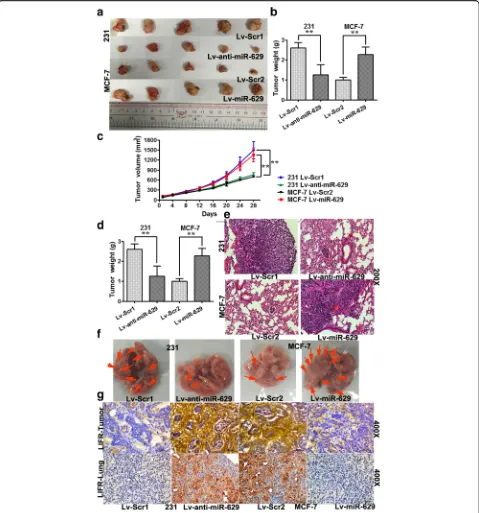
Stably engineered miR-629-3p-inhibiting MDA-MB-231 cells (Lv-anti-miR-629) and pre-miR-629-expressing MCF-7 cells (Lv-miR-629) were inoculated into the mammary gland fat pads of nude mice. We found that the mammary tumors generated from miR-629-3p-inhi-biting MDA-MB-231 cells were significantly smaller than those from scrambled 1-MDA-MB-231 cells (Lv-Scr1) (P< 0.01). Likewise, compared with scrambled 2-MCF-7 cells (Lv-Scr2), ectopic expression of miR-629-3p in MCF-7 cells increased tumor growth significantly (P < 0.01) (Fig. 6a–c).
of miR-629-3p in mice with MDA-MB-231 tumors (Lv-anti-miR-629) (P< 0.01) and was increased by ectopic expression of miR-629-3p in mice with MCF-7 tumors (Lv-miR-629) (P< 0.01). The gross specimens and H&E
staining of the lung sections are shown in Fig. 6e and f. IHC analysis further substantiated that miR-629-3p tar-geted LIFR in both primary tumors and metastatic foci of TNBC with lung metastasis (Fig. 6 g).
[image:10.595.61.537.83.569.2]Discussion
In patients with breast cancer, lung metastasis gene sig-natures have been preliminarily identified [7]. Neverthe-less, the precise molecular mechanisms for the altered expression of miRNAs in lung metastasis of TNBC are unclear. This is the first report showing that miR-629-3p acts as a specific predictor for lung metastasis in TNBC.
Because the characteristics of a primary tumor are usually preserved in metastases [36], we reasoned that
[image:11.595.59.538.86.493.2]normal breast tissues from each patient with lung metas-tasis to evaluate miRNA dysregulation in microarray analyses. miRNA profiling revealed that miR-629-3p is most commonly upregulated in both metastatic lesions and primary carcinomas in patients with TNBC with lung metastasis compared with normal breast tissue. In addition, there was no difference in the expression levels of miR-629-3p between primary tumors and normal breast tissues from patients with TNBC without any re-currence. We then confirmed the predictive capability of miR-629-3p on lung metastasis in two independent TNBC cohorts. Furthermore, in the validation cohort, high expression of miR-629-3p exhibited a strong posi-tive association with lymphatic metastasis and LVI. Not-ably, we also assessed the relationships between miR-629-3p and other distant organs, including the brain, liver, and bone. Interestingly, miR-629-3p was strongly associated with brain metastasis but failed to retain a significant association after multivariate analysis; this suggests that miR-629-3p may act in an organ-specific manner. Subsequent in vitro and in vivo assays sup-ported our findings that miR-629-3p increased the risk of lung metastasis.
Aberrant elevation in the expression of miR-629-3p has been described in various carcinomas, including liver [17], lung [38, 39], colon, lymphoma, ovary, prostate, and testis [20], suggesting that miR-629-3p fulfills a tumor-promoting role in these contexts. However, little is known about the mechanism of miR-629-3p dysregu-lation in breast cancer [18, 19]. In the present study, anti-miR-629-3p drastically reduced the proliferative and migratory capability of MDA-MB-231 cells (TNBC and metastatic). Given that miR-629-3p is expressed at con-siderably higher levels in TNBC cells than in luminal breast cancer cells, we hypothesized that the signaling pathways promoting lung metastasis are partially shared by the two different breast cancer subtypes, in which the miR-629-3p/LIFR axis plays an important role regardless of ER expression, which requires further study. Although miR-629 has been reported to suppress apoptosis through a miRNA-inflammatory feedback loop circuit in hepatocellular carcinoma [17], we found no evidence that apoptosis was affected by miR-629-3p in TNBC cells (data not shown). This discrepancy suggests that the oncogenic roles of miR-629-3p vary in different can-cer types and that these roles are mediated through dif-ferent direct or indirect targets.
With regard to the validated targets of miR-629-3p, Hatziapostolou et al. demonstrated a novel miRNA feed-back inflammatory loop in hepatocellular carcinoma in-volving miR-629, which initiated hepatocellular carcinogenesis by suppressing hepatocyte nuclear factor 4α [17]. miR-629 is also reported to modulate the ex-pression of the Nbs1 gene, which induces DNA repair
and increases lung cancer risk [38]. To identify target genes more precisely, we performed in silico analysis using eight miRNA prediction databases combined with GO enrichment and KEGG pathway analyses. As a re-sult, predicted targets were significantly enriched in many known oncogenic signaling pathways, such as Ras, TGF-β, Hippo, MAPK, 5′-adenosine monophosphate-activated protein kinase, PI3K-Akt, focal adhesion, Wnt, and tumor necrosis factor, providing convincing evi-dence of the oncogenic role of miR-629-3p in TNBC. Moreover, we focused on several tumor suppressor genes; of these, LIFR was a promising candidate target gene of miR-629-3p after evaluation with qRT-PCR, Western blotting, and luciferase reporter assays.
LIFR belongs to the gp130 receptor family, which has been recognized as a tumor suppressor gene in multiple types of cancers [40], including breast [21–23], hepato-cellular [24–26], and pancreatic cancers [41]. Remark-ably, two recent reports have highlighted LIFR as a novel metastasis suppressor in breast cancer. Johnson et al. demonstrated that loss of LIFR allowed dormant breast cancer cells to proliferate and specifically colonize in the bone. Additionally, they found that breast cancer cells which aggressively colonize the lung also lack a functionalLIFRand do not respond to LIF in vitro [21]. In parallel, Ma et al. found thatLIFR was inversely cor-related with lung metastasis in breast cancers, which was identified as the downstream of miR-9 and upstream of Hippo signaling [22]. Additionally, we also note the study by Nandy et al. [42], who demonstrated that miR-125a influenced stem cells by regulating Hippo signaling through LIFR in human primary breast cancer cells. Interestingly, in the discovery set of our study, we found that miR-125b was statistically downregulated in TNBC with good prognosis (fold change 2.3, P= 0.005). These data can be found in the GEO database under accession number [GEO:GSE80038]). Moreover,LIFRhas been re-ported to inhibit metastasis by negatively regulating the PI3K-Akt-matrix metalloproteinase 13 cascade in hepa-tocellular carcinoma [26]. Remarkably, in the present study, KEGG pathway analysis demonstrated that the Hippo signaling pathway (P= 0.00049, fold enrichment 1.99) and PI3K-Akt signaling pathway (P= 0.00327, fold enrichment 1.50) were both significantly affected by miR-629-3p. Taken together, the interactive miRNAs and associated pathways regulating LIFR are of great po-tential to be investigated in future studies.
Conclusions
miR-629-3p on lung metastasis in TNBC and reveals that the suppression of miR-629-3p attenuates pulmon-ary metastasis in experimental breast cancer by directly targeting LIFR, which is an inhibitor of multiple meta-static signaling pathways. Therefore, future studies need to elucidate the specific contributions of miR-629-3p in mediating these predicted pathways, as well as to iden-tify the mechanism of miR-629-3p stimulation in TNBC, which remains crucial to the development of targeted therapies.
Additional files
Additional file 1: Figure S1.Sequences and structures of miR-629-3p mimics, inhibitor of miR-629-3p, andLIFR-expressing andLIFR-interfering lentivirus plasmids. (PDF 280 kb)
Additional file 2: Figure S2.Sequences and structures of anti-miR-629-3p lentiviral vectors, pre-miR-629 lentiviral vectors, and their corresponding scrambled vectors. (PDF 631 kb)
Additional file 3: Table S1.Characteristics of patients with TNBC in the training set and validation set. (PDF 142 kb)
Additional file 4: Table S2.Target genes of miR-629-3p predicted by eight programs. (XLSX 2295 kb)
Additional file 5: Table S3.GO annotation analysis of miR-629-3p. (XLSX 24 kb)
Additional file 6: Table S4.KEGG pathway analysis of miR-629-3p. (XLSX 15 kb)
Additional file 7: Figure S3.Transfection of miR-629, anti-miR-629, Scr1, and Scr2 in MDA-MB-231 and MCF-7 cells. (PDF 53 kb)
Abbreviations
DFS:Disease-free survival; DMFS: Distant metastasis-free survival;
ECM: Extracellular matrix; ER: Estrogen receptor; FFPE: Formalin-fixed, paraffin-embedded; GEO: Gene Expression Omnibus; GO: Gene Ontology;
H&E: Hematoxylin and eosin; HER2: Human epidermal growth factor receptor 2; IHC: Immunohistochemistry; KEGG: Kyoto Encyclopedia of Genes and Genomes; LIFR: Leukemia inhibitory factor receptor; LRRFS: Locoregional recurrence-free survival; LVI: Lymphovascular invasion; Lv-LIFR:LIFR -expressing lentiviral plasmid; MAPK: Mitogen-activated protein kinase; Met: Lung metastasis; miRNA: MicroRNA; Normal: Normal adjacent breast tissue; MTT: 3-(4,5-dimethylthiazol-2-yl)-2,5-diphenyltetrazolium bromide; mut-LIFR: MutantLIFR3′-untranslated region luciferase vector; OS: Overall survival; PI3K: Phosphoinositide 3-kinase; PR: Progesterone receptor; qRT-PCR: Quantitative real-time polymerase chain reaction; Scr1: Scrambled oligonucleotide 1; Scr2: Scrambled oligonucleotide 2; shLIFR:LIFR short-hairpin RNA plasmid; snRNA: Small nuclear RNA; TGF-β: Transforming growth factor-β; TNBC: Triple-negative breast cancer; Tumor: Primary breast carcinoma; UTR: Untranslated region; wt-LIFR: wild-typeLIFR3′-untranslated region luciferase vector
Acknowledgements
The authors thankfully acknowledge the patients for donating their surgical samples that made this work possible.
Funding
This work was supported by the National Natural Science Foundation of China (81402183), the Priming Scientific Research Foundation for the Junior Teachers of Medicine in Sun Yat-sen University (13ykpy48), and the Young Investigator Award from Sun Yat-sen University Cancer Center (YIA201413).
Availability of data and materials
The datasets were generated and analyzed using the miRWalk 2.0 database (http://zmf.umm.uni-heidelberg.de/apps/zmf/mirwalk2/miRretsys-self.html), DAVID Bioinformatics Resources 6.8 (https://david.ncifcrf.gov/tools.jsp), Venny
2.1.0 (http://bioinfogp.cnb.csic.es/tools/venny/), and the TSGene 2.0 database (https://bioinfo.uth.edu/TSGene/download.cgi). Raw microarray data were deposited in the National Center for Biotechnology Information GEO database [GEO:GSE80038] (http://www.ncbi.nlm.nih.gov/geo/query/ acc.cgi?acc=GSE80038).
Authors’contributions
JW conceived of and designed the experiments. CS and HT conducted experiments. CZ performed histopathological review. JT, XL, and BC performed sample collection. JW drafted the manuscript. XX contributed to the analysis of data and revised the manuscript. All authors performed critical review of the manuscript. All authors read and approved the final manuscript.
Authors’information
Not applicable.
Competing interests
The authors declare that they have no competing interests.
Consent for publication Not applicable.
Ethics approval and consent to participate
Ethical approval of the human and animal work was provided by Sun Yat-sen University Cancer Center Institutional Review Board (IRB) (approval number GZR2017-052) and the Institutional Animal Care and Use Committee of Sun Yat-sen University Cancer Center (approval number L102012017000D), respectively. Prior written consent from patients was obtained for the use of clinical materials for research purposes.
Publisher’s Note
Springer Nature remains neutral with regard to jurisdictional claims in published maps and institutional affiliations.
Author details
1
Department of Breast Oncology, Sun Yat-sen University Cancer Center, State Key Laboratory of Oncology in South China, Collaborative Innovation Center for Cancer Medicine, No.651 Dongfeng East Road, Yuexiu District,
Guangzhou, Guangdong 510060, People’s Republic of China.2Department of
Pathology, Sun Yat-sen University Cancer Center, State Key Laboratory of Oncology in South China, Collaborative Innovation Center of Cancer Medicine, Guangzhou, Guangdong 510060, People’s Republic of China.
Received: 4 March 2017 Accepted: 5 June 2017
References
1. Siegel RL, Miller KD, Jemal A. Cancer statistics, 2015. CA Cancer J Clin. 2015; 65(1):5–29.
2. Perou CM, Sorlie T, Eisen MB, van de Rijn M, Jeffrey SS, Rees CA, et al. Molecular portraits of human breast tumours. Nature. 2000;406(6797):747–52.
3. Foulkes WD, Smith IE, Reis-Filho JS. Triple-negative breast cancer. N Engl J Med. 2010;363(20):1938–48.
4. Shah SP, Roth A, Goya R, Oloumi A, Ha G, Zhao Y, et al. The clonal and mutational evolution spectrum of primary triple-negative breast cancers. Nature. 2012;486(7403):395–9.
5. Kennecke H, Yerushalmi R, Woods R, Cheang MC, Voduc D, Speers CH, et al. Metastatic behavior of breast cancer subtypes. J Clin Oncol. 2010;28(20): 3271–7.
6. Nguyen DX, Bos PD, Massagué J. Metastasis: from dissemination to organ-specific colonization. Nat Rev Cancer. 2009;9(4):274–84.
7. Minn AJ, Gupta GP, Siegel PM, Bos PD, Shu W, Giri DD, et al. Genes that mediate breast cancer metastasis to lung. Nature. 2005;436(7050):518–24. 8. Gupta GP, Massagué J. Cancer metastasis: building a framework. Cell. 2006;
127(4):679–95.
9. Joyce JA, Pollard JW. Microenvironmental regulation of metastasis. Nat Rev Cancer. 2009;9(4):239–52.
11. Ma L, Teruya-Feldstein J, Weinberg RA. Tumour invasion and metastasis initiated by microRNA-10b in breast cancer. Nature. 2007;449(7163):682–8. 12. Ma L, Young J, Prabhala H, Pan E, Mestdagh P, Muth D, et al. miR-9, a MYC/
MYCN-activated microRNA, regulates E-cadherin and cancer metastasis. Nat Cell Biol. 2010;12(3):247–56.
13. Liu C, Kelnar K, Liu B, Chen X, Calhoun-Davis T, Li H, et al. The microRNA miR-34a inhibits prostate cancer stem cells and metastasis by directly repressing CD44. Nat Med. 2011;17(2):211–5.
14. Jiang Q, Wang Y, Hao Y, Juan L, Teng M, Zhang X, et al. miR2Disease: a manually curated database for microRNA deregulation in human disease. Nucleic Acids Res. 2009;37(Database issue):D98–104.
15. Lewis BP, Burge CB, Bartel DP. Conserved seed pairing, often flanked by adenosines, indicates that thousands of human genes are microRNA targets. Cell. 2005;120(1):15–20.
16. Costa PM, Pedroso de Lima MC. MicroRNAs as molecular targets for cancer therapy: on the modulation of microRNA expression. Pharmaceuticals (Basel). 2013;6(10):1195–220.
17. Hatziapostolou M, Polytarchou C, Aggelidou E, Drakaki A, Poultsides GA, Jaeger SA, et al. An HNF4α-miRNA inflammatory feedback circuit regulates hepatocellular oncogenesis. Cell. 2011;147(6):1233–47.
18. Lu J, Getz G, Miska EA, Alvarez-Saavedra E, Lamb J, Peck D, et al. MicroRNA expression profiles classify human cancers. Nature. 2005;435(7043):834–8. 19. Volinia S, Calin GA, Liu CG, Ambs S, Cimmino A, Petrocca F, et al. A
microRNA expression signature of human solid tumors defines cancer gene targets. Proc Natl Acad Sci U S A. 2006;103(7):2257–61.
20. Navon R, Wang H, Steinfeld I, Tsalenko A, Ben-Dor A, Yakhini Z. Novel rank-based statistical methods reveal microRNAs with differential expression in multiple cancer types. PLoS One. 2009;4(11), e8003.
21. Johnson RW, Finger EC, Olcina MM, Vilalta M, Aguilera T, Miao Y, et al. Induction of LIFR confers a dormancy phenotype in breast cancer cells disseminated to the bone marrow. Nat Cell Biol. 2016;18(10):1078–89. 22. Chen D, Sun Y, Wei Y, Zhang P, Rezaeian AH, Teruya-Feldstein J, et al. LIFR is
a breast cancer metastasis suppressor upstream of the Hippo-YAP pathway and a prognostic marker. Nat Med. 2012;18(10):1511–7.
23. Iorns E, Ward TM, Dean S, Jegg A, Thomas D, Murugaesu N, et al. Whole genome in vivo RNAi screening identifies the leukemia inhibitory factor receptor as a novel breast tumor suppressor. Breast Cancer Res Treat. 2012;135(1):79–91. 24. Blanchard F, Tracy E, Smith J, Chattopadhyay S, Wang Y, Held WA, et al.
DNA methylation controls the responsiveness of hepatoma cells to leukemia inhibitory factor. Hepatology. 2003;38(6):1516–28. 25. Okamura Y, Nomoto S, Kanda M, Li Q, Nishikawa Y, Sugimoto H, et al.
Leukemia inhibitory factor receptor (LIFR) is detected as a novel suppressor gene of hepatocellular carcinoma using double-combination array. Cancer Lett. 2010;28;289(2):170–7.
26. Luo Q, Wang C, Jin G, Gu D, Wang N, Song J, et al. LIFR functions as a metastasis suppressor in hepatocellular carcinoma by negatively regulating phosphoinositide 3-kinase/AKT pathway. Carcinogenesis. 2015;36(10):1201–12. 27. Goldhirsch A, Wood WC, Coates AS, Gelber RD, Thürlimann B, Senn HJ, et al.
Strategies for subtypes—dealing with the diversity of breast cancer: highlights of the St. Gallen International Expert Consensus on the Primary Therapy of Early Breast Cancer 2011. Ann Oncol. 2011;22(8):1736–47. 28. Livak KJ, Schmittgen TD. Analysis of relative gene expression data using
real-time quantitative PCR and 2−TΔΔCmethod. Methods. 2001;25(4):402–8.
29. Dweep H, Gretz N. miRWalk2.0: a comprehensive atlas of microRNA-target interactions. Nat Methods. 2015;12(8):697.
30. Huang DW, Sherman BT, Lempicki RA. Systematic and integrative analysis of large gene lists using DAVID bioinformatics resources. Nat Protoc. 2009;4(1):44–57.
31. Wang J, Kang WM, Yu JC, Liu YQ, Meng QB, Cao ZJ. Cadherin-17 induces tumorigenesis and lymphatic metastasis in gastric cancer through activation of NF-κB signaling pathway. Cancer Biol Ther. 2013;14(3):262–70.
32. Salvo VA, Boué SM, Fonseca JP, Elliott S, Corbitt C, Collins-Burow BM, et al. Antiestrogenic glyceollins suppress human breast and ovarian carcinoma tumorigenesis. Clin Cancer Res. 2006;12(23):7159–64.
33. Wang J, Zhang C, Chen K, Tang H, Tang J, Song C, et al. ERβ1 inversely correlates with PTEN/PI3K/AKT pathway and predicts a favorable prognosis in triple-negative breast cancer. Breast Cancer Res Treat. 2015;152(2):255–69.
34. Schisterman EF, Perkins NJ, Liu A, Bondell H. Optimal cut-point and its corresponding Youden index to discriminate individuals using pooled blood samples. Epidemiology. 2005;16(1):73–81.
35. Zhao M, Kim P, Mitra R, Zhao J, Zhao Z. TSGene 2.0: an updated literature-based knowledgebase for tumor suppressor genes. Nucleic Acids Res. 2016; 44(D1):D1023–31.
36. Chiang AC, Massagué J. Molecular basis of metastasis. N Engl J Med. 2008; 359(26):2814–23.
37. Shyamsundar R, Kim YH, Higgins JP, Montgomery K, Jorden M, Sethuraman A, et al. A DNA microarray survey of gene expression in normal human tissues. Genome Biol. 2005;6(3):R22.
38. Yang L, Li Y, Cheng M, Huang D, Zheng J, Liu B, et al. A functional polymorphism at microRNA-629-binding site in the 3′-untranslated region ofNBS1gene confers an increased risk of lung cancer in Southern and Eastern Chinese population. Carcinogenesis. 2012;33(2):338–47. 39. Cazzoli R, Buttitta F, Di Nicola M, Malatesta S, Marchetti A, Rom WN, et al.
microRNAs derived from circulating exosomes as noninvasive biomarkers for screening and diagnosing lung cancer. J Thorac Oncol. 2013;8(9):1156–62. 40. Hergovich A. YAP-Hippo signalling downstream of leukemia inhibitory factor
receptor: implications for breast cancer. Breast Cancer Res. 2012;14(6):326. 41. Ma D, Jing X, Shen B, Liu X, Cheng X, Wang B, et al. Leukemia inhibitory
factor receptor negatively regulates the metastasis of pancreatic cancer cells in vitro and in vivo. Oncol Rep. 2016;36(2):827–36.
42. Nandy SB, Arumugam A, Subramani R, Pedroza D, Hernandez K, Saltzstein E, et al. MicroRNA-125a influences breast cancer stem cells by targeting leukemia inhibitory factor receptor which regulates the Hippo signaling pathway. Oncotarget. 2015;6(19):17366–78.
• We accept pre-submission inquiries
• Our selector tool helps you to find the most relevant journal
• We provide round the clock customer support
• Convenient online submission
• Thorough peer review
• Inclusion in PubMed and all major indexing services
• Maximum visibility for your research
Submit your manuscript at www.biomedcentral.com/submit