0022-538X/82/100249-07$02.00/0
Copyright 01982,American Society for Microbiology
Assessment of the Base Sequence Homology
Between the
Two
Subtypes of
Equine
Herpesvirus
it
GEORGEP.ALLEN*ANDLLOYD W. TURTINEN
Departmentof Veterinary Science, University of Kentucky, Lexington, Kentucky 40546
Received 26April1982/Accepted4June1982
The magnitude of the genetic relatedness of the two antigenic subtypes of equine herpesvirus 1 (EHV-1) was determined by DNA-DNA reassociation kinetics. Denatured, labeled viral DNAfromoneEHV-1 subtypewas allowedto
reassociate in thepresence orabsenceof the unlabeledheterologous viral DNA. The initialrateof reassociation of either labeled viralDNA was increased by the
presence of the heterologous viral DNA to an extent indicating 10 to 20%
homology betweenthe two EHV-1 genomes. Similar estimatesof the amountof homology between the genomes of the two EHV-1 subtypes were obtained by determining the maximum fraction of labeled viral DNA that could be made resistant to S1 nuclease by hybridization with a large molar excess of the unlabeled, heterologous viral DNA. Analysis of the thermal stability of the
subtype 1-subtype 2 heteroduplex DNA indicated approximately 30%o basepair
mismatching within the hybrid DNA molecules. Cross-hybridization of 2p_
labeled virion DNA to nitrocellulose blots of restriction endonuclease cleavage fragments of each EHV-1 subtype DNA indicated that the observed homology
betweenthe twoviruses was nonuniformly distributed within the viralgenome. Nohomology couldbe detected between the DNAof either EHV-1 subtype and thatofastrain of equine cytomegalovirus (EHV-2). The datasuggestthat thetwo
biotypes of EHV-1 have arisen by divergent evolution fromacommonprogenitor herpesvirus.
Equine herpesvirus1 (EHV-1) is the
etiologi-cal agent of both upper respiratory infections
(rhinopneumonitis) and contagious virus abor-tion of horses (2). Two antigenic subtypes of EHV-1 have beenrecognized since 1959, when Shimizu and co-workers (12) demonstrated that EHV-1 isolates could be grouped into either of
two distinct but cross-neutralizing serotypes.
Subsequent studies have revealed other major
differences in the growth rate, in vitro host
range, adaptability to hamsters, and
abortigeni-city of the two EHV-1 subtypes (3, 4, 14; R.
Burrows, Abstr. Annu. Cong. Br. Equine Vet.
Assoc. 20th, Cambridge, England, p. 18, 1981).
Moreover, members representative of the two
virus subtypes exhibit differences in the electro-phoretic mobility ofatleast sevenof their
struc-tural glycoproteins when analyzed in sodium
dodecyl sulfate-polyacrylamide gels (16).
Inves-tigations by Burrows and Goodridge (3) first suggested that, whereas the majority of
respira-tory isolates of EHV-1 fall into one antigenic subtype, viruses isolated from aborted equine fetuses belong to the other EHV-1 subtype.
Recent comparisons ofthe restriction
endonu-tPublished as paper no. 824-103 by permission of the Director oftheKentuckyAgriculturalExperiment Station.
clease cleavage patterns of the two viral
ge-nomes suggested a greater degree of genetic
dissimilarity than was originally suspected,
since thetwovirus DNAs haveveryfew
restric-tion enzyme cleavage sites in common (10, 15,
16).
The purpose of the studies described in this
paper was to investigate the degree of base
sequencehomology between the genomes ofthe
twosubtypes of EHV-1,usingthetechniquesof blothybridization, DNA-DNAreassociation
ki-netics, and S1 nuclease analysis. Our results showed that thetwoequineherpesviruses share approximately 17% of theirgenome sequences.
MATERIALS AND METHODS
Viruses and cell culture. The viruses used in this study were the Army 183 (A-183; subtype 1) and Kentucky T2 (Ky-T2; subtype 2) strains of EHV-1 and theKy-T162 strain ofequinecytomegalo-virus
(EHV-2). The origin and propagation of these viruses and
theirpurificationfrom the culture medium of infected
equine dermal fibroblasts (KyED cells) have been
describedpreviously(16).
Isolation and labelingofDNA. The procedures for isolation of DNA from purified EHV virions or from uninfected KyED cells by sodium dodecyl sulfate-pronase digestion and phenol extraction have been
249
on November 10, 2019 by guest
http://jvi.asm.org/
described elsewhere (1). EHV-1 DNAs werelabeledto
specific radioactivities of 3 x10⁶ to4 x106 cpm/,Lg by infecting KyED cells in the presence of 800 ,uaCi of [3H]thymidine (77 Ci/mmol) per ml of culture medium. Viral DNAs for blot hybridization werelabeled invitro by nick translation (9)with[32P]dCTP(7,000Ci/mmol) to a specific activity of108 cpm/,g. All DNAs for hybridization were degraded tofragments 400 to 500 nucleotides in lengthby boiling in 0.3 N NaOH for 20 min (11). After fragmentation, the DNAsolutions were neutralized and dialyzed against TNE buffer (0.1 M Tris-hydrochloride [pH 8.11-0.015 M EDTA-0.15 M NaCl).
Hybridization to nitrocellulose blots of restriction fragments. Unlabeled EHV DNAs were digested to
completion with therestriction endonucleasesBamHI and EcoRl. The resulting cleavage fragments were
separated by electrophoresisin horizontal slab gelsof 0.6% agarose and then blot-transferred to nitrocellu-losefor hybridization (13). The conditions for nitrocel-lulose filter hybridization and subsequent washing were those described by Jeffreys and Flavell (6). Autoradiography of thefilter sheets after hybridization was performed at -80°Cwith Kodak XAR-5 film and Lighting-Plus intensifyingscreens(Du PontCo., Wil-mington, Del.).
Kinetic hybridization.The strategyanddesign of the DNA-DNA reassociation kinetics experiments for quantitation of the extent of base sequencehomology between two viral genomes has been previously de-scribed in considerable detail (1, 5). Hybridizations were performed at 65°C in TNE buffer. Reaction mixtures consisted of 100 ng of labeled viral probe
DNAperml, 0 to 2 ,ug of unlabeled test DNA per ml, and enough calf thymus DNA to give a total DNA concentration of 2pug/ml.The mixturesweresealed in 100-,u microcapillary pipettes, denatured for 7 minat
110°C in an ethylene glycol bath, and allowed to
reanneal at 65°C. Samples were taken after various
periods of incubation, quickly chilled in ice, and
frozen until assayed by chromatographyon hydroxy-apatite columns.
Reassociation of labeled viralDNA wasmonitored by hydroxyapatite (BioGel HTP; Bio-Rad Labora-tories, Richmond, Calif.) chromatographyat60°C,as
described in detail by Sharp et al. (11). The DNA eluting from the columnat0.14M phosphate (single-stranded DNA) or 0.4 M phosphate buffer, pH 6.8
(double-stranded DNA) was precipitated with
tri-chloroacetic acid, collected on nitrocellulose
mem-brane filters (type BA85; Schleicher & Schuell Co., Keene, N.H.), and counted by liquid scintillation spectroscopy.
Thehybridization resultswereplotted bythelinear transformation method of Wetmur and Davidson(17), with the time interval ofhybridizationasthe indepen-dentvariable and thereciprocal ofthefraction ofDNA
remaining single-stranded asthe dependent variable; the method of least-squares regression was used to determine theslopes of the plotted data.
Thermal elution chromatography of DNA-DNA
du-plexes. The extent of base pairmismatching present within the EHV-1 subtype 1-subtype 2heteroduplex
DNAs wasestimatedbycomparison of their thermal stability with that of the homoduplex DNA of each virussubtype. Reannealed viralDNAin0.14 M
sodi-umphosphate (pH6.8)-0.4% sodiumdodecyl sulfate
(PB) waspoured intoajacketed column containing 1 g of hydroxyapatite at 40°C. Single-stranded DNAwas
washed through thecolumn with PB, and the column temperature was then raised in 2°C increments to 90°C. After each temperature equilibration, 2 ml ofa
solution of PBcontaining 8 M urea (7)wasallowedto
flowthrough the column, mixed with RIA-Fluor (New England Nuclear Corp., Boston, Mass.) and assayed forradioactivity.
RESULTS
Blot hybridization. Restriction endonuclease
fragments ofEHV-1, strain A-183 (subtype 1);
EHV-1, strain Ky-T2 (subtype 2); and EHV-2,
strain Ky-T162 were transferred from agarose
gelstonitrocellulose sheets and then hybridized with each of the three viral DNAs that had been
radiolabeledby nick translation with [32P]dCTP,
as described in Materials and Methods. Photo-graphs of the ethidium bromide-stained gels be-fore transfer to nitrocellulose and
autoradio-grams of the blots after DNA transfer and
hybridization are shown in Fig. 1 and reveal several notable features.
No hybridization could be detected, even af-ter prolonged exposure of the film, between
EHV-2DNA and that ofeitherof the two EHV-1 subtypes. Hybridization between the DNAs
from the two subtypes ofEHV-1 was readily
demonstrated but wasconsiderably less efficient
than that between each virus genome and its homologous DNA restriction fragments.
Auto-radiographic exposure times for hybridization between heterologous viralDNAs (i.e., EHV-1
subtype 1 x EHV-1 subtype 2) were approxi-matelyfivetimes longer thanthoserequired for the same degree offilm darkening for
homolo-goushybridizations.
Homology between the DNAs of the two
EHV-1 subtypes was not restricted to any
se-lectedportion of thegenomebutwasdistributed
overthe entire DNA molecule. However, some
restriction fragments appearedtoexhibita great-er degree of cross-hybridization than others,
indicatingthatsomeregions of thegenome may
have been more highly conserved than others during divergent evolution ofthe twovirus
sub-types.
Reassociation kinetics andcomputation of
ho-mology.Foranalysis of the kinetics of
reassocia-tion of mixtures ofDNAsfrom bothsubtypes of EHV-1, labeledDNAfromoneEHV-1subtype
was denatured and allowed to reanneal in the
presenceofa4-, 10-, or20-foldexcessof
unla-beledDNAfrom the heterologous virussubtype
(Fig. 2). In each instance, the initial rate of reassociation of the labeled viral DNA was
increased by the presence ofthe heterologous
DNA in the hybridization mixture, indicating
sequences shared by the two herpesvirus
ge-nomes. However, the additionoftheunlabeled
on November 10, 2019 by guest
http://jvi.asm.org/
A B C
- * w_n--. - ,, .* *_.
a b c a b c a b c
*i|L W.:
v
k _
F* l
4,.
4..:,
_
A B C
a
R
[image:3.496.106.398.73.450.2]1
2
FIG. 1. Hybridization between 32P-labeled EHV DNAs and fragments of the viral DNAs generated by restriction endonucleases. EHV-2, EHV-1 subtype 2, and EHV-1 subtype 1 DNAs (lanes a, b, and c,
respectively) were cleaved with either BamHI (1) or EcoRI (2). The resulting cleavage fragments were
electrophoresedon0.6%agaroseslabgels,stainedwith ethidiumbromide(topframes),and then blot-transferred
tonitrocellulose forhybridization(bottomframes)with either EHV-1 subtype1(A), EHV-1 subtype2(B),or
EHV-2(C)[32P]DNA.
second viral DNA tothe hybridization reaction resulted inabiphasic reassociation curve (Fig.
2). These results indicate thatthe DNA of each
EHV-1 subtype contained sequences
homolo-gous toonlyafractionof theheterologous
EHV-1 genome (5).
Fujinagaetal. (5)havederiveda formula for
quantitating the amount of base sequence
ho-mology betweentwoviral genomes utilizing the
followingtwoparametersfromthe DNA
reasso-ciation kineticstest: (i)thedegreeof increase of
probe reassociationratecausedby the presence
of the second unlabeled viral DNAand (ii) the
molarratio oftest toprobe DNA. Thebasis of theFujinaga equation istoreplotthe
experimen-tal data byamethod thatemphasizes the initial
reassociation reaction of the labeled viral
genome inthe presence of the unlabeled second
viralgenome. Suchplots areshownin Fig. 3 for
thereassociation oflabeled subtype 1 viral DNA
fragments in the presence of three different
concentrations of unlabeled subtype 2 DNA
fragments and vice versa. The initial rate of
probe reassociation, represented by the slopes ofthe lines, was,ineach case,dependentupon
the concentration of added test DNA. The
slopes of the plots of single-stranded DNA/
double-strandedDNAratios(CSJCds)versus lit
duringthe initial period of reassociation of the
probe DNA in the presence (A) and in the 44,
on November 10, 2019 by guest
http://jvi.asm.org/
A
B
ii,8
1.2-1.0
3 6 9 12 3 6 9 12
HOURS AT 650 C
FIG. 2. Reassociation of denatured EHV-1 subtype 1 (A) and subtype 2 (B) [3H]DNA in the presence of unlabeled DNA from the heterologous virus. Hybridizations were performed at65°C in TNEbuffer for various
lengths oftime, and the fraction of DNA remaining single-stranded
(fJs)
was determined byhydroxyapatitechromatography. A 0.1->g amount of denatured EHV-1[3H]DNApermlwasallowedtoreannealin the presence ofnounlabeled viral DNA(0);0.4 ,ug(0),1.0 ,ug(A), or 2.0 ,ug (X)of unlabeledheterologousviral DNA per ml (n=0, 4, 10, or 20, respectively); or 2.0 ,ug of unlabeled KyED cell DNA per ml (+). The total concentration of DNA in eachhybridization reaction was adjusted to 2.1 ,ug/ml with calf thymus DNA.
150.
100-i
50-A
2
B
2
HOURS
FIG. 3. Reassociation of0.1 1lgof EHV-1subtype1(A)orsubtype2(B)[3H]DNAperml inthe presence of different concentrations of the unlabeled heterologous subtypeviral DNA togive unlabeled-to-labeled DNA ratios (n values) of0(0), 4(0),10(A),or20(+). Hybridizationswereperformedat65°Cin TNE buffer for various lengths of time, and the ratio of single- to double-stranded DNA (CSS/CdS) was determined by hydroxyapatitechromatography.
l l
on November 10, 2019 by guest
http://jvi.asm.org/
[image:4.496.56.443.59.311.2] [image:4.496.103.393.403.624.2]absence (AO) of the unlabeled DNA, together
withthe ratiooftest toprobe DNA(n), can then
be used to calculate the fraction ofsequences
shared by the two viral genomes
(xlf)
from thefollowing relationship (5): xlf = [1 -
(A/AO)]/
[n(A/Ao)]. Using this equation, the fraction of
nucleotidesequencesshared betweenthe DNAs
of thetwo EHV-1 subtype viruses was
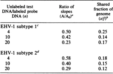
comput-ed(Table 1). Itcanbe seen that the two herpes-viruses share only about 17% of their genome sequences.
Analysis of homology by Si nuclease analysis.
Heteroduplexes of subtype 1-subtype 2 DNAs wereprepared by annealinglow concentrations
of each labeledviral DNA witha100-foldexcess
of unlabeledheterologousviralDNA; hybridiza-tion was allowed to continue at 65°Cfor 12 h.
Under theseconditions, homoduplexes
account-ed for less than 25% of the labeled hybrids,
whereas heteroduplex formation went virtually
to completion. Samplestakenatfrequent
inter-vals during the hybridization reactions were
monitored forreassociation of denatured DNA
by Si nuclease analysis. The values for
hetero-duplex formation were corrected for
self-rean-nealing of labeled DNAmolecules.
The resultsof thistypeofanalysisof homolo-gy indicated that, in the presence of a large
excessofheterologousviralDNA,
approximate-ly 10 to 12% of the EHV-1 genome could be
driven into Si-resistant, double-stranded
mole-cules (datanotshown).
Thermal stability of EHV-1 heteroduplex
DNAs.Because thedegree of nucleotide
comple-mentarity innucleic acid hybridsis reflected in
TABLE 1. Fraction of genome sequences shared by the two subtypes of EHV-1
Unlabeled test Ratio of Shared DNA/labeledprobe slopes fractionof
DNA(n) ~~(A/AO)a genome
DNA(n)
(X/f)b
EHV-1 subtype11
4 0.50 0.25
10 0.42 0.14
20 0.23 0.17
EHV-1 subtype2d
4 0.58 0.18
10 0.40 0.15
20 0.29 0.12
aRatio of slopes of curves for
CSS/Dd,
versuslit
during the intial periods of reassociation of labeled
EHVprobe DNA in the presence (A)and absence(AO)
ofunlabeled heterologous test DNA.
b Calculated by the equation (xlf) = [1 - (A/AO)]/ [n(A/Ao)].
c EHV-1 subtype 1 [3H]DNA was reassociated in the presenceof unlabeled subtype 2 DNA.
d EHV-1 subtype 2 [3H]DNA was reassociated in thepresence of unlabeled subtype 1 DNA.
02 0
1.23
B
D
44.8 32.2
2- .4
1L-40 5060 80 90 40 50 070 80 90
TEMPERATURE(°C)
FIG. 4. Thermal stability of EHV-1 subtype 1-sub-type2heteroduplex DNA. A 0.05->g amount of EHV-1subtype1(Aand C) or subtype 2 (B and D)[3H]DNA
per ml wasdenatured and allowed to reanneal for 4 h
at 65°C with a 200-fold molar excess of unlabeled heterologous viral DNA. A 2-,ug amount of EHV-1
subtype1(A and C) or subtype 2 (B and D)[14C]DNA
per mlwas denatured and allowed to reassociate at 65°Cfor 24 h to form homoduplex DNAs. Hetero- and homoduplexes were diluted into PB and adsorbed onto
ahydroxypatite column at 40°C. The column tempera-turewasraised stepwise to 90°C, and denatured DNA was removed at each step with 2 ml of PB containing 8 M urea. Eachfraction was mixed with 10 ml of RIA-Fluor and counted forboth tritium(0)and carbon-14 (0)radioactivity. Radioactivity present in each eluted fraction at the indicated temperature is plotted in (A) and(B); the cumulative radioactivity is plotted in (C) and (D).
their thermal stability, thermal chromatography of subtype 1-subtype 2 heteroduplexes on
hy-droxyapatitewas used to determine the extent of
mismatchedbase-pairing present within the
hy-bridviral DNAs.
Tritium-labeled heteroduplexes were
ad-sorbedtohydroxyapatite at 40°C along with
14C-labeled homoduplex DNA. By increasing the
temperature of the hydroxyapatite column,
melting ofthe DNA-DNA duplexes was
moni-toredbythe appearance of labeled
single-strand-ed DNA inthe column effluent (Fig. 4).
Homo-VOL 44,1982
on November 10, 2019 by guest
http://jvi.asm.org/
[image:5.496.260.450.69.364.2] [image:5.496.51.244.459.585.2]duplexes of either viral subtypeDNA possessed
steep melting profiles with midpoint
tempera-turesof 76 to78°Cin 8 Murea. EHV-1
heterodu-plex DNAs, in contrast, were eluted from
hy-droxyapatite over a broad temperature range
whose midpoint was 20°C below that of the
homoduplexes.
DISCUSSION
Inthisreport,theextentofgenetichomology
betweenthe tworecognized molecular subtypes of EHV-1 wasdeterminedby akinetic analysis ofviral DNA-DNAreassociation. Results of this studyindicate that the twoherpesviruses share only about 17% of theirgenome nucleotide
se-quences. Similar estimates of homology
be-tween thetwoviralDNAs (11%)wereobtained
by analysis of the maximum fraction of each viral genomethatcould bemade resistanttoS1 nuclease after hybridization with alargeexcess
of theheterologous viral DNA. These valuesare
approximately one-third of theamountof DNA homology (47%) exhibited between the two
types of herpes simplex virus(7).
Because the two EHVs, during their
evolu-tionary descent fromacommon ancestor, have accumulated basesubstitutions (i.e., mutations)
intheir DNAs, heteroduplexes formed between thetwoviral genomeswill contain
non-comple-mentary regions representing the positions at
which base substitutions have occurred. These mispaired bases reduce the stability of the
hy-bridsto an extentthata1.5%mismatch results ina
PC
decreasein the meltingtemperatureof the duplex DNAs (8). The extent ofbase-pairmismatching in thesubtype1-subtype2
duplex-es was therefore estimated by comparing their
thermal stability with that of the homoduplex
viral DNAs.Suchheteroduplexes meltedovera
broad temperature range, with a midpoint 20°C
below that of the homoduplex DNAs. These results indicate the presence ofan average of
30% noncomplementary nucleotides within the
subtype 1-subtype 2 hybrids (8), which
repre-sents, by comparison, approximately twice the
amount ofbase-pair mismatching reported for
herpessimplextype1-type 2heteroduplexes (7). These observations may beinterpreted to
indi-cate alongertimeintervalor a morerapidrateof
evolutionary divergence of thetwo subtypes of EHV-1thanfor thetwotypesofherpessimplex
virus.
Thetechnique ofhybridization ofeach labeled
virionDNAtonitrocellulose blots of restriction
endonuclease DNA fragments from the
heter-ologous virus was employed to determine
whether the observed homology between the
two EHV-1 subtypes was localized to specific
regions of the viral genome. Results of this
procedure indicated a nonuniform distribution within the genome molecule of the sequences
shared by the two virus subtypes. Based upon
therestriction enzymecleavage mapsgenerated
for EHV-1 by Whalleyandco-workers(18),the
evolutionarily conserved sequences of EHV-1
are located both within the unique L- and S-components and within therepeat sequences of the viral genome. Studies currently in progress
to map EHV-1 proteins to specific restriction
fragments should aid in determining the
biologi-calfunctions of the more conserved and diver-gent sequences betweenthe twoviruses.
ACKNOWLEDGMENTS
This investigation was carried out in connection with a project of the Kentucky Agricultural Experiment Station and wassupportedin part by a grant from The Grayson
Founda-tion,Inc.
Wethank Gregory Stratton and Michelle Yeargan for excel-lenttechnical assistance.
LITERATURE CITED
1. Allen, G.,D.O'Callaghan,and C.Randall.1977.Genetic relatedness of equine herpesvirus types 1 and 3. J. Virol. 24:761-767.
2. Bryans, J.1969. The herpesviruses in disease of the horse. Proc. Am. Assoc. Equine Pract. 1968:119-125. 3. Burrows, R., and D.Goodridge. 1973. In vivo and in vitro
studies of equinerhinopneumonitisvirus strains.Equine Infect. Dis.3:306-321.
4. Burrows, R., and D. Goodridge. 1975. Experimental stud-ies on equineherpesvirus type 1 infections. J. Reprod. Fertil.Suppl. 23:611-615.
5. Fujinaga, K., K. Seikakawa, and H. Yamazaki. 1975. Method fordetermination of nucleotide sequence homolo-gybetweenviral genomes by DNAreassociation kinetics. J. Virol. 15:466-470.
6. Jeffreys, A.,and R.Flavell.1977.Aphysical map of the DNA regions flanking the rabbit 0-globin gene. Cell 12:429-439.
7. Kieff, E., B. Hoyer, S.Bachenheimer, and B. Roizman. 1972. Genetic relatedness of type 1 and type 2herpes simplex viruses. J. Virol. 9:738-745.
8. Laird, C.,B.McConaughy, and B. McCarthy. 1969. Rate offixation of nucleotidesubstitution inevolution. Nature (London) 224:149-154.
9. Rigby, P.,M.Diedkmann, C. Rhodes,and P.Berg.1977. Labelling DNA to high specific activity in vitro by nick translation with DNApolymerase. J. Mol. Biol. 113:237-251.
10. Sabine, M., G.Robertson, and J.Whalley. 1981. Differen-tiation ofsubtypes of equine herpesvirus 1 by restriction endonucleaseanalysis. Aust. Vet. J. 57:148-149. 11. Sharp, P., U. Pettersson, and J. Sambrook. 1974. Viral
DNA intransformed cells. I.Astudyof thesequences of adenovirus 2 DNA in a line of transformed rat cellsusing specific fragments of the viral genome. J. Mol. Biol. 86:709-726.
12. Shimizu, R., R. Ishizaki, S. Ishli, Y.Kawakamn,T.Kali, K.Sugimura, and M. Matumoto. 1959. Isolation of equine abortion virus from natural cases ofequine abortionin horsekidney cell culture. Jpn. J. Exp. Med. 29:643-649. 13. Southern, E. 1975. Detection of specific sequences among DNAfragments separated by gelelectrophoresis. J. Mol. Biol.98:503-517.
14. Studdert, M., and M. Blackney. 1979.Equine
herpesvi-ruses: on the differentiation ofrespiratory from foetal strains of type1. Aust.Vet. J.55:488-492.
15. Studdert, M.,T.Simpson,and B.Roizman. 1981.
on November 10, 2019 by guest
http://jvi.asm.org/
entiation ofrespiratory andabortigenic isolates of equine herpesvirus 1 by restriction endonucleases. Science
214:562-564.
16. Turtinen, L., G.Allen,R. Darlington, andJ. Bryans.1982. Serologic and molecular comparisons of several equine herpesvirustype1strains.Am.J. Vet. Res. 42:2099-2104.
17. Wetmur, J., and N. Davidson.1968. Kinetics of renatur-ation of DNA. J. Mol. Biol.31:349-370.
18. Whalley, J., G. Robertson, and A. Davison.1981. Analysis ofthegenomeofequine herpesvirustype1:arrangement of cleavage sites for restriction endonucleases EcoRI, BglII, and BamHI. J. Gen. Virol.57:307-323.
VOL. 44, 1982
on November 10, 2019 by guest
http://jvi.asm.org/

![FIG. 2.chromatography.unlabeledlengthsof(nDNA = no Reassociation of denatured EHV-1 subtype 1 (A) and subtype 2 (B) [3H]DNA in the presence of DNA from the heterologous virus](https://thumb-us.123doks.com/thumbv2/123dok_us/1453919.98033/4.496.103.393.403.624/chromatography-unlabeledlengthsof-reassociation-denatured-subtype-subtype-presence-heterologous.webp)