by Equine Herpesvirus Type 1 pUL56 Is through Dynamin-Dependent
Endocytosis
Teng Huang,aMaik J. Lehmann,bAbdelrahman Said,a,cGuanggang Ma,aNikolaus Osterriedera
Institut für Virologie, Freie Universität Berlin, Zentrum für Infektionsmedizin—Robert von Ostertag-Haus, Berlin, Germanya
; Department of Molecular Parasitology, Humboldt University, Berlin, Germanyb
; Parasitology and Animal Diseases Department, National Research Center, Dokki, Giza, Egyptc
ABSTRACT
Equine herpesvirus type 1 (EHV-1) downregulates cell surface expression of major histocompatibility complex class I (MHC-I)
in infected cells. We have previously shown that pUL56 encoded by the EHV-1 ORF1 gene regulates the process (G. Ma, S.
Feineis, N. Osterrieder, and G. R. Van de Walle, J. Virol. 86:3554 –3563, 2012,
doi:http://dx.doi.org/10.1128/JVI.06994-11
). Here,
we report that cell surface MHC-I in EHV-1-infected cells is internalized and degraded in the lysosomal compartment in a
pUL56-dependent fashion. pUL56-induced MHC-I endocytosis required dynamin and tyrosine kinase but was independent of
clathrin and caveolin-1, the main constituents of the clathrin- and raft/caveola-mediated endocytosis pathways, respectively.
Downregulation of cell surface MHC-I was significantly inhibited by the ubiquitin-activating enzyme E1 inhibitor PYR41,
indi-cating that ubiquitination is essential for the process. Finally, we show that downregulation is not specific for MHC-I and that
other molecules, including CD46 and CD63, are also removed from the cell surface in a pUL56-dependent fashion.
IMPORTANCE
We show that alphaherpesvirus induces MHC-I downregulation through endocytosis, which is mediated by pUL56. The
dy-namin-dependent endocytic pathway is responsible for MHC-I internalization in infected cells. Furthermore, we discovered that
this endocytic process can be disrupted by the inhibiting ubiquitin-activating E1 enzyme, which is indispensable for
ubiquitina-tion. Finally, pUL56 action extends to a number of cell surface molecules that are significant for host immunity. Therefore, the
protein may exert a more general immunomodulatory effect.
T
o achieve productive infection in host cells, viruses have
evolved strategies to evade the recognition by the host
im-mune system. Immunity mediated by cytotoxic CD8
⫹T
lympho-cytes (CTLs) is of critical importance in the defense against
cell-associated pathogens (
1
). CTLs interact with major
histocom-patibility complex class I (MHC-I), onto which antigenic peptides
are loaded in the endoplasmic reticulum (ER). MHC-I molecules
mature while trafficking through the ER and Golgi network before
they eventually reach the cell surface (
2
). Presentation of antigenic
peptides derived from viruses and other nonself proteins results in
specific sensing by CTLs and ultimate elimination of (infected)
cells displaying such peptides. However, a number of viral
pro-teins target the MHC-I antigen presentation pathway, resulting in
the downregulation of cell surface MHC-I and immune evasion
(reviewed in references
3
and
4
).
The adenovirus protein E3-19K was the first identified viral
protein shown to block antigen presentation by MHC-I (
5
). Later,
a number of MHC-I downregulators in the
Herpesviridae
were
identified, with ICP47 of herpes simplex virus 1 (HSV-1) being the
first. ICP47 is a cytoplasmic protein that prevents transport of
proteasome-generated peptides into the ER through irreversible
blockade of the transporter associated with antigen processing
(TAP) (
6
,
7
). Other alphaherpesviruses also encode proteins that
reduce the expression of surface MHC-I. Recently, the central role
of pUL49.5 in interfering with MHC-I assembly and transport was
characterized for pseudorabies virus (PRV), bovine herpesvirus
type 1 (BHV-1), and equine herpesvirus type 1 (EHV-1) and type
4 (EHV-4). This ER-resident membrane protein also inhibits TAP
activity and delays maturation of MHC-I, because in the absence
of antigenic peptides, the trimolecular complex (MHC-I heavy
chain,

2-microglobulin, and peptide) cannot be properly
assem-bled (
8
,
9
).
As a member of the genus
Varicellovirus
, EHV-1 is an
impor-tant pathogen that threatens horse populations worldwide.
EHV-1 infection is characterized by upper respiratory disease,
neurological disorders, and abortion in pregnant mares (
10
). It
has been known that CTL-based immunity confers protection
against EHV-1-induced abortion in mares (
11
). However, EHV-1
subverts this host defense mechanism by reducing cell surface
MHC-I, which may provide an explanation of why vaccination
has failed to provide satisfactory protection against infection and
also clinical disease. Studies have shown that MHC-I
downregu-lation caused by EHV-1 might be associated with endocytosis and
mediated by the expression of an early viral gene(s) (
12
). Recently,
we identified pUL56 of EHV-1 as an early viral protein that plays a
dominant role in this process (
13
). A similar function of the
Received17 July 2014 Accepted19 August 2014
Published ahead of print27 August 2014
Editor:K. Frueh
Address correspondence to Nikolaus Osterrieder, no.34@fu-berlin.de.
G.M. and N.O. are joint senior authors.
Supplemental material for this article may be found athttp://dx.doi.org/10.1128 /JVI.02079-14.
Copyright © 2014, American Society for Microbiology. All Rights Reserved.
doi:10.1128/JVI.02079-14
on November 7, 2019 by guest
http://jvi.asm.org/
pUL56 homologue of EHV-4 was also reported (
14
), suggesting
that MHC-I downregulation caused by pUL56 might be
evolu-tionarily conserved in the genus. However, the mechanism of
pUL56 in downregulating cell surface MHC-I remains unknown.
In the present study, we explored the mechanism underlining
pUL56-induced MHC-I downregulation in more detail. Our
find-ings can be summarized as follows: (i) cell surface MHC-I is
de-graded mainly in the lysosomal compartment of infected cells; (ii)
relocation of surface MHC-I to lysosomes coincides with the
ex-pression of pUL56; (iii) internalization of cell surface MHC-I is
dependent on dynamin but does not occur not through
clathrin-or caveola-dependent endocytosis; (iv) pUL56-induced MHC-I
downregulation requires tyrosine kinase and ubiquitination; (v)
the immune cell surface markers CD46 and CD63 are additional
targets of pUL56 for degradation. We therefore propose that
pUL56 induces degradation of cell surface immune molecules in
lysosomes through dynamin-mediated endocytosis in which
ubiquitination plays a critical role.
MATERIALS AND METHODS
Cells and viruses.Rabbit kidney (RK13) cells and human HeLa cells were maintained in Earle’s minimum essential medium (EMEM) supple-mented with 10% heat-inactivated fetal bovine serum (FBS; Biochrom AG), 100 U/ml penicillin and 0.1 mg/ml streptomycin (1% Pen/Strep; Sigma). The equine skin fibroblast cell line NBL6 (ATCC) was maintained in Iscove’s modified Dulbecco’s medium (Invitrogen) supplemented with 10% FBS, 1% nonessential amino acids (Biochrom AG), and 1% Pen/ Strep. RK13 cells were used to propagate and titrate all the viruses in this study.
EHV-1 strain Ab4, constructed previously as a bacterial artificial chro-mosome (BAC) by insertion of a mini-F sequence in place of the gp2-encoding gene 71 (15), and mutants thereof were used in this study. The pAb4 BAC was maintained inEscherichia coliGS1783 (a gift from Greg Smith, Northwestern University, Chicago, IL), and the BAC-reconsti-tuted virus vAb4G and itsORF1deletion mutant, vAb4G⌬1, were de-scribed previously (13). vAb4G and vAb4G⌬1 both express enhanced green fluorescent protein (EGFP) and allow rapid identification of virus-infected cells. Based on vAb4G and vAb4G⌬1, vAb4 and vAb4⌬1 were generated, in which mini-F sequences containingegfpgenes were re-moved and expression of gp2 was restored (13).
To generate Ab4-dsRed2,egfpin the mini-F vector sequence of pAb4 was replaced with thedsRed2gene using two-step Red-mediated recom-bination (16). Briefly, a kanamycin resistance gene (kan) with an I-SceI restriction site upstream was amplified using the primer pair dsRed2-kan Fw/Rv (Table 1) and cloned into the BstXI site within thedsRed2gene present in plasmid pdsRed2-N1 (Clontech). Using the primers dsRed2-ep Fw and dsRed2-ep Rv (Table 1), the entire cassette was amplified by PCR, and the product was electroporated into GS1783 cells harboring pAb4. After the first Red recombination, kanamycin-resistant colonies were screened by restriction fragment analysis. In the second round of Red
recombination, thekangene was excised and the resolved clones were analyzed by PCR, sequencing, and restriction fragment analysis. The final construct was transfected into RK13 cells to obtain the recombinant virus Ab4-dsRed2.
Antibodies.Anti-MHC class I monoclonal antibody (MAb) H58A (mouse anti-human MHC class I MAb) was purchased from VMRD. Rabbit anti-EHV-1 pUL56 polyclonal antibodies (PAbs) were designed and produced by GenScript Corporation (NJ, USA) (13). Rabbit antise-rum for EHV-1 IR6 was prepared in a previous study (17). A-actin (13E5) rabbit MAb was obtained from Cell Signaling Technologies. Rab-bit PAbs against the hemagglutinin (HA) epitope, LAMP-1, and caveo-lin-1 were obtained from Abcam. Mouse anti-human CD46 and CD63 MAbs were from BioLegend. Mouse anti-human CD58, CD59, and CD95 MAbs were purchased from Genway Biotech. Alexa Fluor 647-conjugated goat anti-mouse IgG, Alexa Fluor 488-conjugated goat anti-rabbit IgG, and Alexa Fluor 568-conjugated goat anti-mouse IgG were from Invitro-gen, while horseradish peroxidase (HRP)-labeled goat anti-rabbit IgG was from Southern Biotech.
Pharmacological inhibitors and flow cytometry.All pharmacologi-cal inhibitors used in this study were purchased from Sigma and dissolved in either water or dimethyl sulfoxide (DMSO) depending on the drug and the recommendation of the supplier. The drug concentrations used were 5M lactacystin, 150 M chloroquine, 10 mM ammonium chloride (NH4Cl), 0.1 to 2M bafilomycin A1, 2 to 80M dynasore, 0.5 to 10
[image:2.585.39.551.77.166.2]g/ml of chlorpromazine, 2 to 50g/ml of genistein, 1 to 5g/ml of filipin, 2 and 20g/ml of nystatin, 10 and 20 mM methyl--cyclodextrin (MCD), and 5M and 10M PYR41. Pitstop2 was dissolved in DMSO and used at 5M and 10M, and Pitstop2-Neg (10M) was used as a negative control (Pitstop2 and Pitstop2-Neg were kindly provided by Volker Haucke, MDC Berlin-Buch, Germany) (18). The biological func-tions of each inhibitor are summarized inTable 2. To examine the effect of drugs on MHC-I surface expression after EHV-1 infection, NBL6 cells were infected with vAb4G at a multiplicity of infection (MOI) of 5. After 2 h of incubation, virus was removed, and fresh medium containing dif-ferent concentrations of drugs was added. After 4 h of inhibition, cells were trypsinized and washed twice with 1⫻phosphate-buffered saline (PBS) containing 2.5% FBS. Cell surface MHC-I was stained with H58A MAb (1/100), followed by incubation with Alexa Fluor 647-conjugated goat anti-mouse IgG (1/500), and analyzed immediately using a FACSCalibur flow cytometer (Becton-Dickinson). For each sample, at least 10,000 cells were recorded. To stain both cell surface and intracellular MHC-I (total MHC-I), cells were fixed in 3.5% paraformaldehyde (PFA) for 10 min before incubation with antibodies in buffer containing 0.02% saponin (Sigma). Cell death was examined in parallel by staining the cells with propidium iodide (PI). When samples were analyzed in a three-color detection mode, compensation between two contiguous channels was ad-justed to subtract the spillover background, as the manufacturer’s instruc-tions suggest (BD Biosciences). MHC-I levels are presented as mean flu-orescence intensities unless stated otherwise. At least three independent experiments were performed for each treatment condition.
TABLE 1Oligonucleotides used for plasmid construction
Primer Sequence (5=–3=)a
DsRed2-kan Fw ATCGTACCCATCTACATGGCCAAGAAGCCCGTGCTAGGGATAACAGGGTAATCGAT DsRed2-kan Rv AAGCTGTCCATGTAGATGGACTTGAACTCCACCACCAGTGTTACAACCAATTAACC DsRed2-ep Fw TAGTGAACCGTCAGATCCGCTAGCGCTACCGGTCGCCACC ATGGCCTCCTCCGAGAACGT DsRed2-ep Rv GTAAAACCTCTACAAATGTGGTATGGCTGATTATGATCAG CTACAGGAACAGGTGGTGGC Cav1 Fw GATCAAGCTTATGTCTGGGGGCAAATACGT
Cav1 Rv CGCGGATCCCGAGCGTAGTCTGGGACGTCGTATGGGTATATTTCTTTCTGCAGGTTGA
Cav1Y14F Fw GATCAAGCTTCGATGTCTGGGGGCAAATACGTGGACTCCGAGGGACATCTCTTCACGGTTCCCATCCGGGAACA
aUnderlined letters indicate restriction sites; bold letters indicate annealing sequences targeting kanamycin; italic letters indicate HA tag sequences; bold italic letters indicate the
point mutation corresponding to the 14th amino acid residue of caveolin-1.
MHC-I Downregulation and Dynamin-Dependent Endocytosis
on November 7, 2019 by guest
http://jvi.asm.org/
Internalization assay.To determine the dynamics of MHC-I on cell surfaces, antibody-based surface labeling was performed as described else-where (19). Briefly, mock- or virus-infected cells were incubated on ice with excess amounts (100g/ml) of MAb CZ3 (kindly provided by Doug-las F. Antczak, Cornell University, USA). After 30 min of gentle rocking, cells were washed with ice-cold PBS three times to remove unbound MAb, and the incubation temperature was shifted to 37°C. At different time points, cells were returned to 4°C and harvested. Following incubation with Alexa Fluor 647-conjugated goat anti-mouse IgG (1/500) for 30 min, cells were analyzed by flow cytometry.
Dominant negative plasmids. EGFP-tagged wild-type (WT) dy-namin (DynII) and dominant-negative (DN) dydy-namin (DynII-K44A) constructs were kindly provided by Mark A. McNiven (Mayo Clinic, Rochester, MN, USA) (20). EGFP-tagged DN Eps15 (DIII) and a control construct (DIII⌬2) were obtained from Alexandre Benmerah (Hôpital Necker-Enfants Malades, Paris, France) (21). An EGFP-fused WT and DN form of equine caveolin-1 (Cav1) were generated. Briefly, total RNA was extracted from NBL6 cells, and cDNA was obtained using RevertAid H minus reverse transcriptase (Fermentas) and an oligo(dT) primer. WT Cav1 with a HA tag at the C terminus was PCR amplified using the for-ward primer Cav1 Fw and the reverse primer Cav1 Rv (Table 1). The PCR product was digested with HindIII and BamHI and cloned into pEGFP-N1 vector (Clontech) to generate pCav1-EGFP. To obtain the DN form of Cav1, PCR was performed using primer Cav1Y14F Fw (Table 1) and Cav1 Rv, which introduced an amino acid substitution (tyrosine 14 to phenylalanine [Y14F]) while maintaining the C-terminal HA tag. The PCR product was cloned into C1 (Clontech), resulting in pEGFP-Cav1(Y14F). Correct insertion of the genes into recombinant plasmids was confirmed by DNA sequencing. The expression of recombinant Cav1-EGFP and EGFP-Cav1(Y14F) was confirmed by Western blotting using the protocol described below.
To verify the biological functions of the caveolin-1 constructs, they were electroporated into NBL6 cells. Electroporation was performed us-ing cuvettes with a 4-mm electrode gap and the GenePulser Xcell (Bio-Rad) with the following settings: 260 V, 1,050F, and 335⍀(22). Elec-troporated cells were resuspended in fresh medium and incubated for 24 h, after which the cells were washed twice with EMEM and kept at 4°C for 30 min in the presence of 0.5g/ml Alexa Fluor 647-conjugated cholera toxin B (CTxB) (Invitrogen). Cells were then shifted to 37°C and incu-bated for 15 min, followed by treatment with citrate-buffered saline (CBS, pH 3.0) for 1 min to remove the unabsorbed remnant of CTxB on the cell surface. After a washing with 1⫻PBS, cells were fixed with 3.5% parafor-maldehyde and inspected with a Zeiss LSM 510 confocal microscope.
To examine whether the DN constructs inhibit pUL56-induced MHC-I downregulation, NBL6 cells were electroporated with either the WT or the DN construct. Twenty-four hours posttransfection, cells were infected with Ab4-dsRed2 at an MOI of 3. At 5 h postinfection (p.i.), cells were trypsinized and cell surface MHC-I was detected by MHC-I MAb H58A and Alexa Fluor 647-labeled goat anti-mouse IgG using a FAC-SCalibur flow cytometer. Cell surface MHC-I expression was determined in cells that had been gated for both EGFP and DsRed2 positivity after compensation.
Western blot analyses. To determine the expression kinetics of pUL56, NBL6 cells were infected with vAb4G or vAb4G⌬1 at an MOI of 5 and incubated at 37°C for 1 h, and then the cells were treated with CBS for 3 min to deactivate remaining virus on the cell surface. Cells were washed twice with 1⫻PBS, and cells were collected at different times p.i. (0.5, 1, 1.5, 2, 2.5, 3, 4, and 6 h p.i.) by trypsinization and lysed with radioimmu-noprecipitation assay (RIPA) buffer (20 mM Tris, pH 7.5; 150 mM NaCl; 1% Nonidet P-40; 0.5% sodium deoxycholate; 0.1% SDS) containing a protease inhibitor cocktail (Roche) and Benzonase (Novagen). After sep-aration by 12% sodium dodecyl sulfate (SDS)-polyacrylamide gel electro-phoresis (PAGE) under reducing conditions, proteins were transferred to polyvinylidene fluoride (PVDF) membranes using the semidry method as previously described (13). After blocking with 5% nonfat dry milk in 1⫻ PBS-T (PBS with 0.05% [vol/vol] Tween 20), the membrane was incu-bated with pUL56 PAbs (1/500), followed by HRP-labeled goat anti-rab-bit IgG. Reactive bands were visualized by enhanced chemiluminescence (ECL Plus; GE Health Care).
To verify the expression of recombinant caveolin-1, plasmids pCav1-EGFP and ppCav1-EGFP-Cav1(Y14F) were transfected into NBL6 cells by elec-troporation. At 48 h after transfection, cells were lysed and analyzed by Western blotting using rabbit anti-HA PAb.
[image:3.585.40.551.80.262.2]Indirect immunofluorescence.NBL6 cells were grown on coverslips coated with 0.5 mg/ml collagen A (Biochrom AG) in double-distilled water (ddH2O) (pH 3.5) and infected with vAb4 at an MOI of 5. At different times p.i., cells were fixed with 3.5% PFA for 5 min at room temperature (RT), followed by permeabilization with 0.1% saponin. After blocking with 3% bovine serum albumin (BSA) in 1⫻PBS for 1 h, cells were incubated with a mixture of mouse anti-MHC-I MAb H58A (1/ 1,000) and rabbit anti-LAMP-1 PAb (1/1,000) or rabbit anti-pUL56 PAb (1/200) for 1 h at RT. After 3 washing steps, cells were incubated with a mixture of Alexa Fluor 568 goat anti-mouse IgG (1/1,000) and Alexa Fluor 488 goat anti-rabbit IgG (1/1,000) for 1 h at RT. After the washes, cover-slips were mounted and inspected using a Zeiss LSM 510 confocal micro-scope. To quantify colocalization, correlation analysis using PSC, a
TABLE 2Summary of inhibitors and their effects on restoration of MHC-I in infected cells
Inhibitor Targeted pathway Concn(s) Specificity
Recovery of MHC-Ia
Lactacystin Proteasomeb 5M Inhibition of proteasome activities by modifying threonine residual ⫺
Endocytosisb
Chloroquine Lysosome 150M Induction of lysosomotropic pH elevation to prevent subsequent fusion of endosomes
⫹⫹
Ammonium chloride Lysosome 10 mM Neutralizing the low pH step required for the progression of lysosome ⫹⫹ Bafilomycin A1 Lysosome 0.1, 0.5, 2M Potent inhibition of acidification by blocking vacuolar proton pump ⫹ Dynasore Dynamin mediated 2, 20, 80M Interference with the GTPase activity of the dynamin family ⫹ Chlorpromazine Clathrin mediated 0.5, 2, 10g/ml Reshuffling of clathrin and its adaptor protein in the endocytic route ⫺ Pitstop2 Clathrin mediated 5, 10M Reversible competition for the clathrin-box binding site in terminal domain ⫺ Genistein Clathrin independent 2, 10, 50g/ml Tyrosine kinase inhibitor that disrupts the actin network of endocytic site ⫹ Filipin Clathrin independent 1, 2, 3, 5g/ml Selectively bound to constitutive cholesterol in caveolae structure ⫹ Nystatin Caveola mediated 2, 20g/ml Sequestration of sterol embedded in lipid bilayers ⫺ Methyl--cyclodextrin Caveola mediated 10, 20 mM Perturbation of lipid raft synthesis essential for caveolin-dependent
endocytosis
⫺
PYR41 Ubiquitinationb 5, 10M Inhibition of ubiquitination by targeting ubiquitin-activating enzyme (E1) ⫹
a⫺, no significant effect;⫹, significant effect;⫹⫹, very significant effect. b
Bold and italics indicate pathways versus individual endocytosis mechanisms.
on November 7, 2019 by guest
http://jvi.asm.org/
plugin for ImageJ (http://www.cpib.ac.uk/tools-resources/software/psc -colocalization-plugin), was applied. Areas of interest were analyzed ac-cording to the program instructions with a threshold setting at 40. The calculations are presented as Pearson and Spearman correlation coeffi-cients. The Pearson coefficient accounts for the linear relationship be-tween the green and the red channel, whereas the Spearman coefficient describes the nonlinear relationship. For both measurements, the degree of overlap of pixels is associated with values from 0 to 1, where 0 indicates that there is no overlap of pixels and 1 indicates complete overlap.
RESULTS
Cell surface MHC-I in EHV-1-infected cells is redirected to and
degraded in the lysosomal compartment.
It was proposed that
downregulation of MHC-I in EHV-1-infected cells might occur
through accelerated endocytosis (
12
), but the evidence was
cir-cumstantial. We sought to address the question of whether surface
MHC-I molecules are indeed internalized and where they are
re-directed. First, we asked whether pUL56 accelerates endocytosis of
MHC-I by internalization assays that labeled MHC-I on the cell
surface with the CZ3 antibody. A decrease of MHC-I levels was
observed at 4 h p.i. in cells infected with vAb4G or vAb4G
⌬
1 virus,
but internalization was significantly slower in cells infected with
vAb4G
⌬
1 lacking pUL56 than in vAb4G-infected cells (
P
⬍
0.05)
(
Fig. 1A
). We concluded that pUL56 indeed plays a role in MHC-I
internalization, and we next began to explore possible endocytic
processes that may be responsible for MHC-I downregulation. We
started with assessing the role of the proteasome in MHC-I
turn-over by using the proteasome inhibitor lactacystin. In
vAb4G-infected NBL6 cells, both surface and total MHC-I expression was
dramatically downregulated at 6 h p.i. With lactacystin treatment,
a slight increase of cell surface MHC-I expression in infected cells
was observed, while total MHC-I was still detected at low levels
that were comparable to those determined in the absence of the
inhibitor (
P
⬎
0.05) (
Fig. 1B
). This result suggested that the
pro-teasome is not responsible for MHC-I degradation in
EHV-1-infected cells.
Lysosomes are responsible for the disposal of many
internal-ized cell surface proteins (
23
). We speculated that MHC-I from
the plasma membrane may be rerouted to lysosomes and
de-graded in these organelles. To test our hypothesis, chloroquine
and ammonium chloride, drugs known to impair lysosomal
func-tion, were used. Both drugs are basic compounds that neutralize
the vesicular transition from endosomes to lysosomes (
24
,
25
). By
FIG 1Cell surface MHC-I in EHV-1-infected cells is degraded in lysosomes. NBL6 cells were mock infected or infected with vAb4G or vAb4G⌬1 at an MOI of 5. (A) Cells were incubated on ice for 30 min with excess amounts of MAb CZ3 directed against equine MHC-I. At 0, 1, 2, 4, and 6 h p.i., cells were stained with Alexa Fluor 647-conjugated goat anti-mouse IgG and subjected to analyses using flow cytometry. The trend curves were fitted based on a logarithmic nonlinear regression model. (B) Cells were incubated with the pharmacological inhibitors or 0.1% DMSO as a control. At 6 h p.i., cells were trypsinized and harvested. Monoclonal antibody against MHC-I (H58A) was used to detect cell surface expression of MHC-I. After fixation and permeabilization, total MHC-I levels were assessed. (C) Bafilomycin A1 counteracted the MHC-I downregulation induced by viral infection. Infected cells were treated with the inhibitor at concentrations of 0.1, 0.5, and 2.0M, respectively. After 4 h of inhibition, cells were collected and analyzed by flow cytometry. A representative histogram for bafilomycin A1 (BFA) treatment at 2M is shown. The data are means⫾standard deviations (error bars) from at least three independent experiments. APvalue of⬍0.05 indicates a statistically significant difference as determined by one-way analysis of variance (ANOVA).
MHC-I Downregulation and Dynamin-Dependent Endocytosis
on November 7, 2019 by guest
http://jvi.asm.org/
[image:4.585.115.470.69.380.2]inactivating lysosomal function, we found that cell surface MHC-I
was still dramatically downregulated after infection, but total
MHC-I levels were restored to those seen in uninfected cells (
Fig.
1B
), indicating that MHC-I is indeed internalized and degraded in
this compartment in EHV-1-infected cells. We also examined the
effect of bafilomycin A1, an inhibitor of vacuolar H
⫹-ATPase that
retards the maturation of early endosomes (
26
). This endocytic
inhibitor also mitigated the effect of virus infection on the levels of
cell surface MHC-I significantly (
P
⬍
0.05,
Fig. 1C
). Overall, the
results obtained with the inhibitors clearly demonstrated that
en-docytosis and lysosomal degradation play a central role in
govern-ing the turnover of MHC-I in EHV-1-infected cells.
Trafficking of MHC-I molecules into the lysosome depends
on the expression of pUL56.
EHV-1 encodes pUL49.5, a type I
transmembrane protein that blocks TAP-mediated peptide
trans-port into the ER lumen, where the MHC-I complex is retained as
a consequence of the absence of antigenic peptides. With respect
to MHC-I downregulation, pUL49.5 and pUL56 are functionally
independent (
14
), suggesting that pUL56 likely targets an
alterna-tive pathway to prevent MHC-I presentation. To examine
whether pUL56 facilitates lysosomal proteolysis, we recorded the
colocalization of MHC-I with lysosomes in a time course
experi-ment by confocal laser scanning microscopy (CLSM). LAMP-1
(lysosome-associated membrane protein 1) was used as a marker
to label the lysosomal compartment. At 3 h p.i., MHC-I molecules
appeared in the form of vesicles, and colocalization of MHC-I
with LAMP-1 was evident (
Fig. 2A
, top). At 4 h p.i., more MHC-I
reactivity was apparent in LAMP-1-containing vesicles, which
also seemed to accumulate at the periphery of the nucleus (
Fig. 2A
,
middle). In contrast, infection with the pUL56-negative mutant
did not affect the distribution of LAMP-1 in the cytoplasm, and
accumulation of MHC-I could not be observed (
Fig. 2A
, bottom).
These findings indicated that degradation of MHC-I is associated
with pUL56 expression and redistribution of lysosomal vesicles.
To further assess the relationship between lysosomal
degrada-tion of MHC-I and expression of pUL56, colocalizadegrada-tion of MHC-I
with pUL56 was examined by CLSM. At 3 h p.i., colocalization of
MHC-I and pUL56 was apparent (
Fig. 2B
, top). At 4 h p.i., vesicles
that were specifically reactive with the H58A anti-MHC-I and the
pUL56 antibodies predominantly clustered in the vicinity of the
nucleus (
Fig. 2B
, middle), whereas no conspicuous relocalization
of MHC-I was triggered in the absence of pUL56 (
Fig. 2B
,
bot-tom). The early expression of pUL56 in vAb4-infected NBL6 cells
was confirmed by Western blot analyses. Consistent with our
pre-vious report (
13
), pUL56 was detectable as early as 2 h p.i. With
the progress of infection, pUL56 appeared as differently
phos-phorylated moieties that had increased apparent molecular
masses (
Fig. 2C
). Higher pUL56 expression levels were detected
from 4 h p.i., a time when dramatic downregulation of MHC-I on
the surface of infected cells was observed (
Fig. 1A
). Based on the
colocalization and expression kinetics data, we surmised that
pUL56 is abundantly produced at early times of infection, which
allows rapid internalization of surface MHC-I and its subsequent
degradation in the lysosomal compartment.
Dynamin-mediated endocytosis is involved in cell surface
depletion of MHC-I.
Prior to the formation of endosomal
ves-icles, dynamin is integral in the formation of the invagination
structure that ultimately leads to membrane abscission (
27
).
We confirmed the role of dynamin in MHC-I downregulation
by chemical interference and the use of dominant negative
molecules, respectively. Dynasore is a synthetic inhibitor that
specifically targets the GTPase domain and is commonly used
to block dynamin-dependent endocytosis (
28
). At low
concen-trations of dynasore, levels of cell surface MHC-I were largely
FIG 2Engulfment of MHC-I molecules is associated with pUL56 expression and followed by lysosomal degradation. (A) Trafficking of MHC-I into the lysosomal vesicles in the presence of pUL56. Lysosome-associated membrane protein 1 (LAMP-1; green) colocalizes with MHC-I (red). Bars, 20m. (B) pUL56 (green) and MHC-I (red) colocalize in vAb4-infected cells, while vAb4⌬1 virus deficient inORF1is unable to trigger internalization and aggre-gation of MHC-I. All images were captured by confocal fluorescence micros-copy. The top panels were inspected using a 63⫻objective. Bars, 10m (top) and 20m (middle and bottom). Areas for quantitative colocalization analysis were masked with dotted curves. The results are shown in scatter plots, and the Pearson correlation coefficient (Rp) and Spearman correlation coefficient (Rs)
are given. Higher values of Rpand Rsindicate stronger colocalization of two
proteins. (C) Early expression profile of pUL56 in infection. vAb4G and vAb4G⌬1 at an MOI of 5 were used to infect NBL6 cells, and whole-cell lysates were extracted at the indicated times p.i. After separation using SDS–12% PAGE, proteins were transferred to PVDF membranes and then probed with the pUL56 PAb.-Actin served as a gel loading control. Molecular mass (kDa) was established by PageRuler Plus from Thermo Scientific.
on November 7, 2019 by guest
http://jvi.asm.org/
[image:5.585.322.518.65.484.2]unaffected in infected cells; however, they were restored at
higher concentrations of the drug. This effect was particularly
apparent when cells were incubated in the presence of 80
M
dynasore (
Fig. 3A
).
DynII-K44A conjugated with EGFP has been confirmed to
specifically target dynamin-mediated endocytosis and can be used
as a dominant negative form of dynamin (
20
). To allow dual-color
fluorescence detection, an EHV-1 mutant expressing dsRed2 as a
marker of infection (Ab4-dsRed2) was generated. MHC-I
down-regulation induced by Ab4-dsRed2 was indistinguishable from
that induced by vAb4G or vAb4 (data not shown). Before
infec-tion with Ab4-dsRed2, NBL6 cells were transfected with plasmids
expressing WT (DynII) or DN (DynII-K44A) dynamin. The sizes
of cell populations expressing DynII and DynII-K44A were largely
identical at 24 h after transfection, and no difference in cell surface
MHC-I levels was observed in uninfected cells transfected with the
WT or DN DynII (
Fig. 3B
, left). In infected cells (5 h p.i.), cell
surface MHC-I expression was analyzed in the cell population
expressing both EGFP and dsRed2. In the presence of
DynII-K44A, expression of cell surface MHC-I in infected cells was
clearly restored (
Fig. 3B
, right), while WT DynII had no effect. We
concluded from the data that EHV-1 enhances MHC-I
internal-ization via dynamin-meditated endocytosis.
Clathrin is not required for surface MHC-I uptake.
The
best-studied endocytic pathways include clathrin- and
caveola-medi-ated endocytosis. To examine which endocytic pathway is
respon-sible for pUL56-mediated MHC-I downregulation, the role of the
clathrin-mediated endocytosis was assessed. In the first set of
ex-periments, we used chlorpromazine, a drug commonly used for
inhibiting clathrin-mediated endocytosis (
29
). In the presence of
chlorpromazine, the levels of MHC-I on the surface of infected
cells remained as low as those in cells without treatment. With
higher concentrations, there was still no significant restoration of
MHC-I levels (
Fig. 4A
). To further examine the role of the
clath-rin-dependent pathway, we included Pitstop2, a novel inhibitor
that blocks clathrin-mediated endocytosis by selectively binding
to the terminal domain (TD) of clathrin (
18
). Consistent with the
results obtained with chlorpromazine treatment, MHC-I levels
in vA4G-infected cells were not restored after treatment with
Pitstop2 and were indistinguishable from those after treatment
with Pitstop2-Neg (
Fig. 4B
).
A dominant negative Eps15 mutant fused to EGFP has been
shown to cause substantial delay of clathrin-mediated uptake
pro-cesses (
21
,
30
). We included this more specific construct to
cor-roborate our findings using the chemical inhibitors. At 24 h after
transfection, approximately 40% of NBL6 cells expressed either
the control (DIII
⌬
2) or the dominant negative form of the protein
(DIII). Expression levels of cell surface MHC-I after transfection
with either construct were virtually identical (
Fig. 4C
, left). Upon
infection with Ab4-dsRed2 (MOI
⫽
3), DIII had no impact on the
inhibition of MHC-I downregulation (
Fig. 4C
, right), as the levels
of surface MHC-I between control cells and those treated with the
DN form were not significantly different. Collectively, these
re-sults suggested that internalization of MHC-I does not rely on
clathrin-mediated endocytosis.
Inhibition of caveola-mediated endocytosis does not affect
internalization of MHC-I.
Like clathrin-mediated endocytosis,
caveolar internalization is another well-characterized
dynamin-dependent endocytic pathway, which is initiated by invagination
of smooth patches of plasma membrane where cholesterol and
sphingolipids are abundant (
31
). Numerous inhibitors have been
reported to effectively block caveolar maturation, including
genis-tein (
31
), filipin (
32
), nystatin (
33
), and M

CD (
34
). Treatment of
cells with genistein resulted in a significant increase of cell surface
FIG 3Dynamin-mediated endocytosis regulates cell surface MHC-I. (A) After entry of vAb4G into NBL6 cells, incubation was continued in the presence of increasing amounts of dynasore (2, 20, and 80M). Statistical significance was analyzed using one-way ANOVA. (B) NBL6 cells were transfected with WT dynamin (DynII; top) or its DN mutant (DynII-K44A; bottom), both of which are EGFP tagged. Twenty-four hours after transfection, cells were infected with the Ab4-dsRed2 virus. At 5 h p.i., infected cells in which dsRed2 was expressed were gated and analyzed using both EGFP (control or dominant negative) and Alexa Fluor 647 (MHC-I) filters. Differential cell populations are highlighted in the ellipses. Results are representative of three replicates.
MHC-I Downregulation and Dynamin-Dependent Endocytosis
on November 7, 2019 by guest
http://jvi.asm.org/
[image:6.585.76.514.67.300.2]MHC-I in infected cells in a dose-dependent manner (
Fig. 5A
).
Therefore, we concluded that virus-mediated endocytosis of
MHC-I depends on tyrosine kinase activity. We then tested the
role of cholesterol in governing MHC-I internalization by
treat-ment with filipin. In the presence of filipin, particularly at higher
concentrations, the recovery of internalized MHC-I was
signifi-cantly increased compared to that in untreated cells or cells treated
with lower concentrations (
P
⬍
0.05) (
Fig. 5B
). Thus,
membrane-bound cholesterol seems to be involved in allowing the uptake of
cell surface MHC-I during EHV-1 infection.
There are multiple factors that control caveolar endocytosis,
and we used two additional drugs that interfere with different
constituents of caveolar structures. When cells were incubated
with nystatin after infection, the levels of MHC-I remained as low
as those in untreated cells (
Fig. 5C
). Similarly, M

CD was unable
to prevent virus-induced MHC-I downregulation in infected cells
at different concentrations (
Fig. 5D
). It was not entirely surprising
to see differences between the four drugs with respect to MHC-I
internalization, because they have different molecular targets and
interfere with different signaling pathways. Some of the drugs are
known to be pleiotropic and affect other endocytic pathways as
well; for example, nystatin and M

CD also interfere with the
clathrin pathway (
33
,
35
). In summary, however, the results
ob-tained with chemical inhibitors suggest that MHC-I
downregula-tion requires the activity of tyrosine kinase and is dependent on
cholesterol, as deduced from the effects of genistein and filipin
treatments, respectively. In contrast, sterol binding and lipid raft
integrity seem to not be required, as nystatin and M

CD
treat-ments did not affect virus-induced internalization of MHC-I.
In order to further substantiate the role of caveolar
endocyto-sis, a more specific tool in the form of a DN caveolin-1 (Cav1)
mutant was employed. We altered the 14th amino acid residue of
Cav1 by PCR-based mutagenesis and replaced the tyrosine with a
phenylalanine residue (Y14F) (
36
,
37
). To facilitate detection of
proteins by flow cytometry, both the WT and the DN mutant were
fused to EGFP and tagged with an HA epitope (
Fig. 6A
). WT Cav1
was positioned upstream of EGFP (Cav1-EGFP), while the DN
Cav1 construct containing the Y14F mutation was positioned
downstream of EGFP [EGFP-Cav1(Y14F)]. We reasoned that the
N-terminal fusion of Cav1 to EGFP together with the Y14F
sub-stitution would ensure the desired dominant negative effect,
be-cause caveolar uptake processes can be blocked by N-terminally
tagged Cav1 (
38
). Expression of both forms of Cav1 was readily
detectable by Western blot analysis in NBL6 cell lysates (
Fig. 6B
).
The effect of EGFP-Cav1(Y14F) was then evaluated by confocal
imaging. Inhibition of caveolar endocytosis was noticeable, as the
uptake of cholera toxin B (CTxB) was strongly attenuated in cells
transfected with the plasmid expressing EGFP-Cav1(Y14F)
FIG 4Clathrin-mediated endocytosis is not essential for MHC-I downregulation. (A) NBL6 cells were mock infected or infected with vAb4G in the presence of chlorpromazine (0.5, 2, or 10g/ml), an inhibitor commonly used to prevent clathrin recycling. (B) Infected cells were incubated with Pitstop2 at 5M or 10
M. Pitstop2-Neg, a structural analogue of Pitstop2, was used as a control at 10M. Similar to chlorpromazine, no effects of Pitstop2 on virus-induced MHC-I downregulation were evident. One-way ANOVA was applied, and aPvalue of⬎0.05 indicates that the difference is not significant. (C) Effect of the dominant negative Eps15 mutant. NBL6 cells were electroporated with equal amounts of pEGFP-DIII⌬2 or pEGFP-DIII plasmids and then cultured for 48 h. At 5 h p.i., infected cells expressing dsRed2 were gated and further examined by analyzing the mean fluorescence intensity of GFP (control or dominant negative Eps15) and Alexa Fluor 647 (MHC-I). Neither the WT nor the DN form restored the cell surface expression of MHC-I. Student’sttest was performed, and no statistical significance between the WT and the DN groups was seen. The dot plots are representative of three independent tests.
on November 7, 2019 by guest
http://jvi.asm.org/
[image:7.585.42.543.70.358.2](
Fig. 6C
). Furthermore, cell surface expression of MHC-I was
an-alyzed in cells overexpressing either of the two forms of Cav1. In
the absence of virus infection, overexpression of Cav1-EGFP or
EGFP-Cav1(Y14F) did not alter levels of MHC-I on the cell
sur-face (
Fig. 6D
, left). Likewise, at 5 h p.i., virus-induced
downregu-lation of MHC-I was not affected, regardless of whether the
wild-type or mutant form of Cav1 was used (
Fig. 6D
, right). Also, by
using CLSM, we did not detect specific colocalization of
caveo-lin-1 and MHC-I in infected cells (data not shown). We concluded
from these experiments that caveolar endocytosis is not involved
in MHC-I downregulation mediated by EHV-1.
Ubiquitination is essential for modulating cell surface levels
of MHC-I in infected cells.
It is known that overexpression of the
gammaherpesvirus (Kaposi’s sarcoma-associated herpesvirus
[KSHV])-encoded ubiquitin E3 ligases K3 and K5 will reduce the
levels of MHC-I at the cell surface. This process is mediated by
linkage of polyubiquitin chains at lysine 63 (K63
polyubiquitina-tion), which is sufficient to activate endocytosis (
39
). Given that
HSV-2 pUL56 strongly increases ubiquitination of host E3 ligase
Nedd4 (
40
), we speculated that downregulation of MHC-I caused
by EHV-1 pUL56 may be dependent on ubiquitination. To test
this hypothesis experimentally, infected cells were incubated with
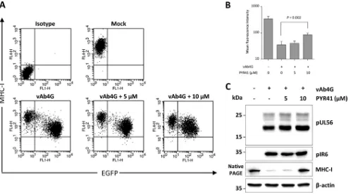
PYR41, which interferes with ubiquitination by E1 blockade (
41
).
Incubation of infected cells with 10
M PYR41 resulted in
recov-ery of cell surface levels of MHC-I at 4 h p.i. compared to
un-treated cells (
Fig. 7A
and
B
). We ruled out the possibility that
inhibition of the ubiquitination machinery might impair the
ex-pression of pUL56. Upon PYR41 treatment, pUL56 and the early
viral protein IR6 (pIR6) remained stable in infected cells. In
con-trast, elevated MHC-I levels were observed in the presence of the
inhibitor (
Fig. 7C
). We concluded from the results of this
experi-ment that MHC-I downregulation and degradation caused by
EHV-1 require ubiquitination, more specifically, an interaction
between EHV-1 pUL56 and components of the cellular
ubiquiti-nation machinery.
Downregulation of additional cell surface markers by
pUL56.
Finally, we determined if pUL56-mediated
internaliza-tion was specific to MHC-I. To this end, cell surface expression of
a number of cell surface markers, specifically, CD46, CD58, CD59,
CD63, and CD95, was analyzed. All the targeted molecules play
critical roles in modulation of host innate or adaptive immunity.
CD46 negatively regulates the pathways for complement
activa-tion (
42
), while CD58 and CD59 facilitate the adhesion and
acti-vation of T cells (
43
). Being a costimulatory factor, CD63 is
re-quired for the steady activation and proliferation of T cells (
44
).
CD95, best known as Fas receptor, initiates apoptosis when the
target cells are recognized by CTLs (
45
). Because the available
antibodies show exclusive reactivity with human and murine but
FIG 5Chemical inhibition of clathrin-independent endocytic pathways. After infection with vAb4G, NBL6 cells were replenished with fresh medium containing genistein at 2, 10, and 50g/ml (A), filipin at 1, 2, 3, and 5g/ml (B), nystatin at 2 and 20g/ml (C), and methyl--cyclodextrin (MCD) at 10 and 20 mM (D). After 4 h of incubation, cells were processed for cell surface MHC-I labeling, and the mean fluorescence intensity of the populations was determined by flow cytometry. Values are expressed as means⫾standard deviations (error bars) from at least three independent experiments. One-way ANOVA was used to analyze the statistical difference between groups.
MHC-I Downregulation and Dynamin-Dependent Endocytosis
on November 7, 2019 by guest
http://jvi.asm.org/
[image:8.585.85.503.67.378.2]not equine CD molecules, our experiments were conducted using
HeLa cells in which MHC-I downregulation dependent on pUL56
had previously been documented (
13
). For testing each marker,
HeLa cells were infected with vAb4G or vAb4G
⌬
1, the mutant
devoid of pUL56. At 24 h p.i., cell surface levels of each marker
were analyzed by flow cytometry and plotted as mean fluorescence
intensity in histograms. Of the selected markers, pUL56 induced a
significant reduction of CD46 and CD63, whereas it did not affect
the levels of CD58, CD59, or CD95 (
Fig. 8
). On the basis of these
data, we concluded that pUL56 is able to reduce cell surface levels
of molecules other than MHC-I and, hence, may have a more
general effect on immunomodulatory molecules in infected cells.
DISCUSSION
Over the past 2 decades, a number of viral inhibitors targeting
different stages of MHC-I presentation have been characterized
(
46
). The majority of such inhibitors were found in herpesviruses,
although their mechanisms of action vary greatly. Recently, we
discovered that pUL56, encoded by the gene
ORF1
of EHV-1,
affects MHC-I expression (
13
). Phylogenetic analyses reveal that
pUL56 is conserved in many alphaherpesviruses and that the
pUL56 homologue of EHV-4 also downregulates MHC-I at the
cell surface (
14
). It was speculated that loss of cell surface MHC-I
is induced by endocytosis in the early stages of EHV-1 infection
(
12
), and here, we investigated the putative relationship between
pUL56 and endocytosis that might result in the reduction of
MHC-I on the cell surface. By using inhibitors that disrupt
somal or lysosomal function, we initially determined that
endo-somal acidification and lysoendo-somal proteolysis govern the fate of
internalized MHC-I. This finding was further corroborated by
confocal microscopy showing that MHC-I colocalized with the
lysosome marker LAMP-1.
Endocytosis is an important intracellular transport
mecha-nism that initiates signal transduction and internalization of a
FIG 6MHC-I downregulation is independent on caveola-mediated endocytosis.(A)Schematic representation of two the forms of caveolin-1 used here, Cav1 (WT) and Cav1(Y14F) (DN), fused to the amino and carboxy termini of EGFP, respectively. (B) Expression of caveolin-1 constructs. At 48 h after transfection, whole-cell lysates were subjected to separation by SDS–12% PAGE followed by Western blot analysis. Specific bands corresponding to the WT (white triangle) or DN (gray triangle) proteins were detected using rabbit anti-HA MAb.-Actin was used as a loading control. PageRuler Plus from Thermo Scientific was run as a molecular ladder. (C) Inhibition of caveola-mediated endocytosis by the DN mutant. EGFP-Cav1(Y14F) (green) effectively blocked the engulfment of cholera toxin B (CTxB), resulting in the reduced intensity of Alexa Fluor 647 (red). Bars, 20m. (D) MHC-I downregulation is not affected by expression of either the WT or the DN mutant after infection. After gating with dsRed2, the infected cells were analyzed by flow cytometry for expression of EGFP and MHC-I. Student’sttest was performed, and no statistical significance between the WT and the DN groups was seen. Representative dot plots from triplicate assays are presented.
on November 7, 2019 by guest
http://jvi.asm.org/
[image:9.585.85.504.67.427.2]number of nutrients, lipids, membrane proteins, and pathogens
(
27
). The endocytic pathways that allow infectious entry have
been extensively studied for a number of viruses (
47
); however, it
is less well understood how viruses induce endocytosis of key
im-mune-related receptors to achieve immune evasion. Notable
ex-ceptions are the KSHV K3 and K5 proteins, which promote
MHC-I endocytosis in infected cells. It was shown in transiently
transfected cells that these two virus-encoded enzymes directly
increase endocytic activity and lead to the uptake of MHC-I
mol-ecules from the cell surface (
19
). Likewise, the HIV Nef protein
was shown to downregulate MHC-I through an endocytic
path-way (
48
), which may imply that enhancing endocytosis as a means
of preventing or reducing presentation of antigenic (viral)
pep-tides by MHC-I might be evolutionarily conserved among many
viruses.
We were able to demonstrate here that EHV-1 pUL56
colocal-izes from 4 h p.i. with MHC-I-containing vesicles and the
lyso-somal marker LAMP-1 in infected cells. This colocalization
cor-relates with decreased cell surface MHC-I levels. The results,
therefore, suggest that it is indeed pUL56 that mediates the
traf-ficking of MHC-I molecules to lysosomes for degradation. The
two known viral MHC-I inhibitors encoded by
alphaherpesvi-ruses, ICP47 and pUL49.5, prevent translocation of antigenic
pep-tide into the ER and formation of the trimolecular complex.
Spe-cifically, ICP47 is present in the cytoplasm and exhibits high
affinity to TAP, which results in functional inactivation of peptide
transport into the ER (
49
,
50
). For pUL49.5, although it also
re-stricts the supply of peptides to the ER, homologues in different
viral species are mechanistically diverse. For example, BHV-1
pUL49.5 reduces TAP stability by promoting its proteasomal
deg-radation (
8
), whereas pUL49.5 of EHV-1 and -4 inhibit the
re-cruitment of ATP, which is indispensable for TAP activity (
9
).
Unlike ER-based interference with MHC-I maturation, pUL56 is
predominantly localized to the Golgi apparatus and endosomal
vesicles. It does not perturb peptide transport to the ER (
13
,
14
),
and its mechanism of action is clearly distinct from those of ICP47
and pUL49.5. Therefore, pUL56 has a novel mode of action with
respect to MHC-I downregulation in members of the
Alphaher-pesvirinae
and functions more like gammaherpesviral K3 and K5
by interfering with endocytosis.
Based on their constituent elements, endocytic pathways are
classified into categories that include clathrin-, caveola-, and
Arf6-dependent endocytosis as well as phagocytosis and
macropinocy-tosis (
51
); however, a growing body of evidence now suggests that
unidentified endocytic pathways might exist, and thus the
classi-fication scheme has been simplified by discriminating only
be-tween clathrin-dependent endocytosis (CDE) and
clathrin-inde-pendent endocytosis (CIE) (
52
). Endocytosis inhibitors are
commonly used to investigate endocytic pathways responsible for
homeostasis of cell surface proteins, but attention should be given
to their possibly pleiotropic effects (
35
). Given that the turnover of
cell surface proteins is dynamically regulated under normal
phys-FIG 7Ubiquitination affects cell surface MHC-I expression in EHV-1-infected cells. (A) Ubiquitination was blocked by PYR41, an E1 inhibitor. NBL6 cells were mock infected or infected with vAb4G for 2 h; afterwards, inhibitor at 5M or 10M was applied. At 4 h of inhibition, the levels of cell surface MHC-I were determined by flow cytometry. (B) The overall inhibitory effects are shown as dot plots and converted into a bar chart to quantitatively compare mean fluorescence intensities obtained with the treatments. Data analyses were performed using ANOVA. (C) PYR41 reduces MHC-I degradation but does not affect the expression of pUL56. At 4 h p.i., cell lysates were prepared with RIPA buffer and fractionated using SDS–12% PAGE. The expression levels of pUL56 and pIR6 were detected by Western blotting. For MHC-I detection, cell lysates were separated by nondenaturing 12% PAGE, and the blot was probed with anti-MHC-I MAb CZ3.-Actin was included as a gel loading control.
MHC-I Downregulation and Dynamin-Dependent Endocytosis
on November 7, 2019 by guest
http://jvi.asm.org/
[image:10.585.42.543.65.341.2]iological conditions, we tested the influence of our inhibitors on
the metabolism of MHC-I, which might distort the interpretation
of our results in infected cells. Except for the significant MHC-I
reduction seen with the treatment of chloroquine, the other
re-agents used showed no or very little effect on the presence of
MHC-I on the cell surface of mock-infected cells (see Fig. S1 in the
supplemental material), ensuring that they target only MHC-I in
infected cells. With inhibition of virus-induced MHC-I
down-regulation by chlorpromazine and Pitstop2 as well by dominant
negative molecules, we demonstrated that MHC-I
downregula-tion is not through the CDE pathway. This observadownregula-tion for EHV-1
differs from that for KSHV, where the K3 protein interferes with
the classical CDE pathway (
39
), while a unique Arf6-dependent
pathway is targeted by HIV-1 Nef to divert mature cell surface
MHC-I to lysosomes for degradation (
48
). Despite the differences,
lysosomes are the ultimate cellular compartment where
degrada-tion of MHC-I occurs in the case of KSHV, HIV-1, and EHV-1.
Caveola-dependent endocytosis is considered an important
CIE pathway. Along with the involvement of dynamin, caveolin-1
is primarily responsible for the formation of the flask-shaped
caveolae (
53
). Moreover, tyrosine kinase activity is necessary for
the aggregation and fusion of caveolae or caveolar vesicles (
54
).
Therefore, it is not surprising that a number of viruses gain access
to host cells by nonclassical endocytic pathways that are all
depen-dent on the action of dynamin and tyrosine kinases but differ in
other factors involved (
55–58
). Here, we studied the role of
caveola-dependent endocytosis in virus-induced MHC-I
down-regulation. Despite the various effects of four inhibitors for CIE,
transfection of a specific dominant negative form of caveolin-1
was unable to rescue cell surface expression of MHC-I, suggesting
that caveola-dependent endocytosis is not involved in MHC-I
in-ternalization. In contrast, we found that the decrease of surface
MHC-I was remarkably attenuated when the action of dynamin
was inhibited with dynasore or a dominant negative form of
dy-namin. These findings led us to conclude that MHC-I
downregu-lation is associated with dynamin, which facilitates vesicle scission
at the plasma membrane and as such is integral to many endocytic
pathways (
59
,
60
). To our knowledge, our report describes the first
example of an alphaherpesvirus that downregulates cell surface
MHC-I through an endocytic pathway that is associated with
dy-namin but unrelated to clathrin and caveolae. However, there
might be another unknown viral protein(s) that can drive MHC-I
downregulation through endocytic processes, as knockout of
pUL56 alone cannot completely prevent endocytosis of MHC-I
mediated by EHV-1 infection.
Ubiquitination determines the destiny of endocytosed
mem-brane proteins (
61
). On the one hand, the formation of vesicles at
different stages of endocytosis requires mono- or
polyubiquitina-tion (
62
). On the other hand, the nature of ubiquitin linkage to
lysine results in cargo sorting and degradation either in the
pro-teasome or in the lysosome (
63
). KSHV K3 and K5 are E3
ubiqui-tin ligases which directly bind to membrane substrates and trigger
endocytosis (
39
,
64
). In the case of HSV-2, pUL56 is able to
in-crease ubiquitination of the E3 ligase Nedd4. This interaction is
thought to affect protein sorting or vesicle trafficking but does not
affect virus release, as originally surmised (
65
). Here, EHV-1
pUL56, which is structurally similar to its HSV-2 orthologue, was
shown to direct MHC-I molecules for lysosomal degradation.
FIG 8A broader range of surface markers is decreased during EHV-1 infection in a pUL56-dependent fashion. HeLa cells were mock infected (black solid line) or infected with vAb4G (gray solid line) or vAb4G⌬1 (black dashed line). Unspecific binding of antibodies was controlled for by staining with corresponding isotype antibodies (gray dashed line). At 24 h p.i., all cells were sampled and incubated with monoclonal primary antibodies against MHC-I, CD46, CD48, CD58, CD59, CD63, and CD95. Alexa Fluor 647-conjugated goat anti-mouse IgG was used as a secondary antibody. Samples were detected by flow cytometry. The FL4-H axis refers to the fluorescence intensity of the given surface markers. Downregulation of MHC-I, CD46, and CD63, but not that of other markers, was rescued by abrogation ofORF1. The histograms are representative of two independent tests.
on November 7, 2019 by guest
http://jvi.asm.org/
[image:11.585.74.513.65.323.2]This process is greatly affected when ubiquitination is chemically
blocked. It is therefore tempting to speculate that the function of
pUL56 depends on its interaction with an E3 ligase, a hypothesis
that we are currently testing.
To address the specificity of pUL56 action, several other cell
surface markers were screened in this study. We found that pUL56
can reduce the expression of CD46 and CD63 on the cell surface.
CD46 is critical for efficient T cell responses and bridges between
the complement system and cellular immunity (
66
); for instance,
expression of CD46 facilitates destruction of the cells infected with
measles virus by CTLs, a process that depends on MHC-I antigen
presentation (
67
). CD63 is a membrane protein containing four
transmembrane domains (tetraspanin) which is widely
distrib-uted on endosomal membranes and known to modulate immune
signaling pathways (
68
,
69
). In dendritic cells, for example,
intra-cellular transport of CD63 is associated with antigen presentation
by MHC-II (
70
). The implication of pUL56 in downregulating
CD46 and CD63 may suggest that it functions as a more
promis-cuous immune modulator, a notion that is supported by the
re-sults obtained in equids infected with the Ab4 mutant unable to
express the protein (
71
).
Removal of cell surface MHC-I seems a wise strategy for
per-sistent infection, as CTLs would be unable to eliminate infected
cells due to a failure to recognize antigens. However, cells devoid
of surface MHC-I are unlikely to survive and might be subject to
clearance by natural killer (NK) cells. KSHV K5 targets other
sur-face molecules to counteract the threat from NK cells. Along with
CD31, CD86, CD144, CD166, and intracellular adhesion
mole-cule 1 (ICAM1) (
64
), KSHV K5 also downregulates
activation-induced C-type lectin (AICL), MHC-I polypeptide-related
se-quence A (MICA), and MICB, which are required for NK cell lysis
(
72
,
73
). Whether pUL56 targets these surface molecules sensed by
NK cells remains to be explored.
In this study, we investigated the pathway by which pUL56
induces downregulation of cell surface MHC-I. The endocytic
process responsible for MHC-1 removal from the cell surface is
mediated by dynamin but not clathrin or caveolae. Importantly,
tyrosine kinase activity and membrane-bound cholesterol are
re-quired for MHC-I endocytosis as is ubiquitination. The latter may
imply that pUL56 function is dependent on the E3 ligase activity.
Since pUL56 is able to regulate other cell surface molecules, it may
have a more comprehensive role in regulation of the immune
response to infection. Future studies will focus on how pUL56
interacts with ubiquitination and the yet-unidentified viral
pro-tein(s) it needs to carry out its functions.
ACKNOWLEDGMENTS
We thank Walid Azab for helpful suggestions on inhibitors and mainte-nance of equine cells. We express gratitude for the Pitstop2 inhibitors, kindly provided by Volker Haucke from the Leibniz-Institut für Moleku-lare Pharmakologie (FMP), Berlin-Buch, Germany, and the anti-MHC MAb CZ3, provided by Douglas F. Antczak (Cornell University, USA). We also gratefully acknowledge the dominant negative mutants kindly provided by Mark A. McNiven (Mayo Clinic, Rochester, MN, USA) and Alexandre Benmerah (Hôpital Necker-Enfants Malades, Paris, France).
T.H. was supported by a grant from the China Scholarship Council. This study was financed by DFG grant OS143/3-1 and unrestricted funds of Freie Universität Berlin to N.O.
REFERENCES
1.Wong P, Pamer EG.2003. CD8 T cell responses to infectious pathogens. Annu.Rev.Immunol.21:29 –70.http://dx.doi.org/10.1146/annurev.immunol. 21.120601.141114.
2.Peaper DR, Cresswell P.2008. Regulation of MHC class I assembly and peptide binding. Annu. Rev. Cell Dev. Biol.24:343–368.http://dx.doi.org /10.1146/annurev.cellbio.24.110707.175347.
3.Fruh K, Gruhler A, Krishna RM, Schoenhals GJ.1999. A comparison of viral immune escape strategies targeting the MHC class I assembly path-way. Immunol. Rev.168:157–166.http://dx.doi.org/10.1111/j.1600-065X .1999.tb01290.x.
4.Lilley BN, Ploegh HL.2005. Viral modulation of antigen presentation: manipulation of cellular targets in the ER and beyond. Immunol. Rev.
207:126 –144.http://dx.doi.org/10.1111/j.0105-2896.2005.00318.x. 5.Burgert HG, Kvist S.1985. An adenovirus type 2 glycoprotein blocks cell
surface expression of human histocompatibility class I antigens. Cell41:
987–997.http://dx.doi.org/10.1016/S0092-8674(85)80079-9.
6.Fruh K, Ahn K, Djaballah H, Sempe P, van Endert PM, Tampe R, Peterson PA, Yang Y.1995. A viral inhibitor of peptide transporters for antigen presentation. Nature375:415– 418.http://dx.doi.org/10.1038 /375415a0.
7.Hill A, Jugovic P, York I, Russ G, Bennink J, Yewdell J, Ploegh H, Johnson D.1995. Herpes simplex virus turns off the TAP to evade host immunity. Nature375:411– 415.http://dx.doi.org/10.1038/375411a0. 8.Koppers-Lalic D, Reits EA, Ressing ME, Lipinska AD, Abele R, Koch J,
Marcondes Rezende M, Admiraal P, van Leeuwen D, Bienkowska-Szewczyk K, Mettenleiter TC, Rijsewijk FA, Tampe R, Neefjes J, Wiertz EJ.2005. Varicelloviruses avoid T cell recognition by UL49.5-mediated inactivation of the transporter associated with antigen processing. Proc. Natl. Acad. Sci. U. S. A.102:5144 –5149.http://dx.doi.org/10.1073/pnas .0501463102.
9.Koppers-Lalic D, Verweij MC, Lipinska AD, Wang Y, Quinten E, Reits EA, Koch J, Loch S, Marcondes Rezende M, Daus F, Bienkowska-Szewczyk K, Osterrieder N, Mettenleiter TC, Heemskerk MH, Tampe R, Neefjes JJ, Chowdhury SI, Ressing ME, Rijsewijk FA, Wiertz EJ.
2008. Varicellovirus UL 49.5 proteins differentially affect the function of the transporter associated with antigen processing, TAP. PLoS Pathog.4:
e1000080.http://dx.doi.org/10.1371/journal.ppat.1000080.
10.Ma G, Azab W, Osterrieder N. 2013. Equine herpesviruses type 1 (EHV-1) and 4 (EHV-4)–masters of co-evolution and a constant threat to equids and beyond. Vet. Microbiol.167:123–134.http://dx.doi.org/10 .1016/j.vetmic.2013.06.018.
11. Kydd JH, Wattrang E, Hannant D.2003. Pre-infection frequencies of equine herpesvirus-1 specific, cytotoxic T lymphocytes correlate with pro-tection against abortion following experimental infection of pregnant mares. Vet. Immunol. Immunopathol.96:207–217.http://dx.doi.org/10 .1016/j.vetimm.2003.08.004.
12. Rappocciolo G, Birch J, Ellis SA.2003. Down-regulation of MHC class I expression by equine herpesvirus-1. J. Gen. Virol.84:293–300.http://dx .doi.org/10.1099/vir.0.18612-0.
13. Ma G, Feineis S, Osterrieder N, Van de Walle GR.2012. Identification and characterization of equine herpesvirus type 1 pUL56 and its role in virus-induced downregulation of major histocompatibility complex class I. J. Virol.86:3554 –3563.http://dx.doi.org/10.1128/JVI.06994-11. 14. Said A, Azab W, Damiani A, Osterrieder N.2012. Equine herpesvirus
type 4 UL56 and UL49.5 proteins downregulate cell surface major histo-compatibility complex class I expression independently of each other. J. Virol.86:8059 – 8071.http://dx.doi.org/10.1128/JVI.00891-12.
15. Goodman LB, Loregian A, Perkins GA, Nugent J, Buckles EL, Mer-corelli B, Kydd JH, Palu G, Smith KC, Osterrieder N, Davis-Poynter N.
2007. A point mutation in a herpesvirus polymerase determines neuro-pathogenicity. PLoS Pathog.3:e160. http://dx.doi.org/10.1371/journal .ppat.0030160.
16. Tischer BK, von Einem J, Kaufer B, Osterrieder N.2006. Two-step red-mediated recombination for versatile high-efficiency markerless DNA manipulation in Escherichia coli. Biotechniques40:191–197.http://dx .doi.org/10.2144/000112096.
17. O’Callaghan DJ, Colle CF, III, Flowers CC, Smith RH, Benoit JN, Bigger CA.1994. Identification and initial characterization of the IR6 protein of equine herpesvirus 1. J. Virol.68:5351–5364.
18. von Kleist L, Stahlschmidt W, Bulut H, Gromova K, Puchkov D, Robertson MJ, MacGregor KA, Tomilin N, Pechstein A, Chau N,
MHC-I Downregulation and Dynamin-Dependent Endocytosis
on November 7, 2019 by guest
http://jvi.asm.org/
Chircop M, Sakoff J, von Kries JP, Saenger W, Krausslich HG, Shu-pliakov O, Robinson PJ, McCluskey A, Haucke V.2011. Role of the clathrin terminal domain in regulating coated pit dynamics revealed by small molecule inhibition. Cell146:471– 484.http://dx.doi.org/10.1016/j .cell.2011.06.025.
19. Coscoy L, Ganem D.2000. Kaposi’s sarcoma-associated herpesvirus en-codes two proteins that block cell surface display of MHC class I chains by enhancing their endocytosis. Proc. Natl. Acad. Sci. U. S. A.97:8051– 8056. http://dx.doi.org/10.1073/pnas.140129797.
20. Cao H, Thompson HM, Krueger EW, McNiven MA.2000. Disruption of Golgi structure and function in mammalian cells expressing a mutant dynamin. J. Cell Sci.113:1993–2002.
21. Benmerah A, Lamaze C, Begue B, Schmid SL, Dautry-Varsat A, Cerf-Bensussan N.1998. AP-2/Eps15 interaction is required for receptor-mediated endocytosis. J. Cell Biol.140:1055–1062.http://dx.doi.org/10 .1083/jcb.140.5.1055.
22. Stroh T, Erben U, Kuhl AA, Zeitz M, Siegmund B.2010. Combined pulse electroporation—a novel strategy for highly efficient transfection of human and mouse cells. PLoS One5:e9488.http://dx.doi.org/10.1371 /journal.pone.0009488.
23. Luzio JP, Pryor PR, Bright NA.2007. Lysosomes: fusion and function. Nat. Rev. Mol. Cell Biol.8:622– 632.http://dx.doi.org/10.1038/nrm2217. 24. Fredericksen BL, Wei BL, Yao J, Luo T, Garcia JV.2002. Inhibition of endosomal/lysosomal degradation increases the infectivity of human im-munodeficiency virus. J. Virol. 76:11440 –11446. http://dx.doi.org/10 .1128/JVI.76.22.11440-11446.2002.
25. Dutta D, Donaldson JG.2012. Search for inhibitors of endocytosis: in-tended specificity and uninin-tended consequences. Cell Logist.2:203–208. http://dx.doi.org/10.4161/cl.23967.
26. Drose S, Altendorf K.1997. Bafilomycins and concanamycins as inhibi-tors of V-ATPases and P-ATPases. J. Exp. Biol.200:1– 8.
27. Doherty GJ, McMahon HT.2009. Mechanisms of endocytosis. Annu. Rev. Biochem.78:857–902.http://dx.doi.org/10.1146/annurev.biochem .78.081307.110540.
28. Macia E, Ehrlich M, Massol R, Boucrot E, Brunner C, Kirchhausen T.
2006. Dynasore, a cell-permeable inhibitor of dynamin. Dev. Cell10:839 – 850.http://dx.doi.org/10.1016/j.devcel.2006.04.002.
29. Wang LH, Rothberg KG, Anderson RG.1993. Mis-assembly of clathrin lattices on endosomes reveals a regulatory switch for coated pit formation. J. Cell Biol.123:1107–1117.http://dx.doi.org/10.1083/jcb.123.5.1107. 30. Benmerah A, Bayrou M, Cerf-Bensussan N, Dautry-Varsat A. 1999.
Inhibition of clathrin-coated pit assembly by an Eps15 mutant. J. Cell Sci.
112:1303–1311.
31. Nabi IR, Le PU.2003. Caveolae/raft-dependent endocytosis. J. Cell Biol.
161:673– 677.http://dx.doi.org/10.1083/jcb.200302028.
32. Schnitzer JE, Oh P, Pinney E, Allard J.1994. Filipin-sensitive caveolae-mediated transport in endothelium: reduced transcytosis, scavenger en-docytosis, and capillary permeability of select macromolecules. J. Cell Biol.127:1217–1232.http://dx.doi.org/10.1083/jcb.127.5.1217. 33. Chen Y, Wang S, Lu X, Zhang H, Fu Y, Luo Y. 2011. Cholesterol
sequestration by nystatin enhances the uptake and activity of endostatin in endothelium via regulating distinct endocytic pathways. Blood117:6392– 6403.http://dx.doi.org/10.1182/blood-2010-12-322867.
34. Mundy DI, Li WP, Luby-Phelps K, Anderson RG. 2012. Caveolin targeting to late endosome/lysosomal membranes is induced by perturba-tions of lysosomal pH and cholesterol content. Mol. Biol. Cell23:864 – 880.http://dx.doi.org/10.1091/mbc.E11-07-0598.
35. Vercauteren D, Vandenbroucke RE, Jones AT, Rejman J, Demeester J, De Smedt SC, Sanders NN, Braeckmans K.2010. The use of inhibitors to study endocytic pathways of gene carriers: optimization and pitfalls. Mol. Ther.18:561–569.http://dx.doi.org/10.1038/mt.2009.281.
36. Orlichenko L, Huang B, Krueger E, McNiven MA. 2006. Epithelial growth factor-induced phosphorylation of caveolin 1 at tyrosine 14 stim-ulates caveolae formation in epithelial cells. J. Biol. Chem.281:4570 – 4579.http://dx.doi.org/10.1074/jbc.M512088200.
37. Shatz M, Lustig G, Reich R, Liscovitch M.2010. Caveolin-1 mutants P132L and Y14F are dominant negative regulators of invasion, migration and aggregation in H1299 lung cancer cells. Exp. Cell Res.316:1748 –1762. http://dx.doi.org/10.1016/j.yexcr.2010.02.006.
38. Pelkmans L, Kartenbeck J, Helenius A.2001. Caveolar endocytosis of simian virus 40 reveals a new two-step vesicular-transport pathway to the ER. Nat. Cell Biol.3:473– 483.http://dx.doi.org/10.1038/35074539. 39. Duncan LM, Piper S, Dodd RB, Saville MK, Sanderson CM, Luzio JP,
Lehner PJ. 2006. Lysine-63-linked ubiquitination is required for endolysosomal degradation of class I molecules. EMBO J.25:1635–1645. http://dx.doi.org/10.1038/sj.emboj.7601056.
40. Ushijima Y, Luo C, Kamakura M, Goshima F, Kimura H, Nishiyama Y.
2010. Herpes simplex virus UL56 interacts with and regulates the Nedd4-family ubiquitin ligase Itch. Virol. J.7:179.http://dx.doi.org/10.1186/1743 -422X-7-179.
41. Yang Y, Kitagaki J, Dai RM, Tsai YC, Lorick KL, Ludwig RL, Pierre SA, Jensen JP, Davydov IV, Oberoi P, Li CC, Kenten JH, Beutler JA, Vousden KH, Weissman AM.2007. Inhibitors of ubiquitin-activating enzyme (E1), a new class of potential cancer therapeutics. Cancer Res.
67:9472–9481.http://dx.doi.org/10.1158/0008-5472.CAN-07-0568. 42. Liszewski MK, Post TW, Atkinson JP.1991. Membrane cofactor protein
(MCP or CD46): newest member of the regulators of complement activa-tion gene cluster. Annu. Rev. Immunol.9:431– 455.http://dx.doi.org/10 .1146/annurev.iy.09.040191.002243.
43. Deckert M, Kubar J, Bernard A.1992. CD58 and CD59 molecules exhibit potentializing effects in T cell adhesion and activation. J. Immunol.148:
672– 677.
44. Pfistershammer K, Majdic O, Stockl J, Zlabinger G, Kirchberger S, Steinberger P, Knapp W. 2004. CD63 as an activation-linked T cell costimulatory element. J. Immunol.173:6000 – 6008.http://dx.doi.org/10 .4049/jimmunol.173.10.6000.
45. Krammer PH.2000. CD95’s deadly mission in the immune system. Na-ture407:789 –795.http://dx.doi.org/10.1038/35037728.
46. Hansen TH, Bouvier M.2009. MHC class I antigen presentation: learning from viral evasion strategies. Nat. Rev. Immunol.9:503–513.http://dx.doi .org/10.1038/nri2575.
47.