HIV Protease-Generated Casp8p41, When Bound and
Inactivated by Bcl2, Is Degraded by the Proteasome
Sekar Natesampillai,
aNathan W. Cummins,
aZilin Nie,
aRahul Sampath,
aJason V. Baker,
b,cKeith Henry,
cMarilia Pinzone,
dUna O’Doherty,
dEric C. Polley,
eGary D. Bren,
aDavid J. Katzmann,
fAndrew D. Badley
a,gaDivision of Infectious Diseases, Mayo Clinic, Rochester, Minnesota, USA
bDivision of Infectious Diseases, University of Minnesota, Minneapolis, Minnesota, USA
cHIV Program, Hennepin County Medical Center, Minneapolis, Minnesota, USA
dDepartment of Laboratory Medicine and Therapeutic Pathology, University of Pennsylvania, Philadelphia,
Pennsylvania, USA
eDepartment of Biostatistics, Mayo Clinic, Rochester, Minnesota, USA
fDepartment of Biochemistry and Molecular Biology, Mayo Clinic, Rochester, Minnesota, USA
gDepartment of Molecular Medicine, Mayo Clinic, Rochester, Minnesota, USA
ABSTRACT
HIV protease is known to cause cell death, which is dependent upon
cleav-age of procaspase 8. HIV protease cleavcleav-age of procaspase 8 generates Casp8p41, which
directly binds Bak with nanomolar affinity, causing Bak activation and consequent
cell death. Casp8p41 can also bind Bcl2 with nanomolar affinity, in which case cell
death is averted. Central memory CD4 T cells express high levels of Bcl2, possibly
explaining why those cells do not die when they reactivate HIV. Here, we determine
that the Casp8p41-Bcl2 complex is polyubiquitinated and degraded by the
protea-some. Ixazomib, a proteasome inhibitor in clinical use, blocks this pathway,
increas-ing the abundance of Casp8p41 and causincreas-ing more cells to die in a
Casp8p41-dependent manner.
IMPORTANCE
The Casp8p41 pathway of cell death is unique to HIV-infected cells
yet is blocked by Bcl2. Once bound by Bcl2, Casp8p41 is polyubiquitinated and
de-graded by the proteasome. Proteasome inhibition blocks degradation of Casp8p41,
increasing Casp8p41 levels and causing more HIV-infected cells to die.
KEYWORDS
apoptosis, HIV, proteasome inhibitor
U
nderstanding pathways of cell death that occur naturally in HIV-infected cells may
instruct interventions that increase the proportion of HIV-infected cells that die.
Although HIV infection kills most CD4 T cells that it infects, a small subset of
HIV-infected cells survive, revert to a resting state, and become a latent reservoir for HIV.
The major barrier to HIV eradication is the existence of this stable reservoir of latent
virus, which resides predominantly in central memory CD4 T cells (1) and in follicular T
helper cells (2). One proposed approach to eliminate these cells posits that reactivating
HIV would allow immune recognition and, consequently, elimination of infected cells
by intrinsic host effector immune mechanisms and/or due to the intrinsic cytotoxic
properties of the HIV proteins themselves (i.e., the “shock-and-kill” hypothesis) (3).
However, experimental testing
in vitro
(4) and
in vivo
has shown with remarkable
consistency that reactivation from latency alone is insufficient to cause the death of the
reactivating cell. For example, vorinostat treatment of antiretroviral therapy
(ART)-suppressed HIV-infected patients caused reactivation of HIV but no reduction in the
frequency of replication-competent HIV within resting CD4
⫹T cells (5). Therefore, the
pathways of cell death that are activated by
de novo
HIV infection are seemingly not
activated during reactivation from latency.
Received7 January 2018Accepted2 April 2018
Accepted manuscript posted online11 April 2018
CitationNatesampillai S, Cummins NW, Nie Z, Sampath R, Baker JV, Henry K, Pinzone M, O'Doherty U, Polley EC, Bren GD, Katzmann DJ, Badley AD. 2018. HIV protease-generated Casp8p41, when bound and inactivated by Bcl2, is degraded by the proteasome. J Virol 92:e00037-18.https://doi.org/10.1128/JVI .00037-18.
EditorGuido Silvestri, Emory University
Copyright© 2018 Natesampillai et al. This is an open-access article distributed under the terms of theCreative Commons Attribution 4.0 International license.
Address correspondence to Andrew D. Badley, badley.andrew@mayo.edu.
S.N. and N.W.C. contributed equally to this work.
crossm
on November 6, 2019 by guest
http://jvi.asm.org/
Multiple pathways have been described by which HIV-infected cells die as a
con-sequence of HIV infection (reviewed in reference 6). One of these pathways is initiated
by the intracellular expression of HIV protease, which, contrary to early reports, is
catalytically active within the cytosol (7, 8). Expression of HIV protease alone in
sufficient amounts is enough to kill some eukaryotic cells, and this phenomenon has
been exploited to screen for inhibitors of HIV protease (9). The normal function of HIV
protease is to cleave Gag-Pol to allow the initial steps of virus packaging. However, due
to its degenerate substrate specificity, HIV protease also cleaves a number of host
proteins (10–12). One host protein cleaved by HIV protease is procaspase 8 (13, 14);
cells expressing a procaspase 8 mutant that is noncleavable by protease do not die
following acute HIV infection
in vitro
(15). Conversely, certain drug resistance mutations
in HIV protease impair its ability to cleave procaspase 8, decreasing Casp8p41 (see
below) expression, and result in less CD4 T cell apoptosis than wild-type HIV protease
(16).
HIV protease cleaves procaspase 8 between phenylalanines at positions 355 and
356, generating a 41-kDa fragment that we have named Casp8p41. Casp8p41 is seen
only in HIV-infected cells (14), and Casp8p41 levels are predictive of future CD4
⫹T cell
losses (16–18). Because Casp8p41 lacks the catalytic cysteine at position 360 of
pro-caspase 8, it is catalytically inert, yet counterintuitively, it maintains the ability to induce
cell death. Once generated, Casp8p41 translocates to the mitochondrion, where it
adopts a BH3-like alpha-helical domain that binds to the BH3 groove of Bak, causing
Bak activation and pore function that leads to loss of mitochondrial transmembrane
potential, release of cytochrome
c
, and activation of downstream executioners of
apoptosis (19–21). Because Casp8p41 is generated at a step in the HIV life cycle that
occurs after integration, it follows that reactivation from latency should also generate
Casp8p41. Indeed, our recent work, using cells from ART-suppressed HIV-infected
patients, has shown that Casp8p41 is generated following HIV reactivation (22).
More-over, those studies showed that the cells in which latent HIV resides (e.g., central
memory CD4 T cells) have an apoptosis-resistant phenotype and elevated expression of
Bcl2. This led to the additional observations that Casp8p41 can also bind Bcl2 with
nanomolar affinity and that, when Casp8p41 directly binds to Bcl2, the HIV-infected cell
is not killed (22). Logically, therefore, inhibiting Casp8p41 binding to Bcl2 (thereby
allowing Casp8p41 to bind Bak) enhances the number of HIV-infected cells that die
(22, 23).
In this study, we tested the hypothesis that, after binding to Bcl2, the Casp8p41-Bcl2
complex is degraded and that blocking the degradation of Casp8p41 leads to increased
Casp8p41 levels, greater proportions of HIV-infected cells dying, and a decrease in the
number of HIV DNA-containing cells.
RESULTS
The Casp8p41-Bcl2 complex is polyubiquitinated.
Regulated protein degradation
is a prerequisite for cellular physiology, and the ubiquitin (Ub) proteasome system
represents a major site of protein turnover. Reversible covalent modification of
sub-strates with polyubiquitin chains drives their association with the proteasome and
subsequent destruction. It has previously been documented that a complex of other
proapoptotic proteins bound to their inhibitors is subject to proteasome-mediated
degradation (e.g., proapoptotic NOXA bound by Mcl1 is degraded by the proteasome)
(24). To test the idea that higher-molecular-weight forms of Ub-modified Casp8p41
exist and might also be degraded by the proteasome, immunoprecipitations were
performed from cells expressing hemagglutinin (HA)-Casp8p41. Western blotting of the
immunoprecipitated material using anti-ubiquitin antibody revealed the existence of
higher-molecular-weight species within cells expressing HA-Casp8p41 (Fig. 1A).
Fur-thermore, these higher-molecular-weight species were susceptible to the catalytic
activities of a subset of deubiquitinating enzymes, specifically, USP2 and YOD1 (Fig. 1B
and C). Whether ubiquitin-modified Casp8p41 was the result of Bcl2 binding was next
addressed through coimmunoprecipitation and Western blotting using anti-HA and
Natesampillai et al. Journal of Virology
on November 6, 2019 by guest
http://jvi.asm.org/
anti-ubiquitin antibodies. Immunoprecipitation of Bcl2 revealed association with
HA-Casp8p41 (Fig. 1D, left), and ubiquitin-reactive species that were sensitive to DUB
activity were detected regardless of whether Bcl2 or HA-Casp8p41
immunoprecipita-tion was first performed (Fig. 1D, right). Together, these data indicate that Casp8p41
can be polyubiquitinated. Furthermore, the observation that these species are sensitive
to the enzymatic activities of YOD1 and USP2 suggests the ubiquitin chains are linked
via lysine 48 (25–27). This suggests a model in which polyubiquitinated Casp8p41 in
complex with Bcl2 is targeted to the proteasome for degradation.
Proteasome inhibition increases Casp8p41 levels.
The clinical development of
proteasome inhibitors (PIs) has altered the course of protein production diseases, such
as multiple myeloma, in which plasma cells produce excessive amounts of monoclonal
immunoglobulins. Long-term treatment with PIs over months to years causes
semise-lective death of myeloma cells (28) and improves the survival of affected patients (29).
We tested whether the currently approved PIs bortezomib and ixazomib would block
the degradation of Casp8p41. Dose-ranging toxicity studies of bortezomib or the active
metabolite of ixazomib (MLN2238, here referred to as ixazomib) were performed in
primary CD4 T cells. At doses at or below 10 nM and 100 nM, respectively, which are
FIG 1The Casp8p41-Bcl2 complex is ubiquitinated and targeted for proteasomal degradation. (A) 293T cells were transfected with HA-empty vector or HA-Casp8p41; immunoprecipitated (IP) with IgG control, anti-HA, or anti-caspase 8; and immunoblotted (IB) for ubiquitin. (B) Cytosolic extracts prepared as described for panel A were treated with various DUBs as indicated, immunoprecipitated with anti-HA, and immunoblotted for ubiquitin. (C) Cytosolic extracts prepared as described for panel A were treated with the DUBs OTUD3 and Trabid, the Lys48-specific YOD1, or USP2 in the presence or absence of the DUB inhibitorN-ethylmaleimide (NEM); immunoprecipitated with anti-HA; and immunoblotted for ubiquitin. (D) 293 T cells were cotransfected with Bcl-2 and HA-Casp8p41; treated with the DUB Trabid, YOD1, or USP2 in the presence or absence of the DUB inhibitor NEM; immunoprecipitated with anti-HA or anti-Bcl2; and then immunoblotted for HA (left) or ubiquitin (right), with short and long exposures depicted.
on November 6, 2019 by guest
http://jvi.asm.org/
[image:3.585.44.548.76.439.2]levels that are achievable in the sera of treated patients (30, 31), there was minimal
nonspecific toxicity with bortezomib and less toxicity with ixazomib, as determined by
active caspase 3 staining (Fig. 2A), consistent with the superior toxicity profile of
ixazomib. Consequently, we used bortezomib at 5 and 10 nM and ixazomib at 50 and
100 nM in future experiments. Jurkat CD4
⫹T cells were transfected with green
fluorescent protein (GFP)-tagged Casp8p41, treated with PIs, and analyzed for
GFP-Casp8p41 positivity 6 h later; 10 nM bortezomib increased the percentage of cells
positive for GFP-Casp8p41 by 2.5-fold relative to control cells (
P
⫽
0.009), and 100 nM
ixazomib resulted in a 2.4-fold increase (
P
⫽
0.045) (Fig. 2B and C). This effect was
confirmed in primary CD4 T cells infected
in vitro
with HIV
IIIb, treated with bortezomib
or control, and assessed for intracellular Casp8p41 positivity using a Casp8p41-specific
monoclonal antibody (MAb) (Fig. 2D). Consistent with our previous reports (14, 17),
Casp8p41 is present in HIV-infected T cells and not in uninfected cells. Furthermore,
consistent with proteasome inhibitors increasing GFP-Casp8p41 in transfected cells
FIG 2Proteasome inhibitors increase Casp8p41 levels and kill HIV-infected CD4 T cell cultures more than uninfected cultures. (A) Uninfected primary CD4⫹T cells were treated with bortezomib or ixazomib at increasing concentrations for 48 h, and cell death was assessed by activated caspase 3
detection by intracellular flow cytometry. Depicted are the means and SD of the results of two experiments. (B and C) Jurkat CD4⫹T cells were
transfected with empty vector or GFP-Casp8p41 and then treated with control (DMSO), bortezomib, or ixazomib, and the percentage of cells that were GFP positive was analyzed 6 h later. (C) Mean (plus SD) data from three independent replicates of the experiment shown in panel B compared by a Kruskal-Wallis test. (D) Primary CD4 T cells were infected with HIVIIIb, treated with control or bortezomib, and assessed for intracellular Casp8p41
expression by flow cytometry. (E) Primary CD4 T cells were infected with HIV-Luc; treated with DMSO, bortezomib, or ixazomib; and monitored for viability by ATP content over time. (F) Primary CD4 T cells infected with HIV-Luc were treated with DMSO, bortezomib, or ixazomib and monitored for luciferase activity over time. (G and H) Jurkat T cells were mock or GFP-HIV infected, treated with DMSO or ixazomib, and monitored for GFP (G) or cell death (by cell permeability dye) (H) over time. (I and J) Cell death selectively measured in GFP-expressing cells (I) and GFP-negative cells (J) in panels G and H was monitored over time.*,P⬍0.05;**,P⬍0.01.
Natesampillai et al. Journal of Virology
on November 6, 2019 by guest
http://jvi.asm.org/
[image:4.585.46.509.76.448.2](Fig. 2B and C), bortezomib treatment increased Casp8p41 expression in HIV-1-infected
cells (Fig. 2D).
Proteasome inhibitors kill HIV-infected CD4 T cells.
Because Casp8p41 is present
only in HIV-infected cells (14, 17), increasing its expression might selectively cause the
death of those HIV-infected cells as opposed to uninfected cells. This hypothesis was
tested using 2 separate models of
in vitro
HIV infection. First, activated primary CD4 T
cells were acutely infected with luciferase-expressing HIV and then treated with
di-methyl sulfoxide (DMSO) control, bortezomib, or ixazomib. Bortezomib or ixazomib
significantly increased killing of HIV-infected T cell cultures over time (
P
⬍
0.001 each)
(Fig. 2E) at doses that did not alter the viability of uninfected primary CD4 T cells (Fig.
2A). Interestingly, the addition of bortezomib and ixazomib to the HIV-Luc-infected cells
initially increased luciferase activity (see below for further discussion), but at later time
points, the amount of luciferase activity decreased, consistent with HIV-infected cells
dying (
P
⫽
0.023 and
P
⫽
0.008, respectively) (Fig. 2F). In our second model system,
Jurkat T cells infected with GFP-HIV were treated with DMSO or ixazomib, and GFP
expression was monitored over time (Fig. 2G). GFP-HIV-infected Jurkat cells treated with
ixazomib had more cell death (
P
⬍
0.0001) than cells treated with control DMSO (Fig.
2H), but viability on mock-infected (uninfected) cells was not altered by ixazomib
treatment (
P
⫽
0.72) (Fig. 2H). As not all cells in HIV-infected cultures are directly
infected, we measured death specifically in the GFP-positive cells and found that
ixazomib increased the number of dead GFP-positive cells compared to DMSO (
P
⬍
0.0001) (Fig. 2I). Conversely, ixazomib also increased cell death in GFP-negative
HIV-exposed cells (
P
⬍
0.0001) (Fig. 2J). However, we have previously shown that GFP
expression is lost during the process of apoptosis, so it is possible that some of the
apparently GFP-negative cells in these cultures were indeed infected (22). Thus, in these
experiments, PIs caused minimal toxicity in uninfected and unexposed T cells (Fig. 2A)
yet enhanced killing of HIV-infected and, to a lesser extent, HIV-exposed cells.
Proteasome inhibitors activate the HIV LTR predominantly via NF-
B.
Above,
we observed that ixazomib causes a transient increase in HIV Luc (Fig. 2E) and HIV GFP
(Fig. 2F) expression in infected cultures. Since bortezomib has been previously shown
to activate NF-
B (32) and since the HIV long terminal repeat (LTR) is driven by NF-
B,
we questioned whether ixazomib might directly activate HIV replication through NF-
B.
To begin, we used J-Lat 10.6 cells, which express GFP upon HIV reactivation, to assess
if ixazomib directly impacts HIV replication. Treatment with ixazomib and with
bort-ezomib led to an increase in GFP expression over time (Fig. 3A) that was coupled with
HIV p55 Gag protein expression by Western blot analyses (Fig. 3B).
We next evaluated whether these effects on HIV replication were mediated by NF-
B
transactivation of the HIV LTR. Luciferase reporter constructs in which either the HIV LTR
was linked to luciferase (HIV-Luc) or the HIV LTR missing the NF-
B-binding sites was
linked to luciferase (HIVΔ
B-Luc) were transfected into Jurkat T cells, and the
transfec-tion efficacy was normalized to that of cotransfected
Renilla
, as previously described
(33). Treatment with bortezomib or ixazomib resulted in a dose-dependent increase in
luciferase expression (normalized to that of
Renilla
luciferase), demonstrating a direct
effect on HIV LTR-mediated replication in cells containing HIV-Luc (Fig. 3C). Consistent
with this effect being mediated predominantly through NF-
B transactivation of the
HIV LTR, Luc activity was significantly less augmented in the cells containing the
HIVΔ
B-Luc reporter (Fig. 3D). Internally consistent with these findings, the negative
repressor of NF-
B, I
B, was inactivated (through Ser32 phosphorylation [34]), and the
p65 subunit of NF-
B was activated (through Ser536 phosphorylation) within 6 h of
treating J-Lat 10.6 cells with bortezomib or ixazomib (Fig. 3E).
Selective killing of HIV-infected cells by proteasome inhibitors depends upon
Casp8p41.
Next, we assessed whether the ixazomib-induced death of HIV-infected
cells was dependent upon the presence of Casp8p41. HIV-infected J-Lat 10.6 cells or
parental Jurkat CD4 T cells were treated with vehicle control, bortezomib, or ixazomib.
Bortezomib and ixazomib increased activated caspase 3 in HIV-infected J-Lat 10.6 cells
on November 6, 2019 by guest
http://jvi.asm.org/
in a dose- and time-dependent manner (
P
⫽
0.014 for 5 nM bortezomib;
P
⫽
0.004 for
10 nM bortezomib;
P
⬍
0.0001 for 50 nM and 100 nM ixazomib) (Fig. 4A and B). The
same treatments only minimally increased activated caspase 3 expression in parental,
uninfected Jurkat cells (Fig. 2H and 5D). In the J-Lat cells, bortezomib and ixazomib
induced intracellular p24 expression (Fig. 4C, top), cleavage of caspase 3 (Fig. 4C,
middle), and cleavage of PARP (Fig. 4C, bottom), consistent with these treated cells
undergoing apoptosis.
In order to determine if the increased apoptosis in HIV-infected cells induced by
ixazomib was dependent on Casp8p41 expression, we utilized JB-6 cells, which are
procaspase 8 deficient and therefore resistant to HIV-induced apoptosis (15). Our
strategy exploited our previous observation that procaspase 8-deficient JB-6 cells
reconstituted with HIV protease-resistant procaspase 8 (Casp8RN) do not die following
HIV infection, whereas JB-6 cells reconstituted with HIV protease-cleavable wild-type
procaspase 8 (Casp8) do die from HIV infection (15). JB6 cells were infected with
vesicular stomatitis virus (VSV)-G-pseudotyped HIV and 2 days later transfected with
enhanced green fluorescent protein (EGFP)-Casp8, EGFP-Casp8RN, or EGFP empty
-ector control (Fig. 4D and E). The cells were then treated with ixazomib or DMSO, and
apoptosis (active caspase 3) in infected (p24
⫹) cells expressing GFP (a marker of
procaspase 8) was measured. Ixazomib increased active caspase 3 expression in
HIV-infected (p24
⫹, -bright, and -dim) cells expressing wild-type procaspase 8 (
P
⫽
0.005)
(Fig. 4F), but not in HIV-infected cells expressing the mutant, noncleavable procaspase
8 or in cells expressing GFP alone. The same effect was noted when the analysis was
restricted to p24-bright cells only (Fig. 4G, active caspase 3 staining) (mean fluorescence
intensity [MFI] of 564 versus 385, comparing ixazomib- versus control-treated wild-type
FIG 3Proteasome inhibitors act directly on the HIV LTR to activate HIV in an NF-B-dependent manner. (A) Chronically HIV-infected J-Lat 10.6 cells were treated with DMSO, bortezomib, or ixazomib and assessed for GFP expression over time. Depicted are means (⫾SD) of the results of four independent experiments. (B) J-Lat 10.6 cells treated as for panel A were assessed for HIV p55 Gag expression by Western blotting. (C) Jurkat CD4⫹T cells transfected
with an HIV-1 LTR firefly luciferase reporter construct were treated with control, bortezomib, or ixazomib, and luciferase activity was measured and normalized to that ofRenillaluciferase. Depicted is fold luciferase induction relative to that of control-treated cells (means and SD of the results of five independent experiments). (D) Jurkat CD4⫹T cells transfected with the HIV LTR luciferase construct with or without deletions in the NF-B-binding site
(ΔKb) were treated with control, bortezomib, or ixazomib and assessed for luciferase activity, normalized to that ofRenillaluciferase. Depicted is fold luciferase induction relative to that of control-treated cells (means and SD of the results of two independent experiments). (E) J-Lat 10.6 cells were treated with control, bortezomib, or ixazomib for 6 or 24 h, as indicated, and expression of NF-B p65, Ser536 phospho-NF-B, IB␣, and Ser32 phospho-IB␣ was assessed by Western blot analysis.*,P⬍0.05;**,P⬍0.01.
Natesampillai et al. Journal of Virology
on November 6, 2019 by guest
http://jvi.asm.org/
[image:6.585.43.522.69.327.2]FIG 4Proteasome inhibitors induce apoptosis in HIV-infected T cells, but not uninfected T cells, that depends upon procaspase 8, which is cleavable by HIV protease. (A) Chronically HIV-infected J-Lat 10.6 cells and uninfected parental Jurkat cells were treated with DMSO, bortezomib, or ixazomib and monitored for apoptosis by intracellular expression of active caspase 3. (B) Polled data depicting means (⫾SD) of the results of four independent experiments, as shown in panel A. (C) J-Lat 10.6 cells treated with DMSO, bortezomib, or ixazomib were analyzed by immunoblotting for HIV p24, cleaved caspase 3, cleaved PARP, and GAPDH. (D) Caspase 8-deficient JB-6 cells were transduced with VSV-G-pseudotyped HIV and then transfected with EGFP, EGFP-caspase 8, or EGFP-caspase8RN. Caspase 8 expression was confirmed by Western blotting (with a mutant of caspase 8 that is not cleaved by HIV protease and therefore does not generate Casp8p41). (E) HIV-infected JB-6 cells as in panel D were treated with ixazomib or control, and cell death was determined by active caspase 3 in caspase 8-expressing (GFP⫹) and HIV-infected (p24⫹) cells. (F) Pooled data from three replicates of the
experiment shown in panel E showing mean and SD. (G) Active caspase 3 expression was also compared in the subset of HIV p24-bright cells (boxed in panel E).*,P⬍0.05;**,P⬍0.01.
on November 6, 2019 by guest
http://jvi.asm.org/
[image:7.585.43.424.71.603.2][WT]-Casp8-positive cells, respectively, as opposed to active caspase 3 staining [MFI of
285 in both ixazomib- and control-treated Casp8RN-expressing cells]) in order to
decrease the possibility that the p24-positive cells were false positive. These data
indicate that the increase in apoptosis induced by ixazomib is dependent upon
expression of procaspase 8 and the ability of procaspase 8 to be cleaved by HIV
protease.
Proteasome inhibitor treatment kills infected cells more than uninfected cells
in coculture.
We next evaluated the effects of bortezomib and ixazomib on
HIV-infected J-Lat 10.6 cells compared to the parental non-HIV-HIV-infected Jurkat cells using a
clonotypic assay in which we evaluated cell survival within mixed populations.
First, we confirmed that HIV-infected J-Lat 10.6 cells and uninfected Jurkat T cells
have similar propensities to undergo apoptosis in response to non-HIV apoptotic
stimuli (CH-11, a Fas agonist, and camptothecin, a topoisomerase inhibitor) (Fig. 5A and
B). Since J-Lat 10.6 and Jurkat cells are phenotypically indistinguishable, we labeled the
chronically HIV-infected J-Lat cells with the lipophilic dye CellTracker Orange (CTO) and
mixed the cells 50:50 with uninfected, unlabeled Jurkat T cells. These cell mixtures were
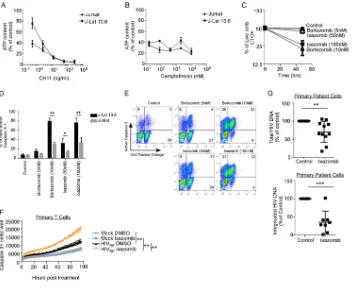
FIG 5Mixtures of HIV-infected and uninfected T cells, treated with proteasome inhibitors, select for the preferential survival of uninfected cells. (A) Jurkat T cells and chronically HIV-infected J-Lat 10.6 cells were treated with apoptosis-inducing anti-Fas (CH11), and cell survival was monitored by intracellular ATP. Depicted are means (⫾SD) of the results of triplicate experiments. (B) Jurkat T cells and J-Lat 10.6 cells were treated with camptothecin, and cell survival was monitored by intracellular ATP content. Depicted are means (⫾SD) of the results of triplicate experiments. (C) J-Lat 10.6 cells were labeled with the lipophilic dye CTO to distinguish them from unlabeled Jurkat T cells, and the two cell types were mixed at a 1:1 ratio. T20 was added to prevent spreading HIV infection, and then the cells were treated with control, bortezomib, or ixazomib and analyzed for active caspase 3 staining. Depicted is the proportion of live (active caspase 3-negative) cells that were also CTO-positive (J-Lat 10.6) cells (means⫾SD of the results of three independent experiments). (D) Pooled data from three replicates of the experiment shown in panel C depicting means and SD. (E) Representative flow data from panels C and D; cell death in the CTO-positive (J-Lat) and CTO-negative (Jurkat) cells can be seen in the same sample. (F) Primary activated CD4 T cells were mock infected or infected with HIV-1RF, treated with ixazomib (100 nM) or vehicle
control, and assessed for active caspase 3 expression over time. (G) Freshly isolated CD4⫹T cells from HIV-infected ART-suppressed patients were treated with ixazomib or control and ART (T20, EFV, or RAL) to prevent spreading infection. After 96 h, cells were harvested, both total (top) (n⫽11 patients) and integrated (bottom) (n⫽8 patients) HIV DNA copies per million cells were determined, and the values were compared to the control by one-samplettest.*,P⬍0.05;**,P⬍0.01;***,P⬍0.001.
Natesampillai et al. Journal of Virology
on November 6, 2019 by guest
http://jvi.asm.org/
[image:8.585.42.398.70.359.2]then treated with vehicle control, bortezomib, or ixazomib. Conceptually, if the
treat-ments cause similar degrees of killing in uninfected J-Lat 10.6 cells and in Jurkat cells,
then the proportion of labeled CTO
⫹J-Lat cells should remain at
⬃
50% over time.
Bortezomib and ixazomib treatments both resulted in a decrease in the proportion of
viable CTO-positive J-Lat 10.6 cells (Fig. 5C) (
P
⬍
0.001). Flow cytometric analysis
demonstrated that, despite low-level caspase 3 induction in Jurkat cells, there were
significantly more active caspase 3-positive HIV-infected J-Lat cells than uninfected
Jurkat cells under the same culture conditions (
P
⬍
0.01 for 10 nM bortezomib and 100
nM ixazomib) (Fig. 5D and E).
We have previously shown in primary CD4
⫹T cells from ART-suppressed patients
that reactivation from latency induces Casp8p41 expression (15). Therefore, we
postu-lated that proteasome inhibitor treatment of cells from ART-suppressed HIV-infected
donors might (i) reactivate HIV, (ii) stabilize Casp8p41, and (iii) result in the death of
more HIV-infected cells than uninfected cells. Since clinical experience indicates that
ixazomib does not cause significant leukopenia (35, 36) whereas bortezomib is
asso-ciated with significant gastrointestinal and hematological toxicity (37), we used
ix-azomib for these studies. To first assess the clinical relevance of our findings, we
infected activated primary CD4 T cells with a clinical dual-tropic HIV-1 isolate (HIV-1
RF),
treated with ixazomib or control, and assessed for caspase 3 activation over time (Fig.
5F). Ixazomib increased active caspase 3 expression in HIV-1
RF-infected and -exposed
cells (
P
⬍
0.0001), but not in mock-infected cells. This is consistent with our findings in
laboratory-adapted HIV-1 strains (compare Fig. 2E and H) and suggests that ixazomib’s
effect may be clinically relevant.
Given the very low frequency of HIV-infected cells in blood circulation (38), it is not
possible to monitor the death of latently infected cells compared to uninfected cells; we
therefore analyzed the HIV DNA levels before and after treatment in patient samples.
CD4 T cells from 11 HIV-positive ART-suppressed patients were treated with ixazomib
or control, and the HIV DNA content was measured by assessing total cell-associated
HIV DNA content with digital-droplet PCR (ddPCR) (39). Ixazomib treatment reduced
total cell-associated HIV DNA by a median of 35% (interquartile range [IQR], 7% to 42%;
P
⫽
0.007) (Fig. 5G, top). To independently verify this result, we treated cells from an
additional 8 patients with ixazomib, and an independent laboratory, blinded to sample
identification, analyzed the integrated HIV DNA content using a previously validated
assay (40, 41). Ixazomib treatment also reduced integrated HIV DNA in the second
group (median reduction in integrated HIV DNA, 69% [IQR, 58 to 89%];
P
⬍
0.001) (Fig.
5G, bottom).
DISCUSSION
There is increasing appreciation that the biological processes that favor survival of
cancer cells also operate in cells containing the transcriptionally silent reservoir for HIV;
examples are epigenetic modifications of DNA and enhanced expression of checkpoint
inhibitors (42, 43). Following the initial description of the HIV protease Casp8p41
pathway of HIV-infected-cell killing (13), the simultaneous advances in understanding
of the control of proapoptotic Bak (44) allowed mechanistic insights into how the
catalytically inert Casp8p41 protein might bind and activate Bak to initiate cell death.
Indeed, we now know that Casp8p41 adopts an alpha-helical conformation that
functions as a BH3-like domain that binds with nanomolar affinity to the BH3 groove in
Bak (21), causing the death of HIV-infected cells. As other Bcl2 family members contain
BH3 grooves and therefore can bind proteins containing BH3 domains (45), our finding
that Casp8p41 binds Bcl2 and Bak with similar affinities (22) provides a mechanistic
basis for how cells that generate Casp8p41 might escape the prodeath effects of
Casp8p41. Indeed, selectively occupying the BH3 groove in Bcl2 with venetoclax allows
Casp8p41 to interact with Bak, and infected-cell death is favored (23). In the current
report, we extend this understanding to show that the Casp8p41-Bcl2 complex is
polyubiquitinated and targeted to the proteasome for degradation. Blocking this
pathway with ixazomib or bortezomib allows Casp8p41 to accumulate, thereby
on November 6, 2019 by guest
http://jvi.asm.org/
ing the Bcl2 restriction of HIV-infected-T cell death. Also of additional benefit is the
previously unreported finding that ixazomib independently drives HIV reactivation,
which is, of course, a necessary step in generating Casp8p41 production. At present, it
is unknown if Casp8p41 is expressed in HIV-infected macrophages or if proteasome
inhibitor treatment of infected macrophages would have the same effect as in T cells.
This will be important to study, as macrophages are naturally resistant to HIV-induced
cell death and contribute to the HIV-1 reservoir (46).
Our data demonstrate that an oral proteasome inhibitor that is clinically dosed once
weekly functions dually to reactivate HIV and to increase Casp8p41 expression, thereby
inducing apoptotic death of T cells in which HIV replicates (Fig. 6). Killing is preferential
for HIV-infected cells because cell death is dependent in part upon the presence of
functional Casp8p41 (Fig. 4F), which is produced only by HIV protease (13, 14). It should
be noted that cell death is also increased to some degree in ixazomib-treated,
GFP-negative Jurkat T cells exposed to HIV-GFP (Fig. 2J), as well as ixazomib-treated,
GFP-negative J-Lat cells (Fig. 4A). However, we have previously shown that GFP
expression alone does not differentiate infected versus uninfected dying cells, as GFP
expression is lost during apoptosis (22). Also, these GFP-negative cells were exposed to
HIV; therefore, individual cells, even if uninfected, could be exposed to other
death-inducing stimuli, such as FasL, HIV gp120, or HIV Vpr. Therefore, the important
exper-imental controls used were uninfected, unexposed cells.
This preferential, proapoptotic effect of ixazomib causes two surrogate measures of
HIV reservoir size (total cell-associated and integrated HIV DNA levels) to decrease
following single-dose treatment
ex vivo
. Acknowledging that the full clinical
signifi-cance of
ex vivo
HIV DNA content reductions is unknown, and given that a significant
fraction of HIV DNA is replication or otherwise defective (47), additional studies
investigating the impact of multidose ixazomib on HIV dynamics using large-volume
leukophoresis samples are ongoing via a pilot clinical trial of ixazomib in
ART-suppressed HIV-positive patients (ClinicalTrials.gov identifier NCT02946047).
FIG 6Schematic model of the effects of proteasome inhibitors of HIV-infected cells. Proteasome inhibitors directly induce viral replication, which generates Casp8p41, which is bound and inhibited by Bcl2 and then targeted to the proteasome for degradation. Proteasome inhibitor-mediated blockade of Casp8p41 degradation favors accumu-lation of Casp8p41, which overcomes Bcl2 restriction, causing death of the reactivated, infected cell. Because Casp8p41 is not present in uninfected cells, this pathway does not operate in uninfected bystander cells.
Natesampillai et al. Journal of Virology
on November 6, 2019 by guest
http://jvi.asm.org/
[image:10.585.44.403.76.326.2]MATERIALS AND METHODS
Cells.All HIV-positive donors were on antiretroviral therapy and had a plasma HIV RNA load of⬍48 copies/ml. Where indicated, peripheral primary CD4 T cells were purified by negative selection, using RosetteSep human CD4⫹T cell enrichment cocktail (Stemcell Technologies) or an EasySep human CD4⫹
T cell isolation kit (Stemcell Technologies); we routinely achieve⬎95% CD4 T cell purity by these techniques. The following cell lines and sources were used: Jurkat T cells (Jurkat e6.1; ATCC; catalog number TIB-152), 293T cells (HEK-293T; ATCC; catalog number CRL-3216), J-Lat 10.6 cells (NIH AIDS Reagent Program), and JB-6 cells (provided by S. Nagata [48]).
Antibodies. The following antibodies were used for immunoblotting or immunoprecipitation: anti-HA peroxidase (3F10) rat monoclonal antibody (catalog number 12013819001; Roche, Indianapolis, IN), mouse anti-caspase 8 antibody (received from Marcus E. Peter, Northwestern University, Chicago, IL), mouse monoclonal anti-ubiquitin (P4D1) antibody (sc-8017; Santa Cruz), anti-Bcl-2 (C21) rabbit poly-clonal IgG (catalog number sc-783; Santa Cruz), monopoly-clonal anti-HIV p24 (catalog number 530; NIH AIDS Reagent Program), anti-GAPDH (glyceraldehyde-3-phosphate dehydrogenase) (14C10) rabbit MAb (cat-alog number 2118; Cell Signaling), histone deacetylase (HDAC) antibody sampler kit (cat(cat-alog number 9928; Cell Signaling), anti-phospho-NF-B-p65 (Ser 536) rabbit MAb (catalog number 3033; Cell Signal-ing), anti-NF-Bp65 (D14E12) rabbit MAb (catalog number 8242; Cell Signaling), anti-phospho-IB␣ (Ser32) (14D4) rabbit MAb (catalog number 2859; Cell Signaling), anti-IB␣(L35A5) mouse MAb (amino-terminal antigen; catalog number 4814; Cell Signaling), anti-cleaved caspase 3 (Asp175) antibody (catalog number 9661; Cell Signaling), and mouse anti-human PARP (catalog number 556494; BD Pharmingen). The following antibodies were used for flow cytometry: phycoerythrin (PE)-conjugated mouse HIV core antibody (Beckman Coulter) and allophycocyanin (APC)-conjugated rabbit anti-active caspase 3 antibody (BD Pharmingen; 1:100).
DNA and viral constructs.The following reagents were obtained through the NIH AIDS Reagent Program, Division of AIDS, NIAID, NIH: HIV-1IIIB, from Robert Gallo, and HIV-1RF, from Dean Winslow. The
envelope-defective proviral GFP-HIV-Nef-Vpr 426-bp plasmid used for Fig. 2F to H has been previously described (21). The HIV-1 envelope-defective proviral plasmid HxBRUR⫺/Env⫺was obtained from É.
Cohen (University of Montreal, Montreal, Canada) (49). Individual proviral constructs were transfected into 293T cells, along with VSV-G expressor (SVCMV-VSV-G); 48 h later, culture supernatants were collected, clarified, and ultracentrifuged to pellet pseudotyped virus. Plasmids pEGFPC1, pEGFP-procaspase 8 Wt, and pEGFP-pEGFP-procaspase 8 RN (caspase 8 that is resistant to HIV protease cleavage due to mutation of Arg [R] 355, Asn [N] 356, Phe [F] 355, and Phe [F] 356) have been previously described (20).
Flow cytometry.Jurkat T cells were transfected with GFP-empty vector and GFP-Casp8p41 con-structs, as previously described (21). Intracellular expression of active caspase 3, HIV p24 antigen, and Casp8p41 were assessed by flow cytometry, as previously described, using appropriate isotype controls for specific determination of expression (17, 22). For the clonotypic assay, J-Lat 10.6 cells were stained with CellTracker Orange (Molecular Probes) according to the manufacturer’s protocol. The labeled J-Lat 10.6 cells were washed and cocultured with unlabeled Jurkat cells at a ratio of 1:1. The cocultured cells were treated with DMSO, bortezomib, or ixazomib at the indicated concentrations for 3 days before being washed and fixed with 2% paraformaldehyde for 24 h prior to analysis. For the experiments shown in Fig. 4D to F, the following gating strategy was used. GFP-positive cells were determined using untransfected controls and used for downstream analyses. HIV-1 p24- and active caspase 3-positive cells were determined using isotype controls. HIV-1 p24-bright cells were identified on the flow plot as those cells which were present between 104and 105on the relevant axis by visual cloud gating. For the analysis
shown in Fig. 4F, the calculation performed was as follows: (percentage in quadrant 2)/(percentage in quadrant 2⫹percentage in quadrant 3)⫻100. Fluorescence-activated cell sorting (FACS) analysis was performed on either a FACScan or Fortessa flow cytometer (BD Biosciences) as needed, based on multicolor parameters. Data analysis was performed using FlowJo software (Tree Star, Inc.).
IncuCyte analysis.Jurkat cells were infected at a multiplicity of infection (MOI) of⬃1.0 with VSV-G GFP HIV in the presence of 6g/ml Polybrene (Sigma-Aldrich) for 6 h, washed, and treated or not with ixazomib (100 nM). Cell death was measured with a real-time imaging IncuCyte system (Essen BioScience) using the cell permeability dye IncuCyte cytotox red reagent (catalog number 4632; Essen BioScience). Analysis of the data was done using IncuCyte Zoom software (2016B).
Analysis of ubiquitination.Casp8p41 protein was analyzed for ubiquitin linkage specificity with a UbiCrest deubiquitinase (DUB) enzyme set (catalog number K-400; Boston Biochem, Inc.) (50). 293T cells transfected with Casp8p41-HA were lysed and immunoprecipitated using an anti-HA affinity matrix (Roche), and Casp8p41 bound to the HA matrix was mixed with 1⫻DUB reaction buffer with 1⫻ concentrations of enzymes OTUB1, GST_AMSH, OTUD3, His6-TRABID, CEZANNE, YOD1, OUTLIN, and USP2
and incubated at 37°C for 30 min. Casp8p41-HA protein was eluted and resolved by SDS-PAGE and immunoblotted with HA-horseradish peroxidase (HRP) (Roche).
HIV-LTR-Luc and HIV-LTR⌬KB-Luc reporter constructs.HIV-1 luciferase-expressing HIV-1 coex-pressingRenilla reniformis (pNL-LucR) was obtained from C. Ochsenbauer (University of Alabama at Birmingham, Birmingham, AL), as previously reported (51). The pNL-LucR stocks were prepared by Lipofectamine LTX transfection of 293T cells, and 60 to 72 h posttransfection, virus was concentrated from culture supernatants by ultracentrifugation, followed by determination of the p24 concentration. All the viral aliquots used for infection underwent one freeze-thaw cycle. Primary CD4⫹T cells were HIV
infected after activation using phytohemagglutinin (2g/ml) and interleukin-2 (IL-2) (50 IU/ml) for 48 h; then,⬃100 ng of p24 in 0.5 ml was added to 106cells for 6 h and then washed and cultured with 10
IU/ml IL-2. When indicated, cells were lysed with 1⫻Renillaluciferase lysis buffer and analyzed forRenilla luciferase activity. Viability was assessed using a CellTiter-Glo luminescent cell viability assay (Promega).
on November 6, 2019 by guest
http://jvi.asm.org/
Total cell-associated HIV DNA measurement and integrated HIV DNA measurement. (i) Quan-tification of HIV-1 DNA using Raindance digital droplet PCR.DNA for analysis was extracted using a Qiagen DNA blood minikit (Qiagen, CA), linearized using a Covaris M220 focused ultrasonicator with 3.0 kb minitube blue (Covaris, MA), and quantified using a NanoDrop 2000 spectrophotometer. The fragmentation length was confirmed via gel electrophoresis. Raindance digital PCR was performed using HIV primers and probes specific for HIV Pol (Hxb2 positions 2536 to 2662) and an RPP30 (RNase P) primer-probe set as previously reported (39). PCRs were prepared by mixing 1g of sonicated genomic DNA, 900 nM primers, and 250 nM probe mixture with 25l of TaqMan genotyping master mix (Applied Biosystem, CA) and 2.5l of droplet stabilizer in 50l. PCR was conducted in a thermocycler with a heated lid (Genetouch thermal cycler; Bioer, Tokyo, Japan) with the following settings: 2 min at 50°C and a denaturation step of at 95°C for 10 min, followed by 45 cycles of 95°C for 15 s and 60°C for 1 min (using a 0.6°C/min ramp rate). Thermal-cycled samples were loaded onto the Raindrop Sense instrument, which uses a 488-nm laser to read the 6-carboxyfluorescein (FAM) and VIC fluorescence intensities per droplet. Data from each sample were converted to a 2-dimensional scatterplot displaying FAM intensity on the xaxis and VIC intensity on theyaxis. Gates used to count the droplet events with specific fluorescence properties were defined using graphical tools to outline regions from the positive control, and these gates were applied to all the samples. The positive droplet events were counted within each gate for each sample. The droplet counts were normalized to the RPP30 counts. Total HIV DNA was calculated and expressed in 1 million CD4 cells by the estimated number of RPP30 copies [(Pol/RPP30)⫻2,000,000]. We validated the use of ddPCR for total HIV DNA estimation using 10 independent evaluations of samples containing a fixed ratio of uninfected Jurkat T cells and chronically HIV-infected 8E5 T cells. The coefficient of variance of the ddPCR was 3.8%, and the detection limit of the assay was found to be 45 HIV DNA copies per million cells.
(ii) Integrated HIV DNA quantification.DNA was isolated using a Gentra Puregene cell kit (Qiagen). Levels of integrated HIV DNA were measured by Alu-Gag quantitative PCR (qPCR), as previously described (40). Briefly, a first-step PCR mixture containing a forward primer specific for the human Alu element and a reverse primer specific for the HIVgaggene was followed by a nested real-time PCR with primers and a probe specific for the HIV LTR. The numbers of cells assayed per well ranged from 1,813 to 29,000. The number of cells assayed per well was based on total HIV levels to result in the tightest confidence intervals for binomial distribution (we targeted 30 to 80% positive wells at two dilutions) (40).
Statistics.Results are depicted as means and standard deviations (SD) unless specifically noted. The mean values of the results of independent experiments were compared usingttests, analysis of variance (ANOVA), or the nonparametric equivalent, as appropriate. The mean values of matched samples were compared using pairedttests. Time course experiments were compared using linear regression. All tests were two sided, andPvalues of less than 0.05 were considered statistically significant. Statistical analysis was performed using GraphPad Prism 6.
Study approval. Primary human peripheral blood mononuclear cells were obtained following informed consent according to Mayo Clinic Institutional Review Board (IRB)-approved protocols (IRB numbers 1036-03 and 13-005646) and a Hennepin County Medical Center Human Subjects Research Committee-approved protocol (HSR number 15-3934).
ACKNOWLEDGMENTS
We thank the study participants who volunteered to provide blood samples for
analysis.
This study was supported by funding from the National Institute of Allergy and
Infectious Diseases (grants R01 AI110173 and R01 AI120698 to A.D.B. and
R01-AI-120011 and UM1-AI-126617 to U.O.) and by CTSA grant number UL1 TR000135 (N.W.C.)
from the National Center for Advancing Translational Sciences (NCATS), components of
the National Institutes of Health (NIH). The contents are solely our responsibility and do
not necessarily represent the official view of the NIH.
Keith Henry has conducted clinical research for Merck, Janssen, ViiV/GSK, and Gilead
(payments were made to the institution—Hennepin County Medical Center).
S.N., N.W.C., G.D.B., Z.N., U.O., D.J.K., and A.D.B. conceived and designed the
exper-iments. S.N., N.W.C., G.D.B., Z.N., M.P., U.O., E.C.P., J.V.B., K.H., and R.S. performed the
experiments and analyzed data. N.W.C., J.V.B., K.H., and R.S. recruited study participants.
S.N., N.W.C., and A.D.B. wrote the manuscript.
REFERENCES
1. Finzi D, Blankson J, Siliciano JD, Margolick JB, Chadwick K, Pierson T, Smith K, Lisziewicz J, Lori F, Flexner C, Quinn TC, Chaisson RE, Rosenberg E, Walker B, Gange S, Gallant J, Siliciano RF. 1999. Latent infection of CD4⫹T cells provides a mechanism for lifelong persistence of HIV-1, even in patients on effective combination therapy. Nat Med 5:512–517. https://doi.org/10.1038/8394.
2. Perreau M, Savoye AL, De Crignis E, Corpataux JM, Cubas R, Haddad EK,
De Leval L, Graziosi C, Pantaleo G. 2013. Follicular helper T cells serve as the major CD4 T cell compartment for HIV-1 infection, replication, and production. J Exp Med 210:143–156.https://doi.org/10.1084/jem.2012 1932.
3. Deeks SG. 2012. HIV: Shock and kill. Nature 487:439 – 440.https://doi .org/10.1038/487439a.
4. Shan L, Deng K, Shroff NS, Durand CM, Rabi SA, Yang HC, Zhang H,
Natesampillai et al. Journal of Virology
on November 6, 2019 by guest
http://jvi.asm.org/
Margolick JB, Blankson JN, Siliciano RF. 2012. Stimulation of HIV-1-specific cytolytic T lymphocytes facilitates elimination of latent viral reservoir after virus reactivation. Immunity 36:491–501.https://doi.org/ 10.1016/j.immuni.2012.01.014.
5. Archin NM, Liberty AL, Kashuba AD, Choudhary SK, Kuruc JD, Crooks AM, Parker DC, Anderson EM, Kearney MF, Strain MC, Richman DD, Hudgens MG, Bosch RJ, Coffin JM, Eron JJ, Hazuda DJ, Margolis DM. 2012. Admin-istration of vorinostat disrupts HIV-1 latency in patients on antiretroviral therapy. Nature 487:482– 485.https://doi.org/10.1038/nature11286. 6. Cummins NW, Badley AD. 2010. Mechanisms of HIV-associated
lympho-cyte apoptosis: 2010. Cell Death Dis 1:e99.https://doi.org/10.1038/cddis .2010.77.
7. Kaplan AH, Swanstrom R. 1991. Human immunodeficiency virus type 1 Gag proteins are processed in two cellular compartments. Proc Natl Acad Sci U S A 88:4528 – 4532.https://doi.org/10.1073/pnas.88.10.4528. 8. Kaplan AH, Manchester M, Swanstrom R. 1994. The activity of the protease of human immunodeficiency virus type 1 is initiated at the membrane of infected cells before the release of viral proteins and is required for release to occur with maximum efficiency. J Virol 68: 6782– 6786.
9. Debouck C, Metcalf BW. 1990. Human immunodeficiency virus protease: a target for AIDS therapy. Drug Dev Res 21:1–17. https://doi.org/10 .1002/ddr.430210102.
10. Strack PR, Frey MW, Rizzo CJ, Cordova B, George HJ, Meade R, Ho SP, Corman J, Tritch R, Korant BD. 1996. Apoptosis mediated by HIV pro-tease is preceded by cleavage of Bcl-2. Proc Natl Acad Sci U S A 93:9571–9576.https://doi.org/10.1073/pnas.93.18.9571.
11. Adams LD, Tomasselli AG, Robbins P, Moss B, Heinrikson RL. 1992. HIV-1 protease cleaves actin during acute infection of human T-lymphocytes. AIDS Res Hum Retrovir 8:291–295. https://doi.org/10.1089/aid.1992.8 .291.
12. Ventoso I, Blanco R, Perales C, Carrasco L. 2001. HIV-1 protease cleaves eukaryotic initiation factor 4G and inhibits cap-dependent translation. Proc Natl Acad Sci U S A 98:12966 –12971.https://doi.org/10.1073/pnas .231343498.
13. Nie Z, Phenix BN, Lum JJ, Alam A, Lynch DH, Beckett B, Krammer PH, Sekaly RP, Badley AD. 2002. HIV-1 protease processes procaspase 8 to cause mitochondrial release of cytochrome c, caspase cleavage and nuclear fragmentation. Cell Death Differ 9:1172–1184.https://doi.org/10 .1038/sj.cdd.4401094.
14. Nie Z, Bren GD, Vlahakis SR, Schimnich AA, Brenchley JM, Trushin SA, Warren S, Schnepple DJ, Kovacs CM, Loutfy MR, Douek DC, Badley AD. 2007. Human immunodeficiency virus type 1 protease cleaves pro-caspase 8 in vivo. J Virol 81:6947– 6956. https://doi.org/10.1128/JVI .02798-06.
15. Nie Z, Bren GD, Rizza SA, Badley AD. 2008. HIV protease cleavage of procaspase 8 is necessary for death of HIV-infected cells. Open Virol J 2:1–7.https://doi.org/10.2174/1874357900802010001.
16. Natesampillai S, Nie Z, Cummins NW, Jochmans D, Bren GD, Angel JB, Badley AD. 2010. Patients with discordant responses to antiretroviral therapy have impaired killing of HIV-infected T cells. PLoS Pathog 6:e1001213.https://doi.org/10.1371/journal.ppat.1001213.
17. Cummins NW, Jiang W, McGinty J, Bren GD, Bosch RJ, Landay A, Deeks SG, Martin JN, Douek D, Lederman MM, Brenchley J, Badley AD. 2010. Intracellular Casp8p41 content is inversely associated with CD4 T cell count. J Infect Dis 202:386 –391.https://doi.org/10.1086/653705. 18. Cummins NW, Neuhaus J, Sainski AM, Strausbauch MA, Wettstein PJ,
Lewin SR, Plana M, Rizza SA, Temesgen Z, Touloumi G, Freiberg M, Neaton J, Badley AD. 2014. Short communication: CD4 T cell declines occurring during suppressive antiretroviral therapy reflect continued production of Casp8p41. AIDS Res Hum Retrovir 30:476 – 479.https:// doi.org/10.1089/aid.2013.0243.
19. Algeciras-Schimnich A, Belzacq-Casagrande AS, Bren GD, Nie Z, Taylor JA, Rizza SA, Brenner C, Badley AD. 2007. Analysis of HIV protease killing through caspase 8 reveals a novel interaction between caspase 8 and mitochondria. Open Virol J 1:39 – 46.https://doi.org/10.2174/ 1874357900701010039.
20. Sainski AM, Natesampillai S, Cummins NW, Bren GD, Taylor J, Saenz DT, Poeschla EM, Badley AD. 2011. The HIV-1-specific protein Casp8p41 induces death of infected cells through Bax/Bak. J Virol 85:7965–7975. https://doi.org/10.1128/JVI.02515-10.
21. Sainski AM, Dai H, Natesampillai S, Pang YP, Bren GD, Cummins NW, Correia C, Meng XW, Tarara JE, Ramirez-Alvarado M, Katzmann DJ, Ochsenbauer C, Kappes JC, Kaufmann SH, Badley AD. 2014. Casp8p41
generated by HIV protease kills CD4 T cells through direct Bak activation. J Cell Biol 206:867– 876.https://doi.org/10.1083/jcb.201405051. 22. Cummins NW, Sainski AM, Dai H, Natesampillai S, Pang YP, Bren GD, de
Araujo Correia MC, Sampath R, Rizza SA, O’Brien D, Yao JD, Kaufmann SH, Badley AD. 2016. Prime, shock, and kill: priming CD4 T cells from HIV patients with a BCL-2 antagonist before HIV reactivation reduces HIV res-ervoir size. J Virol 90:4032– 4048.https://doi.org/10.1128/JVI.03179-15. 23. Cummins NW, Sainski-Nguyen AM, Natesampillai S, Aboulnasr F,
Kauf-mann S, Badley AD. 2017. Maintenance of the HIV reservoir is antago-nized by selective BCL2 inhibition. J Virol 91:e00012-17.https://doi.org/ 10.1128/JVI.00012-17.
24. Nakajima W, Hicks MA, Tanaka N, Krystal GW, Harada H. 2014. Noxa determines localization and stability of MCL-1 and consequently ABT-737 sensitivity in small cell lung cancer. Cell Death Dis 5:e1052.https:// doi.org/10.1038/cddis.2014.6.
25. Komander D, Clague MJ, Urbe S. 2009. Breaking the chains: structure and function of the deubiquitinases. Nat Rev Mol Cell Biol 10:550 –563. https://doi.org/10.1038/nrm2731.
26. Locke M, Toth JI, Petroski MD. 2014. Lys11- and Lys48-linked ubiquitin chains interact with p97 during endoplasmic-reticulum-associated deg-radation. Biochem J 459:205–216.https://doi.org/10.1042/BJ20120662. 27. Bozza WP, Liang Q, Gong P, Zhuang Z. 2012. Transient kinetic analysis of
USP2-catalyzed deubiquitination reveals a conformational rearrange-ment in the K48-linked diubiquitin substrate. Biochemistry 51: 10075–10086.https://doi.org/10.1021/bi3009104.
28. Vincenz L, Jager R, O’Dwyer M, Samali A. 2013. Endoplasmic reticulum stress and the unfolded protein response: targeting the Achilles heel of multiple myeloma. Mol Cancer Ther 12:831– 843. https://doi.org/10 .1158/1535-7163.MCT-12-0782.
29. Kumar SK, Rajkumar SV, Dispenzieri A, Lacy MQ, Hayman SR, Buadi FK, Zeldenrust SR, Dingli D, Russell SJ, Lust JA, Greipp PR, Kyle RA, Gertz MA. 2008. Improved survival in multiple myeloma and the impact of novel therapies. Blood 111:2516 –2520.https://doi.org/10.1182/blood-2007-10 -116129.
30. Papandreou CN, Daliani DD, Nix D, Yang H, Madden T, Wang X, Pien CS, Millikan RE, Tu SM, Pagliaro L, Kim J, Adams J, Elliott P, Esseltine D, Petrusich A, Dieringer P, Perez C, Logothetis CJ. 2004. Phase I trial of the proteasome inhibitor bortezomib in patients with advanced solid tu-mors with observations in androgen-independent prostate cancer. J Clin Oncol 22:2108 –2121.
31. Smith DC, Kalebic T, Infante JR, Siu LL, Sullivan D, Vlahovic G, Kauh JS, Gao F, Berger AJ, Tirrell S, Gupta N, Di Bacco A, Berg D, Liu G, Lin J, Hui AM, Thompson JA. 2015. Phase 1 study of ixazomib, an investigational protea-some inhibitor, in advanced non-hematologic malignancies. Invest New Drugs 33:652– 663.https://doi.org/10.1007/s10637-015-0230-x.
32. Hideshima T, Ikeda H, Chauhan D, Okawa Y, Raje N, Podar K, Mitsiades C, Munshi NC, Richardson PG, Carrasco RD, Anderson KC. 2009. Bort-ezomib induces canonical nuclear factor-kappaB activation in multiple myeloma cells. Blood 114:1046 –1052. https://doi.org/10.1182/blood -2009-01-199604.
33. Bren GD, Trushin SA, Whitman J, Shepard B, Badley AD. 2009. HIV gp120 induces, NF-kappaB dependent, HIV replication that requires procaspase 8. PLoS One 4:e4875.https://doi.org/10.1371/journal.pone.0004875. 34. Sasaki CY, Barberi TJ, Ghosh P, Longo DL. 2005. Phosphorylation of
RelA/p65 on serine 536 defines an I{kappa}B{alpha}-independent NF-{kappa}B pathway. J Biol Chem 280:34538 –34547. https://doi.org/10 .1074/jbc.M504943200.
35. Kumar SK, Berdeja JG, Niesvizky R, Lonial S, Laubach JP, Hamadani M, Stewart AK, Hari P, Roy V, Vescio R, Kaufman JL, Berg D, Liao E, Di Bacco A, Estevam J, Gupta N, Hui AM, Rajkumar V, Richardson PG. 2014. Safety and tolerability of ixazomib, an oral proteasome inhibitor, in combina-tion with lenalidomide and dexamethasone in patients with previously untreated multiple myeloma: an open-label phase 1/2 study. Lancet Oncol 15:1503–1512.https://doi.org/10.1016/S1470-2045(14)71125-8. 36. Moreau P, Masszi T, Grzasko N, Bahlis NJ, Hansson M, Pour L, Sandhu I,
Ganly P, Baker BW, Jackson SR, Stoppa AM, Simpson DR, Gimsing P, Palumbo A, Garderet L, Cavo M, Kumar S, Touzeau C, Buadi FK, Laubach JP, Berg DT, Lin J, Di Bacco A, Hui AM, van de Velde H, Richardson PG. 2016. Oral ixazomib, lenalidomide, and dexamethasone for multiple myeloma. N Engl J Med 374:1621–1634.https://doi.org/10.1056/NEJM oa1516282.
37. Eskandary F, Regele H, Baumann L, Bond G, Kozakowski N, Wahrmann M, Hidalgo LG, Haslacher H, Kaltenecker CC, Aretin MB, Oberbauer R, Posch M, Staudenherz A, Handisurya A, Reeve J, Halloran PF, Bohmig GA. 2018.
on November 6, 2019 by guest
http://jvi.asm.org/
A randomized trial of bortezomib in late antibody-mediated kidney transplant rejection. J Am Soc Nephrol 29:591– 605.https://doi.org/10 .1681/ASN.2017070818.
38. Siliciano JM, Siliciano RF. 2015. The remarkable stability of the latent reservoir for HIV-1 in resting memory CD4⫹T cells. J Infect Dis 212: 1345–1347.https://doi.org/10.1093/infdis/jiv219.
39. Strain MC, Lada SM, Luong T, Rought SE, Gianella S, Terry VH, Spina CA, Woelk CH, Richman DD. 2013. Highly precise measurement of HIV DNA by droplet digital PCR. PLoS One 8:e55943.https://doi.org/10.1371/ journal.pone.0055943.
40. Liszewski MK, Yu JJ, O’Doherty U. 2009. Detecting HIV-1 integration by repetitive-sampling Alu-Gag PCR. Methods 47:254 –260.https://doi.org/ 10.1016/j.ymeth.2009.01.002.
41. Graf EH, O’Doherty U. 2013. Quantitation of integrated proviral DNA in viral reservoirs. Curr Opin HIV AIDS 8:100 –105.https://doi.org/10.1097/ COH.0b013e32835d8132.
42. Porichis F, Kwon DS, Zupkosky J, Tighe DP, McMullen A, Brockman MA, Pavlik DF, Rodriguez-Garcia M, Pereyra F, Freeman GJ, Kavanagh DG, Kaufmann DE. 2011. Responsiveness of HIV-specific CD4 T cells to PD-1 blockade. Blood 118:965–974. https://doi.org/10.1182/blood-2010-12 -328070.
43. Archin NM, Kirchherr JL, Sung JA, Clutton G, Sholtis K, Xu Y, Allard B, Stuelke E, Kashuba AD, Kuruc JD, Eron J, Gay CL, Goonetilleke N, Margolis DM. 2017. Interval dosing with the HDAC inhibitor vorinostat effectively reverses HIV latency. J Clin Invest 127:3126 –3135. https://doi.org/10 .1172/JCI92684.
44. Dai H, Smith A, Meng XW, Schneider PA, Pang YP, Kaufmann SH. 2011. Transient binding of an activator BH3 domain to the Bak BH3-binding groove initiates Bak oligomerization. J Cell Biol 194:39 – 48.https://doi .org/10.1083/jcb.201102027.
45. Cao X, Yap JL, Newell-Rogers MK, Peddaboina C, Jiang W,
Papaconstanti-nou HT, Jupitor D, Rai A, Jung KY, Tubin RP, Yu W, Vanommeslaeghe K, Wilder PT, MacKerell AD, Jr, Fletcher S, Smythe RW. 2013. The novel BH3 alpha-helix mimetic JY-1-106 induces apoptosis in a subset of cancer cells (lung cancer, colon cancer and mesothelioma) by disrupting Bcl-xL and Mcl-1 protein-protein interactions with Bak. Mol Cancer 12:42. https://doi.org/10.1186/1476-4598-12-42.
46. Kumar A, Abbas W, Herbein G. 2014. HIV-1 latency in monocytes/ macrophages. Viruses 6:1837–1860.https://doi.org/10.3390/v6041837. 47. Bruner KM, Murray AJ, Pollack RA, Soliman MG, Laskey SB, Capoferri AA,
Lai J, Strain MC, Lada SM, Hoh R, Ho YC, Richman DD, Deeks SG, Siliciano JD, Siliciano RF. 2016. Defective proviruses rapidly accumulate during acute HIV-1 infection. Nat Med 22:1043–1049.https://doi.org/10.1038/ nm.4156.
48. Kawahara A, Ohsawa Y, Matsumura H, Uchiyama Y, Nagata S. 1998. Caspase-independent cell killing by Fas-associated protein with death domain. J Cell Biol 143:1353–1360. https://doi.org/10.1083/jcb.143.5 .1353.
49. Yao XJ, Mouland AJ, Subbramanian RA, Forget J, Rougeau N, Bergeron D, Cohen EA. 1998. Vpr stimulates viral expression and induces cell killing in human immunodeficiency virus type 1-infected dividing Jurkat T cells. J Virol 72:4686 – 4693.
50. Hospenthal MK, Mevissen TE, Komander D. 2015. Deubiquitinase-based analysis of ubiquitin chain architecture using ubiquitin chain restriction (UbiCRest). Nat Protoc 10:349 –361.https://doi.org/10.1038/nprot.2015 .018.
51. Edmonds TG, Ding H, Yuan X, Wei Q, Smith KS, Conway JA, Wieczorek L, Brown B, Polonis V, West JT, Montefiori DC, Kappes JC, Ochsenbauer C. 2010. Replication competent molecular clones of HIV-1 expressing Re-nilla luciferase facilitate the analysis of antibody inhibition in PBMC. Virology 408:1–13.https://doi.org/10.1016/j.virol.2010.08.028.
Natesampillai et al. Journal of Virology
on November 6, 2019 by guest
http://jvi.asm.org/