Eidgenössische Technische Hochschule, Zürich, Switzerland; INRS-Institut Armand-Frappier, Université du Québec, Québec, Canada; Facultad de Ciencias Biológicas, Universidad Andrés Bello, Santiago, Chilef
How hantaviruses assemble and exit infected cells remains largely unknown. Here, we show that the expression of Andes (ANDV) and Puumala (PUUV) hantavirus Gn and Gc envelope glycoproteins lead to their self-assembly into virus-like particles (VLPs) which were released to cell supernatants. The viral nucleoprotein was not required for particle formation. Further, a Gc endodomain deletion mutant did not abrogate VLP formation. The VLPs were pleomorphic, exposed protrusions and reacted with patient sera.
H
antaviruses are rodent-borne, enveloped, negative-strand,tripartite RNA viruses that form a separate genus in the
Bu-nyaviridaefamily. Their transmission to humans can produce se-vere diseases, such as hemorrhagic fever with renal syndrome caused by Puumala hantavirus (PUUV) in Northern Europe and hantavirus pulmonary syndrome caused by Andes hantavirus
(ANDV) in Argentina and Chile (1–3).
Hantavirus virions are roughly spherical and highly hetero-genic, varying from 120 to 160 nm in size. They expose glycopro-tein spikes locally ordered into tetramers and also contain naked
membrane patches on their surfaces (4,5). The viral envelope
membrane is acquired from infected cells during virus budding and encloses the three single-stranded RNA (ssRNA) segments that encode four structural proteins: the Gn and Gc glycoproteins, nucleocapsid protein (N), and RNA-dependent RNA polymerase. As other bunyaviruses, hantaviruses do not have a matrix protein that mediates assembly and budding; hence, a role for the
cyto-plasmic tails of glycoproteins has been proposed (6–8). Previous
studies of orthobunyavirus mutant glycoproteins showed that the endodomains of both glycoproteins are required for virus-like
particle (VLP) and virus assembly (9). Further, studies on the
Uukuniemi phlebovirus Gn tail showed that the Gn endodomain
plays a crucial role in genome packaging into virus particles (10).
Bunyavirus glycoproteins originate from a single glycoprotein precursor (GPC) through cotranslational cleavage in the
endo-plasmic reticulum (11–14). Hantaviruses are believed to bud at
internal membranes, most probably derived from the Golgi appa-ratus, and exit cells via exocytosis; alternatively, they may bud
directly from the plasma membrane (11,15,16).
To study viral assembly and budding processes, individual or isolated viral components are expressed in cells to test their release into the culture medium as VLPs corresponding to
membrane-containing viral structures (17). Previously, it has been reported
that hantavirus VLPs are produced when Gn, Gc, and N proteins
are coexpressed (18,19). However, not all of these proteins may be
necessary for VLP production. This notion is supported by the observation that animals elicit high neutralizing antibody
re-Received22 October 2013Accepted26 November 2013 Published ahead of print11 December 2013
Address correspondence to Nicole D. Tischler, ntischler@cienciavida.cl.
R.A. and N.C.-M. contributed equally to this work.
Copyright © 2014, American Society for Microbiology. All Rights Reserved.
doi:10.1128/JVI.03118-13
Gn
Gc
Actin
170 kDa 72 kDa
53 kDa 42 kDa 170 kDa
N
A
ANDVPUUV GPC GPC/N
B
55 kDa
170 kDa
55 kDa
Lysates Supernatant
Gc
Actin 42 kDa
pI.18 pI.18GPC GPC/N
GPC
Lysates Supernatant
pI.18 pI.18 GPC
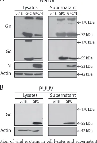
FIG 1Detection of viral proteins in cell lysates and supernatants. Western blots of lysates and concentrated supernatant of 293FT cells transfected with different plasmids. (A) Transfection of empty plasmid, pI.18/ANDV-GPC, or cotransfection of pI.18/ANDV-GPC and pCMV-Bios/ANDV-N. (B) Trans-fection of empty plasmid or pWRG/PUU-M(s2). ANDV Gn and N proteins were detected with anti-Gn 6B9/F5 and anti-N 7B3/F7, respectively. ANDV and PUUV Gc was detected with MAb anti-Gc 2H4/F6. No MAb against PUUV Gn was available.
on November 7, 2019 by guest
http://jvi.asm.org/
[image:1.585.79.248.402.650.2]sponses after DNA vaccination solely using a hantavirus Gn and
Gc coding plasmid (20–23), which may be indicative for VLP
for-mationin vivo, in the absence of the N protein. In addition, for
other members of theBunyaviridae, such as phleboviruses, it has
been reported that the glycoproteins are the only viral
compo-nents required for the formation of VLPs (24,25). To test whether
the hantavirus N protein is required for the assembly and budding of hantavirus-like particles, in the present work, hantavirus VLP formation was assessed by plasmid-driven expression of hantavi-rus Gn and Gc glycoproteins. To this end, 293FT cells (Invitrogen) grown in 10-cm dishes were transfected by the calcium phosphate
protocol (26) using 8g of pI.18/ANDV-GPC plasmid coding for
ANDV-GPC under the control of the cytomegalovirus promoter
(27). As a positive control for hantavirus VLP formation, 293FT
cells were cotransfected with pI.18/ANDV-GPC and
pCMV-Bios/N (28) coding for ANDV-N under the cytomegalovirus
pro-moter. The expression of ANDV Gn, Gc, and N was analyzed by
Western blotting of cell lysates at 48 h posttransfection (Fig. 1A).
To this end, the monoclonal antibodies (MAbs) anti-Gn 6B9/F5
(29), anti-Gc 2H4/F6 (30), and anti-N 7B3/F7 (31) and secondary
antibody anti-mouse IgG peroxidase conjugate (Sigma) were used. In the lysates of cells transfected with one or both plasmids,
ANDV Gn monomers were detected at⬃70 kDa and ANDV Gc
monomers at ⬃55 kDa (Fig. 1A, left). Higher-molecular-mass
bands of Gn and Gc were also observed as reported previously (27,
29). N could be detected in cells cotransfected with pCMV-Bios/N
with a size of⬃50 kDa. To analyze whether the viral proteins were
also present in cell supernatants, they were concentrated by
ultra-centrifugation at 100,000⫻g, as described previously (27). As
seen inFig. 1A(right), Gn and Gc were detected in concentrated
supernatants of cells expressing both envelope proteins alone or in combination with the viral N protein, confirming our hypothesis that N was not required for the release of glycoproteins into the cell supernatant. To test whether the release of ANDV
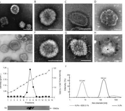
glycopro-FIG 2Characterization of hantavirus VLPs. Negative-stain EM of concentrated supernatants from cells transfected with pI.18/ANDV-GPC (A to D) or pWRG/PUU-M(s2) (E to H). Negative-stain EM using phosphotungstic acid of unfixed ANDV VLPs (B), PUUV VLPs (E, F), glutaraldehyde-fixed ANDV VLPs (A, C), and PUUV VLPs (G). Immunogold EM of ANDV VLPs (D) and PUUV VLPs (H) stained with uranyl acetate using patient sera (1:100) and protein A conjugated to gold beads (20 nm). (I) Sucrose gradient sedimentation of ANDV VLPs and detection of VLPs through Western blotting using anti-ANDV Gc MAb 2H4/F6. Buoyant density was determined by refractometry, and relative intensity of Gc bands was quantified as arbitrary units using ImageJ (40). (J) Dynamic light scattering of ANDV VLPs at pH 7.4 and ANDV VLPs treated for 30 min with SDS 0.1%. Bars, 100 nm.
on November 7, 2019 by guest
http://jvi.asm.org/
[image:2.585.79.509.65.447.2]teins was a particular property of ANDV or if it was a more general feature of hantaviruses, plasmid-driven expression of PUUV gly-coproteins was next tested. To this end, 293FT cells were
trans-fected with 8g of the plasmid pWRG/PUU-M(s2) coding for
PUUV-GPC under the cytomegalovirus promoter (32). When cell
lysates and the concentrated supernatant of pWRG/PUU-M(s2)-transfected cells were analyzed by Western blotting, PUUV Gc was
detected in both fractions with its expected molecular mass (Fig.
1B), indicating that, as for ANDV, the glycoproteins of PUUV
were also released into the cell supernatant in the absence of ad-ditional viral molecules. To test whether the presence of viral gly-coproteins in cell supernatants was related to virus-like structures, concentrated supernatants of cells transfected with plasmids cod-ing for ANDV-GPC or PUUV-GPC were next analyzed by elec-tron microscopy (EM) using phosphotungstic acid at a pH of
⬃7.4 for negative staining, as described previously (33). Electron
micrographs of concentrated supernatants of ANDV or PUUV glycoprotein-expressing cells showed virus-like structures that
were variable in size and shape (Fig. 2AtoDandEtoH,
respec-tively). No apparent morphological differences could be detected between ANDV VLPs produced with or without the N protein (data not shown). It has been well described that EM of
hantavi-ruses reveals a characteristic grid-like pattern (34–36).
Negative-staining EM of ANDV and PUUV VLPs fixed with 0.5%
glutaral-dehyde allowed to discern a grid-like pattern (Fig. 2C andG,
respectively). Further, isolated surface projections could be
distin-guished (Fig. 2G, arrows), resembling a Y-shape similar to the
described molecular structure of Hantaan virus and Tula virus spikes that are composed of a tetrameric, globular head domain
connected to the membrane by a thinner, central stalk region (4,
5).
The observed virus structures were further characterized by immunogold EM. For this purpose, VLPs were adsorbed to Form-var/carbon-coated copper grids and blocked with 5% bovine se-rum albumin. Subsequently, the immobilized VLPs were
incu-PUUV VLPs were recognized by the respective specific patient
sera. Normal patient sera were used as negative controls (n⫽3;
data not shown). To further characterize the VLPs in terms of density, sucrose gradient sedimentation was performed by ultra-centrifugation for 16 h at 38,000 rpm using an SW55 rotor. The refractive index of each fraction was analyzed at 20°C, and the presence of VLPs was examined by Western blotting using MAb anti-Gc 2H4/F6. VLPs derived from ANDV glycoprotein expres-sion peaked in fraction 8, corresponding to a buoyant density of
1.15 g/ml (Fig. 2I) that coincides with the density range of 1.15 to
1.18 g/ml, which has been reported for infectious hantaviruses and
other bunyaviruses (34,37). The size range of particles generated
by glycoprotein expression was next determined by dynamic light scattering (Zetasizer Nano ZS; Malvern Instruments). The size of
over 90% of VLPs varied within the range of 90 to 255 nm (Fig. 2J).
When VLPs were incubated with 0.1% SDS, their size diminished
below 20 nm, confirming their membranous composition (Fig.
2J). Taken together, these data indicate that the release of
glyco-proteins into cell supernatants in the form of virus-like structures does not require the participation of the viral N protein. Further, the hantavirus glycoproteins are the only viral components re-quired for the assembly and release of VLPs.
The hantavirus VLPs were further characterized in terms of their antigenicity using the sera of hantavirus patients. For this purpose, ANDV or PUUV VLPs contained in concentrated super-natants were immobilized on enzyme-linked immunosorbent as-say (ELISA) plates by incubation for 1 h at room temperature (RT). Subsequently, wells were blocked with 4% casein-sucrose for 2 h at RT, and human sera were then added at a dilution of 1:250. After 1.5 h of incubation at RT, the wells were washed 5 times with 0.05% Tween 20 –phosphate-buffered saline (PBS). Next, wells were incubated for 1 h with anti-human immunoglob-ulins G, A, and M conjugated to peroxidase and finally revealed with a tetramethylbenzidine peroxidase substrate (KPL). The re-action was stopped within 10 min by the addition of 1 M
phos-phoric acid, and the absorbance was read at 450 nm. As seen inFig.
3, ANDV VLPs reacted with sera derived from Chilean patients.
Weak cross-reactivity with ANDV VLPs was detected with the sera of patients from North America infected with Sin Nombre virus or Sin Nombre virus-related species. No reactivity against ANDV VLPs was observed with sera from PUUV-infected patients and with negative-control sera. When PUUV VLPs were incubated with patient sera, reactivity was detected with PUUV-infected pa-tient sera, and a weak cross-reactivity was observed with the sera of patients from America. No reactivity of PUUV VLPs was observed with the negative-control sera. In summary, these data confirm that the ANDV and PUUV VLPs contain glycoproteins on their surfaces that expose epitopes that are recognized by patient sera reactive against the respective native hantavirus.
To test the requirement of the Gc endodomain for VLP
assem-bly, a Gc endodomain deletion mutant (GPC⌬GcCT) was
gener-ated, based on the prediction of the Gc transmembrane region (38,
39). DNA mutagenesis was performed by Pfx polymerase
(Invit-rogen)-driven PCR amplification of the ANDV-GPC coding
re-***
Serum (-) SNV
PUUV ANDV Seru
m (-) SNV PUUV ANDV
0 1 2
VLPs ANDV VLPs PUUV
**
**
***
O
ptic
al Densit
FIG 3Antigenicity of hantavirus VLPs. ELISA plates were activated with con-centrated ANDV or PUUV VLPs, and their reactivity was tested with sera derived from patients infected with different hantavirus species (ANDV,
PUUV, Sin Nombre virus [SNV]). A Studentttest was used for statistical
evaluation: ***,P⬍0.00025; **,P⬍0.0025; *,P⬍0.025.
on November 7, 2019 by guest
http://jvi.asm.org/
[image:3.585.62.264.66.265.2]gion using the forward primer 5=-TAGATCTATTATGGAAGGG
T G G T A T C T G G T T G C - 3= a n d t h e r e v e r s e p r i m e r 5=
-CTCGAGCTAGCACAGAACACTGAACATTATGATAGAG AG-3=. Subsequently, the PCR product was subcloned into the pI.18 expression vector. To test expression and folding of the
GPC⌬GcCT mutant, 293FT cells were surface biotinylated 48 h
post-transfection, using a cell surface protein isolation kit (Pierce) as
de-scribed previously (29). Previous to biotinylation, the cell
superna-tant was collected for particle concentration, and the cell lysates were separated into two fractions: the unbiotinylated fraction containing intracellular proteins and the biotinylated fraction containing surface proteins. When these fractions were analyzed by Western blotting, no apparent difference among the wild-type (WT) and mutant proteins was detected in terms of Gc surface localization or the presence of
glycoproteins in the concentrated supernatant (Fig. 4A). Further,
negative-stain EM of the concentrated supernatant revealed virus structures similar to those of VLPs formed by the WT glycoproteins (Fig. 4B). These data indicate that in contrast to the orthobunyavirus Gc endodomain, the ANDV Gc endodomain is not required for the assembly of virus structures. This difference may be explained in part by the different length of these regions, conserving nine residues
among hantaviruses (39) compared to 26 residues among
orthobu-nyaviruses (9).
In the absence of a reverse-genetics system for hantaviruses, the VLP formation system is an important tool for the characteriza-tion of the maturacharacteriza-tion and exit funccharacteriza-tions as well as cell entry of hantavirus glycoproteins. Further mutagenesis studies are cur-rently assessing glycoprotein properties required for their assembly and cell exit. It remains to be determined whether
self-assembly is a general feature of glycoproteins of theHantavirus
genus and theBunyaviridae.
ACKNOWLEDGMENTS
We thank Jay Hooper (USAMRIID) for providing the plasmid pWRG/ PUU-M(s2). Furthermore, we acknowledge Brian Hjelle (University of New Mexico, USA) and Hector Galeno (Instituto de Salud Púbica, Chile) for providing patient sera. Our thanks also extend to Alejandro Muñizaga (Advanced Microscopy Unit, Pontificia Universidad Católica, Chile) for his advice on immunogold EM.
This work was financed by CONICYT through grant FONDECYT 1100756 and basal funding PFB-16. R.A. was supported by a CONICYT doctoral fellowship.
REFERENCES
1.Macneil A, Nichol ST, Spiropoulou CF.2011. Hantavirus pulmonary
syndrome. Virus Res.162:138 –147.http://dx.doi.org/10.1016/j.virusres
.2011.09.017.
2.Krautkramer E, Zeier M, Plyusnin A. 2013. Hantavirus infection: an
emerging infectious disease causing acute renal failure. Kidney Int.83:23– 27.http://dx.doi.org/10.1038/ki.2012.360.
3.Schmaljohn C, Hjelle B.1997. Hantaviruses: a global disease problem. Emerg. Infect. Dis.3:95–104.http://dx.doi.org/10.3201/eid0302.970202. 4.Huiskonen JT, Hepojoki J, Laurinmaki P, Vaheri A, Lankinen H,
Butcher SJ, Grunewald K.2010. Electron cryotomography of Tula han-tavirus suggests a unique assembly paradigm for enveloped viruses. J. Vi-rol.84:4889 – 4897.http://dx.doi.org/10.1128/JVI.00057-10.
5.Battisti AJ, Chu YK, Chipman PR, Kaufmann B, Jonsson CB, Ross-mann MG.2011. Structural studies of Hantaan virus. J. Virol.85:835– 841.http://dx.doi.org/10.1128/JVI.01847-10.
6.Wang H, Alminaite A, Vaheri A, Plyusnin A.2010. Interaction between hantaviral nucleocapsid protein and the cytoplasmic tail of surface glyco-protein Gn. Virus Res.151:205–212.http://dx.doi.org/10.1016/j.virusres .2010.05.008.
7.Hepojoki J, Strandin T, Wang H, Vapalahti O, Vaheri A, Lankinen H. 2010. Cytoplasmic tails of hantavirus glycoproteins interact with the nu-cleocapsid protein. J. Gen. Virol.91:2341–2350.http://dx.doi.org/10.1099 /vir.0.021006-0.
8.Strandin T, Hepojoki J, Vaheri A.2013. Cytoplasmic tails of bunyavirus Gn glycoproteins— could they act as matrix protein surrogates? Virology 437:73– 80.http://dx.doi.org/10.1016/j.virol.2013.01.001.
9.Shi X, Kohl A, Li P, Elliott RM. 2007. Role of the cytoplasmic tail domains of Bunyamwera orthobunyavirus glycoproteins Gn and Gc in
virus assembly and morphogenesis. J. Virol.81:10151–10160.http://dx
.doi.org/10.1128/JVI.00573-07.
10. Overby AK, Pettersson RF, Neve EP.2007. The glycoprotein cytoplasmic tail of Uukuniemi virus (Bunyaviridae) interacts with ribonucleoproteins and is critical for genome packaging. J. Virol.81:3198 –3205.http://dx.doi .org/10.1128/JVI.02655-06.
11. Pettersson R, Melin L.1996. Synthesis, assembly and intracellular
trans-port ofBunyaviridaemembrane proteins.InElliott RM (ed), The
Bunya-viridae. Plenum Press, New York, NY.
12. Spiropoulou CF, Goldsmith CS, Shoemaker TR, Peters CJ, Compans
RW.2003. Sin Nombre virus glycoprotein trafficking. Virology308:48 –
63.http://dx.doi.org/10.1016/S0042-6822(02)00092-2.
13. Shi X, Elliott RM.2004. Analysis of N-linked glycosylation of Hantaan virus glycoproteins and the role of oligosaccharide side chains in protein folding and intracellular trafficking. J. Virol.78:5414 –5422.http://dx.doi .org/10.1128/JVI.78.10.5414-5422.2004.
14. Lober C, Anheier B, Lindow S, Klenk HD, Feldmann H.2001. The Hantaan virus glycoprotein precursor is cleaved at the conserved
penta-peptide WAASA. Virology289:224 –229.http://dx.doi.org/10.1006/viro
.2001.1171.
15. Goldsmith CS, Elliott LH, Peters CJ, Zaki SR. 1995. Ultrastructural characteristics of Sin Nombre virus, causative agent of hantavirus
pulmo-nary syndrome. Arch. Virol.140:2107–2122.http://dx.doi.org/10.1007
/BF01323234.
16. Xu F, Yang Z, Wang L, Lee YL, Yang CC, Xiao SY, Xiao H, Wen L.2007. Morphological characterization of hantavirus HV114 by electron micros-copy. Intervirology50:166 –172.http://dx.doi.org/10.1159/000098959. 17. Lyles DS.2013. Assembly and budding of negative-strand RNA viruses.
Adv. Virus Res.85:57–90.http://dx.doi.org/10.1016/B978-0-12-408116-1
.00003-3.
18. Betenbaugh M, Yu M, Kuehl K, White J, Pennock D, Spik K, Schmal-john C.1995. Nucleocapsid- and virus-like particles assemble in cells infected with recombinant baculoviruses or vaccinia viruses expressing FIG 4Deletion analysis of ANDV Gc endodomain mutant. (A) Western blot analysis using anti-Gc and anti-actin MAbs of different fractions corresponding to the nonbiotinylated fraction (intracellular proteins), the biotinylated fraction (surface proteins), or the concentrated supernatant of 293FT cells that were
transfected with pI.18/ANDV-GPC, pI.18/ANDV-GPC⌬GcCT, or empty vector and biotinylated 48 h posttransfection. (B) Negative-stain EM analysis of
concentrated supernatants derived from 293FT cells transfected with pI.18/ANDV-GPC⌬GcCT. Bar, 100 nm.
on November 7, 2019 by guest
http://jvi.asm.org/
[image:4.585.139.449.63.143.2]protects against Seoul virus infection. Virology255:269 –278.http://dx .doi.org/10.1006/viro.1998.9586.
21. Hooper JW, Custer DM, Thompson E.2003. Four-gene-combination DNA vaccine protects mice against a lethal vaccinia virus challenge and elicits appropriate antibody responses in nonhuman primates. Virology 306:181–195.http://dx.doi.org/10.1016/S0042-6822(02)00038-7. 22. Hooper JW, Custer DM, Smith J, Wahl-Jensen V.2006. Hantaan/Andes
virus DNA vaccine elicits a broadly cross-reactive neutralizing antibody
response in nonhuman primates. Virology347:208 –216.http://dx.doi
.org/10.1016/j.virol.2005.11.035.
23. Boudreau EF, Josleyn M, Ullman D, Fisher D, Dalrymple L, Sellers-Myers K, Loudon P, Rusnak J, Rivard R, Schmaljohn C, Hooper JW. 2012. A phase 1 clinical trial of Hantaan virus and Puumala virus M-seg-ment DNA vaccines for hemorrhagic fever with renal syndrome. Vaccine 30:1951–1958.
24. Overby AK, Popov V, Neve EP, Pettersson RF.2006. Generation and analysis of infectious virus-like particles of Uukuniemi virus (bunyaviri-dae): a useful system for studying bunyaviral packaging and budding. J. Virol.80:10428 –10435.http://dx.doi.org/10.1128/JVI.01362-06. 25. Mandell RB, Koukuntla R, Mogler LJ, Carzoli AK, Freiberg AN,
Hol-brook MR, Martin BK, Staplin WR, Vahanian NN, Link CJ, Flick R. 2010. A replication-incompetent Rift Valley fever vaccine: chimeric virus-like particles protect mice and rats against lethal challenge. Virology397: 187–198.http://dx.doi.org/10.1016/j.virol.2009.11.001.
26. Graham FL, van der Eb AJ.1973. Transformation of rat cells by DNA of
human adenovirus 5. Virology 54:536 –539.http://dx.doi.org/10.1016
/0042-6822(73)90163-3.
27. Cifuentes-Munoz N, Darlix JL, Tischler ND.2010. Development of a lentiviral vector system to study the role of the Andes virus glycoproteins. Virus Res.153:29 –35.http://dx.doi.org/10.1016/j.virusres.2010.07.001. 28. Gupta S, Braun M, Tischler ND, Stoltz M, Sundstrom KB, Bjorkstrom
NK, Ljunggren HG, Klingstrom J.2013. Hantavirus-infection confers resistance to cytotoxic lymphocyte-mediated apoptosis. PLoS Pathog. 9:e1003272.http://dx.doi.org/10.1371/journal.ppat.1003272.
29. Cifuentes-Muñoz N, Barriga GP, Valenzuela PDT, Tischler ND.2011.
.1128/JVI.02409-08.
31. Tischler ND, Rosemblatt M, Valenzuela PD.2008. Characterization of cross-reactive and serotype-specific epitopes on the nucleocapsid proteins of hantaviruses. Virus Res.135:1–9.http://dx.doi.org/10.1016/j.virusres .2008.01.013.
32. Brocato RL, Josleyn MJ, Wahl-Jensen V, Schmaljohn CS, Hooper JW. 2013. Construction and nonclinical testing of a Puumala virus synthetic M
gene-based DNA vaccine. Clin. Vaccine Immunol.20:218 –226.http://dx
.doi.org/10.1128/CVI.00546-12.
33. von Bonsdorff CH, Pettersson R.1975. Surface structure of Uukuniemi virus. J. Virol.16:1296 –1307.
34. White JD, Shirey FG, French GR, Huggins JW, Brand OM, Lee HW. 1982. Hantaan virus, aetiological agent of Korean haemorrhagic fever, has
Bunyaviridae-like morphology. Lanceti:768 –771.
35. Lee HW, Cho HJ.1981. Electron microscope appearance of Hantaan virus, the causative agent of Korean haemorrhagic fever. Lanceti:1070 – 1072.
36. Martin ML, Lindsey-Regnery H, Sasso DR, McCormick JB, Palmer E. 1985. Distinction between Bunyaviridae genera by surface structure and comparison with Hantaan virus using negative stain electron microscopy. Arch. Virol.86:17–28.http://dx.doi.org/10.1007/BF01314110.
37. Obijeski JF, Murphy FA. 1977. Bunyaviridae: recent biochemical
developments. J. Gen. Virol.37:1–14.http://dx.doi.org/10.1099/0022
-1317-37-1-1.
38. Hofmann K, Stoffel W.1993. TMbase—a database of membrane
span-ning protein segments. Biol. Chem.374:166.
39. Tischler ND, Fernandez J, Muller I, Martinez R, Galeno H, Villagra E, Mora J, Ramirez E, Rosemblatt M, Valenzuela PD.2003. Complete sequence of the genome of the human isolate of Andes virus CHI-7913:
comparative sequence and protein structure analysis. Biol. Res.36:201–
210.
40. Schneider CA, Rasband WS, Eliceiri KW.2012. NIH Image to ImageJ: 25
years of image analysis. Nat. Methods9:671– 675.http://dx.doi.org/10
.1038/nmeth.2089.


![FIG 3 Antigenicity of hantavirus VLPs. ELISA plates were activated with con-centrated ANDV or PUUV VLPs, and their reactivity was tested with seraderived from patients infected with different hantavirus species (ANDV,PUUV, Sin Nombre virus [SNV])](https://thumb-us.123doks.com/thumbv2/123dok_us/147336.24480/3.585.62.264.66.265/antigenicity-hantavirus-activated-centrated-reactivity-seraderived-different-hantavirus.webp)
