0022-538X/86/110470-06$02.00/0
CopyrightC)1986,AmericanSociety forMicrobiology
Binding of Complement Component
C3b
to
Glycoprotein gC
of
Herpes
Simplex
Virus Type
1:
Mapping
of
gC-Binding
Sites and
Demonstration
of Conserved C3b
Binding
in
Low-Passage
Clinical Isolates
HARVEY M.
FRIEDMAN,"2*
JOSEPH C. GLORIOSO,3'4GARY H. COHEN,5 JEFFREY C. HASTINGS,2 SANDRA L. HARRIS,1 ANDROSELYN J. EISENBERG6Department of Medicine, School
of Medicine,1
Departmentof
Microbiology,
Schoolof
DentalMedicine,5
andDepartment
ofPathobiology, School
of
VeterinaryMedicine,6 University of
Pennsylvania,
and TheJoseph
Stokes Jr. ResearchInstitute, TheChildren's
Hospital
of
Philadelphia,2 Philadelphia, Pennsylvania 19104,
and Unitfor
Laboratory
AnimalMedicine3
andDepartmentof
Microbiology
andImmunology,4 University of
Michigan
Medical School,AnnArbor, Michigan48109
Received4April 1986/Accepted8July 1986
The sites onglycoproteingCofherpessimplexvirus type1
(HSV-1)
which bindcomplement
component C3b were evaluated byusing (i)anti-gC monoclonalantibodies and(ii) mutantswhich have alterationsatdefined regionsof theglycoprotein. Monoclonal antibodieswereincubated with HSV-1-infectedcells inacompetitive assay to blockC3b binding. Each of 12differentmonoclonals, whichrecognizethe fourmajor antigenicsites ofgC,completely inhibited C3bbinding.With thisapproach,no oneantigenic
groupongC
could beassigned as theC3b-binding region. Next, 21 gCmutants wereevaluatedfor C3bbinding,including 1 which failed to synthesizegC,4whichsynthesizedtruncatedforms of theglycoproteinsuchthatgCdidnotinsert intothecell's membrane, and 16 which expressed gC onthe cell's surface but which had mutations in various antigenic groups. Elevenstrains didnotbind C3b. Thisincluded
the 1strain which didnotsynthesizegC,
the 4strains which secreted gC without inserting the glycoprotein into the cell membrane, and 6 of 16 strains which expressed gConthecellsurface.Inthese sixstrains,the mutationswere atthreedifferentantigenic
sites.One hypothesistoexplain thesefindingsis thatC3bbindingismodifiedby changesin theconformation ofgC
whichdevelopeitherafter antibodies bindtogCorasaresultof mutations in the
gC
gene. Attachment of C3btogCwasalso evaluated in 31low-passage clinicalisolatesof HSV-1.Bindingwasdetected with eachHSV-1isolate, but notwith nineHSV-2 isolates. Therefore, althoughmutants thatlackC3b
binding
arereadily
selectedin vitro, the C3b-binding function ofgCis maintainedinvivo. These resultsindicate that the sitesongCthatbind C3baredifferent from those thatbind monoclonalantibodies, thatantibodies directedagainstall sitesongC blockC3bbinding, and thatC3bbinding isaconservedfunction ofgC invivo.Herpes simplex virus type 1(HSV-1) encodesat leastsix
viralglycoproteins, designated gB,gC,gD, gE (40), gH (4),
and a newly recognized glycoprotein not yet given an
alphabetical designation (35).Theseglycoproteinsare
incor-porated into the virion envelope and are expressed on the
surface of infected cells. Although knowledge remains
in-complete, certainfunctionshavebeen assignedtoseveralof
these glycoproteins. gB is involved in the penetration of
virus into cells (27, 37), gC serves as a receptor for
comple-ment component C3b (16), gD plays a role in inducing cell
fusion (30), and gE functionsas a receptorfor the Fc portion
ofimmunoglobulin G (IgG) (2, 32, 33). HSV glycoproteins
serve as targets of the host's cellular and humoral immune attack (3, 5, 17, 26; reviewed in reference 31). It is of interest that twoof the viral glycoproteins, gC and gE, have binding
functionsthat arelikely to diminish the effectiveness of this
attack. The binding of nonimmune IgG to the viral Fc receptor, gE, protects the virus from neutralization by
immune serum (12). In experimental models using
aggre-gated IgG, the Fc receptor also protects HSV-infected cells
from complement-dependent and cell-mediated immune
lysis (1). Current work in our laboratory indicates that the
C3b receptor, gC, has similar functions. It diminishes the
* Correspondingauthor.
efficiency ofcomplement-mediated viral neutralization and
inhibits complement-mediated lysis of HSV-1-infected cells
(S. Harris, G. Cohen, R. Eisenberg, and H. Friedman,
manuscript in preparation).
HSV-1 is the first infectious agent shown to have C3b receptoractivity. Blood cells, includingmonocytes,
macro-phages, neutrophils, eosinophils,mastcells, B lymphocytes,
someTlymphocytes, and erythrocytes, have C3b receptors
on their plasma membrane (reviewed in reference 15). On thesecells, C3b receptorshave several well-described func-tions. First, the receptorsinhibitcomplement activation by
accelerating thedecay ofboth the classical and alternative
pathwayC3convertaseenzymes (14,22). These convertases
aregeneratedduring activationof the complement cascade;
C4b2a serves as the convertase of the classical pathway, while C3bBb functions as the alternative pathway C3 convertase. C3b receptors on the membrane of human blood cells enhance thedecayofC2aand Bbfrom the convertases, whichrenders them enzymatically inactive. By accelerating
thedecay oftheC3 convertases, C3b receptors decrease the
amount of C3b generated which has an inhibitory effect on
complementactivation.Asecondmechanism by which C3b
receptors inhibit complement activation is by interacting withserumfactor I to cleave C3b to its degradation product iC3b (14). This decreases the amount of C3b available for
470
on November 10, 2019 by guest
http://jvi.asm.org/
participation in the complement cascade. A third function
for
C3b receptors on neutrophils, monocytes, andmacro-phages is that they promote binding and phagocytosis of
C3b-coated particles, although phagocytosis may not occur
unless IgG-bearing particles are also bound to the cell's Fc receptors (13).
Our previous workto characterize the binding of C3b to
gC showedthat C3b receptors are expressed on a variety of
different celltypes following HSV-1 infection. These include
endothelial, epithelial, and fibroblastic cells (39). The extent
ofC3b binding, however, varied in the different cell types.
Wenoted that the binding ofC3b to gC is greatly increased
if sialic acid isstripped from the glycoprotein on the surface
ofHSV-1-infected cells (38). This indicates that
glycosyla-tion affectsthe binding of C3b to gC, which may account for
the variation in C3b binding following HSV-1 infection of
various celltypes. Although HSV-1 and HSV-2 both induce
receptorsforthe Fc portion of IgG, HSV-2-infected cells do
notbind C3b(16). Infection with other viruses also fails to
induce C3b receptors. Viruses examined include
cytomeg-alovirus, varicella-zostervirus, measles virus, mumps
virus,
adenovirus type 7 (39), pseudorabies virus, and infectious
bovine
rhinotracheitis
virus (H. Friedman, unpublishedob-servation).
In thisreport, we examine the effects of mutations in gC
on C3b binding. Recent studies have shown that the
anti-body-binding sites on gC are clustered into two distinct
antigenic regions(29). We evaluated whetherC3b binding to
gCmaps to aparticular antigenic site.Wealso examined the roleofmutations at various positions in gC in modifying C3b
binding to gC. Defining the sites on gC which bind C3b is
necessary if we are to distinguish those regions of this
glycoprotein which serve as targets ofthe immune attack
from those that help the virusescapefrom immune
surveil-lance.
MATERIALS AND METHODS
Virusstrains.
Wild-type
HSV-1(strain KOS 321)wasusedfor selection of
gC
mutants aspreviously
described(21). Allmutants were selected onthebasis of resistance to
neutral-ization by anti-gC monoclonal
antibody
and complement.Mutants with the gC-negative phenotype included strains
which failed to synthesize gC and those which secreted
truncated forms of the
glycoprotein
(20, 24). Monoclonalantibody-resistant (mar)mutants, whichexpressan
antigen-ically
alteredform ofgC
onthecellsurface,
werecharacter-ized as described
previously (29).
Viruses were grown onAfricangreen
monkey kidney
cells(Vero)
at37°C,
and titersweredeterminedby plaqueassay.
Cell cultures and infection.Studiestodetect
binding
of C3b togC wereperformed
on cultures of human umbilical veinendothelial cells which were grown in 24-well
plates
on afibronectin matrix as
previously
described(39).
Humanumbilical vein endothelial cells were chosen for
study
be-cause C3b-coated
erythrocytes
form rosettes which aredistinctandeasy to evaluate after HSV-1 infection(16, 39).
Confluent
monolayers
were infected with virus at amulti-plicity of infection of3 to 10. At 24 to 48 h
postinfection,
infected cells were evaluated for the
expression
of C3breceptors as describedbelow.
Detection of C3b receptors by
erythrocyte-binding
assays.C3b-coated
erythrocytes
wereprepared
aspreviously
de-scribed (16,
39).
Briefly, sheep
erythrocytes
wereradiola-beled with
51Cr
(Na2CrO4;
NewEngland
NuclearCorp.,
Cambridge, Mass.)
and were sensitizedwithsubagglutinat-ing concentrations of rabbit anti-sheep erythrocyte IgM
(Cordis Laboratories Inc., Miami, Fla.). IgM-coated
eryth-rocytes(108/ml) were then incubated in a stepwise manner
with purifiedcomplement components (Cordis Laboratories
Inc.) consisting of 1,000 U of humanCl, 100Uof human C4, 75 U of human C2, and 200 U of human C3 per ml. The
complement-coatederythrocytes were pelleted "y
centrifu-gation and suspended in calcium- and magnesium-free
gelatin Veronal (Winthrop Laboratories, New York,
N.Y.)-buffered saline containing10 mM EDTA. The final
prepara-tionconsisted of EIgMC1423 cells which were stored at 4°C forup to3weeks. As a controlforthe requirement ofC3b,
erythrocyteswere prepared as described above except that
C3 was omitted, which resulted in the preparation of
EIgMC142 cells.
To increase the sensitivity of the C3b-binding assay to
KOS-infected cells,theendothelial monolayers were treated
with0.05 U of neuraminidase per well (type x, from
Clos-tridium perfringens; Sigma Chemical Co., St. Louis, Mo.)
(160 U per mg of protein) for 30 min at 37°C (38). The enzyme was removed by washing, and
51Cr-labeled
C3b-coatederythrocytes, orcontrolerythrocytes,wereaddedto
the
monolayer
for 2 h at 37°C (9). Unbound erythrocytes were removed by washing, and the monolayer was viewed for rosettes. At least four erythrocytes adherent to thesurface ofa cell were required for the identification of a
rosette. After viewing, adherent erythrocytes were lysed
with distilled water and thelysate was counted forgamma
emission (Minnigamma 1275; LKB, Hicksville, N.Y.). The
51Cr
bindingwas calculated as the ratio ofbound/total51Cr
added (16).
Blocking of C3b binding. Thepreparation and
character-ization of the monoclonal antibodiesthat recognize gD and
definedantigenic sitesongC have previously been described
(16, 29). Ascitic fluidwas diluted 1:10or1:20andincubated
for30minwithendothelial cells thathad beeninfected with
virus24 to 48 h earlier.Unboundantibodywasremovedby
washing
andC3b-coated erythrocyteswereadded.Rosetting assays were performed as described above. A monoclonalantibody
was considered to block C3b attachment if theantibody reducedthebinding of C3b-coated erythrocytesto
background levels, that is, to those levels obtained when
infected monolayers were incubated with EIgMC142
(con-trol) erythrocytes.
Immunofluorescence for detection of gC or gD on the surface of infected cells. Immunofluorescence
experiments
were performedtodetermine whethermutants
negative
forC3b
binding expressed
eithergC
orgD
on the surface ofinfected cells. Endothelial cells were grown on 12-mm
cir-cular glass cover
slips
and were inoculated with virus.Twenty-four
hourslater,
anti-gC
oranti-gD
monoclonalantibodies,
diluted1:100,
wereaddedtounfixed cells for 30 min at37°C.
Unboundantibody
was removedby washing
and bound
antibody
was detected withfluorescein-con-jugated
F(ab')2
goat anti-mouseIgG (Cooper
Biomedical,
Inc.,West
Chester, Pa.).
DetectionofC3breceptorsonlow-passageclinical isolatesof HSV-1 and HSV-2. HSV-1 and HSV-2 were isolated from oral or
genital
clinicalsamples.
Viruses were grown on humanembryonic lung
fibroblast cells(MRC-5
strain),
and isolates were identified as type 1 or 2by
immunofluores-cence,using
commercially
available monoclonal antibodies(Syva
Corp.,
PaloAlto,
Calif.).
The isolates werepassaged
once ortwicein MRC-5 cellstobuild up the titers and
t,k'n
were inoculated ontoendothelial cells for detection of C3b
receptorsby
rosetting
assays.on November 10, 2019 by guest
http://jvi.asm.org/
RESULTS
Blocking C3b bindingtoKOS-infectedcells,using monoclo-nalantibodies. Previouslywe showed that monoclonal
anti-bodiestogCblock C3bbindingwhile monoclonalantibodies to gB, gD, and gE have no effect (16). The anti-gC
mono-clonal antibodies evaluated included antibodies
5S, 17S,
19S,and27S,whichbindtothegroupIlcsiteongC(29),and antibody 1C8 (39), which has notbeenmapped to a
partic-ulargC epitope. Apossibleinterpretation of these resultsis thatC3b bindssolely orpredominantly tothegrouplIc site
ofgC. To evaluatethis inmoredetail,weattemptedtoblock the binding of C3b-coated erythrocytes to gC on
KOS-infected cells, using monoclonal antibodies which bind to eachof the defined antigenic sites ontheglycoprotein.
Table 1 shows that monoclonal antibodies which bind to antigenicgroupI(antibodies C2,C14),group hIa (antibodies C3, C16), group Ilb (antibodies C13, C17), and group IIc (antibodies C10, 17S, 18S, 19S, 26S, 27S)allblocked roset-ting of C3b-coatederythrocytes to KOS-infectedcells. The percentage of51Cr-labeled C3b-coated erythrocytes which bound to the monolayer, in the absence of monoclonal antibodyblocking, ranged from 7.6to24.1% (meanof three experiments, 16.6%). Eachof the monoclonalantibodiesto gC blocked binding of C3b-coated erythrocytes to <2.0%, whichwas similartocontrol erythrocytes (EIgMC142 cells) bindingtoinfectedmonolayers (meanof threeexperiments, 1.2%). Incontrast,monoclonalantibodiestogB, gD,andgE did not reduce binding of C3b-coated erythrocytes. There-fore, antibodies to each of four sites on gC block C3b
binding, while antibodiestoother HSV-1glycoproteinshave no effect.
These resultshave severalpossibleinterpretations. First, C3bmaybindtositesongCthatinclude domains from each antigenic group. Second, monoclonal antibody binding to any siteongC mayblock C3b binding by steric hindrance.
Third, antibodybindingtoanyantigenicsiteongCmayalter the conformation of the glycoprotein which reduces C3b binding.
C3b binding to gC on mar mutants. To further evaluate these possibilities, we examined 21 KOS strainswhich had
mutations in the gC gene. Three general categories of gC mutants were examined, including (i) a gC-minus strain
which failed to synthesize gC in infected cells, and (ii) truncated gC mutants in which various sized fragments, including the transmembrane insertion piece, were missing
from thecarboxy end of the glycoprotein. Cells infected with thesemutants secretedgC into the culture supernatantand failed to insert gConto the plasma membrane (20, 24). (iii) Mutantsin whichgC was presentonthe plasma membrane
butinan alteredform (29) werealso included.
Each ofthe21 marmutants was inoculated onto human umbilicalvein endothelialcultures. At 24to48h postinocu-lation, infected cellswereevaluated for expression of gCon
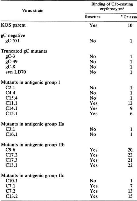
thecell surfaceandforbinding of C3b-coated erythrocytes, by rosetting and 51Cr binding. Table 2 shows that C3b receptors were detected on the parent KOS strain. As expected, C3breceptorswerenotdetectedonthegC-minus
mutant or on the fourmutants which secretedgC into the supernatant withoutinserting the glycoprotein into the cell membrane. Of the remaining 16 mar mutants, 10 were
positive for C3b binding. This included three mutants in antigenicgroupI, four in group Ilb, and three ingroupIlc.
[image:3.612.321.560.90.367.2]On cellsinfected with these 10mutants, the binding ofC3b was to gC because anti-gC monoclonal antibodies totally blocked C3bbinding.
TABLE 1. Role ofanti-gCmonoclonal antibodies inblocking C3bbindingtogConKOS-infected cells
BindingofC3b-coated erythrocytes Blocking reagenta
b
51Cr-binding
Rosetteassayb assaycMonoclonalantibody (MCA)togC AntigenicgroupI
MCA C2 No 1
MCA C14 No 1
AntigenicgroupIla
MCA C3 No 1
MCA C16 No 1
AntigenicgroupIlb
MCA C13 No 1
MCA C17 No 1
Antigenicgroup Ilc
MCA C10 No 1
MCA 17S No 1
MCA18S No 1
MCA19S No 1
MCA 26S No 1
MCA27S No 1
MonoclonalantibodytogB, Yes 6
MCA24S
MonoclonalantibodytogD, Yes 3
MCA 1D3
Monoclonal antibodytogE, Yes 5
MCA 1E3
Saline control Yes 4
aA 200-,ul portion ofa 1:20 dilution of ascites was used in blocking
experiments. ForantibodiesC2, C3, C17, andC10,asciteswasalsodiluted 1:40,1:80,and1:160. Eachdilutiontotally blocked C3b binding. Antibodies wereadded 24 hpostinfection and incubated for 30 min at37°C.Theywere then removedby washing before addingC3b-coatederythrocytes.Results are the mean of two separateblocking experiments.
bRosettes wereviewedbylight microscopy(x 100magnification).To be counted as a rosette, the binding of four or more erythrocytes to an endothelialcell wasrequired.
c51Crassay results are expressedas the ratio ofbinding of C3b-coated
erythrocytes (EIgMC1423 cells) to infected cells which are blocked with monoclonalantibodycompared with background binding, that is,binding of C2-coated erythrocytes (EIgMC142 cells) to infected cells. A ratio of 1 indicates nobinding of C3b-coated erythrocytes; a ratio of 2 or more indicates C3bbinding.
Thesix mar mutantswhich did not express C3b receptors included three of six mutants inantigenicgroup I, two of two mutantsin group Ila,and one offour mutants in group lIc.
Immunofluorescence was performed on unfixed cells to
document that gC was expressed on the cell surface after
infection with these six mutants.Antibodies to group II sites were used to detect gC on cells infected with group I mutants, while antibodies to group I sites were used to detect gC after infection with group Ila or lIc mutants.
Bright immunofluorescence was noted with each mutant,
indicating thatgC was expressed on the cell surface. This
indicates that the mutation in C3b had converted these six mutantsto C3breceptor-negative strains.
Three possible interpretations were discussed above to
explainthe resultsshowninTable 1. The findings shown in
Table 2 help to exclude one of these interpretations: that whichpostulatesthatC3bbinds to each of the fourantigenic groups on gC. The support for this exclusion is that muta-tions at sitelIb(C9.6, C17.2,C17.3, C13.1) have no effect on
C3b binding, suggesting that this domain is nonessential.
Nevertheless, monoclonal antibody tothis domain blocked
C3bbinding.Whether this blockoccurredbecause of steric
on November 10, 2019 by guest
http://jvi.asm.org/
TABLE 2. Detection of C3breceptors on cells infected with HSV-1 strain KOS andKOS mutants
Binding ofC3b-coating
Virus strain erythrocytesa
Rosettes 51Crassay
KOS parent Yes 10
gC negative
gC-551 No 1
Truncated gC mutants
gC-3 No 1
gC-49 No 1
gC-8 No 1
synLD70 No 1
Mutantsin antigenic group I
C2.1 No 1
C4.4 No 1
C15.4 No 1
C11.1 Yes 12
C14.1 Yes 9
C15.1 Yes 6
Mutants in antigenic group Ila
C3.1 No 1
C16.1 No 1
Mutantsin antigenic group IIb
C9.6 Yes 20
C17.2 Yes 22
C17.3 Yes 21
C13.1 Yes 22
Mutants in antigenic groupIIc
C10.1 No 1
C7.1 Yes 7
C7.2 Yes 13
C13.2 Yes 15
aResults are the mean of two to three separate infections for each virus
strain exceptfor KOS parent strain, which is the mean of sevenexperiments. Rosettes are measured as described in footnote b, Table 1. 51Crassay is expressed as the ratio of binding of C3b-coated/C2-coated erythrocytes. Ratiosarerounded to the nearest whole number.Aratio of>2indicatesthat C3b receptors are present. In all cases, the actual binding of C2-coated erythrocyteswas<2%.To prove that C3b-coatederythrocytesarebindingto gC, blockingassayswereperformedoncells infected withmarmutantsthat demonstrate C3bbinding. Infected cellswereincubated withamonoclonal antibody to either group I or II antigenic determinants prior to adding C3b-coated erythrocytes. In all cases, C3bbinding was reduced to back-ground levels (i.e., those obtained with C2-coated erythrocytes). Mutants which do not bind C3b were showntoexpressgConthecellsurface, using indirect immunofluorescenceandantibodiestogroupIIsitestodetectgCafter infection with group Imutants orantibodiestogroup IsitestodetectgCafter infection with group Ila and Ilcmutants.
hinderance orbecause of
antibody-induced changes
ingC
conformation cannotbe determined fromtheseresults.
Within
antigenic
site 1,considerableheterogeneity
existssincesome mutants bindwhile others failtobindC3b. Itis
of interest that antibody C14, which
recognizes
antigenic
group 1, blocks C3b
binding (Table
1), yet themutantvirusselectedby
growth
in thepresenceof thisantibody (C14.1)
maintains the property of
binding
C3b. Thisfinding
alsosupports the
hypothesis
thatanti-gC
antibody
canblockC3bbinding without
occupying
adomainongC
essentialfor thisfunction.
Eleven of the 21 mar mutants examined have lost the
abilitytobindC3b
(Table 2).
These resultsindicate thatC3bbinding isnot
required
for viral survival in vitro. Todeter-mine whether thisfunction is conserved in vivo, we
exam-ined low-passage clinical isolates for C3b binding.
Endothelial cell monolayers were infected with either HSV-1 orHSV-2. Twenty-four hours postinfection, C3b-coated or control erythrocytes(EIgMC142 cells) were added. Specific
bindingof C3b-coated erythrocytes wasconfirmed by
block-ingassays, using anti-gC monoclonal antibodies. Each of 31 HSV-1 clinical isolates bound C3b (Table 3). As expected,
each of theHSV-1 isolates expressed both glycoproteins gC
and gD on the surface of infected cells as monitored by
monoclonalantibodies in an immunofluorescence assay. The
HSV-2 isolates expressed gD but not the type-specific
epitope on gC recognized by monoclonal antibody 1C8.
Noneofthe nineHSV-2 isolates bound C3b (Table 3).
DISCUSSION
We attempted to define the regions of gC that bind C3b.
Multiple monoclonal antibodies were evaluated for their
ability to block C3b binding to gC. All antibodies to gC
blocked C3b binding. KOS strain mutants in the gC gene
were thenexamined forC3bbinding. Unexpectedly, strains
with mutationsat onesite(Ilb) had no effect on C3b binding
andmutations at another site (Ila)eliminated C3b binding,
whilemutations atthe remaining two sites (I andlIc) had a
variableeffect.Theseresults indicate that antigenic groups I
andlIc, which were definedby resistence to neutralization
by monoclonal antibodies (29), are not homogeneous, since
withineach group is a subpopulation which either binds or
fails to bind C3b. More important to the purpose of this
investigation,theresultsindicatethat the antigenicgrouping
ofgCdoes notdefineoneparticular domain ofthe
glycopro-teinthatbinds C3b.
Two interpretations are offerred toexplain the results of
blocking experimentswithmonoclonalantibodies (Table 1).
Oneisthatantibodies block by steric hinderance, such that
C3b
binding
isprevented evenifantibody bindsto anones-sentialdomain on gC, such as site lIb. An alternate
hypo-thesis isthatantibody bindingtogC alterstheconformation
of the glycoprotein, which affects C3b binding.
Several interpretations are also possible to explain the
results of mar mutant binding studies shown in Table 2.
Perhaps C3b binds to several sites on gC, such as those
composed, in part,ofgroups I, Ila, and lIc. Alterations in
any one ofthese sites may theneliminate C3b
binding.
An alternateexplanation
is that mutations ingC
affect C3bbinding by
altering
the conformation of theglycoprotein.
Thelatter
explanation
provides
aunifying
hypothesis
withthe results obtained by monoclonal
antibody
blocking
as-says. In supportof this
hypothesis
isrecentDNAsequenc-ing results.The
gC
genesof fourmarmutantswhichexpressTABLE 3.
C3b
bindingtolow-passageclinical isolates of HSV-1orHSV-2No.of strainspositive
HSVtype No.tested Expressionoftype- Expressionof Binding commongDepitope HSV-1gCon of C3bc
oninfectedcellsa infected cellsb of C3bC
HSV-1 31 31 31 31
HSV-2 9 9 0 0
agDwasdetectedwithmonoclonalantibody1D3(16),whichrecognizesa
type-commonepitopeongD.
bgCwasdetected withanti-gCantibody 1C8, C10, C13,orC16.
cBinding ofC3bwasmeasuredasdescribedin thefootnotetoTable 2. For eachHSV-1isolate,thespecificityofthebindingof C3btogCwasshownby
blockingassays,usinganti-gCmonoclonalantibody 1C8,C10, C13,orC16.
on November 10, 2019 by guest
http://jvi.asm.org/
gC
on the cell'ssurface,
but which fail to bindC3b,
have beensequenced (J.
C. Glorioso and M.Levine, manuscript
in
preparation).
The strains include group I mutantC2.1,
group Ila mutants C3.1 andC16.1,
and group Ilc mutantC10.1.
Each strain has a mutation in asingle
nucleotidewhich is
predicted
toproduce
asingle
amino acidsubstitu-tion. The mutations span a
length
separated by
244 aminoacids,
from amino acids 129 to 373. Ofinterest,
group I mutant C11.1 also has asingle
nucleotidesubstitution,
resulting
in apredicted single
amino acidchange,
yet thisstrain maintains
C3b-binding
activity.
Themostlikely
expla-nation for the results is that certain
point
mutations alteracritical conformation of
gC required
for C3bbinding.
Evi-dence
using protein-denaturing
agents,suchasFormalin andparaformaldehyde
(18),
indicate thatC3bbinding
togC
canbe eliminated
by fixing
infected cells with these reagents(Friedman, unpublished observations).
Definitive evidence to supporttheimportance
ofgC
conformation in C3bbind-ing, however,
awaits results of studiesexamining
C3bbind-ing
to thepurified glycoprotein
which is either in its nativestate ordenatured.
Our
previous
studies indicatedthat removalof sialic acidfrom
gC
oninfected cells increasedthebinding
of C3btogC
(38).
In view of theDNA-sequencing
results mentionedabove,
it is apparent thatchanges
intheprotein
backbonecanalso
modify
C3bbinding.
None ofthemutationsinvolv-ing sinvolv-ingle
amino acid substitutions occurredat sites whichwould be
predicted
tomodify
N-linked or 0-linkedglycosylation.
Inaddition,
neuraminadasetreatmentdidnotmodify
C3bbinding
to these mutants. Theseresults,
there-fore,
indicate that bothcarbohydrate
and amino acidvaria-tions of
gC
affect C3bbinding.
The mar mutant results reveal that the virus need not
retain
C3b-binding ability
for it to survive in vitro. This isconsistent with the observation that
gC-negative
mutantsalso
replicate efficiently
in vitro(19, 28).
Complement
com-ponents are not part of the constituents present in cellculture
fluids; therefore,
noselectivepressure isexertedonthe virus to maintain
C3b-binding
function.However,
invivo,
a different situation exists. Each of 31low-passage
clinical isolates of HSV-1 examined in this
study expressed
gC
onthe cell'ssurfaceandboundC3b(Table 3).
Glycopro-tein
gC
was also detected in each of63 isolatestested in astudy performed by
Pereiraetal.(34).
Theseresults suggest animportant
function forgC
andfor C3bbinding
in vivo.Studies which documentan
important
role in vivo forgC
as a
C3b-binding
protein
arelacking. Experiments
in miceindicate that
gC
is notessentialfor virus-induced disease ofthe centralnervoussystem
(11). However,
it isinteresting
tonote that in the murine studies of Dix et
al.,
when thegC-minus
strainsMiyama
and MP were inoculatedby
thefootpad
route,they
were avirulent(11).
When inoculateddirectly
into thebrain,
these strainswere virulent, although theMiyama
strainhaddiminished virulence compared withwild-type
virus.Kumeletal. alsonoteddecreased virulence ofgC
mutants after intracerebral inoculation (25). Theintracerebral route ofinfectionis not natural and bypasses
certain
advantages
thatgC
may confer forviral survivalat moreproximal
sites ofentryof virus intothehost. In rabbits, thegC-negative
MPstrainwasfoundtobevirulent, but theviruswasinoculateddirectly onto ascarified cornea (6). An
important
consideration for future evaluations of gC inanimal
pathogenesis
studies is, first, to determine theopti-mal route of inoculation for these studies and, second, to
examine the
'species
specificity
ofC3bforgC. Thebinding ofC3bto
gC
maynotcrossspecies
barriers. Asimilarsituationhas recently been described for the Fcreceptor of
HSV-1,
whichbinds human but not murineIgG (23).
Although the function ofgC in vivo remains undefined,
recentin vitro studies have advanced ourunderstandingof
this glycoprotein. Experiments in our laboratory indicate
thatgCon the surface of infected cellsprotectsthe cell from
lysis by antibody and complement or complement alone
(Harrisetal., inpreparation). Cytotoxicitywasexamined in
BHK-21 cells infected with wild-type HSV-1 or with a mutant which failed to bind C3b. Cells infected with the mutantviruswere morereadily lysed byanti-HSVantibody
and complement or complement alone. Studies
performed
with purified gC indicate the possible mechanism by which
thisglycoprotein prevents complement-mediated lysis.
Gly-coprotein gC inhibits alternative pathway complement
acti-vation by accelerating the decay of the alternative pathway C3 convertase. Purified gC also reduces the efficiency of
complement-mediated lysis by competing with C5orC5bfor
binding sites on C3b (L. H. Fries, H. M. Friedman, G. H.
Cohen,R. J. Eisenberg,C. H. Hammer, and M. M.
Frank,
submitted forpublication).
The results ofthis study indicate thatasantibody bindsto gC it alters the ability of the glycoproteinto reactwith C3b. Recently, Ross etal. showed thatserumfrom patients with recurrentherpes labialis blocksthebinding of antibody C16 togC (36). This indicates that human antibody recognizesa similar siteon gC asantibody C16. Since C16 inhibits C3b binding (Table 1), this implies that following HSV-1 infection thehost's humoral immune responsetogC is likelytoblock thebinding of C3b. Whether sufficient quantities ofanti-gC antibody are produced in vivo to occupy all available gC sites iscurrently unknown. However, itseemsreasonableto conclude from our results that, once the host mounts an antibody response to gC, the C3b-binding property ofthe glycoprotein will be diminished. An implication of this conclusion is that any selective advantage gC offers in survival of the virus is likely to occur early in infection, before anti-gC antibody is made. Alternatively, C3b may compete with antibody for binding sites on gC, thereby decreasing the efficiency of antibody-directed attack on virus-infected cells.
ACKNOWLEDGMENTS
This work was supported by Public Health Service grants HL 28220 from the National Heart and Lung Institute and DE 02623 from the National Institute of Dental Medicine.
WethankCindy Seidel-Dugan, Pat Pane, and Randee Kravetz for technical assistance and Martin Zweig for generously providing monoclonal antibodies 17S, 18S, 19S, 24S, 26S, and 27S.
LITERATURE CITED
1. Adler, R., J. C. Glorioso, J. Cossman, and M. Levine. 1978. Possible role of Fc receptors on cells infected and transformed by herpesvirus:escapefromimmune cytolosis. Infect. Immun. 21:442-447.
2. Baucke, R. B., and P. G. Spear. 1979. Membrane proteins specified by herpes simplex viruses. V. Identification of an Fc-bindingglycoprotein. J. Virol. 32:779-789.
3. Bishop, G. A., G. Kumel, S. A. Schartz, and J. C. Glorioso. 1986.Specificity ofhumannatural killer cells in limiting dilution culture fordeterminants of herpes simplex virus type 1 glyco-proteins.J. Virol. 57:294-300.
4. Buckmaster, E. A., U. Gompels, and A. Minson. 1984. Characterisation and physical mapping of an HSV-1 glycopro-tein ofapproximately 115 x 103 molecular weight. Virology 139:408-413.
5. Carter, V. C., P. A. Schaffer, and S. S. Tevethia. 1981. The
on November 10, 2019 by guest
http://jvi.asm.org/
involvement of herpes simplex virus type 1 glycoproteins in cell-mediated immunity. J. Immunol. 126:1655-1660.
6. Centifanto-Fitzgerald, Y. M., T. Yamaguchi, H. E. Kaufman, M. Tognon, and B. Roizman. 1982. Ocular diseasepattern induced by herpes simplex virus is genetically determined by a specific region of viral DNA. J. Exp. Med. 155:475-489.
7. Chou,P.Y., and G. D. Fashman. 1974.Conformational param-eters for amino acids in helical beta-sheet and random coil regions. Biochemistry 13:211-222.
8. Chou, P. Y., and G. D. Fashman. 1974. Prediction of protein conformation. Biochemistry 13:222-245.
9. Cines, D.B., A.P. Lyss,M. Bina,R.Corkey,N. A. Kefalides, and H. M. Friedman. 1982. Fc and C3 receptors induced by herpes simplex virus on cultured human endothelial cells. J. Clin. Invest. 69:123-128.
10. Cohen, G. H., B. Dietzschold, M. Ponce de Leon, D. Long, E. Golub, A. Varrichio, L. Pereira, and R.J. Eisenberg. 1984. Localization andsynthesis ofanantigenic determinant of herpes simplexvirusglycoprotein D that stimulates the production of neutralizing antibody. J. Virol. 49:102-108.
11. Dix, R. D., R. R. McKendall, and J. R. Baringer. 1983. Com-parativeneurovirulence of herpes simplex virus type 1 strains afterperipheral or intracerebral inoculation ofBALB/c mice. Infect. Immun. 40:103-112.
12. Dowler, K. W., and R. W. Veltri. 1984. In vitro neutralization of HSV-2: inhibitionby binding of normalIgG and purifiedFcto virion Fc receptor (FcR). J. Med. Virol. 13:251-259.
13. Ehlenberger, A. G., and V. Nussenzweig. 1977. The role of membrane receptors forC3b and C3d inphagocytosis.J. Exp. Med. 145:357-371.
14. Fearon, D. T. 1979. Regulation of the amplification C3 convertase of human complement by an inhibitory protein isolated from humanerythrocytemembranes. Proc.Natl. Acad. Sci. USA 76:5867-5871.
15. Fearon, D. T. 1983. The humanC3b receptor. SpringerSemin. Immunopathol.6:159-172.
16. Friedman, H.M., G. H. Cohen, R.J. Eisenberg, C.A. Seidel, and D. B.Cines. 1984. GlycoproteinC ofherpessimplexvirus 1 acts as a receptor for the C3b complement component on
infected cells. Nature(London)309:633-635.
17. Glorioso, J., U. Kees, G. Kumel, H. Kirchner, and P. H. Krammer. 1985. Identification ofherpes simplex virus type 1 (HSV 1) glycoprotein gC as the immunodominant antigen for HSV1 specificmemorycytotoxicTlymphocytes.J. Immunol. 135:575-582.
18. Hayat, M. A. 1981. Fixation for electron microscopy, p. 130-136. AcademicPress, Inc., NewYork.
19. Heine, J. W.,R.W. Honess,E.Cassai,and B. Roizman. 1974. Proteins specified by herpes simplex virus. XII. The virion polypeptidesof type 1strains. J. Virol. 14:640-651.
20. Holland, T. C., F. L.Homa, S. D. Marlin, M. Levine, andJ. Glorioso. 1984. Herpes simplex virus type 1 glycoprotein C-negative mutantsexhibitmultiple phenotypes,including
secre-tion oftruncatedglycoproteins.J. Virol. 52:566-574.
21. Holland,T.C., S.D.Marlin,M.Levine,andJ.Glorioso. 1983. Antigenicvariantsofherpessimplexvirus selected with
glyco-protein-specificmonoclonal antibodies. J. Virol. 45:672-682. 22. Iida, K.,and V.Nussenzweig.1981.Complementreceptorisan
inhibitor of the complement cascade. J. Exp. Med. 153: 1138-1150.
23. Johansson, P. J. H., E. B. Myhre, and J. Blomberg. 1985. Specificityof Fc receptors inducedby herpes simplexvirustype 1: comparison of immunoglobulin G from different animal species. J.Virol. 56:489-493.
24. Kikuchi, G.E., J. E.Coligan,T. C.Holland,M. Levine, J. C. Glorioso, and R. Nairn. 1984. Biochemical characterization of
peptides from herpes simplex virus glycoprotein gC: loss of
CNBrfragments from thecarboxy terminus oftruncated, se-cretedgCmolecules. J. Virol. 52:806-815.
25. Kumel, G.,H.C.Kaerner,M.Levine, C. H.Schroder,andJ.C. Glorioso. 1985. Passiveantibody protection by herpes simplex virus-specificmonoclonalantibodies and monoclonal antibody-resistantmutantsaltered inpathogenicity.J.Virol.56:930-937. 26. Lawman, M. J. P., R. J. Courtney, R. Eberle,P.A. Schaffer, M.K. O'hara,andB. T. Rouse. 1980. Cell-mediatedimmunity
toherpes simplexvirus: specificityofcytotoxicTcells. Infect. Immun. 30:451-461.
27. Little, S. P., J. T. Jofre, R.J. Courtney, and P. A. Schaffer. 1981. Avirion-associatedglycoprotein essential forinfectivity ofherpes simplexvirus type 1. Virology115:149-160. 28. Manservigi, R., P. G.Spear, and A. Buchan.1977. Cell fusion
inducedby herpessimplexvirus ispromotedandsuppressed by different viral glycoproteins. Proc. Natl. Acad. Sci. USA 74:3913-3917.
29. Marlin, S. D., T. C. Holland, M.Levine, and J.C. Glorioso. 1985. Epitopesofherpes simplexvirus type1 glycoprotein gC
areclustered intwodistinctantigenicsites. J. Virol. 53:128-136. 30. Noble,A.G.,G. T.-Y.Lee,R.Sprague,M. L.Parish,and P.G. Spear. 1983. Anti-gD monoclonalantibodies inhibit cell fusion inducedby herpessimplexvirus type1. Virology 129:218-224. 31. Norrild, B. 1985. Humoral response to herpes simplex virus infections, p. 69-86. In B. Roizman and C. Lopez (ed.), The herpes viruses, vol. 4. Immunobiology and prophylaxis of humanherpesinfections. Plenum Press,NewYork.
32. Para,M.F.,R. B.Baucks,and P.G.Spear.1982.Glycoprotein
gEofherpes simplexvirus type 1: effects ofanti-gE onvirion
infectivityandonvirus-inducedFc-bindingreceptors. J. Virol.
41:129-136.
33. Para, M. F., L. Goldstein, and P.G. Spear. 1982. Similarities and differences in the Fc-binding
glycoprotein
(gE)ofherpes
simplex virus types 1 and 2 andtentative
mapping
of the viralgenefor thisglycoprotein. J. Virol.41:137-144.
34. Pereira, L., D. V. Dondero, D. Gallo, V. Devlin, and J.D. Woodie. 1982. Serologicanalysisofherpes
simplex
virustypes 1and2withmonoclonal antibodies. Infect.Immun.35:363-367. 35. Richman,D.D.,A.Buckmaster,S.Bell,C.Hodgman,and A. C. Minson. 1986. Identification ofa newglycoprotein
ofherpes
simplexvirus type1andgenetic mappingof the genethatcodes for it. J. Virol. 57:647-655.
36. Ross, C., J. Glorioso, S. Sacks, C. Lavery, and W. E. Rawls. 1985. Competitiveinhibitionbyhumanseraofmouse
monoclo-nalantibodybindingtoglycoproteinsC andDofherpes
simplex
virus types 1and 2.J. Virol. 54:851-855.
37. Sarmiento, M., M. Haffey, and P.G. Spear. 1979. Membrane
proteins specified by herpes simplex viruses. III. Role of
glycoprotein VP7 (B2) in virion
infectivity.
J. Virol. 29:1149-1158.
38. Smiley,M. L.,and H. M. Friedman. 1985.
Binding
ofcomple-mentcomponentC3b to
glycoprotein
C is modulatedby
sialicacid on herpes simplex virus type 1-infected cells. J. Virol.
55:857-861.
39. Smiley,M.L.,J.A. Hoxie,and H. M. Friedman.1985.
Herpes
simplex virus type 1 infection of endothelial,
epithelial,
andfibroblast cells induces a receptor for C3b. J. Immunol. 134:2673-2678.
40. Spear, P. G. 1976. Membrane
proteins
specified by herpes
simplexviruses. I.Identification of four
glycoprotein
precursorsandtheirproductsintype 1-infectedcells. J. Virol.17:991-1008.