EVALUATION OF FUNCTIONAL OUTCOME OF
PROSTHETIC REPLACEMENT OF COMMINUTED
RADIAL HEAD AND NECK FRACTURE
DISSERTATION SUBMITTED FOR MS (ORTHOPAEDICS) MADURAI MEDICAL COLLEG
MADURAI
THE TAMILNADU
Dr.M.G.R. MEDICAL UNIVERSITY
CHENNAI–600032.
CERTIFICATE
This is to certify that the work “EVALUATION OF FUNCTIONAL OUTCOME OF PROSTHETIC REPLACEMENT OF COMMINUTED RADIAL HEAD AND NECK FRACTURE” which is being submitted for M.S. Orthopaedics, is a bonafide work of Dr.P.SIVAKARAN , Post Graduate Student at Department of Orthopaedics, Madurai Medical College
The Dean ,
Madurai Medical College
Madurai.
CERTIFICATE
This is to certify that this dissertation titled “EVALUATION OF FUNCTIONAL OUTCOME OF PROSTHETIC REPLACEMENT OF COMMINUTED RADIAL HEAD AND NECK FRACTURE” is a bonafide work done by Dr.P.SIVAKARAN, postgraduate student of Madurai Medical College, Govt Rajaji Hospital.
Prof.Dr.R.ARIVASAN, M.S Ortho.D.Ortho
Professor and Head, Department of Orthopaedics & Traumatology
Madurai Medical College, Madurai.
CERTIFICATE
This is to certify that this dissertation “EVALUATION OF FUNCTIONAL OUTCOME OF PROSTHETIC REPLACEMENT
OF COMMINUTED RADIAL HEAD AND NECK FRACTURE” is the bonafide work done by Dr.P.SIVAKARAN under my direct guidance and supervision in the Department of Orthopaedic Surgery, Madurai Medical College, Madurai-20.
Prof. DR.N.THANAPPAN, M.S Ortho.,
Professor and Chief Ortho unit-III Department of Orthopaedics & Traumatology
ACKNOWLEDGEMENT
At the very outset I would like to thank PROF. Dr.D.Marudhupandian M.S, the Dean, Madurai Medical College and Govt. Rajaji Hospital, Madurai for permitting me to carry out this study in this hospital.
I am greatly indebted and thankful to my beloved chief, and my guide Prof.DR.N.THANAPPAN M.S.ortho ortho- III unit and Head of department, Prof.Dr.R.ARIVASN M.S.,Ortho, D.Ortho., Ortho-I unit, Department of Orthopaedic Surgery and Traumatology, Madurai Medical College for his invaluable help, encouragement and guidance rendered to me in preparing this dissertation.
I am most indebted and take immense pleasure in expressing my deep sense of gratitude to Prof. Dr. V. R. Ganesan, M.S., Ortho D.Ortho, Prof. Dr.B.Sivakumar, M.S., Ortho., D.Ortho for their easy accessibility and timely suggestion, which enabled me to bring out this dissertation. I take immense pleasure to thank my co-guide Dr.J.MAHESWARAN M.S Ortho,D Ortho.,for his timely help and encouragement.
Kumar M.S.Ortho., Dr.Karthikeyan M.S Ortho, Dr.Singaravel M.SOrtho, Dr.Gokulnath, M.S Ortho, Dr.Anbarasan, M.S Ortho Dr.Gopi Manohar DNB ortho, DR.Saravanan M.S.ortho., Assistant Professors, Department of Orthopaedics, Madurai Medical College, for their timely help and guidance given to me during all stages of the study. Last but not the least, I express my gratitude to the patients for their kind co-operation.
DECLARATION
I, Dr.P.SIVAKARAN, solemnly declare that the dissertation titled “EVALUATION OF FUNCTIONAL OUTCOME OF PROSTHETIC REPLACEMENT OF COMMINUTED RADIAL HEAD AND NECK FRACTURE” has been prepared by me. This is submitted to “The Tamil Nadu Dr. M.G.R. Medical University, Chennai, in partial fulfillment of the regulations for the award of M S
degree branch II Orthopaedics.
Dr.P.SIVAKARAN Post Graduate in Orthopaedics, Madurai Medical College, Hospital, Madurai.
CONTENTS
PART A
CONTENTS Page No.
Introduction 1
Aim and Objective 2
Review of Literature 3
Anatomy 9
Classification 30
Treatment 32
PART –B
CONTENTS Page No.
Materials and Methods 41
cases 49
Observation & Results 66
Discussion 69
Conclusion 73
ANNEXURES :
a. MASTER CHART b. BIBLIOGRAPHY
c. PATIENT PROFORMA d. CONSENT FORM
e. ETHICAL COMMITTEE APPROVAL f. PLAGIARISM FIRST PAGE & DIGITAL
1
INTRODUCTION
Radial head fractures are the most common fractures of the elbow
with an estimated incidence of 2.5 to 2.9 / 10,000 people / year. Radial
head fractures are more common in women than men and most frequently
occur between the age of 20 & 60 years .Undisplaced and minimally
displaced radial head fractures typically occur as isolated injuries while
more displaced and comminuted fractures commonly have associated
injuries to the collateral ligaments and may have associated fractures of
the coronoid, capitellum, or proximal ulna. In high energy trauma,
dislocations of the elbow and/or forearm can also occur. Disruption of the
interosseous membrane and distal radial ulnar joint ligaments may result
in axial instability of the forearm, termed the Essex–Lopresti lesion. The
majority of radial head and neck fractures are minimally displaced and
are isolated injuries. These fractures typically have a good functional
outcome with nonsurgical treatment.
2
AIMS AND OBJECTIVES
• To evaluate the functional outcome of prosthetic replacement of comminuted Radial head and neck fracture Objectives
1. To analyse the clinical ,radiological & functional outcome of patients treated with prosthetic replacement of comminuted radial head and neck fractures
3
REVIEW OF LITERATURE
Over the years, the indication for the use of a radial head prosthesis changed from the prevention of heterotopic ossification to the prevention of proximal migration of the radius and instability of the elbow.
Currently, the optimal indication for the use of radial head prostheses is a non-reconstructable radial head fracture with associated injuries that would leave the elbow, or forearm unstable if the radial head were resected.
In the early twentieth century, radial head resection became the treatment of choice for displaced radial head fractures
In 1924, Speed even stated that : “In adults, unless the lesion is
only a mere crack, there is no doubt that removal of the head is primarily
indicated”. Others reserved Regrowth of bone at the proximal radius was
4
Indications slowly changed and ten years later, Carr et al were the first to comment that the prosthesis increased elbow stability, when compared to radial head resection.
In 1951, Essex- Lopresti described two cases where the radial head fractures were associated with a distal radioulnar dislocation. Although he did not use a prosthesis in either of these cases, he suggested the temporary use of a radial head prosthesis, until the forearm had healed and became stable.
In 1953, Cherry described a second type of radial head prosthesis made of an acrylic resin, to prevent proximal translation of the radius and consequent strain on the distal radio-ulnar joint and to maintain the normal carrying angle of the elbow and prevent cubitus valgus. The use of a radial head prostheses was however still quite rare at that time
5
Long-term results of the Speed prosthesis published in 1964 were shown to be similar to those of patients treated with radial head resection, with decreased pronation and supination in the prosthesis group
In 1969, the Swanson Silastic® radial head prosthesis became available (Dow Corning Corporation, USA. Unfortunately the prosthesis did not prevent proximal migration of the radius and a large degree of distortion was found during movement of the elbow .Timing of the prosthetic replacement was first discussed in 1974. Early replacement of the radial head showed better functional results, but good pain relief was still achieved in the late group.
The first structural complications with the Swanson prosthesis were reported in 1979. In a series of eighteen patients, the prosthesis had broken in three, subluxed in one and tilted in six Resected fragments of a comminuted radial head fracture. The fracture was complicated by a medial collateral ligament injury and it was judged that this fracture was not amenable to reconstruction. The radial head was replaced with a metal prosthesis.
6
following the use of the silastic prosthesis . Biomechanical studies showed that radial head replacements could restore some of the stability of the elbow , and longitudinal stability of the forearm but that a stiffer implant would be necessary. Due to the abundance of objective data against the Swanson radial head prosthesis, new types of prostheses were developed and indications were again adjusted.
Morrey et al limited the indications for the use of a radial head implant to instability following radial head resection and acute dissociation of the distal radio-ulnar joint.
The development and clinical trial of a vitallium prosthesis (Howmedica, London, UK), were described by Knight et al in 1993. Looseningof the prosthesis was described in two patients and the authors commented that replacement was not clearly better than resection for simple radial head fractures .
7
In 1996, Charnley et al used the floating radial head prosthesis after release of the elbow for heterotopic ossification following head injury. No recurrence of the heterotopic ossification was found at a follow-up of 3.5 years . Interestingly, prevention of heterotopic ossification was the main indication for Speed to use a radial head prosthesis.
Allograft replacement of the radial head was published in 1997. One of the allografts dislocated three weeks following surgery, while the arm was still in a long arm cast. Another concern was degeneration and collapse of the graft that was found in follow-up radiographs. No long-term results were published.
In contrast to the results of radial head prostheses, excellent long-term (average follow-up of17 years) results of resection of the radial head,were published in 1997
8
In 2000 ‘safe and effective’ short-term results were found with a new type of metal radial head prosthesis used for non reconstructible radial head fractures . Short-term results of the floating radial head prosthesis were also promising and the prosthesis was found to restore clinical stability . However, degenerative changes of the elbow were found in approximately 50%, while the prosthesis had to be removed in a patient due to pain and functional impairment at the elbow . The first long-term results of a ‘monoblock’ metal radial head prosthesis (Smith & Nephew,Inc., Memphis, TN, USA) were published in 2001.
9
ANATOMY OF THE ELBOW JOINT
10
THE BONES:
THE DISTAL END OF HUMERUS:
11
THE PROXIMAL END OF ULNA :
The proximal ulna consists of the olecranon and the ellipsoid anterior surface of the ulnar notch. The notch is covered by articular cartilage except for the mid portion which is usually covered by fatty tissue. The olecranon is the insertion site for the triceps brachii muscle tendon. The distal end of the ulnar notch is the coronoid process which is the insertion site for the brachialis muscle tendon and the anterior bundle of the medial collateral ligament. At the medial aspect of the coronoid process, the sublime tubercle is a bony prominence that serves as the insertion site for the medial collateral ligament. At the lateral aspect of the coronoid process, the supinator crest is a rather elongated bony prominence that serves as the attachment for the lateral ulnar collateral ligament. It has been stated that the coronoid process is an important restraint for elbow stability.
A)THE PROXIMAL END OF RADIUS :
12
of cartilage. The radial neck length is of 13 mm (range 9–19mm). The radial head is angulated 55o (range 45–65º).
This radial torsion is calculated by comparing a line drawn on the radial head, perpendicular to the radial notch, to a line drawn between the center of the radial styloid process and the center of the ulnar notch. Likewise the radial head is angulated when compared to the diaphysis as shown by a proximal diaphysis–neck angle of 17* (range 6–28º). These considerations are important when restoring radial anatomy in cases of radial head fractures or prosthetic replacements. The radial neck connects the radial head to the shaft. At the medial and distal aspect of the neck, the radial tuberosity can be found; it is a bony prominence that serves as the insertion point of the biceps tendon.
THE ARTICULAR CAPSULE :
13
cases of chronic instability and decreased in the presence of joint contractures. The maximum volume capacity of the capsule is of 25–30 ml in adults occurring at 80*of flexion .This enables to predict the position of greatest comfort when effusion or hemarthroses are present. Gray and Morrey in their anatomic studies provided detailed description of the elbow capsule. The anterior capsule inserts proximally above the coronoid and radial fossas. Distally it is attached to the anterior margin of the coronoid process medially and to the annular ligament laterally. Fibrous bands have been described within the capsule: three anteriorly and three distinct bands posteriorly.
14
radius and capitulum which adopts a meniscoid structure and may assist in humeroradial joint motion.
THE ULNAR COLLATERAL (MEDIAL CUBITAL) LIGAMENT :
The medial collateral ligament complex (MCL) consists of three bundles with different points of origin and insertion forming a triangular shape: the anterior, posterior, and transverse . Because of the multiplicity of the bundles and their functions, the MCL has been compared to the anterior cruciate ligament of the knee .
The anterior bundle (or anterior oblique ligament) is the most significant component of the MCL, being the main stabilizer to valgus stress of the elbow. Its origin is at 5 mm anterior and inferior to the tip of the medial epicondyle and inserts on the sublime tubercle,18 mm distal to the coronoid tip, along the medial aspect of the coronoid process. The width at its midpoint averages 5mm and the mean length is of 27 mm. The anterior bundle can be further divided into anterior and posterior bands.
15
Neither the anterior nor posterior bands are isometric. Between the anterior and posterior bands, the valgus stress is resisted from 30*to 120*. The anterior band is taut in extension and relaxes in flexion. The posterior band is taut at intermediate positions and relaxed in extension.
16
THE RADIAL COLLATERAL (LATERAL CUBITAL) LIGAMENT :
17
THE ANNULAR LIGAMENT:
20
MOVEMENTS :
21
22
23
THE SUPERIOR RADIO-ULNAR JOINT :
It is a uni-axial pivot joint between the circumference of the radial head and osseo-fibrous ring made by the ulnar radial notch and annular ligament. The annular ligament is a strong band that encircles the radial head and holding it against the ulnar radial notch. It forms about four-fifths of the ring . It is attached anteriorly behind the posterior margin of radial notch. The proximal annular border blends with the cubital capsule.
MOVEMENTS :
24
MUSCLES PRODUCING THE MOVEMENTS
Pronation : Pronator Quadratus, Pronator Teres and Flexor Carpi Radialis.
Supination : Biceps Brachii and Supinator .
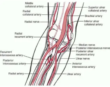
BLOOD SUPPLY OF THE ELBOW JOINT :
The elbow joint is supplied by the articular branches from the anastomotic networks around the joint. The anastomosis is formed by:
a) Anterior descending and posterior descending branches of profunda brachii artery.
b) Radial recurrent branch of radial artery
c) Inferior and superior ulnar collateral branch of brachial artery.
d) Anterior and posterior ulnar recurrent branches of ulnar artery.
25
NERVE SUPPLY OF THE ELBOW JOINT :
The elbow joint is supplied mainly from articular branches of musculocutaneous and radial nerves, but the ulnar, median and sometimes the anterior interosseous nerve also contribute. The articular branches from the musculocutaneous nerve arises from the nerve to brachialis and supply the anterior part of the capsule.
[image:34.595.111.488.357.654.2]
26
Ossification centre Appearance Fusion
Capitulum 1 Year 14-16 Yrs
Head of Radius 4 Year 16-18 Yrs
Medial Epicondyle 4 Years (Female)
6 Years (Male)
16-18 Yrs
Trochlea 9 Years (Female)
10 Years(Male)
14-16 Yrs
Olecranon 10 Years 14-16 Yrs
Lateral Epicondyle 12 Years 14-16 Yrs
27
FUNCTIONAL ANATOMY:
The radial head consists of a concave dish which articulates with
the capitellum and a flattened articular margin which articulates with the
lesser sigmoid (radial) notch of the ulna. The nonarticular margin
comprises about one-third of the diameter and is more rounded and often
devoid of cartilage. A “safe zone” for placement of a plate on the
nonarticular margin of the proximal radius has been defined, best
identified during surgery by positioning the forearm in neutral rotation
and placing the plate 10-degree anterior to the mid-axial line The radial
head is not circular but is somewhat elliptical in shape. The
radiocapitellar dish is elliptical and typically offset from the neck of the
radius. An understanding of the complex geometric shape of the radial
head is required when repairing more comminuted fractures and when
performing radial head replacement. Vascular supply of the radial head is
supplied by branches of the radial recurrent artery and branch of the
ulnar artery which form a pericervical arterial ring. A branch of the
interosseous artery supports the neck and the nutrient artery provides
28
BIOMECHANICS OF THE ELBOW
Elbow joint is near equivalent of a mechanical hinge although not a perfect one. The predominant movements of the elbow joint are flexion and extension which are movements of forearm in respect of humerus as levers and elbow joint as the fulcrum. When the elbow is in full extension, the axes of the arm and forearm produce carrying angle, flexion it disappears. Elbow joint is considered in full extension when the long axis of the humerus and that of the ulna become collinear found that most of the activities of daily living could be accomplished .
29
30
CLASSIFICATION
Numerous classifications have been described for fractures of the
radial head. Mason described a Type I fracture as a fissure or marginal
sector fracture without displacement; Type II as a marginal sector fracture
with displacement; and a Type III as a comminuted fracture involving the
whole head. Type IV injury was subsequently described which includes
any radial head fracture associated with an elbow dislocation. The most
popular classification is the Broberg and Morrey modification of the
original Mason classification. Type I fracture is undisplaced or displaced
less than 2 mm and involves less than 30% of the articular surface. Type
II fracture is displaced greater than 2 mm and involves greater than 30%
of the articular surface.Type III fractures is comminuted. Van Reit further
modified this classification describing the Mayo–Mason classification
whereby a suffix is added to the original modified Mason classification
for concomitant soft tissue injuries, fractures, or dislocations of elbow
31
MECHANISM OF INJURY
Most radial head fractures occur as the result of low-energy
mechanisms such as a trip and fall on an outstretched hand. Sporting
activities as well as motor vehicle collisions cause higher-energy
fractures typically with greater displacement and a higher incidence of
concomitant injuries. Mechanisms of fracture vary but include three
common patterns: (1). A valgus load causes impaction of the radial head
into the capitellum, commonly with rupture of the MCL. (2).
Posterolateral rotatory subluxation of the radial head with respect to the
capitellum causes a partial articular shear fracture of the anterior portion
of the radial head often with rupture of the LCL. (3). An axial forearm
load causes impaction of the radial head into the capitellum with more
severe trauma producing a fracture of the coronoid or rupture of the
interosseous membrane and distal radioulnar joint ligaments; the
32
METHODS OF TREATMENT
Nonoperative Treatment of Radial Head Fractures Indications/Contraindications
Patients with undisplaced or minimally displaced radial head
fractures without a block to forearm rotation should be treated
nonsurgically. In the setting where there is a block to forearm rotation in
a patient with radiographically undisplaced or minimally displaced
fracture, the patient should be re-evaluated several days after injury when
the elbow is less painful. Alternatively, aspiration of the hemarthrosis and
injection of local Anesthetic can be used to check for the presence of a
mechanical block to rotation. A chondral flap of capitellar cartilage can
be the cause of limited rotation and cannot be detected on imaging
typically noted at surgery. It is unclear as to how large and how displaced
a radial head fracture can be and still have a good outcome with
nonsurgical management. While it has been suggested that partial
articular fractures of the radial head which are displaced more than 2 mm
and involve more than 30% of the articulation should be considered for
open reduction and internal fixation, there are no comparative
randomized clinical trials demonstrating an improved outcome relative to
33
crepitus with forearm rotation may also be considered a relative
indication for surgery
OPERATIVE METHODS :
Operative Treatment of Radial Head Fractures Indications/Contraindications
Patients with displaced radial head fractures with a block to
motion, those who have concomitant injuries which require surgical
intervention such as unstable fracture-dislocations, or those with retained
intra-articular loose bodies are best treated surgically. Treatment options
include radial head fragment excision, radial head excision, open
reduction and internal fixation, and radial head arthroplasty. Fragment
excision is indicated in patients with a block to forearm motion by a small
(less than 25% of the articular diameter) nonreconstructable displaced
articular fracture of the radial head. The excision of large fragments of
the radial head can cause painful clicking and contribute to instability in
the setting of concomitant bony and ligament injuries as a consequence of
loss of concavity–compression stability of the radiocapitellar joint. Radial
head excision may be considered for isolated displace fractures of the
radial head that are not amenable to internal fixation. Given the
documented high incidence of concomitant soft tissue injuries in patients
with comminuted radial head fractures, primary excision of the radial
34
examination under anesthesia is mandatory to evaluate for the presence of
elbow or forearm instability. Even in the presence of intact collateral
ligaments ,radial head excision has been documented to alter load transfer
and kinematics across the elbow; however, the benefit of routine
replacement of the radial head versus radial head excision has not been
evaluated in randomized clinical trials. The indications for open reduction
and internal fixation remain controversial. Clear indications include
displaced, non comminuted fractures of the radial head limit forearm
rotation, or radial head fractures fixed as a component of the surgical
repair of an elbow fracture-dislocation. It has been suggested that
fractures displaced greater than 2 mm and involving greater than 30% of
the articular surface (a Type II fracture in the modified Mason
classification) might be best treated with surgery; however, this remains
unproven. The best candidates for internal fixation are younger patients
with good-quality bone with three or fewer fragments. The management
of partial articular fractures tends to be more successful than complete
fractures of the radial head and neck likely due to both improved stability
with partial articular fractures and compromised vascularity with
complete fractures of the radial neck. Low-profile tripod screw fixation
has been shown to provide improved results relative to plate fixation;
however, screw fixation alone is only indicated for radial neck fractures
35
setting of unreconstructable comminuted radial head fractures due to the
high incidence of associated ligamentous and bony injuries. Radial head
arthroplasty should not be performed in the setting of gross wound
contamination, if the radial neck cannot be reconstructed to accept an
implant, or if the capitellum is deficient or missing from an associated
injury.
Radial Head Arthroplasty
Preoperative Planning. A modular metallic radial head arthroplasty
system should always be available when operating on displaced radial
head fractures because commination is often more severe than predicted
by plain radiographs or CT. In the setting of neck commination, small
plates or cerclage wires should be available to allow for neck
reconstruction and the use of a standard prosthesis. Alternatively a long
stem bipolar prosthesis should be available in the uncommon situation
where reconstruction of the radial neck to accept a standard prosthesis is
not possible Positioning.
The radial head can be replaced with the arm placed across the
chest with the surgeon standing or using an arm table with the surgeon
sitting. When treating concomitant proximal ulna and olecranon fractures,
36
Surgical Technique:
After deep surgical dissection the annular ligament must be
sectioned to adequately expose the radial head and neck and to facilitaten
the prosthesis insertion. The exact replacement technique will depend on
the prosthesis system to be employed. After removing any loose
fragments, section the radial neck at the junction of the radial head and
neck or at the level of the fracture using an oscillating saw. Most radial
head systems are modular to improve size matching with the native radial
head and neck. Reassemble the excised fragments of radial head on the
back table to ensure all fragments are removed from the elbow and to
determine the optimal diameter and thickness of the radial head prosthesis
to be employed. The native radial head is somewhat elliptical and is
offset relative to the radial neck. Most commercially available implants
are axisymmetric and nonanatomic; some employ a bipolar articulation.
The optimal diameter of a radial head implant is not known but the
articular dish of the implant should likely approximate the articular dish
of the native radial head. The optimal implant diameter is typically the
minor diameter of the elliptical native radial head, most commonly 2 mm
smaller than the maximum diameter. An implant whose diameter is too
large may cause an erosion of the lateral trochlea, prevents optimal
closure of the annular ligament and may contribute to residual instability.
37
well as thickness. Measurement of radial head thickness should be
performed using the excised fragments of the radial head where
available. Over-lengthening (over-stuffing) with the placement of a radial
head prosthesis that is too thick may be associated with the development
of pain, stiffness, and capitellar wear. Place a Homan retractor around the
posterior aspect of the radial neck and lever it against the ulna to translate
the proximal radius laterally to prepare the radial canal and to insert the
assembled modular prosthesis. If a smooth stem prosthesis is to be used
choose a stem 1 mm smaller than the maximum-sized diameter neck rasp
to allow the stem to move slightly in the neck such that the articular
surface of the implant tracks optimally with the capitellum. After placing
the trial implants, the radial head should articulate at the level of the
proximal radial ulnar joint which is typically 2 mm distal to the tip of the
coronoid.
A careful evaluation for congruent tracking of the radial head
implant on the capitellum both visually and fluoroscopically is required.
Radiographic parameters are not very useful to detect over lengthening of
the radial head. The medial ulnohumeral joint space should be parallel;
over-lengthening causes the medial ulnohumeral joint to open laterally.
However, this may not be evident until there is 6- to 8-mm
over-lengthening of the radial head insert. Following insertion of the definitive
38
concomitant osseous and ligament injuries are required maintain elbow
39
Postoperative Care.
Immediate active motion in a soft dressing is permitted if there are
no associated injuries. Concomitant ligament injuries will direct the
rehabilitation plan as outlined in the section on the operative treatment of
elbow dislocations.
Pitfalls and Preventative Measures:
Incorrect implant sizing is a common problem following radial
head arthroplasty. Unfortunately radiographic parameters are unreliable
intra operatively making the use of the excised radial head and the
relationship of the radial head to the proximal ulna the most useful sizing
tools. Do not use the distance between the capitellum and radial neck cut
to size the implant as partial and complete LCL tears are common and
this may lead to over-lengthening of the radial head implant.
Contralateral radiographs can be helpful to make the diagnosis of radial
head over-lengthening postoperatively and to assist in planning
preoperatively if the radial head has previously been removed. A
measurement technique has been developed based on contralateral
radiographs which can accurately quantify radial head implant length
within 1 mm. Radial neck fractures can occur with overzealous
retraction, reaming, and prosthesis insertion. Meticulous surgical
technique should prevent this complication in most circumstances. Repair
40
insertion. Stiffness is prevented by encouraging early active motion.
Indomethacin can be used in an effort to prevent heterotopic ossification
in patients without contraindications; however, its effectiveness remains
unproven. Posterior interosseous nerve injuries can be avoided by
maintaining the forearm in pronation during the surgical approach and
avoidance of Homan retractors placed anterior to the radial neck.
Capitellar wear or erosions can occur, particularly with over lengthening
of a radial head prosthesis or maltracking of implant. Management can
include revision or removal of the implant if the elbow is stable.
Mechanical failure of the prosthesis can arise from failure to link the
modular implant correctly, or a failure of the coupling mechanism such as
a screw or polyethylene in the setting of a bipolar device. Revision should
be corrective. Polyethylene wear with secondary osteolysis and implant
at the head–neck junction.
POST-OPERATIVE CARE AND REHABILITATION :
41
MATERIALS AND METHODS
The present study includes 20 cases of comminuted radial head fractures admitted in GOVERNMENT RAJAJI HOSPITAL, Madurai medical college , between November 2016 to October 2019.
Collection of data for patients presenting with fracture of radial head are as follows :-
• History
• Clinical examination- local and systemic.
• Radiological examination by routine and other imaging modalities.
• Investigations- Baseline investigations • Fracture anatomy assessed with X-rays. • Diagnosis – Clinical and Radiological.
• Informed written consent will be taken for Surgical procedure. • Surgery – Radial Head Replacement
• Complications :- • Follow up :- Assessment at 6 weeks
42
Assessment at 12 weeks
- Assessment of Radiological and Functional ability of the elbow Assessment at 6 months
- Assessment of Radiological and Functional ability of the elbow. - Assessment of any complications.
- Assessment of function using Mayo Elbow performance score. Inclusion and exclusion criteria :
Inclusion criteria :
1.Mason type II&III Radial head fracture 2.Radial neck fracture
3.Age >20 years
patients without any coexisting major comorbid conditions Exclusion criteria :
• Mason type I radial head fracture • Age <20 years
• Concomitant neurovascular injuries • Compound fractures
. The patients were then assessed clinically to evaluate their general condition and the local injury.
43
Radiographic study was done taking anteroposterior and lateral xray of the involved elbow. The limb was then immobilized in above elbow plaster of paris slab with sling.
Preoperative planning :
• Informed written consent of the patient or relative was taken prior to the surgery.
• Single dose of tetanus toxoid and antibiotic 1 hour prior to surgery were given preoperatively.
• Preparation of the elbow and axilla was done a day before the surgery.
• The injured elbow was immobilised in an above elbow slab during preoperative period.
• Instruments and implants to be used were checked and sterilised. Position :
• In supine position on operating table ,with affected arm positioned over chest with pronation
• Limp is exsanguinated by applying a soft rubber bandage or exsanguinator or by elevating it for 3-5minutes
44
OPERATIVE PROCEDURE :
• Type of anaesthesia : supra clavicular block • Pneumatic tourniquet was used in all cases
• curved incision beginning over the posteroir surface of lateral humeral epicondyle and continuing downward and medially to a point over the posterior border of ulna, about 6cm distal to tip of olecranon
45
• superficial surgical dissection superior origin of anconeus detached from lateral epicondyle of humerus &anconeus and extensor carpi ulnaris muscle is separated by using retractors
Deep surgical dissection forearm is fully pronated to move posterior interosseous nerve away from the operative field capsule of elbow joint incised longitudinally to reveal the
46
IMPLANTS
After treatment :
47
were removed on the 10th or 12th postoperative day check Xray in
anteroposterior and lateral views were taken. Later patients were discharged with the forearm in an arm pouch advised to perform shoulder, elbow, wrist and finger movements. Patients advised not to lift heavy weight or exert the affected upper limb.
Follow-up :
48
GRADING OF RESULTS
a) MAYO ELBOW PERFORMANCE SCORE
Figure 14 : Mayo Elbow Performance Score
In this Study, we have used the Mayo Elbow Performance Score to evaluate the functional outcome.
49
CASES
50
51
52
53
CASE:5 SATHYA MAYO SCORE AT 1 YEAR
54
55
56
CASE:8 DEIVAMANI MAYO SCORE AT 1 YEAR FOLLOW UP: EXCELLENT
57
STATISICAL ANALYSIS
1.AGE INCIDENCE: In this study among 20 patients 50% of patients are in 31-40 age group
AGE IN YEARS FREQUENCY PERCENTAGE
20-30 4 20%
31-40 10 50%
41-50 5 25%
51-60 1 5%
58
2.SEX INCIDENCE: This study includes 65% male patients and 35% Female patients
SEX FREQUENCY PERCENTAGE
MALE 13 65%
FEMALE 7 35%
59
3. SIDE OF INJURY: Right side involved in 70% of patients & left side involved in 30% of patients
SIDE FREQENCY PERCENTAGE
RIGHT 14 70%
LEFT 6 30%
60
4. MODE OF INJURY: Majority of patients (65%) had history of accidental fall
MODE OF INJURY FREQUENCY PERCENTAGE
ACCIDENTAL FALL 13 65%
RTA 7 35%
61
5. FRACTURE DISRIBUTION TYPE:
FRACTURE TYPE FREQENCY PERCENTAGE
MASON TYPE II 3 15%
MASON TYPE III 17 85%
62
6. INTERVAL BETWEEN INJURY AND SURGERY: Most of the patients operated with in 7days after injury
TIME INTERVAL NO.OF CASES PERCENTAGE
0-7 13 65%
8-14 6 30%
15-21 1 5%
TOTAL 20 100
0 2 4 6 8 10 12 14
63
7. COMPLICATIONS: 13 patients developed elbow stiffness and 3 paitientd developed PIN palsy
COMPLICATIONS FREQENCY PERCENTAGE
PIN PALSY 3 15%
ELBOW STIFFNESS 13 65%
HETERTOPIC CALCIFICATION
2 10%
CAPITO HUMERAL ARTHRITIS
0 0
IMPLANT LOOSENING 0 0
64
8. FUNCTIONAL OUTCOME:
FOLLOW UP NO OF CASES WITH FAIR MAYO SCORE NO OF CASES WITH GOOD MAYO SCORE
NO OF CASES WITH EXCELLENT MAYO SCORE
3MONTHS 14 4 2
6MONTHS 3 4 13
12MONTHS 0 2 18
0 5 10 15 20
12 MONTH FOLLOW UP 6 MONTH FOLLW UP 3 MONTH FOLLOW UP
65
66
OBSERVATION AND RESULTS
The following observations were made in our study.
From November 2016 to October 2018, 20 patients with radial head fractures underwent radial head replacement in our institution..
Of the 20 cases all cases fulfilled the inclusion criteria .No one denied to take part in the study.
Twenty patients of radial head fractures were treated surgically with radial head replacement and analysed with an average follow up of 12 months.
The mean age of the cases was 35 years (range 21 – 60 years). 70% of the patients were less than 40 years.
There were 13 males & 7 females. Males dominated our study . 14 fractures affected right side & 6on left upper limb
65 % fractures were due to accidental fall. 35% were due to RTA. All types of fractures were simple (closed) fractures.
The fractures were labelled according to MASON radial head fracture classification.
67
NO patients had associated injuries .
None of them had fracture related pre-operative nerve injuries. None of them had pre existing elbow problems.
13 Patients (65%) operated with in 7 days. 6 patients (30%) operated between 8-14 days.1 patient (5%) operated after 2weeks ALL of the patients were operated with radial head replacement
with bone cementing . No one developed intra operative complications
The average surgical time was 60 minutes ranging from 45 minutes to 90minutes.
13 Patients developed elbow stiffness which were managed by regular physiotherapy and CPM .Most of the patients improved later.
Three patients had transient PIN palsy post operatively which spontaneously recovered after 3 weeks. 2 patients developed heterotopic calcification.
None of them had implant loosening or capitio humeral arthritis during the one year follow up.
68
At 3 months follow up 14 patients had FAIR MEPS score,4 patients had GOOD MEPS score , only 2 patients had EXCELLENT MEPS score.
At 6 months follow up 3 patients had FAIR MEPS score,4 patients had GOOD MEPS score 13 patients had EXCELLENT MEPS score.
69
DISCUSSION
70
or a medial collateral ligament rupture. In these situations, a radial head implant is recommended to help stabilize the joint. If the radial head is fractured and the distal radioulnar joint is dislocated, proximal migration of the radius after simple radial head excision may be minimized by a radial head implant. In each of these situations, a radial head implant may be indicated to stabilize the elbow joint and allow range-of-motion exercises to begin early.
71
Janssen et al performed a long term retrospective study to evaluate the outcome of 21 patients who underwent radial head resection. They used a modified version of the Morrey scoring system to assess the patients . From the data including all 21 patients initially enrolled in this study, 17 scored excellent, 3 good and 1 fair at their most recent evaluation (ranging from 6 to 30 years). No patients had symptoms of instability, wrist degeneration, or limited elbow flexion. Eleven patients had mild elbow degeneration, four had ongoing pain, eight had loss of extension, one had limited pronation and two had limited supination. The authors recommend early resection and mobilization for Mason type III fractures to avoid future functional impairment.
Goldberg et al. completed a retrospective study to evaluate the outcome of 36 radial head fractures, 16 of which were Mason type III fractures and were treated with excision of the radial head. 31 of 36 patients reported subjective satisfaction with their elbow function. Nine of the 36 reported varying levels of pain. Nineteen of the 36 reported weakness of the affected arm.
72
The one poor result was reported in a patient who had sustained other multiple injuries to the affected upper limb.
Ashwood et al. published the outcomes of 16 patients with Mason type III fractures treated with a titanium radial head prosthesis. Eight patients had excellent outcomes, five were good, three were fair and no cases had a poor outcome. All three fair results occurred in patients who had delayed surgery. The authors concluded that this method of treatment for Mason type III fractures was satisfactory and noted that early mobilization is important for preserving the elbow range of movement and function.
73
CONCLUSION
Previously considered satisfactory radial head excision in comminuted radial head fracture will not affect elbow, radio ulnar joint, wrist joint. But numerous study highlighted the importance of intactness of superior radio ulnar joint is mandatory for stability of elbow joint ,as well as integrity of DRUJ complex. The prosthetic replacement of non reconstructable radial head fracture provides the platform for effective longterm function of the affected limb.
74
MASTER CHART
No Name
Age/ Sex Side Of Injury Mode Of Injury Mason Type
Injury And Surgery Interva;L Pin Palsy Elbow Stiffness Heterotrophic Calcification 3 Month Follow Up 6month Follow Up 1 Year Follw Up
1 Arulmurugan 42/M Right Rta Type Iii 9th Day - + - Fair Good Excellent
2 Aashanammal 39/F Left Af Type Iii 11 Th Day - + + Fair Fair Excellent 3 Madhan 22/M Right Rta Type Iii 14 Th Day - - - Good Good Excellent 4 Sethu 48/M Left Af Type Iii 2nd Day - + - Fair Excellent Excellent
5 Sathya 26/F Right Af Type Iii 4th Day - + + Fair Fair Excellent
6 Gopal 41/M Right Af Type Iii 10 Th Day - + - Fair Excellent Excellent 7 Lakshmi 49/F Right Rta Type Iii 18 Th Day - + - Fair Excellent Excellent 8 Raja 29/M Left Af Type Iii 6th Day - + - Fair Excellent Excellent
9 Siva Perumal 43/M Right Af Type Iii 12th Day + - - Fair Good Good
10 Munisamy 37/M Left Af Type Ii 5th Day - - - Fair Excellent Excellent
11 Dhavamani 48/M Right Rta Type Iii 7th Day - - - Fair Excellent Excellent
12 Bala Subrama 38/M Right Af Type Iii 6th Day - + - Fair Excellent Good
75
No Name
Age/Se x Side Of Injury Mode Of Injury Mason Type
Injury And Surgery Interva;L Pin Palsy Elbow Stiffness Heterotrophic Calcification 3 Month Follow Up 6month Follow Up 1 Year Follw Up
14 Jothi 32/F Right Af Type Iii 3rd Day - - - Good Excellent Excellent
15 Mani 36/M Left Af Type Iii 2nd Day - + - Fair Excellent Excellent
16 Selvi 27/F Right Rta Type Iii 7th Day - + - Fair Excellent Excellent
17 Palani 35/M Right Af Type Iii 3 Rd Day + + - Fair Excellent Excellent 18 Murugeshwari 39/F Right Af Type Iii 5 Th Day - - - Fair Excellent Excellent 19 Pandiyammal 33/F Left Rta Type Ii 2nd Day - + - Good Good Excellent
76
BIBLIOGRAPHY
1. Mason ML. Some observations on fracture of the head of the radius with a review of one hundred cases. Br J Surg 1954;42:123-32. [PUBMED]
2. Group. OLoEW. The Oxford 2011 Levels of Evidence. Available from: http://www.cebm.net/index/aspx?o=5653. [Last cited in 2011].
3. Smets S, Govaers K, Jansen N, Van Riet R, Schaap M, Van Glabbeek F. The floating radial head prosthesis for comminuted radial head fractures: A multicentric study. Acta Orthop Belg 2000;66:353-8.
[PUBMED]
4. Longo UG, Franceschi F, Loppini M, Maffulli N, Denaro V. Rating systems for evaluation of the elbow. Br Med Bull 2008;87:131-61. [PUBMED]
5. Moro JK, Werier J, MacDermid JC, Patterson SD, King GJ.
Arthroplasty with metal radial head for unreconstructible fractures of the radial head. J Bone Joint Surg Am 2001;83:1201-11.
[PUBMED]
6. Chen XW, Cao LH, Yang GQ, Li M, Su JC. Comparison between radial head replacement and open reduction and internal fixation in clinical treatment of unstable, multi-fragmented radial head
fractures. Int Orthop 2011;35:1071-6.
7. Broberg MA, Morrey BF. Results of delayed excision of the radial head after fracture. J Bone Joint Surg Am 1986;68:669-74. [PUBMED]
8. Ruan HJ, Fan CY, Liu JJ, Zeng BF. A comparative study of internal fixation and prosthesis replacement for radial head fractures of Mason type III. Int Orthop 2009;33:249-53.
[PUBMED]
9. Koslowsky TC, Mader K, Gausepohl T, Pennig D Reconstruction of Mason type-III and type-IV radial head fractures with a new fixation device: 23 patients followed 1-4 years. Acta Orthop 2007;78:151-6.
77
11. Ikeda M, Sugiyama K, Kang C, Takagaki T, Oka Y. Comminuted fractures of the radial head. Comparison of resection and internal fixation. J Bone Joint Surg Am 2006;88(Suppl 1 Pt 1):11-23. [PUBMED]
12. Janssen RP, Vegter J. Resection of the radial head after Mason type-III fractures of the elbow: Followup at 16 to 30 years. J Bone Joint Surg Br 1998;80:231-3.
[PUBMED]
13. Goldberg I, Peylan J, Yosipovitch Z. Late results of excision of the radial head for an isolated closed fracture. J Bone Joint Surg Am 1986;68:675-9.
[PUBMED]
14. Morrey BF, An KN Biomechanics of the elbow. In: Morrey BF, editor. The elbow and its disorders. Philadelphia: WB Saunders; 1985. p. 43-61.
15. Iftimie PP, Calmet Garcia J, de Loyola Garcia Forcada I, Gonzalez Pedrouzo JE, Gine Goma J. Resection arthroplasty for radial head fractures: Long term followup. J Shoulder Elbow Surg 2011;20:45-50.
16. Anturia SA, Sanchez-Marquez JM, Long term results of radial head resection following isolated radial head fractures in patients younger than forty years old. J Bone Joint Surg Am 2010;92:558-66.
17. Leppilahti J, Jalovaara P. Early excision of the radial head for fracture. Int Orthop 2000;24:160-2.
[PUBMED]
18. Herbertsson P, Josefsson PO, Hasserius R, Karlsson C, Besjakov J, Karlsson M. Uncomplicated Mason type-II and III fractures of the radial head and neck in adults. A long term followup study. J Bone Joint Surg Am 2004;86:569-74.
19. Faldini M, Nanni M, Leonetti D, Capra P, Bonomo M, Persiani V, et al. Early radial head excision for displaced and comminuted radial head fractures: Considerations and concerns at long term followup. J Orthop Trauma 2012;26:236-40.
78
21. Popovic N, Gillet P, Rodriquez A, Lemaire R. Fracture of the radial head with associated elbow dislocation: Results of treatment using a floating radial head prosthesis. J Orthop Trauma 2000;14:171-7.
22. Ashwood N, Bain GI, Unni R. Management of Mason type-III radial head fractures with a titanium prosthesis, ligament repair and early mobilization. J Bone Joint Surg Am 2004;86:274-80.
[PUBMED]
23. Maripuri SN, Debnath UK, Rao P, Mohanty K. Simple elbow dislocation among adults: A comparative study of two different methods of treatment. Injury 2007;38:1254-8.
[PUBMED]
24. Isaack PS, Egol KA. Posttraumatic contracture of the elbow. Bull Hosp Jt Dis 2006;63:129-36.
25. Nandi S, Maschke S, Evans PJ, Lawton JN. The Stiff Elbow. Hand 2009;4:368-79.
[PUBMED]
26. Adams JE, Steinmann SP, Osterman AL. Management of injuries to the Interosseous Membrane. Hand Clin 2010;26:543-8.
[PUBMED]
27. Ruch DS, Chang DS, Koman LA. Reconstruction of longitudinal stability of the forearm after disruption of the interosseus ligament and radial head excision (Essex-Lopresti lesion). J South Orthop Assoc 1999;8:47-52.
[PUBMED]
28. Morrey BF, Chao EY, Hui FC. Biomechanical study of the elbow following excision of the radial head. J Bone Joint Surg Am 1979;61:63-8.
[PUBMED]
29. Duckworth AD, Watson BS, Will EM, Petrisor BA, Walmsley PJ, Court-Brown CM, et al. Radial head and neck fractures: Functional results and predictors of outcome. J Trauma 2011;71:643-8. [PUBMED]
30. Nalbantoglu U, Gereli A, Kocaoglu B, Aktas S, Turkmen M.
Capitellar cartilage injuries concomitant with radial head fractures. J Hand Surg Am 208;33:1602-7.
31. Duckworth AD, Clement ND, Aitken SA, Ring D, McQueen MM. Essex-Lopresti lesion associated with an impacted radial neck
79
[PUBMED]
32. Bain GI, Ashwood N, Baird RU. Management of mason type-III radial head fractures with a titanium prosthesis, ligament repair and early mobilization. surgical technique. J Bone Joint Surg Am
2005;87(Suppl 1Pt 1):136-47.
33. Postacchini F, Morace GB. Radial head fracture treated by resection. Long term results. Ital J Orthop Traumatol 1992;18:323-30.
[PUBMED]
34. Mikic ZD, Vukadinovic SM. Late results in fractures of the radial head treated by excision. Clin Orthop Relat Res 1983;181:220-8.
35. Geel CW, Palmer AK. Radial head fractures and their effect on the distal radioulnar joint. A rationale for treatment. Clin Orthop Relat Res 1992;275:79-84.
[PUBMED]
36. Jungbluth P, Frangen TM, Muhr G, Kalicke T. A primarily
overlooked and incorrectly treated Essex-Lopresti injury: What can this lead to? Arch Orthop Trauma Surg 2008;128:89-95.
37. Cooney WP. Radial head fractures and the role of radial head prosthetic replacement: Current update. Am J Orthop 2008;37 (8 suppl):21-5.
38. Herbertsson P, Josefsson PO, Hasserius R, Besjakov J, Nyqvist F, Karlsson MK. Fractures of the radial head and neck treated with radial head excision. J Bone Joint Surg Am 2004;86:1925-30. [PUBMED]
80
81
CONSENT FORM
FOR OPERATION/ANAESTHESIA
I_________ Hosp. No.______ in my full senses hereby give my full consent for ______ or any other procedure deemed fit which is a diagnostic procedure / biopsy / transfusion / operation to be performed on me / my son / mydaughter / my ward_____age under any anaesthesia deemed fit. The nature,risks and complications involved in the procedure have been explained to me in my own language and to my satisfaction. For academic and scientific purpose theoperation/procedure may be photographed or televised.
Date:
Signature/Thumb Impression of Patient/Guardian
Name:
84
ANTI PLIAGRISM CERTFICATE
CERTIFICATE II
This is to certify that this dissertation work titled
“EVALUATION OF FUNCTIONAL OUTCOME OF
PROSTHETIC REPLACEMENT OF COMMINUTED RADIAL
HEAD AND NECK FRACTURE” of the candidate Dr. P. SIVAKARAN with Registration Number 221612106 for
the award of MASTER DEGREE in the branch of ORTHOPAEDICS. I have personally verified the urkund.com website for the purpose of plagiarism check. I found that the uploaded thesis file contains from introduction to conclusion pages and result shows 14% of plagiarism in the dissertation.