JOURNAL OFVIROLOGY, Aug. 2005, p. 9588–9596 Vol. 79, No. 15 0022-538X/05/$08.00⫹0 doi:10.1128/JVI.79.15.9588–9596.2005
Copyright © 2005, American Society for Microbiology. All Rights Reserved.
Utilization of Homotypic and Heterotypic Proteins of Vesicular
Stomatitis Virus by Defective Interfering Particle Genomes
for RNA Replication and Virion Assembly: Implications
for the Mechanism of Homologous
Viral Interference
Gyoung Nyoun Kim and C. Yong Kang*
Siebens-Drake Research Institute, The University of Western Ontario, London, Ontario, Canada N6G 2V4
Received 3 February 2005/Accepted 20 April 2005
Defective interfering (DI) particles of Indiana serotype of vesicular stomatitis virus (VSVInd) are capable of
interfering with the replication of both homotypic VSVInd and heterotypic New Jersey serotype (VSVNJ)
standard virus. In contrast, DI particles from VSVNJdo not interfere with the replication of VSVIndstandard
virus but do interfere with VSVNJreplication. The differences in the interfering activities of VSVIndDI particles
and VSVNJDI particles against heterotypic standard virus were investigated. We examined the utilization of
homotypic and heterotypic VSV proteins by DI particle genomic RNAs for replication and maturation into infectious DI particles. Here we show that the RNA-nucleocapsid protein (N) complex of one serotype does not utilize the polymerase complex (P and L) of the other serotype for RNA synthesis, while DI particle genomic RNAs of both serotypes can utilize the N, P, and L proteins of either serotype without serotypic restriction but with differing efficiencies as long as all three proteins are derived from the same serotype. The genomic RNAs
of VSVIndDI particles assembled and matured into DI particles by using either homotypic or heterotypic viral
proteins. In contrast, VSVNJDI particles could assemble only with homotypic VSVNJviral proteins, although
the genomic RNAs of VSVNJDI particles could be replicated by using heterotypic VSVIndN, P, and L proteins.
Thus, we concluded that both efficient RNA replication and assembly of DI particles are required for the heterotypic interference by VSV DI particles.
The mechanism of homologous viral interference mediated by defective interfering (DI) particles has been the subject of investigation for several decades, with the expectation that an understanding of homologous viral interference at the molec-ular level may lead us to the development of virus-specific, antiviral therapeutic agents. DI particles are subgenomic virus particles, which are generated from the standard virus during undiluted, high-multiplicity passages. DI particles depend on the viral proteins provided by the standard virus for their replication and maturation, yet they interfere with the replica-tion of standard virus in vitro and in vivo. Vesicular stomatitis virus (VSV) has been frequently utilized to study the mecha-nism of the homologous viral interference, since VSV DI par-ticles are physically separable from the standard virus. We isolated two panhandle-type DI particles from the two
differ-ent serotypes of VSV, VSVIndDI particle (IDI) from Indiana
serotype (26) and VSVNJDI particle (NJDI) from New Jersey
serotype (4), and characterized their genomic structures and interfering activities in our laboratory (4). Among several types of DI particles of VSV, the panhandle-type DI particles are the most commonly isolated. Panhandle-type DI particles contain
various lengths of inverse complementary sequences at the 3⬘
and 5⬘termini of their genome (13, 21). The 3⬘ genomic
ter-minus of the panhandle-type DI particle is exactly the same as
the 3⬘terminus of the plus-strand antigenome of the standard
virus. The essential role of the genomic termini for replication and maturation of the panhandle-type DI particles was dem-onstrated by replacing the internal region with sequences of
non-VSV origin in the VSVIndDI particle genome (23). The
significance of the terminal sequences of panhandle-type DI particle genomes in homologous viral interference has not yet been examined directly.
The VSV genome encodes five proteins that are essential for its transcription, replication, and assembly: nucleocapsid pro-tein (N), phosphopropro-tein (P), matrix propro-tein (M), glycopropro-tein (G), and a large RNA-dependent RNA polymerase (L). Al-though the mechanism of homologous viral interference has not been illustrated, it is widely believed that VSV DI particles outcompete the standard virus for certain viral proteins, par-ticularly the polymerase complex (comprised of the P and L proteins), during their replication (7). It has been reported that DI particles inhibit the synthesis of full-length genomic RNA of the standard virus rather than primary transcription of mRNAs (9, 10, 14). The transcription of VSV mRNA from the ribonucleoprotein (RNP) complex of one serotype is carried out only by the homotypic polymerase complex, which has been demonstrated by in vitro reconstitution assay (2, 5, 11). However, it has not been determined whether or not the RNA-N protein complex of one serotype can be utilized as a template for RNA replication by the heterotypic polymerase complex. To determine how genomic RNAs and RNPs of DI
* Corresponding author. Mailing address: Department of Microbi-ology and ImmunMicrobi-ology, Siebens-Drake Research Institute, The Uni-versity of Western Ontario, 1400 Western Road, London, Ontario N6G 2V4, Canada. Phone: (519) 661-3226. Fax: (519) 661-3359. E-mail: [email protected].
9588
on November 8, 2019 by guest
http://jvi.asm.org/
particles from one serotype can utilize the proteins from the other serotype of VSV, we constructed four different cDNA plasmids encoding chimeric DI RNA genomes representing
VSVInd [pIDI(50-50) and pIDI(255-268)] and VSVNJ [pNJDI
(50-50) and pNJDI(227-188)]. To understand the molecular
mechanism that leads to the different outcomes of the
hetero-typic interference by VSVInd and VSVNJDI particles, we
in-vestigated the replication and maturation of DI particles in the presence of homotypic and heterotypic VSV proteins using the VSV reverse genetics system. Here we show the difference
between VSVIndDI particles and VSVNJDI particles in terms
of the ability to utilize heterotypic proteins for genome repli-cation as well as particle assembly and maturation.
MATERIALS AND METHODS
Plasmids. Plasmids containing the VSVIndDI particle genome with
se-quences [pIDI(50-50) and pIDI(255-268)] and plasmids containing the VSVNJDI
particle genome (Hazelhurst strain) withsequences [pNJDI(50-50) and pNJDI
(227-188)] were generated by replacing internal sequences of the VSV DI par-ticle genomes with 2,322 bp of bacteriophagesequence as shown in Fig. 1.
The cDNA clones of VSV genes from VSVIndand the Hazelhurst strain of
VSVNJwere constructed in the pBluescript II KS vector (Stratagene, La Jolla,
CA). The clones are nucleocapsid protein N gene clones (pBKS-NIand
pBKS-NNJ), phosphoprotein P gene clones (pBKS-PIand pBKS-PNJ), matrix protein M
gene clones (pBKS-MI and pBKS-MNJ), and glycoprotein G gene clones
(pBKS-GI and pBKS-GNJ). The expression of the proteins from the cDNA
clones has been confirmed previously in our laboratory (4). We have described construction, expression, and functional analyses of the plasmids encoding L proteins of VSVNJ(pBKS-LNJ) recently (15). A plasmid encoding VSVIndL
(pGem4-LI) was kindly provided by M. Schubert (National Institutes of Health
[NIH], Bethesda, MD). Expression of the VSV proteins from these plasmids was under the control of the T7 transcriptional promoter.
Recovery of chimeric DI particles from cDNA transfection.Chimeric DI par-ticles were recovered from cDNAs by transfection using the calcium phosphate precipitation method and reverse genetics of VSV (16, 24).
Virus, chimeric DI particle preparation, and antibody.A heat-resistant strain of VSVInd(originally obtained from L. Prevec, McMaster University, Hamilton,
Ontario, Canada) and the Hazelhurst strain of VSVNJ(a gift from S. Emerson,
NIH) were used for the study. Recombinant vaccinia virus, vTF7-3 (6), was a
kind gift from Bernard Moss (NIH) and was used as the source of T7 DNA-dependent RNA polymerase in the VSV reverse genetics system. Stock of vTF7-3 was prepared in TK⫺143 cells as described by Mackett et al. (20).
The chimeric DI particles of VSV recovered from cDNA transfection were amplified by coinfecting BHK-21 cells with the corresponding, homotypic VSV standard virus at a multiplicity of infection (MOI) of 3. The amplified chimeric DI particles were purified by a gradient centrifugation using 5% to 30% linear sucrose gradients prepared in TNE buffer (10 mM Tris-HCl, pH 7.4, 100 mM NaCl, 1 mM EDTA). The collected chimeric DI particles were stored at⫺80°C. To estimate DI particle concentration, the total protein of the DI particle was quantified with a protein assay kit (Bio-Rad, Hercules, CA).
Polyclonal rabbit antibodies against total proteins of VSVIndand VSVNJwere
prepared previously in our laboratory using sucrose gradient-purified and lysed whole viruses (4).
Analysis of RNA synthesis.For the analysis of chimeric DI RNA replication in the presence of the N, P, and L proteins of VSV, one of the chimeric pDIs encoding the positive-strand antigenomic RNA of chimeric DI and plasmids encoding N, P, and L of VSVIndor VSVNJwere transfected into BHK-21 cells,
which were preinfected with recombinant vaccinia virus vTF7-3, at an MOI of 5. Transfection was carried out using Lipofectin (Invitrogen, Carlsbad, CA) accord-ing to manufacturer’s protocol. The concentrations of the plasmids used for the transfection were 1.4 pmol (5 to 5.5g) of chimeric pDIs and 6g, 3g, and 1
g of pN, pP, and pL, respectively. Chimeric DI RNA-N protein complexes were isolated from cell lysates prepared at 30 h posttransfection by immunoprecipi-tation. Immunoprecipitation of RNA-N protein complexes and RNA extraction were done as described previously (15). The level of chimeric DI RNA synthesis was determined by Northern blot analysis usingsequence-specific riboprobes
-E/E1 and-E/E2 (15). The RNA bands were detected by autoradiography, and the intensity of the bands was measured using a densitometer (ImageMaster; Amersham-Pharmacia, Piscataway, NJ).
Genomic RNA and mRNA synthesis of the standard virus and genomic RNA synthesis of the chimeric DI particles in cells coinfected with the standard virus were examined by labeling infected cells with [3
H]uridine (30Ci/ml) (NEN, Boston, MA) in the presence of 10g/ml actinomycin D. The cells were radio-labeled for 2 h at 4 h postinfection. RNAs were isolated from the cells and analyzed by electrophoresis in a 1% agarose formaldehyde gel (3). After the completion of gel electrophoresis, the gel was fixed with a solution containing 30% methanol and 10% acetic acid for 60 min. The gel was washed twice with methanol for 30 min each and then treated with a 3% solution of 2,5-diphenyl-oxazole (Sigma-Aldrich, St. Louis, MO) for 3 h. The gel was rehydrated in distilled H2O for 60 min and dried using a gel drier (Bio-Rad). The RNA bands
[image:2.585.69.510.70.263.2]were detected by autoradiography.
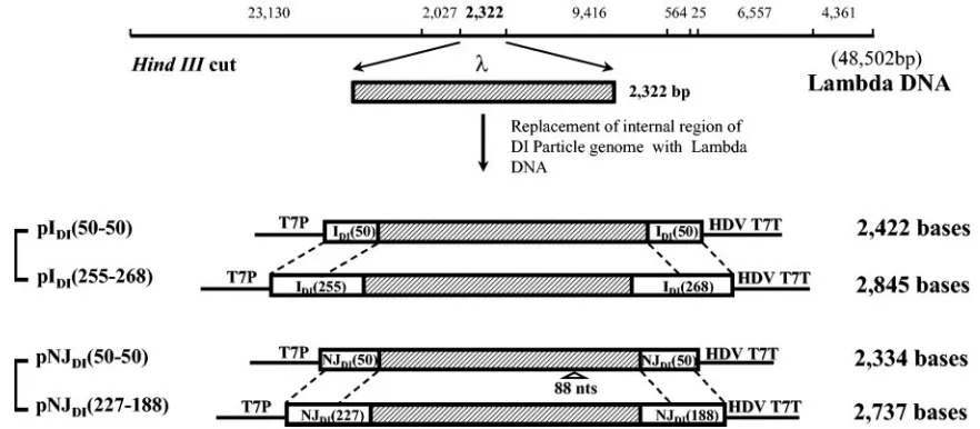
FIG. 1. Schematic representation of chimeric pDIs containing bacteriophagesequences. The internal region of the 2,322-bp HindIII fragment ofDNA is shown as a shaded bar. Numbers in the names of the plasmids indicate the length (in nucleotides) of wild-type sequences at the 3⬘ and 5⬘genomic termini of chimeric DI RNAs. The sizes of chimeric DI RNAs encoded from the plasmids are shown at the right side of the plasmids. Eighty-eight-nucleotide deletions (88 nts) in theDNA of pNJDI(50-50) are shown under the shaded bar. T7P, HDV, and T7T indicate the T7 transcriptional promoter, hepatitis delta virus ribozyme sequences, and T7 transcriptional terminator, respectively. Numbers in the bracket indicate conserved 3⬘and 5⬘VSV-specific nucleotide sequences of DI particles.
on November 8, 2019 by guest
http://jvi.asm.org/
Interference assay.The interfering activities of purified DI particles were examined by the standard virus yield reduction assay as described previously (26, 27, 31).
RESULTS
Usage of P and L proteins by RNA-N protein complex of DI
particles.The functional template for the replication of the DI
particle is the genomic RNA encapsidated with N protein. It has been demonstrated that the polymerase complex (P and L proteins) of isolated RNP can be interchanged with the added P and L proteins (8). It may be possible that the capability of RNPs of DI particles from one serotype to utilize P and L proteins provided by the standard VSV of the other serotype determines the heterotypic interfering activities of DI parti-cles. Therefore, we examined the utilization of P and L
pro-teins of both VSVIndand VSVNJby DI genomic RNA-N
pro-tein complex from both serotypes of VSV. We analyzed the
synthesis of two of the DI RNAs [IDI(50-50) and NJDI(50-50)]
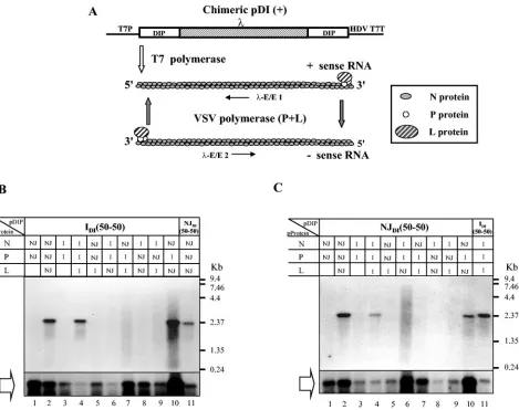
(Fig. 1) in various combinations of N, P, and L proteins from the two different serotypes. The detection of RNA synthesis by Northern blot analysis is depicted in Fig. 2. Figure 2B shows
the replication of IDI(50-50) in the presence of different
com-binations of N, P, and L proteins from VSVIndand VSVNJ. T7
RNA polymerase-mediated positive-strand RNA synthesis and encapsidation were detected in all combinations (Fig. 2B, open block arrow); however, VSV RNA polymerase-derived nega-tive-strand RNA synthesis was detected with only three
com-binations of N, P, and L proteins; (i) NInd, PInd, and LInd; (ii)
NNJ, PNJ, and LNJ; and (iii) NNJ, PInd, and LNJ(Fig. 2B, lanes
2, 4, and 10). NJDI(50-50) replicated in the presence of the
same combinations of N, P, and L proteins as IDI(50-50) (Fig.
2C), although the efficiency of replication in the presence of
NInd, PInd, and LIndwas lower than that in the presence of NNJ,
PNJ, and LNJ. Synthesis of NJDI(50-50) RNA in the presence of
NNJ, PInd, and LNJwas also detected, indicating that not only
IDI(50-50) but also NJDI(50-50) can be replicated by the
com-FIG. 2. Replication of IDI(50-50) and NJDI(50-50) RNAs with combinations of N, P, and L proteins from VSVIndand VSVNJ. The encapsidated positive-sense and negative-sense chimeric DI RNAs were detected by Northern blot analysis. (A) Schematic representation of DI RNA synthesis after transfection.-E/E1 and-E/E2 represent the riboprobes for the detection of RNA synthesis by Northern blot analysis. (B) Replication of IDI(50-50) RNA with combinations of N, P, and L proteins from VSVIndand VSVNJ. (C) Replication of NJDI(50-50) RNA with combinations of N, P, and L proteins from VSVIndand VSVNJ. Combinations of N, P, and L proteins from VSVIndand VSVNJare shown in the boxes. The open arrows indicate the RNA synthesized by T7 RNA polymerase and encapsidated by N proteins. The RNA-N protein complexes were immuno-precipitated with anti-VSV sera. I and NJ denote proteins from VSVIndand VSVNJ, respectively.
9590 KIM AND KANG J. VIROL.
on November 8, 2019 by guest
http://jvi.asm.org/
[image:3.585.59.528.68.439.2]bination of NNJ, PInd, and LNJproteins. These results
demon-strate that N, P, and L proteins of one serotype do not exclude the genomic RNAs of VSV DI particles as long as all three proteins are from the same serotype or three proteins are in
the combination of NNJ, PInd, and LNJ.The results also
dem-onstrate that DI RNP complex from one serotype does not utilize P and/or L proteins from the other serotype. It was interesting to see that replication of chimeric DIs was carried
out by the combination of NNJ, PInd, and LNJbut not with the
combination of NInd, PNJ, and LInd. These results clearly
dem-onstrate that PIndcan interact with N and L proteins from both
serotypes of VSV. In contrast, PNJcan interact only with N and
L proteins of VSVNJ.
Usage of homotypic and heterotypic N, P, and L proteins by
DI particle genomic RNA for RNA replication.The genomic
RNAs of DI particles from both serotypes containing only 50 nucleotides of terminal sequences can utilize N, P, and L proteins as long as all three proteins are from the same serotypes (Fig. 2). The panhandle-type DI particles contain
cis-acting promoter sequences at the 3⬘ termini of both
posi-tive- and negaposi-tive-strand genomes. It has been previously
dem-onstrated that the VSVIndDI particle genome requires only 45
nucleotides of 3⬘and 5⬘genomic terminal sequences to
repli-cate efficiently (18). The minimal number of terminal
nucleo-tides required for efficient replication of VSVNJDI particles
has not been determined previously. The NJDI genome
con-tains 71-nucleotide-long inverse complementary sequences
(15), which are longer than that of IDI(54 nucleotides). We
compared the genomic RNA replication between DI particles
containing 50 nucleotides of genomic end sequences, IDI
(50-50) and NJDI(50-50), and DI particles containing more than 50
nucleotides of genomic end sequences, IDI(255-268) and
NJDI(227-188). To determine the replication efficiency of DI
RNAs using homotypic and heterotypic N, P, and L proteins, DI RNA synthesis was analyzed in the presence of N, P, and L
proteins from either VSVIndor VSVNJas depicted in Fig. 2.
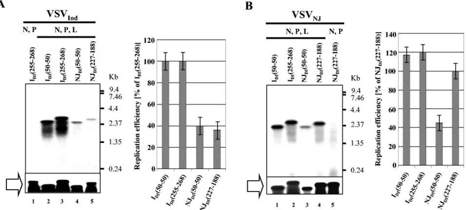
The amounts of encapsidated plus-strand RNA synthesized from the chimeric pDI by T7 RNA polymerase were similar among the chimeric DI particles (Fig. 3, open block arrow), indicating that approximately equal numbers of templates were available for negative-strand RNA synthesis by the VSV RNA polymerase. All chimeric DI RNAs were synthesized by the
VSVInd N, P, and L proteins with differing efficiencies.
Al-though chimeric VSVNJ DI RNAs were synthesized by the
heterotypic VSVIndN, P, and L proteins, the amount of
chi-meric VSVNJDI RNA synthesis was significantly lower than
that of chimeric VSVIndDIs (Fig. 3A). In contrast, the N, P,
and L proteins of VSVNJ supported replication of chimeric
IDI(50-50) and IDI(255-268) as efficiently as its homotypic
NJDI(227-188) particle (Fig. 3B). Our results demonstrate that
genomic RNAs of DI particles can utilize N, P, and L proteins
from either serotype, but only IDIRNA can replicate with high
efficiency using N, P, and L proteins from both serotypes.
In the presence of VSVInd N, P, and L proteins, similar
amounts of negative-strand RNAs were synthesized from
IDI(50-50) and IDI(255-188), indicating that 50 nucleotides of
IDI at both the 3⬘ and 5⬘ genomic termini are sufficient for
replication and encapsidation (Fig. 3A, lanes 2 and 3). In
contrast, NJDI(50-50) particles were only 30% as efficient in
the synthesis of negative-strand RNA as NJDI(227-188) (Fig.
3B, lanes 3 and 4). These results suggest that the VSVNJDI
[image:4.585.55.534.73.289.2]particles require more than 50 nucleotides of terminal genomic sequences for the optimum level of genomic RNA replication.
FIG. 3. Replication of chimeric DI RNAs in the presence of homotypic and heterotypic N, P, and L proteins. Open block arrows indicate the chimeric DI RNAs synthesized by T7 RNA polymerase and encapsidated with N proteins. VSV polymerase-specific (second strand) RNA synthesis of chimeric DI RNAs in the presence of homotypic and heterotypic N, P, and L proteins is shown in the left panels. Average values relative to IDI(255-268) and NJDI(227-188) from three experiments are shown in the graphs. Error bars represent standard deviations of the mean. (A) Replication of chimeric DI RNAs in the cells expressing VSVIndN, P, and L proteins. (B) Replication of chimeric DI RNAs in the cells expressing VSVNJN, P, and L proteins.
on November 8, 2019 by guest
http://jvi.asm.org/
Assembly of DI particles using homotypic and heterotypic
proteins.Assembly and maturation of VSV is mediated by the
interaction of RNP complexes with matrix proteins (M) and
glycoproteins (G) (19, 22, 34). VSVIndDI particles are able to
replicate and mature in cells coinfected with either VSVNJor
VSVIndstandard virus (26, 28). On the other hand, VSVNJDI
particles are able to replicate and mature only in cells
coin-fected with the homotypic VSVNJ standard virus. It is now
clear that genomic RNAs of VSVNJDI particles can be
en-capsidated by the N protein of VSVIndand can be associated
with the polymerase complex (P and L) of VSVInd to form a
functional RNP complex (Fig. 3A). Therefore, we decided to
examine whether or not genomic RNA of the VSVNJDI
par-ticle could be assembled into DI parpar-ticles in the presence of N,
P, L, M, and G proteins of VSVInd. Recovery of VSVInd DI
particles from cDNA was also performed with the same set of proteins using the reverse genetics of VSV (16, 24). In the
presence of N, P, L, M, and G proteins of VSVInd, only VSVInd
DI particles were recovered from transfected cells and
ampli-fied by coinfection with standard VSVInd or VSVNJ (Fig. 4,
lanes 1 and 2). We were unable to recover VSVNJDI particles
in the presence of VSVInd proteins (Fig. 4, lanes 3 and 4),
indicating that VSVNJ DI particles cannot assemble using
VSVIndproteins although their genomic RNAs can be
repli-cated in the presence of N, P, and L proteins of VSVInd. In
contrast, all chimeric DIs [NJDI(50-50), NJDI(227-188), IDI
(50-50), and IDI(255-268)] were recovered from cDNAs when all
five VSVNJproteins were coexpressed (Fig. 4, lanes 6, 7, and
8). The amount of IDI(50-50) particle recovered was extremely
low, necessitating a longer exposure to see a band on the
Northern blot. These findings demonstrate that the IDI
ge-nome replicates and assembles using proteins from VSVNJ, as
expected. However, genomes of NJDI particles cannot be
as-sembled using VSVInd proteins, despite the fact that the
ge-nomes of these DI particles can be replicated at a low level in
the presence of N, P, and L proteins of VSVInd. We also found
that IDI(50-50) and NJDI(50-50) matured poorly with VSVNJ
proteins. These results suggest that the VSVInd proteins can
assemble only with VSVInd genomes and that an appropriate
length of genomic terminal sequences is required in the
assem-bly of VSVNJand VSVIndvirions.
Genomic terminal sequences of DI particles confer
interfer-ing activity.We examined the interfering activities of purified
IDI(50-50) and NJDI(227-188) against the homotypic and
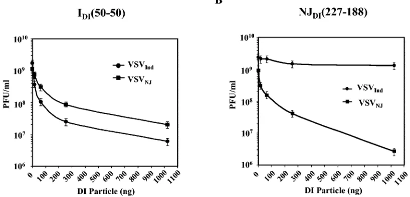
het-erotypic standard viruses to determine the role of genomic termini in homologous viral interference. BHK-21 cells were
infected at an MOI of 3 with standard VSV (VSVInd or
VSVNJ) and superinfected with various concentrations (16 ng,
64 ng, 256 ng, and 1,024 ng) of DI particles. At 18 h after infection, culture medium was harvested and the titer of the
standard virus was measured by plaque assay. IDI(50-50)
inter-fered with the replication of both VSVInd and VSVNJ. The
yield reduction of the standard virus was correlated with the increased amount of input DI particles in both homotypic and
heterotypic viral interference assays (Fig. 5). Although IDI
particles could interfere heterotypically with the replication of
VSVNJ, 6- to 20-fold more IDI(50-50) particles were required
to achieve the same level of interference as with the homotypic
standard virus (Fig. 5A). NJDI(227-188) particles also showed
the same level of interfering activity as their homotypic
wild-type VSVNJDI particles (data not shown). These VSVNJDI
particles interfered with the replication of the homotypic
VSVNJstandard virus, resulting in a standard virus yield from
300- to 1,000-fold less at the highest concentrations of input DI
particles. However, these VSVNJDI particles did not interfere
with the replication of the heterotypic VSVIndstandard virus,
even with the highest concentration (Fig. 5B). These results demonstrate that the interfering activity of the panhandle-type
DI particles of VSV was conferred by the 3⬘ and 5⬘genomic
termini, which contain promoter sequences, but not by the internal region of the DI particle genome.
In cells coinfected with VSV DI particles and the standard virus, DI particles inhibit the replication of full-length stan-dard virus genomic RNA and secondary transcription of the mRNAs, while genomic RNA of DI particles is synthesized preferentially to a certain saturation point (4). Therefore, we examined the synthesis of standard virus genomic RNA and genomic RNA of DI particles in cells infected with both stan-dard virus and chimeric DI particles. BHK-21 cells were infected
with an MOI of 3 of VSVIndor VSVNJand superinfected with
various concentrations of IDI(50-50) or NJDI(227-188) (1 ng, 4
[image:5.585.54.271.72.338.2]ng, and 16 ng for the homotypic standard virus and 64 ng, 240 ng, and 960 ng for the heterotypic standard virus). Cells were
FIG. 4. Assembly and maturation of chimeric DI particles using homotypic and heterotypic VSV proteins. The recovered chimeric IDI and NJDIparticles from the transfections were concentrated by ultra-centrifugation and were amplified by coinfecting BHK-21 cells with either VSVIndor VSVNJstandard viruses at an MOI of 3. VSVIndwas used as a helper virus for the DI particles recovered by using VSVInd N, P, M, G, and L proteins. VSVNJwas used as a helper virus for the DI particles recovered by using VSVNJN, P, M, G, and L proteins. The presence of chimeric DI particles in the culture fluid was determined by infecting fresh BHK-21 cells with the culture fluid from the first amplification and Northern blot analysis using thesequence-specific riboprobe-E/E2.
9592 KIM AND KANG J. VIROL.
on November 8, 2019 by guest
http://jvi.asm.org/
labeled with [3H]uridine (30 Ci/ml) for 2 h. Total cellular
RNAs were isolated and analyzed in a 1% agarose formalde-hyde gel. The RNA bands were visualized by autoradiography.
IDI(50-50) showed a dramatic inhibition of VSVInd genomic
RNA and mRNA synthesis with increasing amounts of DI
particles (Fig. 6). The synthesis of IDI(50-50) genomic RNA
increased gradually with larger amounts of input DI particles
(Fig. 6A). IDI(50-50) inhibited genomic RNA and mRNA
syn-thesis of VSVNJ as well, but much greater amounts of DI
particles were required to obtain the same level of interfering
activity compared to the VSVIndRNA synthesis (Fig. 6A). The
amount of IDI(50-50) genomic RNA synthesized with the help
of VSVNJwas not as large as that of IDI(50-50) genomic RNA
synthesized in the presence of homotypic VSVInd. Similar
lev-els of IDI(50-50) genomic RNA were synthesized in cells
coin-fected with 240 ng and 960 ng of the DI particles and the
VSVNJstandard virus (Fig. 6A, lanes 7 and 8). NJDI(227-188)
inhibited genomic RNA and mRNA synthesis of VSVNJin a
gradual manner with increasing concentration of the DI
par-ticles (Fig. 6B). In contrast, NJDI(227-188) did not affect the
genomic RNA and mRNA synthesis of VSVInd, although we
used as much as 960 ng per culture dish (Fig. 6B).
To our surprise, a small amount of NJDI(227-188) RNA was
replicated with the help of the VSVIndstandard virus (Fig. 6B).
We have confirmed that the low level of NJDI(227-188) RNA
synthesis was not the result of copurified VSVNJstandard virus
[image:6.585.93.502.76.273.2]FIG. 5. Interfering activity of chimeric DI particles. The yields of the standard viruses represent the mean value of duplicate plaque assays. (A) Yield reduction of VSV standard viruses by IDI(50-50). (B) Yield reduction of VSV standard viruses by NJDI(227-188). Error bars represent the standard deviation of the mean of three separate assays.
FIG. 6. Analysis of VSV RNA synthesized in cells coinfected with chimeric DI particles and the standard viruses. (A) Synthesis of IDI(50-50) genomic RNA and genomic RNA and mRNA of VSVIndand VSVNJstandard virus in the presence of various concentrations of input IDI(50-50). (B) Synthesis of genomic RNA of NJDI(227-188) and genomic RNA and mRNA of VSVInd and VSVNJ with various concentrations of NJDI(227-188). Arrows indicate the genomic RNAs of the chimeric DIs. Std, standard virus; L, G, N, and M/P, mRNA of each gene.
on November 8, 2019 by guest
http://jvi.asm.org/
in the DI particle preparation (data not shown). The result was
confirmed by Northern blot analysis using the
sequence-specific riboprobe, which is much more sensitive and detects
internalsequences in the NJDI(227-188) RNA directly (data
not shown). These results show that VSVNJDI particles can
replicate at a very low efficiency with the help of the VSVInd
standard virus, although NJDIfailed to interfere heterotypically.
DISCUSSION
Both standard VSV and DI particles incorporate all five VSV proteins during assembly and maturation (1, 12, 32). Although panhandle-type DI particles cannot produce any VSV proteins, it is assumed that they can initiate their genomic RNA synthesis by using the polymerase complex incorporated into the DI particles (30). If DI particles are to interfere with the replication of the standard virus homotypically and/or het-erotypically, the DI particle RNPs or genomic RNAs synthe-sized from the initial templates should be able to utilize the proteins from the standard virus efficiently. Because only the genomic RNAs encapsidated with N protein as a form of RNP complex function as templates for transcription and replica-tion, it has been difficult to demonstrate in vitro whether or not the DI genomic RNA of one serotype can utilize the proteins
of the other serotype. It has been demonstrated that VSVInd
DI particles could utilize proteins from the VSVNJstandard
virus for their replication in infected cells (28). However,
genomic RNA synthesis of the VSVNJDI particle has not been
demonstrated in cells coinfected with the VSVInd standard
virus. It has also been demonstrated that P and L proteins in the RNP are interchangeable with the newly added P and L proteins in vitro (8). Therefore, we expected that the RNP
complex of VSVIndDI particles could exchange with P and L
proteins of VSVNJbut that the RNP complex of VSVNJDI
particles could not do the reverse. DI particle genomic RNAs
of both VSVIndand VSVNJreplicated when N, P, and L
pro-teins were from the same serotype or a combination of NNJ,
PInd, and LNJ(Fig. 2B and C). Any other combinations of N, P,
and L proteins did not support the replication of VSVInd DI
particle or VSVNJDI particle genomes. Combinations of NInd,
PInd, and LNJor NInd, PNJ, and LNJdid not support the
rep-lication of the IDI genome, demonstrating that the RNA-N
protein template of the VSVInd DI particle or the VSVNJDI
particle cannot utilize the P and L proteins of the heterotypic standard virus directly. Overall, the data shown in Fig. 2 dem-onstrate that protein interactions among RNA-N protein com-plexes, P proteins, and L proteins play a crucial role in con-ferring serotypic specificity of the VSV.
It was not known previously whether or not the genomic
RNAs of VSVNJcould utilize VSVIndproteins for their
repli-cation. Our results shown in Fig. 3 demonstrate that genomic
RNAs of VSVNJDI particles utilized N, P, and L proteins of
VSVInd for their replication. However, the efficiency of the
VSVNJDI particle RNA replication in the presence of VSVInd
N, P, and L proteins was much lower. This shows that DI particle genomic RNAs from both serotypes can utilize VSV proteins without serotypic exclusion but with differing efficien-cies.
It has previously been demonstrated that panhandle-type
VSVIndDI particle genomes containing 45 nucleotides of the
3⬘ and 5⬘terminal genomic sequences replicated efficiently in
the presence of homotypic proteins (23). Our data in Fig. 3
agreed with the previous results by showing that IDI(50-50) and
IDI(255-268) replicated equally well with both homotypic and
heterotypic N, P, and L proteins. VSVNJDI particle genomes
containing only 50 nucleotides of terminal genomic sequences
[NJDI(50-50)] were less efficient in their replication than DI
particle genomes containing more than 188 nucleotides of the
VSVNJ-specific terminal genomic sequences [NJDI(227-188)].
Considering the long 3⬘ and 5⬘ inverse complementary
se-quence of the NJDI particle genome (71 bases), it may be
possible that VSVNJ DI particles containing longer inverse
complementary sequences in their genomes are generated from the standard virus and are selectively amplified because of the advantage they have in utilizing the polymerase complex or other viral proteins involved in the assembly of the VSV virions.
VSV virion matures by budding through the cytoplasmic membrane as a result of the specific interaction of the RNP complex with the matrix protein (M) and glycoprotein (G) (17,
22). Since chimeric VSVNJDI RNAs can replicate using the N,
P, and L proteins of VSVInd (Fig. 3A), which suggests the
assembly of functional RNP, they should be able to mature and
bud out as DI particles if M and G proteins of VSVInd are
provided. However, the results in Fig. 4 show that the
matu-ration of VSVNJDI particles using VSVIndproteins does not
occur. These data can be interpreted in one of two ways. The
first, a very low level of replication of NJDIgenome using N, P,
and L proteins of VSVIndresults in few RNA-N protein
com-plexes which are available for assembly by M and G proteins of
VSVInd. The second is that specific genomic terminal
se-quences of DI particles may be involved in the maturation of VSV particles as described by others (33).
Pattnaik et al. have shown that chimeric VSVIndDI particles
containing 51 nucleotides from the 3⬘and 5⬘genomic termini
of a panhandle-type DI particle with non-VSV internal se-quences could replicate and mature into infectious DI particles (23). These investigators examined the maturation of the chi-meric DI particle without analyzing the interfering activity of the chimeric DI particles. We successfully recovered,
ampli-fied, and purified IDI(50-50), IDI(255-268), and NJDI(227-188)
particles using the homotypic standard viruses (Fig. 4, lanes 1, 2, and 8). The interfering activities of the recovered chimeric DI particles were the same as those of wild-type DI particles, indicating that genomic terminal sequences encompassing the promoter sequences in panhandle-type DI particle genomes are the only elements required for interfering activity.
We propose the following model of homotypic and hetero-typic viral interference mediated by VSV DI particles. In the case of homotypic viral interference, RNPs of VSV DI parti-cles utilize their own as well as newly synthesized polymerase complexes provided by the standard virus to initiate antigeno-mic RNA synthesis. Once the RNAs are synthesized from the RNP of DI particles, the nascent RNA genomes are encapsi-dated by N proteins synthesized by the standard virus and form new RNPs. While DI particle genomic RNA replicates using N, P, and L proteins without the need for transcription, stan-dard viruses have to transcribe mRNAs to provide the viral
proteins necessary for replication. Because its 3⬘and 5⬘inverse
complementarity of panhandle-type DI particle genome
pro-9594 KIM AND KANG J. VIROL.
on November 8, 2019 by guest
http://jvi.asm.org/
vides stronger promoter sequences to both senses of the DI particle genome (18, 23) and the DI particle is dependent on the standard virus for the N, P, and L proteins (25), DI parti-cles have a replicative advantage over the standard virus. Con-sequently, DI particles interfere with the replication of the standard virus.
In the case of heterotypic interference, when cells are coin-fected with DI particles and a heterotypic standard virus, DI particle RNA-N protein complexes from either serotype can-not utilize P and L proteins from different serotypes (Fig. 2). They require initial transcription of leader RNAs through the use of their own DI particle-associated P and L proteins. The
incapability of IDIRNPs to directly use VSVNJP and L
pro-teins may delay the replication of the DI particle genome. The
delay of IDIparticle replication may give the VSVNJstandard
virus a chance to initiate replication using the available N, P, and L proteins and subsequently results in the reduced
inter-fering activity against the VSVNJstandard virus. This
possibil-ity is supported by an earlier observation that adding DI par-ticles at later times of standard virus infection fails to inhibit the genomic RNA and mRNA synthesis of the standard virus
(29). The efficiency of the promoter of the VSVNJDI particle
genome using the polymerase complex from VSVIndwould not
be sufficient to compete for VSVIndP and L proteins (Fig. 3).
Therefore, VSVNJDI particles cannot interfere with the
rep-lication of VSVIndstandard virus, although genomic RNAs of
VSVNJDI particles can be replicated by N, P, and L proteins
of VSVIndwith very low efficiency (Fig. 3).
Another difference between the VSVIndand VSVNJDI
par-ticles in using heterotypic proteins is the inability of VSVNJDI
particles to assemble into virions using VSVInd proteins (Fig.
4). Although we cannot directly demonstrate whether or not the ability to assemble using heterotypic proteins contributes
to heterotypic interference by VSVIndDI particles, we
specu-late that the assembly of VSVInd DI particles using VSVNJ
proteins will amplify the DI particles and the amplified DI particles will spread to neighboring cells, which will result in a
reduction of the standard virus yield. In contrast to the VSVInd
DI particles, the VSVNJDI particles cannot assemble using
heterotypic VSVIndproteins, although their genomic RNA can
be replicated by VSVIndproteins at a low level; therefore, the
input DI particles are restricted to the cells initially infected. Our results suggest that both DI particle genomic RNA repli-cation and particle assembly are prerequisites for efficient ho-mologous viral interference. In addition, the ability of the genomic RNAs of DI particles to replicate and mature as DI particles efficiently using heterotypic VSV proteins allows DI particles to interfere heterotypically.
ACKNOWLEDGMENTS
We thank Chad Michalski and Rosanne Kang for correcting the manuscript.
This study was supported by grants from the Canadian Institute of Health Research (CIHR) and Sumagen Company Limited.
REFERENCES
1.Baltimore, D., A. S. Huang, and M. Stampfer.1970. Ribonucleic acid syn-thesis of vesicular stomatitis virus. II. An RNA polymerase in the virion. Proc. Natl. Acad. Sci. USA66:572–576.
2.Bishop, D. H., S. U. Emerson, and A. Flamand.1974. Reconstitution of infectivity and transcriptase activity of homologous and heterologous viruses:
vesicular stomatitis (Indiana serotype), Chandipura, vesicular stomatitis (New Jersey serotype), and Cocal viruses. J. Virol.14:139–144.
3.Brown, T., K. Mackey, and T. Du.1997. Analysis of RNA by Northern and slot blot hybridization, p. 4.9.1–4.9.16.InR. B. Frederick, M. Ausubel, R. E. Kingston, D. D. Moore, J. G. Seidman, J. A. Smith, and K. Struhl (ed.), Current protocols in molecular biology. John Wiley and Sons, New York, N.Y.
4.Choi, W. Y.1997. Molecular biological characterization of defective inter-fering particles of vesicular stomatitis virus. University of Ottawa, Ottawa, Canada.
5.De, B. P., and A. K. Banerjee.1984. Specific interactions of vesicular stoma-titis virus L and NS proteins with heterologous genome ribonucleoprotein template lead to mRNA synthesis in vitro. J. Virol.51:628–634.
6.Fuerst, T. R., E. G. Niles, F. W. Studier, and B. Moss.1986. Eukaryotic transient-expression system based on recombinant vaccinia virus that syn-thesizes bacteriophage T7 RNA polymerase. Proc. Natl. Acad. Sci. USA
83:8122–8126.
7.Giachetti, C., and J. J. Holland.1989. Vesicular stomatitis virus and its defective interfering particles exhibit in vitro transcriptional and replicative competition for purified L-NS polymerase molecules. Virology170:264– 267.
8.Helfman, W. B., and J. Perrault. 1989. Redistributive properties of the vesicular stomatitis virus polymerase. Virology171:319–330.
9.Holland, J. J.1987. Defective interfering rhabdoviruses, p. 297–360.InR. R. Wagner (ed.), The rhabdoviruses. Plenum, New York, N.Y.
10.Huang, A. S., and E. K. Manders.1972. Ribonucleic acid synthesis of vesic-ular stomatitis virus. IV. Transcription by standard virus in the presence of defective interfering particles. J. Virol.9:909–916.
11.Hunt, D. M., M. G. Mellon, and S. U. Emerson.1979. Viral transcriptase, p. 169–183.InD. H. L. Bishop (ed.), Rhabdoviruses. CRC, Boca Raton, Fla. 12.Kang, C. Y., and L. Prevec.1969. Proteins of vesicular stomatitis virus. I.
Polyacrylamide gel analysis of viral antigens. J. Virol.3:404–413. 13.Keene, J. D., M. Schubert, and R. A. Lazzarini.1979. Terminal sequences of
vesicular stomatitis virus RNA are both complementary and conserved. J. Virol.32:167–174.
14.Khan, S. R., and R. A. Lazzarini.1977. The relationship between autoint-erference and the replication of defective interfering particle. Virology77:
189–201.
15.Kim, G. N., W. Y. Choi, M. Park, and C. Y. Kang.2002. Replication and transcription of viral RNAs by recombinant L proteins of New Jersey sero-type of vesicular stomatitis virus. Virus Res.90:347–364.
16.Lawson, N. D., E. A. Stillman, M. A. Whitt, and J. K. Rose.1995. Recom-binant vesicular stomatitis viruses from DNA. Proc. Natl. Acad. Sci. USA
92:4477–4481.
17.Lenard, J., and R. Vanderoef.1990. Localization of the membrane-associ-ated region of vesicular stomatitis virus M protein at the N terminus, using the hydrophobic, photoreactive probe 125I-TID. J. Virol.64:3486–3491. 18.Li, T., and A. K. Pattnaik. 1997. Replication signals in the genome of
vesicular stomatitis virus and its defective interfering particles: identification of a sequence element that enhances DI RNA replication. Virology232:
248–259.
19.Lyles, D. S., M. McKenzie, and J. W. Parce.1992. Subunit interactions of vesicular stomatitis virus envelope glycoprotein stabilized by binding to viral matrix protein. J. Virol.66:349–358.
20.Mackett, M., T. Yilma, J. K. Rose, and B. Moss.1985. Vaccinia virus re-combinants: expression of VSV genes and protective immunization of mice and cattle. Science227:433–435.
21.Nichol, S. T., and J. J. Holland.1987. Genome RNA terminus conservation and diversity among vesiculoviruses. J. Virol.61:200–205.
22.Odenwald, W. F., H. Arnheiter, M. Dubois-Dalcq, and R. A. Lazzarini.1986. Stereo images of vesicular stomatitis virus assembly. J. Virol.57:922–932. 23.Pattnaik, A. K., L. A. Ball, A. LeGrone, and G. W. Wertz.1995. The termini
of VSV DI particle RNAs are sufficient to signal RNA encapsidation, replication, and budding to generate infectious particles. Virology206:760– 764.
24.Pattnaik, A. K., L. A. Ball, A. W. LeGrone, and G. W. Wertz.1992. Infectious defective interfering particles of VSV from transcripts of a cDNA clone. Cell
69:1011–1020.
25.Pattnaik, A. K., and G. W. Wertz.1990. Replication and amplification of defective interfering particle RNAs of vesicular stomatitis virus in cells expressing viral proteins from vectors containing cloned cDNAs. J. Virol.
64:2948–2957.
26.Prevec, L., and C. Y. Kang.1970. Homotypic and heterotypic interference by defective particles of vesicular stomatitis virus. Nature228:25–27. 27.Rud, E. W., A. K. Banerjee, and C. Y. Kang.1986. Defective interfering
particles of VSVNJ (Ogden), generated by heat treatment, contain multiple internal genomic deletions. Virology155:61–76.
28.Schnitzlein, W. M., and M. E. Reichmann.1977. A possible effect of viral proteins on the specificity of interference by defective vesicular stomatitis virus particles. Virology80:275–288.
29.Stampfer, M., D. Baltimore, and A. S. Huang.1969. Ribonucleic acid syn-thesis of vesicular stomatitis virus. I. Species of ribonucleic acid found in
on November 8, 2019 by guest
http://jvi.asm.org/
Chinese hamster ovary cells infected with plaque-forming and defective particles. J. Virol.4:154–161.
30.Szilagyi, J. F., and L. Uryvayev.1973. Isolation of an infectious ribonucleo-protein from vesicular stomatitis virus containing an active RNA transcrip-tase. J. Virol.11:279–286.
31.Treuhaft, M. W., and M. O. Beem.1982. Defective interfering particles of respiratory syncytial virus. Infect. Immun.37:439–444.
32.Wagner, R. R., T. C. Schnaitman, R. M. Snyder, and C. A. Schnaitman.1969.
Protein composition of the structural components of vesicular stomatitis virus. J. Virol.3:611–618.
33.Whelan, S. P., and G. W. Wertz.1999. The 5⬘terminal trailer region of vesicular stomatitis virus contains a position-dependentcis-acting signal for assembly of RNA into infectious particles. J. Virol.73:307–315.
34.Wilson, T., and J. Lenard.1981. Interaction of wild-type and mutant M protein vesicular stomatitis virus with nucleocapsids in vitro. Biochemistry
20:1349–1354.
9596 KIM AND KANG J. VIROL.