MANAGEMENT OF MANDIBULAR FRACTURES : A COMPARATIVE
RANDOMIZED STUDY
A Dissertation submitted
in partial fulfilment of the requirements for the degree of
MASTER OF DENTAL S UR GER Y
BRANCH – I II
ORAL AND MA XILLOFACI AL SUR GERY
THE TAMILNADU DR. M. G. R. MEDICAL U NIVE RSIT Y
CHENNAI - 600032
2015 – 2018.
ADHIPARASAKTHI DENTAL COLLEGE & HOSPITAL
DEPARTMENT OF ORAL & MAXILLOFACIAL SURGERY,
ADHIPARASAKTHI DENTAL COLLEGE AND HOSPITAL,
MELMARUVATHUR – 603319.
This is to certify t hat Dr. Prithivi Sankar. N, Post Gr aduate student (2015 - 2018) in the Department of Oral and Maxillofacial Surgery , Adhiparasakthi Dental College and Hospital, Melmaruvathur – 603319, has done this dissertation titled “EFFICACY OF TITANIUM LOCKING MINI PLATES IN THE MANAGEMENT OF MANDIBULAR FRACTURES : A COMPARATIVE RANDOMIZED STUDY” Under our direct guidance and supervision in partial fulfilment of the regulations laid down b y the Tamilnadu Dr. M. G. R Medical Universit y, Chennai – 600032 for MDS. , (Branch -III) Oral and Maxillofacial Surgery degree examination.
Guide :
Dr. M. Karthikeyan, M. D. S., Professor,
Department of Oral & Maxillofacial Surgery.
Co Guide :
Dr. S. Gokkulakrishnan, M. D. S., Professor & Head,
Department of Oral & Maxillofacial Surgery.
Dr. S. Thillainayagam, M. D. S., Principal,
I thank God for all his blessings and for giving me everything to pursues my masters in Oral & Maxillofacial Surgery and the strength to complete this dissertation.
I thank our Correspondent Dr. T. Ramesh, MD., for giving me the chance to make my dreams of becoming an Oral & Maxillofacial Surgeon, a reality and giving me everything I needed to complete this dissertation at Adhiparasakthi Dental College & Hospital, Melmaruvathur.
I express my humble gratitude, sincerity & respect to our esteemed Principal, Prof. Dr. S. Thillainayagam, Adhiparasakthi Dental College & Hospital, Melmaruvathur.
I express my sincere solidarity to my esteemed Head of the Department Prof. Dr. S. Gokkulakrishnan, Professor & Head, Department of Oral & Maxillofacial Surgery, Adhiparasakthi Dental College & Hospital, Melmaruvathur. I am thankful for his guidance, constructive criticism, patient hearing & moral support throughout my postgraduation course & without which this dissertation would not have been possible.
Maxillofacial Surgery, Adhiparasakthi Dental College & Hospital, Melmaruvathur for his constant advice & guidance.
I am thankful to my teacher Dr. M. James Antony Bhagat, Reader, Department of Oral & Maxillofacial Surgery, Adhiparasakthi Dental College & Hospital, Melmaruvathur for his constructive criticism and his inputs towards making this dissertation an exceptional one.
I am thankful to my teacher Dr. T. J. Gnanasagar, Reader, Department of Oral & Maxillofacial Surgery, Adhiparasakthi Dental College & Hospital, Melmaruvathur for his ideas for the betterment of this dissertation and myself.
I am indebted to my teacher Dr. Abishek. R. Balaji, Senior Lecturer, Department of Oral & Maxillofacial Surgery, Adhiparasakthi Dental College & Hospital, Melmaruvathur for his moral support and constant guidance.
I am grateful to my teacher Dr. Srinivasulu. P, Senior Lecturer, Department of Oral & Maxillofacial Surgery, Adhiparasakthi Dental College & Hospital, Melmaruvathur for his patient hearing & support.
S. Barath Vikraman, Dr. S. Durai Murugan, Dr. Chinnaiah Rajendran and Dr. M. Veeramuthu for all the help they have rendered.
A special mention of thanks to all my patients for their consent, co-operation and participation in this study.
I am indebted to my aunt / mother, Dr. Shanti Vijay Bose for being a pillar of support for me from the day I was born. Her constant guidance and support has been crucial in many of my life decisions and she has shaped the way I see and perceive many of the things, today.
I am grateful to my family member, Mr. R, Jeevanandham, Mrs. Pavalamani Jeevanandham, Dr. Vijay. C. Bose, Mr. Saravanan Jeevanandham, Mrs. Gana Saraswathy Saravanan for offering me unswerving support throughout my life.
I thank my cousins Sneha Vijay Bose,C. V. Shiv Bose, Mridula Saravanan & Nandita Saravanan for helping me in completing my goals and offering me advice when I had no one else to turn to.
little more easy to complete.
I dedicate this work to my parents, Dr. P. Nanthakumar & Mrs. Uma Nanthakumar, my brother Mr. Pratap Sankar, my sister in law Mrs. Kristina Adair Sankar and my nephew Iniyan Adair Sankar who have been my major support system throughout my life. They have strongly believed in me and have been instrumental in guiding me through the turmoils of life.
Title of the Dissertation
Efficacy of Titanium Locking Mini Plates in the Management of Mandibular Fractures : A Comparative
Randomized Study
Place of the Study Adhiparasakthi Dental College & Hospital, Melmaruvathur-603319.
Duration of the
Course 3 Years
Name of the Guide Dr. M. Karthikeyan, MDS.
Name of the Co - Guide Prof. Dr. S. Gokkulakrishnan, MDS.
I hereby declare that no part of this dissertation will be utilized for gaining financial assistance or any promotion without obtaining prior permission of the Principal, Adhiparasakthi Dental college and Hospital, Melmaruvathur -603319. In addition, I declare that no part of this work will be published either in print or in electronic media without the guide’s knowledge who have been actively involved in this dissertation. The author has the right to reserve & publish this work, solely with the permission of the Principal, Adhiparasakthi Dental College and Hospital, Melmaruvathur-603319.
Guide Head of the Department
This is to certify that this dissertation work titled “Efficacy of Titanium Locking Mini Plates in the Management of Mandibular Fractures : A Comparative Randomized Study” of the
candidate Dr. N. Prithivi Sankar with registration Number 241515402 the award of MDS in the branch of Oral & Maxillofacial Surgery. I personally verified the urkund.com website for the purpose of plagiarism Check. I found that the uploaded thesis file contains from introduction to conclusion pages and result shows 7 percentage of plagiarism in the dissertation.
Introduction
Mandibular fractures accounts for 36-70% of all maxillofacial trauma, as the mandible is the only mobile facial jaw bone, with various functions such as mastication, phonation and respiration. Hence the treatment of facial fractures is important for both functional and cosmetic reasons. Champy’s ideal lines of osteosynthesis with monocortical mini plate system is the time
tested and considered gold standard in the management of mandibular fractures. Transoral placements of miniplates have gained popularity in the last decade. But the quest to improve stability along the fracture fragments has been the need of the hour because of the increased incidence of high velocity road traffic accidents which does not involve normal fracture pattern and in which chances of crush injuries are high, requiring better stability. Conventional screw plating system engages only the bone and may lead to reduced stability by the way of screw loosening which is enhanced by mandibular torsional movements, bony pathologies and age. To overcome this problem newer innovation like locking plates have been developed in which the screws engage both the bone and the plate thus increasing the stability of screws which in turn increases the stability of fracture segments.
Aims & Objectives
Materials
Titanium 2. 0 mm locking miniplates Conventional 2. 0 mm miniplates
8 mm and 10 mm self threading titanium screws 8 mm and 10 mm locking screws
Titanium bone plating kit
Methods
Detailed clinical examination was carried out for all patients and following parameters
were noted.
Facial asymmetry, occlusion, mouth opening were assessed. Radiographic evaluation of the fractured site.
Routine haematological investigations.
Medical assessment of the patient by physician and anesthesiologists was done. Informed consent obtained from patient prior to surgery.
After the routine clinical and radiological examination protocol the fracture sites were
exposed by both intraoral and extra oral approaches with osteosynthesis using 2.0 mm titanium locking plates and screws.
Closure was done with 3 - 0 vicryl and 3 - 0 chromic gut sutures.
Povidine - Iodine mouthwashes and antibiotic cover (Inj. Cefotaxime - 1gm 12th hourly
S. No Title Page
1. Introduction 1
2. Aims & Objectives 4
3. Review of Literature 5
4. Materials & Methods 28
5. Results 33
6. Discussion 57
7. Conclusion 65
8. Bibliography 67
9. Photographs 76
S. No Title Page
1. Pre Operative – Extra Oral 76
2. Pre Operative – Intra Oral 76
3. Pre Operative Radiograph 77
4. Intraoperative Photographs 77
5. Post Operative – Extra Oral 78
6. Post Operative – Intra Oral 78
7. Post Operative Radiograph – 1st POD 79
8. Post Operative Radiograph – 1st POW 79
9. Post Operative Radiograph – 1st POM 80
10. Post Operative Radiograph – 3rd POM 80
11. Post Operative Radiograph – 6th POM 81
12. Locking Plates 81
13. Pre Operative – Extra Oral 82
14. Pre Operative – Intra Oral 82
15. Pre Operative Radiograph 83
16. Intraoperative Photographs 83
17. Post Operative – Extra Oral 84
18. Post Operative – Intra Oral 84
19. Post Operative Radiograph – 1st POD 85
22. Post Operative Radiograph – 3rd POM 86
23. Post Operative Radiograph – 6th POM 87
24. Conventional Mini Plates 87
S. No Title Page
1. Grouping of Samples 28
2. Evaluation of Parameters 32
3. Favorable / Unfavorable Fracture 41
4. Approach 42
5. Pain Control Group 44
6. Pain Experimental Group 44
7. Comparison of Pain 46
8. Mouth Opening Control Group 47
9. Mouth Opening Experimental Group 47
10. Comparison of Mouth Opening 49
11. Occlusion 50
12. Wound Healing 51
13. Infection 52
14. Segmental Mobility 53
15. Plate Exposure 54
S. No Title Page
1. Gender Distribution 34
2. Age Distribution 35
3. Etiology of Injury 36
4. Influence of Alcohol 37
5. Usage of Helmet 38
6. Time from Injury to Hospitalization 39
7. Location of Fracture Site 40
8. Favorable / Unfavorable Fracture 41
9. Approach 42
10. Pain (10 Point VAS Scale) 45
Board
Adhiparasakthi Dental College & Hospital
Melmaruvathur, Tamilnadu – 603 319.
An ISO 9001 : 2008 Certified Institution Accredited by NAAC with “B” Grade
Recognized by DCI, New Delhi
Affiliated to the Tamilnadu Dr. M. G. R. Medical University
CHAIR PERSON
Dr. S. Thillainayagam, MDS
This Ethical Committee has undergone the research protocol submitted by Dr. N. Prithivi Sankar, Post Graduate, Department of Oral & Maxillofacial Surgery under the title “ Efficacy Of Titanium Locking Mini
Plates In The Management Of Mandibular Fractures : A Comparative Randomized Study “, Reference No : 2015 – MD – Br III – KAR – 07 /
APDCH under the guidance of Dr. T. Ramakrishnan for consideration of approval to proceed with this study.
This committee has discussed about the materials being involved with the study. The qualification of the investigator, the present norms and recommendations from the Clinical Research Scientific Body and has come to a conclusion that this research protocol fulfills the specific requirements and the committee authorizes the proposal.
Date Chair Person
MEMBERS
Dr. K. Rajkumar, BSc, MDS
Dr. H. Murali, MDS
Dr. Muthuraj, MSc, MPhil, PhD
Prof. Dr. N. Thilagavathy, MDS
Prof. Dr. S. Gokkulakrishnan, MDS
Prof. Dr. A. Vasanthakumari, MDS
Dr. N. Manisundar, MDS
Shri. Balaji, BA, BL
Shri. E. P. Elumalai
MEMBER SECRETARY
Prof. Dr. T. Ramakrishnan, MDS
Inform IEC / IRB immediately in case of any issue(s) / adverse events
Inform IEC / IRB immediately in case of any change of study procedure, site and investigator.
Annual report to be submitted to IEC / IRB
Traffic Accidents accounting for 10,00,000 lives in the year 2010 to 12,00,000 lives in the year 2015 (9).
Aims
To evaluate the efficacy of 2. 0 mm titanium locking miniplates / screws system in
mandibular fractures and compare the results of locking miniplates to that of 2.0 mm conventional miniplates.
Objectives
The purpose of the study is to evaluate the clinical efficacy of locking titanium miniplates and to compare these outcomes with that of conventional miniplates by :
Pain
Mouth Opening
Occlusion
Paresthesia
Occlusal stability
Post-operative infection
Post-operative fragment rigidity
A historical perspective of maxillofacial trauma improves the understanding of current techniques and provides the basis for the development of new methods. According to the Arbeitsgemeinschaft für Osteosynthesefragen (AO)/Association for the Study of Internal Fixation
(ASIF) principles, the main objective of open reduction and rigid internal fixation in the
management of mandibular fractures is to achieve undisturbed healing and immediate restoration
of form and function without the adjunctive use of maxillomandibular fixation (MMF) (18).
The recent innovation in treatment is incorporation of general anesthesia, pain management and addition of new biomaterials in the form of titanium miniplates which gave an advantage to the patient towards returning to normal function within days of treatment. The advent of modern biomaterials has changed clinical practice towards plating the bone and early restoration of function.
Hippocrates (460 B. C.) not only devised the technique of reducing a dislocated mandible,
but also devised methods of immobilizing a fractured mandible. The ends of the fracture were reduced by hand and the fracture site was immobilized by gold or linen threads tied around the adjacent teeth. He recommended extra oral fixation by strips of Carthaginian leather glued to the skin, the ends of which were tied over the skull (20).
Becker (1952) put forth the basic principles of open reduction of mandibular fractures and
the indications and contra indications for open reduction of mandibular fractures. Once the fractures were approached, the fractures were reduced with transosseous wiring which were further immobilized using intermaxillary fixation for a period of time. The scars were very much noticeable. (21).
Roberts (1964) used plates which were composed of cobalt chrome alloys, which were
5 cm in length and they were used to treat a series of mandibular fractures. After the fractured segments were reduced the plates were applied to the outer cortical plate by means of screws which were 7 mm in length and 1. 5 mm in diameter. These plates provided sufficient reduction of the fractures segments and good post-operative stability (18).
Battersby (1967) later reviewed a large number of cases over a period of twelve years who
had been treated using plates manufactured using the chrome-cobalt-molybdenum alloy manufactured by the Austenal Laboratories of America. He reported that the alloy was inert and did not evoke any significant systemic response. The plates also provided satisfactory reduction of the fractures segments. This treatment protocol proved to be an effective alternative to transosseous wiring, however the patients requires to be put into intermaxillary fixation for a considerable period of time (22).
Spiessl (1972) studied rigid internal fixation of the mandible and stated that it’s necessary
to adapt the plate to the convex surface of the mandible at its lower border. No matter how skillfully the plates were adapted to the lower border there was always a tendency for the upper border and the lingual plate to open with the final tightening of the screws. Ultimately, this caused a distortion of the occlusion and in case of bilateral fractures it caused the opening of fractures on the opposite side. In order to overcome these difficulties, the compression plating system was developed which used a two plate system, one at the lower border and one at the upper border. The compression holes were positioned, one on each side of the fracture (23).
Michelet, Deymes & Dessus (1973) evaluated the use of the mini plates made of cobalt
cases and reported that though the meta-carpal plates provided sufficient reduction and good post-operative results, they were difficult to adapt. Their popularity faded with time (24).
Becker (1974) described the use of a self-compression plate in the treatment of fractured
mandibles. In his study he discovered that the self-compression plate provided stable fixation without rotation. Unlike the compression plates invented by Speissl, both the compression holes were present on the same side of the fracture. Because of the tendency of the upper border to open when compression is applied across the fracture at the lower border, it is necessary to apply a tension band at the level of the alveolus before tightening the screws. An arch bar or a separate plate with screws penetrating the outer cortex only is enough (25).
Schilli (1977) designed a plate with oblique lateral holes which ensured that the
compressing force was in part directed towards the upper border so that when the plate was tightened into place there was less tendency for the fracture line to gape. The dynamic compression plate with oblique lateral holes and the additional inlay portion is indicated in the inlay portion in indicated in oblique fractures and when the alveolar process is weak (26).
Champy et al (1978) modified and improvised the technique of mini-plate osteosynthesis
monocortical screws were sufficient when placed below the roots and either above or below the inferior alveolar canal. At the angle region the plate is fixed on the vestibular flat osseous area located beside the third molar. In case of fractures anterior to mental foramen, in addition to sub apical plate another plate near the lower border of the mandible was fixed. They documented that compression osteosynthesis was not advantageous, as there existed a natural compression along the lower border of the mandible which if excessive could lead to bone necrosis (27).
Branemark et al (1983) used titanium implants in over 400 patients who were treated for
edentulousness over a period of 9 years and reported a 91 % percent positive result when using titanium and gold implants for the correction of edentulousness. The high success rate was due to the inert nature of the titanium metal and its resistance to corrosion when it comes with biological fluids (28).
Prein J & Kellman RM (1987) reported the advantages of rigid internal fixation according
to the AO/ASIF method with Dynamic Compression Plates (DCP). Results showed good postoperative healing in majority of the patients. The most important advantages of using this technique were found to be the, avoidance of IMF which resulted in early, active, pain free mobilization of the jaws, safe and secure airways without tracheostomies particularly in polytrauma patients and shorter periods of hospitalization. However, the main disadvantages associated with this technique were the bulky nature of the plates , wide extra oral incision, uneven compression by the plate which may lead to infection, psuedoarthrosis and malocclusion and requirement of second surgery for plate removal.
Philip L. Maloney et al (1991) evaluated the validity of a treatment protocol for compound
mandibular fractures were treated in a prospective study. Thirty cases were treated with open reductions with rigid fixation. The remaining 22 patients were treated solely with closed reduction. The results concluded that if the initial treatment is delayed for more than 3 days, any infection at the compound fracture site should first be resolved by MMF and intravenous antibiotics before performing an open reduction (29).
J. P. Hayter (1993) presented a review of literature on the treatment of mandibular
fractures with open reduction and rigid internal fixation. Although compression plates were
initially used for bone fixation in the maxillofacial region, miniplates replaced them
because of advantages like small and easily adaptable plates, mono cortical application,
intra-oral approach, functional stability and biomechanical favorability. In miniplate
osteosynthesis the mouth opening is also improved and there is no pulmonary deficit when
compared to cases with inter maxillary fixation (30).
Ellis Edward et al (1996) evaluated 81 patients with fractures of the mandibular angle that
were treated with open reduction and internal fixation using one non compression miniplates with 2.0 mm self-threading screws through a trans-oral approach. None of the patients required post-surgical MMF. Their results suggested that two of the patients required hospitalization and I.V. Antibiotics and one of these patients had a fibrous union that required a bone graft. They concluded that the use of a single miniplate for fractures of angle of mandible is a simple and reliable technique with minimum complications (31).
T. Kawai et al (1997) conducted a study to find out the best time to undertake radiological
follow up examinations after mandibular fracture plating through a retrospective study of
radiographs. Serial radiographs of 325 fracture sites in 231 patients over a 10 year period
The results showed that radiographic changes are seen with 2-3 months in patients less than
18 years of age and it was 3-4 months for elder patients (32).
James W. Sikes et al (1998) tested the hypothesis that increased resistance to displacement
will be obtained when using the locking-head as compared with the number of conventional screws per segment in both fracture and reconstruction models. They concluded that the locking-head screws provided significantly increased resistance to displacement when only 2 screws per segment were used in reconstruction model. When 4 screws per segment were used, there was no significant difference between locking-head and conventional screw types in either model. The effect of bony buttressing was significant and may explain why miniplates often fail in the atrophic mandible but was successful in the fully dentate patients (33).
Fordyce et al (1999) performed a retrospective study of all isolated mandibular fractures
which required active management. 115 patients were selected among which 66 patients had fractures reduced manually to obtain reduction without the use of perioperative IMF and 49 patients were treated with conventional IMF. Both groups were similar in severity and type of fracture. The overall results did not show any significant discrepancies between the groups in healing and the study concluded that IMF is not usually necessary to reduce fractures confined to the mandibular bone (34).
Potter Jason et al (1999) evaluated the results of fractures treated by open reduction and
that the use of this small bone plate for fractures of the angle of the mandible provided adequate fixation in most cases but was associated with an unacceptable incidence of plate fracture, hence this plate is not recommended for routine angle fractures (35).
Pedro M. Villarreal et al (2000) in their prospective study evaluated mandibular fracture
repair after either maxillo-mandibular fixation (MMF) or rigid Internal Fixation (RIF) using the computer-assisted densitometric image analysis (CADIA) system. Out of 52 patients, 32 were treated by MMF and 20 by RIF. They concluded that the use of RIF results in more rapid bone mineralization than the use of MMF in the first 15 days post operatively while the bone density at 30, 60 and 90 days post operatively did not show significant differences (36).
Jose Moreno et al (2000) conducted a study to identify the complication rates associated
with different treatments for mandibular fractures. They compared IMF, 2.0 mm miniplates, 2.4 mm AO plates and 2.7mm AO plates in 245 patients with 386 mandibular fractures. The results showed that the most frequent complication in all the cases was post-surgical infection followed by the other type of fixation postpost-surgical malocclusion. This study did not show any difference in results with any of the methods used. However, the results concluded that the occurrence of complications is fundamentally related to the severity of the fracture rather than the type of the treatment used (37).
Thomas Schug et al (2000) studied the use of titanium mesh in the treatment of fractures
20% there were minor complications such as postoperative hematoma. In only one case did infection occur, a more severe complication. They concluded that because of its geometry and the excellent physical and biomechanical properties, titanium mesh helps to achieve better stabilization of complex mandibular fractures than conventional miniplates do. Complications such as infection and nonunion can largely be avoided and bony continuity of the mandible can be restored (38).
Hisanori Hirai et al (2001) obtained specimens from 14 who were diagnosed with
mandibular fractures, who had undergone open reduction and internal fixation using titanium miniplates. The specimens were removed with bone along with screws are stained with toluidine blue and were observed under light microscopy. The mean ratio of direct contact between cortical bone and the titanium bone screws was analyzed and was found to be 82.4% for cortical bone and the mean percentage of bone contacts for all screws was 64.4%. The results concluded that titanium bone screws used for mandibular fracture fixation develop almost complete contact with new bone (39).
Valfrido Antonio Filho et al (2001) evaluated the effect of multiple sterilization cycles
upon mechanical properties of titanium miniplates (system 2.0, Engimplan). Four groups
Richard H. Haug et al (2002) conducted a study to determine whether the degree of plate
adaptation influences the mechanical behavior of the plate / screw / substrate system. A total of 130 polyurethane mandible replicas were used and were subjected to incisal and molar loading. 2.4mm locking and non-locking reconstruction plates and 2.0mm locking and non-locking plates were adapted on the superior border of the angle region. Load and displacement data was recorded accordingly. The results of the study concluded that the degree of adaptation affected the mechanical behavior of the non-locking system and it did not affect the locking system (41).
A.P. Silvia et al (2002) studied the failure of titanium bone plates used for oral and
maxillofacial surgery during use. Microstructural examination of a titanium plate revealed the presence of equiaxed a grains and intergranular platelets, which were identified as b phase. Fractographic examination revealed that fracture happened by a transgranular cleavage mechanism associated with secondary intergranular cracking (brittle fracture). Selective attack was observed to occur on the surfaces of the implant. These results indicated that the premature fracture of the miniplate was caused by hydrogen embrittlement (42).
Edward Ellis III et al (2002) examined the use of the 2 mm locking plate/screw system
all others were managed in an outpatient basis. Four patients required removal of their plates for varying reasons. The possible advantages in the use of locking plate/screw system were found to be theoretical but this system provided sound fixation in all cases (43).
Yadranko Ducic (2002) described the use of titanium mesh and hydroxyapatite cement
for the treatment of large through-and-through calvarial defects. Twenty consecutive calvarial defects (10 to 156 cm2) that resulted from surgical removal of neoplasms or were secondary to trauma were reviewed retrospectively after reconstruction with titanium mesh and hydroxyapatite cement. There was no evidence of adverse healing, wound infection, or implant exposure or extrusion in any of the patients reviewed. Adequate 3-dimensional aesthetic restoration of calvarial contour was noted in each case. There was evidence of osseous ingrowth into the titanium mesh and hydroxyapatite cement construct in all 3 patients who underwent biopsy. Titanium mesh and hydroxyapatite cement Cranioplasty appears to be a reasonable method for the reconstruction of significant calvarial defects (44).
Ralf Gutwald et al (2003) compared a new internal mini locking system with conventional
Marisa Aparecida et al (2003) reviewed 191 patients with a total of 280 mandibular
fractures that were treated with 2. 0 mm miniplates. Twenty two fractures developed infection, with an overall incidence of 7.85%. One patient (0.89%) developed inferior alveolar nerve paresthesia, facial asymmetry was observed in 2.67% and incidence of malunion was 1.78%. The overall incidence of complications, including infections, was similar to that of AO fixation (compression plates) (16).
Chad P. Collins et al (2004) compared standard 2. 0 mm monocortical plates to 2. 0 mm
locking plates in the treatment of mandible fractures. Ninety patients with 122 fractures
were reviewed. Locking plates were used in 64 fractures and standard plates were placed
in 58 fractures. Complication rates were similar in both the groups and it was concluded
that mandible fractures treated with 2. 0 mm locking plates and standard 2. 0 mm plates
present similar short-term complication rates (12).
Richard J. Shaw et al (2004) conducted a study to compare complication rates of
Mario Roccuzzo et al (2004) evaluated a surgical protocol for vertical ridge augmentation
in the maxilla and mandible using autogenous onlay bone graft associated with a titanium mesh. A group of 18 partially edentulous patients, presenting the need for vertical bone augmentation of at least 4 mm, were treated before implant placement. Particulate bone was added and a titanium micro-mesh was used to stabilize and protect the graft. After a mean interval of 4. 6 months, meshes and screws were removed and 37 endosseous implants were successfully placed. Mean vertical bone augmentation obtained was 4. 8mm (range 4–7 mm). The preliminary results suggested that, by using the presented technique, patients can be successfully rehabilitated by means of implant-supported prosthesis 6–7 months after the first surgery, even in case of severely atrophied maxilla (47).
Ayman Chritab et al (2005) conducted a prospective study in 50 mandibular fractures which were treated with 2. 0 mm locking plates placed according to Champy’s lines of
ideal osteosynthesis. All patients were observed for complications such as soft tissue
infections, non-union, mal-union, malocclusion, osteomyelitis, plate fracture and
iatrogenic nerve injuries. Results showed 3 complications (6%) which healed uneventfully
and primary bone healing has been achieved in 98% of cases. Finally, the study concluded
that Locking plate system plus 1 week MMF is a reliable and effective treatment modality
for mandibular fractures (48).
Hiroshi Mugino et al (2005) conducted a study to analyze the treatment of fractures of the
edentulous mandible and to discuss this method in relation to the mandibular height at the
fracture site. Fifteen fracture sites in 11 patients with an edentulous mandible were
retrospectively examined, 9 fractures in the mandibular body, 3 in the paramedian region,
vertical height were treated with one miniplate. Fractures in an extremely atrophic
mandible with 10 mm or less were treated using one or two miniplates. The studies
concluded that miniplate osteosynthesis is a less invasive treatment and it is suitable for
fractures of the atrophic edentulous mandible, except for comminuted or defect fractures.
To obtain stable fixation in severely atrophic mandibles, we need to consider the use of
two miniplates or a combination with microplates (49).
Constantin A. Landes et al (2006) evaluated the 5-year outcome stability and
complications in orthognathic surgery using resorbable versus titanium osteofixation. Twenty-two cleft lip and palate maxillary retrognathia cases were operated on using either poly (70L-lactide-co-30DL-lactide) or titanium miniplate osteofixation. Average operative movement and postoperative instability recorded for maxillary horizontal movement (A-point-Nasion) were 2. 5 mm and 2. 1 mm for the study group, compared with 6. 3 mm and 1. 9 mm for the control group. For maxillary vertical movement (ANS-Nasion), measured values were 4. 9 mm and 1. 3 mm for the study group and 2.3 mm and 0.9 mm for the controls. For mandibular horizontal movement, measured values were 10. 7 mm and 2. 8 mm for the study group and 1. 9 mm and 0. 8 mm for the controls. Gonial angle measures were 7. 18 mm and 3. 58 mm for the study group and 6. 78 mm and 3.18 mm for the controls. They concluded horizontal maxillary stability appeared inferior to vertical stability, but mandibular stability was more reliable. Because groups were not matched for magnitude or direction of movement, the results of this study are preliminary and should be interpreted cautiously (50).
Michael. A. Miranda et al (2007) reviewed the usage of innovative locking plate for the
contraindication for internal fixation because of poor results. In osteoporotic bone the quality of bone becomes the prime determinant of screw holding power in normal conventional non locking screws. In summary, locking plate technology has been shown to improve the anchorage of plates in osteoporotic fractures, but its application is technique sensitive (51).
Domenick P. Coletti et al (2007) used a red oak model and compared the locking
mechanism of tapered design to the more well established threaded design by measuring the seating torque, plate flexure, fracture displacement, screw stripping, and screw head deformation of the various systems. Observation and comparison of the hardware failures of the two different plate-screw designs were the goal of the study. The results showed that there was less screw stripping and higher seating torque in the tapered systems when compared with threaded systems which provided an effective locking mechanism. Plate flexure was an unanticipated finding in all designs, with a higher incidence noted in a tapered design. Fractured displacement should theoretically be eliminated when using a locking plate. Interestingly, there was no statistical difference between the two designs (52).
C. N. Elias et al (2008) presented a review of literature on the composition of titanium.
Alparslan Esen et al (2008) conducted an experimental study on sheep hemimandibles to
compare the stability of titanium and absorbable plate and screw fixation system using 3 different plating techniques (single titanium plate, single absorbable plate, double absorbable plates). The study demonstrated that the titanium plate and screw fixation system had greater resistance to occlusal loads than absorbable plate and screw systems. In addition a second absorbable plate orientation provides a more favorable biomechanical behavior than a single absorbable plate system (54).
Nayak et al (2008) analyzed the benefit of primary reconstruction of depressed fractures
and compared the various options available today. The various techniques adopted by the
author like simple elevation, apposition by nylon suture; or rigid fixation by titanium and
absorbable mini plate were undertaken. Twenty two patients were included in this study
out of which ten were male and ten were female. Eighteen (56%) cases underwent titanium
miniplate fixation and eleven (34%) apposition using nylon sutures. In two cases simple
elevation of fracture segments was carried out and in one fixation with absorbable
(Poly-L- lactide) miniplate, was performed. Cosmetic deformity correction to acceptable level
was achieved better with miniplates. Primary reconstruction of depressed fracture segment
should be attempted whenever possible. The biodegradable miniplates are implant of
choice, but the titanium mini plates are cost effective and a better option when compared
to any other available measures (55).
Charles. H. C et al (2009) conducted a study in 9 cases with atrophic nonunion of humoral
diaphysis treated with locking plate fixation and recombinant human bone morphogenetic proteins. All cases were diagnosed with atrophic nonunion prior to surgery. All patients
allow for stable fixation without directly compressing the bone. The results showed that
this technique is useful and reliable in patients with atrophic bone or osteopenia (56).
J. O ‘Connell et al (2009) performed a 10 year retrospective study to evaluate the
indications for the removal of titanium miniplates following osteosynthesis in maxillofacial trauma and orthognathic surgery. The following variables were recorded : patient gender and age, number of plates inserted, indications for plate placement, location of plates, number and location of plates removed, indications for plate removal, time between insertion and removal, medical co-morbidities, and the follow-up period. During the 10 years of the study, 1247 titanium miniplates were placed in 535 patients. A total of 32 (3%) plates were removed from 30 patients. Superficial infection accounted for 41% of all plates removed. All complications were minor and most plates were removed within the first year of insertion. A low removal rate of 3% suggests that the routine removal of asymptomatic titanium miniplates is not indicated (57).
Allan S Herford et al (2009) examined the use of the locking reconstruction bone
plate/screw system for fractures of the mandible or continuity defects during an 18 month period. 102 locking bone plates were placed in 84 patients. There were no cases of malocclusion or difficulties encountered in using the plate/screw system. Loss of fixation was seen in only one patient. The use of a locking plate/screw system was found to be simple, and it offers advantages over conventional bone plates by not requiring the plate to be compressed to the bone to provide stability (58).
Rudolf Seemann et al (2009) conducted a prospective randomized multicenter study for
fractures and non-locking systems were used in 74 patients. Data were acquired during 3 distinct periods 1. Peri-operatively; 2. 5 - 7 weeks after surgery; 3. 5-7 months after surgery. In 142 condyles excellent healing was seen (overall success rate of 97.3%), 4 cases of osteosynthesis failure rates were observed (failure rate of 2.7%). Plate fracture occurred twice in the locking and once in the once in non-locking group. Screw loosening occurred twice exclusively in the nonlocking group. Their results concluded that the Medartis Trilock locking and Medartis non locking condylar plates show equivalent complication rates (15).
Baohui Ji et al (2010) evaluated the stress distribution and stress shielding effect of
titanium miniplates used for the treatment of symphyseal fractures using the finite element analysis. The results showed that ratio of lower miniplates in technique 2 (reduction with 2 miniplates) were much higher than upper miniplates and the mini plates in technique 1 (reduction with a single miniplate), and the value of lower miniplates gained a maximum value of 83.34% during left unilateral molar clenching. The study demonstrated that miniplates stress distribution and stress shielding effect ratio were affected not only by the way in which the mandible was loaded but also by the number of miniplates fixing the fracture (59).
Samrat Sabhlok et al (2010) evaluated the efficacy and stability of 2. 0 mm titanium plates
post-operative phase as compared to the traditional 2. 5mm plate without post-post-operative MMF (60).
Sauerbier et al (2010) evaluated the use of a 2. 0-mm locking plate system in mandibular
surgery. 53 patients (42male, 11female) with a total of 56 mandibular fractures were treated with a 2. 0mm mini-locking-plate system and retrospectively examined. Gender, age, cause of fractures, surgical access, classification of fractures, osteosynthesis, postsurgical findings and complications were evaluated. The use of a 2.0-mm locking plate system with its advantages of improved handling characteristics, increased stability, shorter surgical time and the preservation of bony perfusion is a viable alternative to conventional miniplates in the management of mandibular fractures (61).
Dogan Dolanmaz et al (2010) evaluated a study in which six unembalmed adult sheep
Paolo Scolozzi et al (2010) prospectively evaluated the use of a single
Arbeitsgemeinschaft für Osteosynthesefragen (AO) 2.0-mm locking reconstruction plate
for linear noncomminuted mandibular fractures without the use of a second plate. They
analyzed the clinical and radiologic data of 45 patients with 74 fractures (21 single
fractures, 22 double fractures, and 2 triple fractures). Fracture locations were the symphysis
(n - 35, 47.3%), body (n - 15, 20.3%), and angle (n - 24, 32.4%). All patients had
satisfactory fracture reduction and a successful treatment outcome without major
complications. Ten patients (22.2%) developed minor complications. The results has
demonstrated that treating linear noncomminuted mandibular fractures with a single AO
2.0-mm locking reconstruction plates is associated with no major complications and sound
bone healing in all patients (18).
Paulo Domingo et al (2010) conducted an in vitro study to access the biomechanical
stability of 9 different osteosynthesis methods which included various combinations of locking and non-locking plate / screw systems after sagittal-split ramus osteotomies in 45 polyurethane hemi mandibles. All 9 combinations were tested on a universal testing machine with increasing compressive loads until a 3mm displacement was observed. The results concluded that there was no statistically significant difference of stability between locking miniplates and non-locking mini plates but locking miniplates presented a better performance in bone fixation in all groups (63).
Verma A et al (2011) conducted a clinical study to evaluate the efficacy of locking plates
and conventional mini plates in 43 mandibular fracture patients. 22 patients were treated
with 2 mm conventional mini plates (Group A) and 21 patients with 2 mm locking mini
% complication rate in when compared with 13.6% Group A. The study supports the
concept of higher stability of the locking plates over non-locking plates (64).
Chandan Prabhakar et al (2011) evaluated the efficacy of locking miniplates/screw
system in the treatment of mandibular fractures without maxillomandibular fixation. He concluded that the locking miniplates system was found to be reliable and effective in management of mandibular fractures without postoperative intermaxillary fixation (65).
Shivani Jain et al (2011) compared the clinical and radiological outcomes of open
treatment of mandibular fractures using titanium miniplates or intraosseous wires. The study also aimed to find out whether internal fixation with titanium miniplates can effectively reduce the period of maxillomandibular fixation (MMF). 40 patients who sustained mandibular fractures were divided into 2 groups. Group I included 20 patients who were treated using intraosseous wires and post-operative MMF for 4 weeks. Group II A included 10 patients who were treated using titanium miniplates and pot-operative MMF for 1 week and Group II B comprised of 10 patients who were treated using titanium miniplates and no post-operative. MMF. Results concluded that the use of bone plates assures early restoration of normal form and function as compared with the use of intra osseous wiring which was associated with extended period of MMF (66).
Deepak et al (2011) studied the versatile nature and the biocompatibility of the titanium
the patients treated with stainless steel plates and screws had local infection while the success rate for titanium plates was 100%. 20% of cases treated with titanium system encountered the complication of shearing and fracture of the titanium screw head while fitting the screw. Wound dehiscence in case of stainless steel bone plates was noted in one out of ten patients (10%) while in the group treated with titanium plates it was 0%. They concluded that titanium plates were found to be very ideal in the management of mandibular fractures. Titanium plates were more biocompatible when compared to stainless steel plates as evidenced by the rate of infection. In all cases the plates were found to be rigid, stable and satisfactory for use in the facial skeleton. Titanium plates being more malleable were easily adapted to the varying contours of the mandible which clinically translated into reduced time required for plating (67).
Singh et al (2011) conducted a prospective controlled study to evaluate the efficacy of a
2. 0 mm locking plate / screw system compared with a 2. 0 mm non locking plate / screw system in mandibular fractures. Patients were evaluated 12 weeks post operatively. Complications were assessed according to the type of plate used and the site of the fracture. The study concluded that the number of patients requiring post operative inter maxillary fixation was significantly higher in the control group. The overall statistical analysis showed no significant changes in the complication rates when both the groups were compared (17).
Laxmi Gandi et al (2012) evaluated numerous advances in microsurgical technique;
instrumentration and faster application. Preclinical testing has shown them to be substantially more retentive in cancellous bone, a significant advance in cancellous block bone grafting. Locking 2. 0 mm miniplates utilize double threaded screws which both lock to the bone and the plate creating a mini-internal fixator and are designed for midface application in the repair of fractures, osteotomies and defects. Locking plates / screws system proved to be more rigid, reliable and increased stability than conventional Plates / screws system, thereby reducing the need and duration of IMF. Shorter surgical time and preservation of bony perfusion is a viable alternative to conventional miniplates in the management of mandibular fractures (68).
Babu S. Parmar et al (2014) prospectively evaluated the use of Arbeitsgemeinschaft fur
Osteosynthesefragen (AO) 2. 0 mm locking reconstruction plate for linear non-comminuted mandibular fractures without the use of a second plate. The study has demonstrated that treating linear non-comminuted mandibular fractures with a single AO 2. 0 mm locking reconstruction plate provides excellent stability at the fracture site which in turn leads to sound bone healing and early functional rehabilitation (19).
Balakrishnan et al (2014) studied the use of three dimensional titanium miniplates to
superior and inferior borders giving 3 - dimensional stability at fracture site. Results concluded that 3-D titanium mini plate system may be considered as a viable treatment option for mandibular fracture management (42).
Chrcanovic (2014) conducted a meta analysis to determine whether there was a significant
This is a prospective study conducted on 30 patients who were clinically and radiographically diagnosed with mandibular fractures, from 2015 - 2017. The cases were planned for Open Reduction & Internal Fixation ↓ GA (ORIF) in the Department of Oral and Maxillofacial
Surgery, Adhiparasakthi dental college and hospital, Melmaruvathur. IRB Approval:
Before the start of the study, the methodology was presented to the IRB and approval was obtained. (2015 – MD – Br III – KAR – 07 / APDCH)
Grouping of Samples
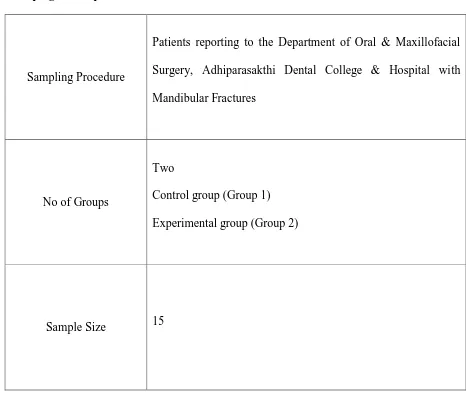
Sampling Procedure
Patients reporting to the Department of Oral & Maxillofacial Surgery, Adhiparasakthi Dental College & Hospital with Mandibular Fractures
No of Groups
Two
Control group (Group 1) Experimental group (Group 2)
[image:45.612.67.533.303.696.2]Sample Size 15
Patient Selection
Patients reporting to The Department of Oral & Maxillofacial Surgery, Adhiparasakthi Dental College & Hospital, Melmaruvathur 603 319, Tamilnadu with mandibular fractures will be included in this study.
Inclusion Criteria
- Patients between ages 18-60 years diagnosed with mandibular fractures - Dentate patients
- Both the sexes will be included - Keys of occlusion to be present
- Patients comes under ASA (American Society of Anaesthesiologists ) Type – I & II Exclusion Criteria
- Polytrauma Patients - ASA III & IV - Paediatric Patients - Edentulous Patients
- Fracture with mal-union/non-union/infected sites - Condylar & Coronoid Fractures
Materials
- 2. 0 mm titanium locking miniplates - 8 mm and 10 mm locking titanium screws. - 2. 0 mm conventional miniplates
Methodology
The study subjects were first monitored for any vital changes and any underlying medical conditions. If there were any abnormalities in the vitals, they were first addressed before any definitive management of the fractures could be addressed. In the case of presence of any active haemorrhage, the haemorrhage was first addressed. Detailed clinical examination was carried out both by inspection and palpation for all patients and the following parameters were noted.
- Facial asymmetry, occlusion, mouth opening, pain were assessed. - Radiographic evaluation of the fractured site.
Study subjects are divided into two groups.
- Group A - ORIF with conventional 2. 0 mm miniplates
- Group B - ORIF with titanium interlocking 2. 0 mm miniplates Following clinical and radiological diagnosis the following were done
- Placement of Erich’s arch bar ↓ LA. - Routine haematological investigations.
- Medical assessment of the patient by physician and anesthesiologists was done. - Informed consent obtained from patient prior to surgery.
Operative Procedure
- Patient intubated using Naso Endo Tracheal Intubation. - Painting of the surgical site using 5 % Povidone Iodine.
- Administration of 2 % Lignocaine with 1 : 2,00,000 Adrenaline using Local Infiltration at the site of the fracture.
- Incision marking.
- Layer by layer dissection.
- Exposure of fracture site & fracture reduction - Maxillomandibular fixation placed.
- Fixation of fractured segments using conventional miniplates or locking plates. - Irrigation of the surgical site with Normal Saline & Metronidazole.
- Layer by layer closure done using 3 – 0 vicryl in case of intra oral approach or 4 – 0 ethilon in case of extra oral approach.
- Extubation of the patient. Post Operative Care
- Control Group – MMF was placed for a period of three weeks. - Experimental Group – MMF was not placed.
- Patients were administered 1 gram of Cefotaxime 12th hourly and 500 mg of Metronidazole
8th hourly intravenously for three days.
- Following discharge, post operative instructions were given to the patient.
- Patients were advised to take 200 mg of Cefotaxime and 400 mg of Metronidazole orally for a period of five days.
Post Operative Evaluation
Each group was divided into six comparative stages - Pre Operative
- Immediate post-operative state - First week
- Sixth month
Evaluation of Parameters
Pain : Pain was evaluated using a 10 point VAS scale.
Mouth Opening : Maximal mouth opening was evaluated by measuring the inter incisal distance
Infection : Presence of pus discharge from the surgical wound was indicative of infection.
Wound Dehiscence : The wound healing was evaluated based on the presence or absence of wound gaping
Segmental Mobility : Segmental mobility was evaluated by bimanual palpation
Occlusion : Pre operative and post operative occlusion was evaluated.
Plate Exposure : Surgical site was evaluated for the presence or absence of plate exposure
Paresthesia Nerve paresthesia was evaluated using a pin prick test
Assessment of Reduction :
[image:49.612.71.543.90.654.2]The reduction of the fractured segments was evaluated by the surgeon, radiographically
A total of 30 patients, who met the inclusion criteria, were included and treated for mandibular fractures in the Department of Oral & Maxillofacial Surgery, Adhiparasakthi Dental College & Hospital, in this study. All the cases were diagnosed to have mandibular fractures after thorough clinical and radiological examination. 15 patients were allocated to Group A (Experimental Group) and 15 patients were allocated to Group B (Control Group). After informed consent was obtained all pre-operative parameters were measured. After completion of the hematological investigations and the radiological investigations, the case was operated and during the post-operative period all the parameters were again measured. The data collected were compiled using Microsoft Excel 2016 and was subjected to Statistical Analytical Tests, performed using IBM Corporation Statistical Package for Social Science, Version 22. 0 (Armonk, NY).
Fisher’s Exact Test, Mann Whitney U Test & Paired T Test were used to calculate the
statistical difference between the control and the experimental group. In all the tests conducted, a P value less than 0.05 was regarded as statistically significant. None of the patients reported any life threatening adverse events. A total of 30 patients were included in this study and they were split into two groups.
Control Group - 2. 0 mm Conventional Miniplates
Experimental Group - 2. 0 mm Locking Miniplates
1. Sex
In the control group, out of the 15 patients who were treated 14 were male (94 %) and 1 was female (6 %). In the experimental group, out of the 15 patients who were treated all 15 were male (100 %).
Chart 1 – Gender Distribution
2. Age
This study included patients between the age of 16 and 80. When the age distribution was analyzed the patients were grouped into five categories for easy analysis. They were divided into five groups namely16 – 20 Years, 21 – 30 Years, 31 – 40 Years, 41 – 50 Years, 51 – 60 Years.
When the age distribution was studied it was discovered that in the control group 26.7 % of the participants were in the age group of 16 – 20 years (n = 4), 33.3 % of the participants were in the age group of 21 – 30 years (n = 5), 13.3 % of the participants were in the 31 – 40 age group (n = 2), 20 % of the participants were in the age group 41 – 50 (n = 3) and 6.7 % of the participants
0 2 4 6 8 10 12 14 16
Male Female
Gender Distribution
belonged to the 51 – 60 age group (n =1). In the experimental group 6.7 % of the participants were in the age group of 16 – 20 years (n = 1), 40 % of the participants were in the age group of 21 – 30 years (n = 6), 33.3 % of the participants were in the 31 – 40 age group (n = 5), 6. 7 % of the participants were in the age group 41 – 50 (n = 1) and 13. 3 % of the participants belonged to the 51 – 60 age group (n =2).
Chart 2 – Age Distribution
The mean age of the participants in the control group was 30. 41 years with a standard deviation of 13. 30 years. The mean age of the participants in the experimental group was 34. 21 years with a standard deviation of 15. 13 years.
3. Etiology of Injury
Mandibular trauma can have varied etiology and in this particular study the etiology of the trauma was also elicited. In the control group, 85.5 % cases reported with mandibular fractures due to a road traffic accident (n = 13) and 14. 5 % of cases reported with mandibular fractures
0 1 2 3 4 5 6 7
16 - 20 21 - 30 31 - 40 41 - 50 51 - 60
Age Distribution
reported because of domestic violence (n = 2). In the experimental group, 86. 7 % of cases were due to road traffic accidents (n = 13), 6. 7 % of cases reported because of an accidental fall (n = 1) and 6. 7 % of cases reported because of domestic violence (n=1). The results obtained from this particular study showed that road traffic accidents were a leading cause of mandibular fractures.
Chart 3 – Etiology of Injury
4. Influence of Alcohol
In the control group, cases who reported with mandibular fractures as a result of a road traffic accident were examined further to determine whether or not the individual was under the influence of alcohol. In the control group, 13 out of 15 mandibular fractures were due to a road traffic accident, out of which 46. 7 % cases were under the influence of alcohol (n = 6) and 53. 3 % of cases were not under the influence of alcohol (n = 7). In the experimental group, 13 mandibular fractures were caused due to road traffic accidents. In these cases 66. 7 % of cases
0 2 4 6 8 10 12 14
RTA Fall Domestic Violence Assault Sports Injury
Etiology of Injury
were under the influence of alcohol (n = 10) and 33. 3 % of cases were not under the influence of alcohol. A strong predilection for consumption of alcohol and road traffic accidents was noted from the findings of this study.
Chart 4 – Influence of Alcohol
5. Usage of Helmet
In cases of road traffic accidents which were included in this prospective study, whether or not the patients were wearing a helmet at the time of the accident was noted. Out of the 13 case of road traffic accidents in the control group only 9. 7 % of the cases wore a helmet (n = 1) whereas 90. 3 % of cases did not wear a helmet (n = 12). In the experimental group 100 % of the mandibular
0 2 4 6 8 10 12
Yes No
Influence of Alcohol
fractures due to road traffic accidents were not using a helmet at the time of the accident (n = 13).
Chart 5 – Usage of Helmet
6. Time for Injury to Hospitalization
The time taken for the individual to report to the hospital following the injury was noted in this study. In the control group, 33. 3 % of cases reported within 24 hours of the accident (n = 5), 46. 7 % of the patients reported within 24 – 48 hours (n = 7), 13. 3 % of patients reported between 448 – 72 hours after the accident (n = 2) and 6. 7 % of patients reported later than 96 hours (n = 1). In the experimental group 26. 7 % of the study population reported to the Department of Oral & Maxillofacial Surgery within the first twenty four hours of the accident (n = 4), 60 % of the patients reported to the hospital between 24 – 48 hours (n = 9), 6. 7 % of the patients reported between 48 – 72 hours (n = 1) and 6. 7 % of the patients reported between 72 – 96 hours (n = 1).
0 2 4 6 8 10 12 14
Yes No
Usage of Helmet
Chart 6 – Time from Injury to Hospitalization
7. Location of Fracture Site
In both groups the location of the fracture site was noted according to Dingman & Natvig’s
Classification. In the Control Group, 53. 3 % of fractures occurred at the Parasymphysis region (n = 8), 13. 3 % of the fractures occurred at the body region (n = 2). 33. 3 % of fractures occurred at the region of the angle (n = 5). In the experimental group, 6. 7 % of fractures occurred at the symphysis region (n = 1), 60 % of fractures occurred at the site of the parasymphysis (n =9), 26. 7 % of the cases at the region of the body of the mandible (n = 4) and 6. 7 % of the fractures occurred at the region of the angle (n = 1). According to the findings from this study the most common site of the fracture was the Parasymphysis of the mandible. Fractures of the Parasymphysis also presented with fractures of the condyle of the mandible.
0 1 2 3 4 5 6 7 8 9 10
0 - 24 Hours 24 - 48 Hours 48 - 72 Hours 72 - 96 Hours > 96 Hours
Time from Injury To Hospitalization
Chart 7 – Location of Fracture Site
8. Favorable / Unfavorable Fractures
In both the groups whether or not the fracture was favorable was taken into account and the results were tabulated. A p value co-efficient lesser than 0. 05 was considered significant. In the control group 66. 7 % of cases were favorable fractures (n = 10) and 33. 3 % of cases were unfavorable fractures (n = 5). In the experimental group 71. 5 % of cases were favorable fractures (n = 11) and 28. 5 % of cases were unfavorable fractures (n = 4). Fisher’s exact test was used for statistical purposes.
0 1 2 3 4 5 6 7 8 9 10
Symphysis Parasymphysis Body Angle
Location of Fracture Site
Group
Fracture
Total p-Value Favorable Unfavorable
Control 10 5 15
Experimental 11 4 15 1.000*
[image:58.612.87.527.284.671.2]Total 21 9 30
Table 3 – Favorable / Unfavorable Fracture
Chart 8 – Favorable / Unfavorable Fractures 0
2 4 6 8 10 12
Favorable Unfavorable
Fracture
9. Approach
Some cases in this study were operated using an intra oral approach and some cases were operated using an extra oral approach. Fisher’s exact test was used for statistical purposes.
Group
Approach
Total P -Value Intra Oral Extra Oral
Control 11 4 15
Experimental 13 2 15 0.651*
[image:59.612.78.536.201.674.2]Total 24 6 30
Table 4 – Approach
Chart 9 – Approach 0
2 4 6 8 10 12 14
Intra Oral Extra Oral
Fracture
10.Time from Injury – Surgery
The time taken for the patient from the accident till the start of the surgery was calculated. In the control group the mean time was 118. 33 hours with a standard deviation of 62. 55 hours. In the experimental group, the mean time was 168. 60 hours with a standard deviation of 100. 09 hours.
11.Intra Operative Time
Intra operative time in this study was calculated as the time from the incision till the time the last suture was completed. In the control group the mean intra operative time was 108. 13 minutes with a standard deviation of 28. 83 minutes. In the experimental group the mean intra operative time was 85. 67 minutes with a standard deviation. Hence, the experimental group had significantly reduced intra operative times. This factor is in favor of the experimental group.
12.Hardware Cost
The total cost of the plates and the screws was recorded in this study and it was tabulated. In the control group the mean cost of the hardware used for the surgery was around Rs. 4,668. 13 with a standard deviation of Rs. 1883. 11. In the experimental group the mean cost of the hardware was around Rs. 3,113. 33 with a standard deviation of Rs. 776. 08. These parameters show that there is a significant difference in the cost of the hardware used for both groups. This parameter is in favor of the experimental group.
13.Pre Operative & Post Operative Pain
severity of pain between the groups. The pain was calculated using a ten point VAS scale and the results were documented as follows.
Pre Operative
1st Post Operative
Day
1st Post Operative
Week
1st Post Operative
Month
3rd Post Operative
Month
6th Post Operative
Month
Mean 6.67 7.53 6.40 3.80 1.87 .73
Median 7.00 8.00 6.00 4.00 2.00 .00
[image:61.612.71.543.134.369.2]Std. Deviation 1.397 .990 .986 1.265 1.598 1.387
Table 5 – Pain Control Group
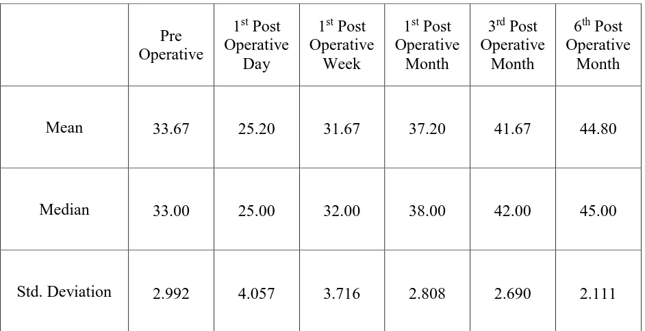
Pre Operative
1st Post
Operative Day
1st Post
Operative Week
1st Post
Operative Month
3rd Post
Operative Month
6th Post
Operative Month
Mean 6.07 6.73 5.00 2.73 1.20 .13
Median 6.00 7.00 5.00 3.00 1.00 .00
Std. Deviation 1.438 .961 1.069 1.100 .414 .352
[image:61.612.73.540.442.674.2]Chart 10 – Pain (10 Point VAS Scale)
0 1 2 3 4 5 6 7 8
PRE OP IM OP 1 WEEK 1 MONTH 3 MONTH 6 MONTH
Pain - VAS Scale
The pain scores in each group were compared to each other to find whether there was any statistical significance in the two groups using the Mann Whitney U Test. The results are tabulated as below.
Group Mean Rank P Value
Pre Operative
Control 16.90
0.372*
Experimental 14.10
1st Post Operative
Day
Control 18.83
0.031*
Experimental 12.17
1st Post Operative
Week
Control 20.87
0.000*
Experimental 10.13
1st Post Operative
Month
Control 18.80
0.041*
Experimental 12.20
3rd Post Operative
Month
Control 17.20
0.305*
Experimental 13.80
6th Post Operative
Month
Control 17.13
0.325*
[image:63.612.72.543.180.573.2]Experimental 13.87
Table 7 – Comparison of Pain between Control & Experimental Group