0022-538X/11/$12.00
doi:10.1128/JVI.00425-11
Copyright © 2011, American Society for Microbiology. All Rights Reserved.
Detection of Chronic Wasting Disease Prions in Salivary, Urinary,
and Intestinal Tissues of Deer: Potential Mechanisms of
Prion Shedding and Transmission
䌤
Nicholas J. Haley,
1Candace K. Mathiason,
1Scott Carver,
1Mark Zabel,
1Glenn C. Telling,
2and Edward A. Hoover
1*
Department of Microbiology, Immunology, and Pathology, College of Veterinary Medicine and Biomedical Sciences,
Colorado State University, Fort Collins, Colorado,
1and Department of Molecular Biology and Genetics,
University of Kentucky, Lexington, Kentucky
2Received 2 March 2011/Accepted 12 April 2011
Efficient horizontal transmission is a signature trait of chronic wasting disease (CWD) in cervids.
Infectious prions shed into excreta appear to play a key role in this facile transmission, as has been
demonstrated by bioassays of cervid and transgenic species and serial protein misfolding cyclic
amplifi-cation (sPMCA). However, the source(s) of infectious prions in these body fluids has yet to be identified.
In the present study, we analyzed tissues proximate to saliva, urine, and fecal production by sPMCA in an
attempt to elucidate this unique aspect of CWD pathogenesis. Oropharyngeal, urogenital, and
gastroin-testinal tissues along with blood and obex from CWD-exposed cervids (comprising 27 animals and >350
individual samples) were analyzed and scored based on the apparent relative CWD burden. PrP
CWD-generating activity was detected in a range of tissues and was highest in the salivary gland, urinary
bladder, and distal intestinal tract. In the same assays, blood from the same animals and unseeded normal
brain homogenate controls (
n
ⴝ
116 of 117) remained negative. The PrP-converting activity in peripheral
tissues varied from 10
ⴚ11- to 10
0-fold of that found in brain of the same animal. Deer with highest levels
of PrP
CWDamplification in the brain had higher and more widely disseminated prion amplification in
excretory tissues. Interestingly, PrP
CWDwas not demonstrable in these excretory tissues by conventional
Western blotting, suggesting a low prion burden or the presence of protease-sensitive infectious prions
destroyed by harsh proteolytic treatments. These findings offer unique insights into the transmission of
CWD in particular and prion infection and trafficking overall.
Chronic wasting disease (CWD) is an efficiently transmitted
prion disease of cervids (e.g., deer, elk, and moose) and is the
only known prion disease affecting free-ranging, nondomestic
animals. While the origins of CWD are uncertain, the disease
has been present in wild cervid populations of northern
Colo-rado and southern Wyoming for over 40 years (56, 57) and has
now been identified in both captive and free-ranging cervids in
17 states, 2 Canadian provinces, and the Republic of Korea
(44). With intensified national surveillance efforts, CWD
con-tinues to be identified in areas previously thought to be free of
infection, including recent discoveries in Virginia and Missouri
(20, 21). The prevalence of CWD varies across North America
but can be as high as 30% in some areas of Colorado or up to
100% in captive populations (23).
While much remains to be learned about the mechanisms of
horizontal CWD transmission, the environment and
environ-mental fomites were previously shown to be capable of
har-boring infectious prions in both outdoor and, later, indoor
cervid housing facilities (31, 33). Our previous work
demon-strated that both saliva and blood from CWD-positive
(CWD
⫹) animals contained infectious prions able to transmit
infection to naïve white-tailed deer (32). Subsequently, the
application of a transgenic mouse bioassay and serial protein
misfolding cyclic amplification (sPMCA) to saliva, urine, and
fecal samples from infected cervids and to deer inoculated with
urine and feces has demonstrated lower levels of infectious
prions in these excreta (17, 18, 50).
The identification of infectious prions in excreta has shed
light on the transmission and pathogenesis of CWD; however,
the proximal source of the excreted prions remains elusive, i.e.,
whether they are produced in organs of excretion or,
alterna-tively, are derived from central nervous or lymphoid tissues
and excreted into bodily fluids and feces, questions pertinent to
our understanding of prion transmission and pathogenesis in
nature.
Limited evidence exists for protease-resistant,
disease-asso-ciated forms of the prion protein (PrP
RES/Sc/d) in glandular
epithelial and excretory tissues. However, researchers have
reported the presence of PrP
RESby immunohistochemistry
(IHC), paraffin-embedded tissue (PET) blotting, or Western
blotting (WB) in the kidneys of scrapie-affected sheep (28, 35,
46, 47) and CWD
⫹deer (19), the lingual tissues of hamsters
inoculated with the HY (hyper) strain of transmissible mink
encephalopathy (10, 34), and the salivary glands of
scrapie-affected sheep (55) and CWD-infected cervidized transgenic
mice (43). While the identification of protease-resistant prion
proteins in cervid excretory tissues using conventional assays
* Corresponding author. Mailing address: Department of
Microbi-ology, ImmunMicrobi-ology, and PathMicrobi-ology, College of Veterinary Medicine
and Biomedical Sciences, Colorado State University, Fort Collins, CO
80523. Phone: (970) 491-7587. Fax: (970) 491-0523. E-mail: Edward
.Hoover@colostate.edu.
䌤
Published ahead of print on 27 April 2011.
6309
on November 7, 2019 by guest
http://jvi.asm.org/
has proven difficult (2, 13, 14, 24, 49), bioassay studies
identi-fying genuine prion infectivity in excretory tissues, albeit
lim-ited, date to the 1960s (16) and implicate both the salivary
glands of Creutzfeldt-Jakob disease (CJD)-infected mice (41)
and kidneys of scrapie-infected mice (12). sPMCA (38, 48)
now offers a more time- and animal-efficient means to detect
low levels of infectious prions and thereby reexamine questions
and mechanisms of prion excretion and transmission.
In the present study, we have applied standardized sPMCA
to examine tissues associated with saliva, urine, and fecal
ex-cretion (e.g., salivary glands, tongue, nasal mucosa, kidney,
ureter, urinary bladder, and intestinal tract) from 27
CWD-exposed versus naïve white-tailed deer to gain insight into the
potential peripheral mechanisms underlying the facile
horizon-tal transmission of chronic wasting disease.
MATERIALS AND METHODS
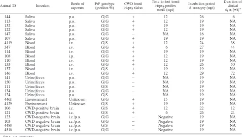
Infected cervids.Twenty-seven white-tailed deer (Odocoileus virginianus) were exposed to CWD from positive and negative sources in various forms (e.g., urine and feces, saliva, environmental fomites, blood, or brain tissue) and by various routes (e.g., orally [p.o.], intravenously [i.v.], intraperitoneally [i.p.], intracranially [i.c.], or through environmental exposure) (30–32). All animals were inoculated and maintained in dedicated, restricted-access, indoor CWD research facilities in accordance with Colorado State University IACUC guidelines. The sources of
inoculum included terminally ill mule deer (Odocoileus hemionus) of an
un-known PrP genotype (courtesy of Michael Miller, Colorado Division of Wildlife) or white-tailed deer utilized in previous and concurrent studies of either of two genotypes: homozygous for glycine (i.e., G/G) at cervid PrP position 96 or heterozygous at that position, with alleles for both glycine and serine (G/S). The deer were monitored for 16 to 27 months postinoculation (mpi), during which time the majority became tonsil biopsy positive for CWD by immunohistochem-istry (IHC). The duration of clinical disease prior to necropsy ranged from 0
weeks (i.e., no clinical signs) to 72 weeks. At scheduled necropsy dates, or when exhibiting signs of terminal disease, deer were euthanized and subjected to an extensive necropsy using fresh instruments and collection vessels for each indi-vidual, at which time bodily fluids and an array of tissues were collected and
frozen at⫺80°C. Animals that were not tonsil biopsy positive for CWD prior to
euthanasia were thoroughly evaluated for CWD infection after necropsy, includ-ing an evaluation of neural and lymphoid tissues, which have been shown to be early diagnostic sites for CWD (e.g., obex, retropharyngeal lymph nodes, and tonsils) by IHC and Western blotting (WB). A complete description of animals, inoculation routes and sources, CWD status, time of first tonsil biopsy specimen positivity in mpi, incubation period at necropsy (in mpi), and duration of clinical signs (in weeks) is shown in Table 1.
Study samples and preparation.Obex, parotid salivary gland, tongue, nasal respiratory epithelium, kidney, ureter, urinary bladder, proximal and distal
in-testinal tract, and blood were collected at necropsy and frozen at⫺80°C. Tissue
samples were thawed briefly, and 10 to 50 mg of each sample was trimmed individually and homogenized as a 1% (wt/vol) solution in PMCA buffer (1% [vol/vol] Triton X-100, 5 mM EDTA, and 150 mM NaCl in phosphate-buffered saline [PBS] adjusted to a pH of 7.2) by using a FastPrep tissue homogenizer for 60 s at power setting 6.5. Whole-blood samples remained suspended in 5% EDTA until sPMCA evaluation. All tissues were prepared in individual micro-centrifuge tubes and homogenized in parallel in the same machine concurrent with both positive and negative controls. Prepared homogenates were coded to allow for blinded evaluation by sPMCA.
Western blotting of peripheral tissues.Peripheral tissue homogenates,
pre-pared as described above, were blindly analyzed for the presence of PrPCWD
using conventional Western blotting. Fifteen microliters of the 1% sample
ho-mogenate was mixed with 7l of sample buffer (0.1% [vol/vol] Triton X-100 and
4% [wt/vol] SDS in PBS); pretreated with 2l collagenase A, 2l dispase II, and
1l DNase I (Boehringer-Mannheim, Germany) (final concentrations, 1 mg/ml,
1 mg/ml, and 0.05 mg/ml, respectively) for 30 min at 37°C; and ultimately
digested with 3l proteinase K at 500g/ml (Invitrogen) (final concentration, 60
g/ml) for 30 min at 37°C. Ten microliters of 4⫻running buffer was then added
to the sample, followed by denaturation for 3 min at 95°C. Twenty microliters of this preparation was run on a precast 12% SDS-PAGE gel (Invitrogen) in a Bio-Rad electrophoresis apparatus for 1 h at 160 mV. Samples were then
trans-TABLE 1. Summary of source animals with tissues evaluated by sPMCA
Animal ID Inoculum Route of
exposure
PrP genotype (position 96)
CWD tonsil biopsy status
Time to first tonsil biopsy-positive
result (mpi)
Incubation period at necropsy (mpi)
Duration of clinical
signs (wk)a
144
Saliva
p.o.
G/G
⫹
12
26
6
113
Saliva
p.o.
G/G
⫹
12
19
NA
132
Saliva
p.o.
G/G
⫹
19
19
NA
122
Saliva
p.o.
G/G
⫹
12
19
NA
147
Saliva
p.o.
G/G
⫺
NA
16
NA
107
Saliva
p.o.
G/G
⫹
19
19
NA
4119
Blood
i.v.
G/S
⫹
12
24
38
347
Blood
i.v.
G/G
⫹
6
27
44
114
Blood
i.v.
G/G
⫹
19
19
NA
108
Blood
i.p.
G/G
⫹
12
19
NA
110
Blood
i.v.
G/G
⫹
12
19
NA
133
Blood
i.v.
G/G
⫹
12
26
50
137
Blood
i.v.
G/S
⫹
19
19
NA
146
Blood
i.v.
G/G
⫹
12
29
72
141
Urine/feces
p.o.
G/G
⫺
NA
19
NA
150
Urine/feces
p.o.
G/G
⫺
NA
19
NA
111
Urine/feces
p.o.
G/S
⫺
NA
19
NA
134
Urine/feces
p.o.
G/G
⫺
NA
19
NA
124
Urine/feces
p.o.
G/S
⫺
NA
19
NA
4461
Environment
Unknown
G/G
⫹
15
19
NA
4129
Environment
Unknown
G/S
⫹
19
19
NA
106
CWD-positive brain
i.c.
G/S
⫹
12
22
12
121
CWD-positive brain
i.c.
G/S
⫹
6
26
24
123
CWD-negative brain
i.c./p.o.
G/G
⫺
Negative
19
NA
103
CWD-negative brain
i.c./p.o.
G/G
⫺
Negative
19
NA
4488
CWD-negative brain
i.c./p.o.
G/S
⫺
Negative
19
NA
4516
CWD-negative brain
i.c./p.o.
G/G
⫺
Negative
19
NA
aNA, not applicable.
on November 7, 2019 by guest
http://jvi.asm.org/
[image:2.585.42.541.84.363.2]ferred onto a polyvinylidene difluoride (PVDF) membrane (Millipore) for 1 h at 115 mV in a Bio-Rad transfer apparatus. PVDF membranes were subsequently probed with a PrP-specific monoclonal antibody (BAR224) conjugated to horse-radish peroxidase, diluted 1:20,000 in 5% (wt/vol) powdered milk in 0.2% Tween 20 in Tris-buffered saline (TBST) for 1 h. Following washing with TBST, immu-noreactivity was detected by using an enhanced chemiluminescence detection system (ECL-Plus; Amersham Biosciences) with an LAS 3000 imaging system (Fuji Inc., Valhalla, NY).
Preparation of normal brain homogenate for sPMCA.Normal brain
homog-enate (NBH), the substrate for prion conversionin vitro, was prepared from
Tg(CerPrP)5037mice in a room that had not previously been used for prion research (18, 26). Following euthanasia and perfusion with 5 mM EDTA in PBS,
whole brain was collected from naïveTg(CerPrP)5037mice and placed on ice.
Brain homogenates were prepared as a 10% (wt/vol) solution in PMCA buffer with the addition of Complete protease inhibitors (Roche Pharmaceuticals, In-dianapolis, IN) using a Dounce homogenizer. Homogenates were then centri-fuged for 1 min at 2,000 rpm, and the supernatant was frozen in
single-experi-ment aliquots at⫺80°C in a “prion-free” room until use for sPMCA. Each
preparation was composed of brains from 4 to 6 mice to minimize the potential influence of expression variation in CerPrP or other cofactors (1, 7); multiple batches of NBH were utilized over the course of the experiments.
sPMCA of tissues.Tissue homogenates from CWD-exposed deer were blindly
assayed for PrPCWD, in duplicate, by sPMCA. All test samples were prepared in
parallel with tissue-matched positive and negative controls as a 1% homogenate in PMCA buffer, as described above, and subsequently spiked into normal brain homogenate for amplification (17, 18, 25, 26). Ten microliters of test or control
tissue homogenate was added to 50l of NBH and assayed, in parallel and in
adjacent wells of a 96-well plate (USA Scientific, Ocala, FL), along with normal
brain homogenate prepared from unexposedTg(CerPrP)5037mice as additional,
unseeded negative controls. Plates were then sonicated by using an ultrasonic processor (Misonix, Farmingdale, NY) and incubated at 37°C. Sonication pa-rameters were set at 40-s bursts at power level 7.0, followed by 30 min of incubation. Ninety-two cycles of sonication were performed over 48 h, with a
10-l aliquot of sonicated material transferred into 50l of fresh NBH for serial
amplification. Following each round of amplification, samples were evaluated by
Western blotting, as described below, for the presence of PrPCWD
. Each sample received a score based on the number of rounds in which that particular sample was positive (i.e., a maximum of 3 in a three-round experiment), and the scores for each duplicate run were totaled to arrive at a final score, with “0” being the lowest and “6” being the highest score that a given sample could receive. In each experimental run, between 25 and 50% of samples evaluated were tissue-matched negative controls. Additionally, 10 to 20% of the samples in a run were unspiked, NBH negative controls.
sPMCA of blood.EDTA-preserved whole-blood samples from CWD-exposed deer were blindly analyzed in duplicate as described above for tissues, with some modifications. Using a protocol initially described by Tattum and colleagues (51),
we spiked 1l of whole blood into 100l of normal brain homogenate. Sixty
microliters of this preparation was added to a 96-well plate and analyzed by sPMCA. Sonication parameters were set at 20-s bursts at power level 7.5, fol-lowed by 30 min of incubation at 37°C. One hundred forty-four cycles of
soni-cation were performed over 72 h, with a 10-l aliquot of sonicated material
transferred into 50l of fresh NBH for serial amplification. Each sample was
evaluated by WB as described below for PrPCWDsignals following each round of
amplification.
sPMCA of obex dilution series and approximation of tissue scores.Obex tissue dilutions from each deer, collected and processed as described above, were made by diluting each of the initial 1% obex homogenates into 10% normal
Tg(CerPrP)5037brain homogenate (NBH) at 1:10 dilutions ranging from 100
to
10⫺14(i.e., final dilutions of 10⫺2to 10⫺16). Prior to dilution, the initial 1% obex
homogenates were sonicated for 20 s at power setting 7.0 in an ultrasonic
processor. Ten microliters of each dilution was added to 50l of NBH and
analyzed for three rounds of sPMCA as described above for tissues. Each dilu-tion then received an sPMCA score as described above for peripheral tissues; these scores were then used to estimate the relative log obex dilution equivalent (LODE) scores for individual tissues within animals. Tissue LODE scores were conservatively assigned based on the lowest obex dilution yielding a given score.
[image:3.585.47.538.90.376.2]WB of sPMCA-amplified tissues.Following each round of sPMCA, an aliquot of each sonicated sample was subjected to Western blotting for the evaluation of
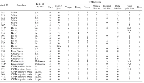
TABLE 2. Summary of sPMCA results for blood and tissues associated with production and excretion of saliva, urine,
and feces in CWD-exposed deer
aAnimal ID Inoculum Route of
exposure
sPMCA score
Obex Salivary
gland Tongue Kidney Ureter
Urinary bladder
Proximal intestine
Distal intestine
Nasal
epithelium Blood
144
Saliva
p.o.
6
6
2
0
2
6
2
5
3
0
113
Saliva
p.o.
6
6
0
0
0
5
2
3
0
0
132
Saliva
p.o.
6
0
0
0
0
3
2
3
1
0
122
Saliva
p.o.
6
0
0
5
0
1
1
3
0
0
147
Saliva
p.o.
4
0
0
1
0
0
0
0
0
0
107
Saliva
p.o.
4
3
1
0
0
0
1
1
3
0
4119
Blood
i.v.
6
6
2
0
5
4
2
6
NA
0
347
Blood
i.v.
4
2
0
2
0
0
1
1
NA
0
114
Blood
i.v.
6
5
0
4
2
6
1
2
2
0
108
Blood
i.p.
3
0
0
0
0
0
0
2
1
0
110
Blood
i.v.
6
6
3
5
3
6
1
2
3
0
133
Blood
i.v.
6
6
6
5
4
1
2
4
1
0
137
Blood
i.v.
4
0
0
0
0
1
0
1
1
0
146
Blood
i.v.
6
NA
1
5
2
6
1
4
2
0
141
Urine/feces
p.o.
4
0
0
0
0
0
0
0
1
0
150
Urine/feces
p.o.
4
0
0
0
0
0
0
0
0
0
111
Urine/feces
p.o.
3
0
0
0
0
1
1
1
1
0
134
Urine/feces
p.o.
4
0
0
1
0
0
0
0
0
0
124
Urine/feces
p.o.
0
0
0
1
0
0
0
0
1
0
4461
Environment
Unknown
6
0
4
3
4
6
3
5
NA
0
4129
Environment
Unknown
3
0
0
0
0
0
0
1
NA
0
106
CWD-positive brain
i.c.
6
5
0
0
0
4
3
4
5
0
121
CWD-positive brain
i.c.
6
6
0
0
1
6
2
4
NA
0
123
CWD-negative brain
i.c./p.o.
0
0
0
0
0
0
0
0
0
0
103
CWD-negative brain
i.c./p.o.
0
0
0
1
0
0
0
0
0
0
4488
CWD-negative brain
i.c./p.o.
0
0
0
0
0
0
0
0
0
0
4516
CWD-negative brain
i.c./p.o.
0
0
0
0
0
0
0
0
0
0
aAcross individuals, all tissues were variously positive, with scores ranging from 0 to 6. Surprisingly, scores for peripheral tissues occasionally rivaled or exceeded
those of the obex (i.e., salivary gland and urinary bladder of deer 144 and tongue and ureter of deer 4461). NA, sample not available.
on November 7, 2019 by guest
http://jvi.asm.org/
the PrPCWDsignal. Fifteen microliters of sample homogenate was mixed with 7
l of sample buffer (0.1% [vol/vol] Triton X-100 and 4% [wt/vol] SDS in PBS),
digested with 3l proteinase K at 500g/ml (final concentration, 60g/ml) for
20 min at 37°C followed by 10 min at 45°C, and analyzed for PrPCWDusing the
Western blotting protocol described above.
Evaluation of intrarun variability in duplicate experiments.As all samples were run in duplicate on two different sonicating machines, we sought to com-pare the intrarun variabilities in experimental results. A commercially available calculator for the categorical Cohen’s kappa value was used to define the agree-ment of results between the two different experiagree-ments (28a). Because scores progressed linearly in the sPMCA evaluation, a linear weighted approach was used to calculate Cohen’s kappa. The significance of the correlation was deter-mined by using a method described previously by Landis and Koch (27).
Statistical analysis of data.To broadly analyze the factors affecting the
pe-ripheral PrPCWD
distribution and intensity, we used a permutational-based mul-tivariate analysis of variance (PERMANOVA). We used this test to evaluate (i)
the effect of the obex score on the PrPCWDdistribution and intensity among
peripheral tissues and (ii) the relationship between the source and route of
inoculum on the PrPCWD
distribution and intensity among peripheral tissues. We conducted these analyses on two presentations of our data: a semiquantitative scoring measure (raw scoring of sPMCA intensity) and a more quantitative
measurement of sPMCA intensity relative to the amount of amplifiable PrPCWD
found in the brain of that animal (LODE score or relative PrPCWD
burden).Post
hocanalyses were used to evaluate pairwise comparisons of treatment groups
within each PERMANOVA. As the distribution of PrPCWDintensity (measured
by sPMCA scores) across tissues was fairly limited (ranging from 0 to 6 for the
raw sPMCA score and 0 to 10 for the LODE score, i.e., relative PrPCWD
burden), we did not transform the data prior to PERMANOVAs. PERMANOVAs were based on Bray-Curtis similarity matrices and were graphically represented using nonmetric multidimensional scaling (NMDS) plots.
RESULTS
[image:4.585.98.484.67.366.2]In an effort to determine the proximal source of CWD
pri-ons in saliva, urine, and feces, and to add to our understanding
of CWD pathogenesis, we blindly evaluated, in duplicate, a
range of organs and tissues associated with saliva, urine, and
fecal production and excretion for PrP
CWDamplification using
a standardized sPMCA assay. Tissues were collected at
nec-ropsy from 27 deer experimentally exposed to various sources
of CWD by several different routes (Table 1). These tissues
were then scored based on the number of rounds
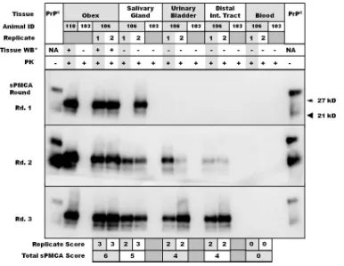
demonstrat-FIG. 1. Representative Western blots of replicate salivary gland, urinary bladder, distal intestinal tract, and blood samples from study deer.
Obex samples from deer 110 and 103 are used as positive and negative controls, respectively, while replicate tissues are those of deer 106.
Tissue-matched negative controls from deer 103 were included. Tissue replicate scores appear below the Western blots, with cumulative scores
summed to derive sPMCA scores (e.g., deer 106 obex, “6”; salivary gland, “6”).
ⴱ
Tissues were Western blotted prior to amplification to determine
whether PrP
CWDcould be identified in unamplified homogenates. PK, proteinase K.
TABLE 3. Summary of scores achieved in duplicate
sPMCA experiments
aSonicator A score
No. of samples with sonicator B score of:
0 1 2 3
0
220
13
0
1
1
9
9
3
1
2
3
3
9
3
3
0
0
5
24
a
Tissues were evaluated in duplicate on two different sonicators, sonicators “A” and “B,” receiving a score ranging from 0 to 3 in each replicate. Corre-sponding scores from duplicated experiments were tallied and evaluated
cate-gorically to derive Cohen’s kappa value (0.80⫾0.031).
on November 7, 2019 by guest
http://jvi.asm.org/
[image:4.585.299.541.621.692.2]ing amplification; individual tissue scores were then grouped
according to the source of inoculum and correlated with
intra-animal obex dilution scores.
Substantial CWD prion/PrP
CWD-amplifying activity
de-tected in excretory tissues by sPMCA.
Salivary glands, tongue,
nasal mucosa, kidney, ureter, urinary bladder, and both
prox-imal and distal intestinal tract were assayed for evidence of
prion amplification activity by a standard three-round sPMCA
protocol. Tissues were then assigned a score based on the total
number of positive rounds in each of two replicates. In total,
over 300 individual samples were evaluated, including 118
neg-ative-control samples. Animals were initially grouped based on
inoculum source (e.g., saliva, blood, urine, and feces); specific
tissue scores for each of these groups were averaged to
deter-mine the mean tissue score for each group. In exposed animals,
PrP
CWDwas amplified to various degrees in all tissues
evalu-ated but was most predominant in salivary gland, urinary
blad-der, and distal intestinal tract (Table 2 and Fig. 1; see also Fig.
4). Amplification scores ranged from 1 to 6; animals inoculated
i.v. or i.p. with whole blood appeared to exhibit the most
widespread distribution of PrP
CWD-amplifying activity, while
those inoculated orally with either saliva or urine and feces
demonstrated a more limited distribution. Interestingly, the
amplification activity from some peripheral tissues, such as
salivary gland and urinary bladder, rivaled that observed for
the obex of the same animal (e.g., salivary gland and urinary
bladder from deer 144 and urinary bladder from deer 4461,
etc.) (Table 2). Among 118 negative-control samples, a single
false-positive result was identified for a kidney section from
deer 103. This particular sample was evaluated, in duplicate, in
four separate experiments and received a total score of “1” in
a single sPMCA experiment; no amplification was observed
in the three remaining experiments.
[image:5.585.41.541.70.400.2]Despite clear evidence that blood and its components
har-bor infectious CWD prions (30, 32), an effective sPMCA
pro-tocol has not yet been identified for amplifying PrP
CWDfrom
blood. sPMCA protocols have been described for the
amplifi-cation of scrapie from the blood of hamsters and mice (e.g., see
references 7a and 34a); however, for the present study we
chose a protocol that was recently shown to be effective for
sheep scrapie (51). In three rounds of sPMCA, all 27
whole-blood samples from both CWD-exposed and -naïve deer
re-mained negative for PrP
CWDamplification (Fig. 1 and Table 2)
despite the amplification of concurrent CWD
⫹brain positive
controls. Thus, we surmised that it was unlikely that blood
contamination was responsible for the positive data obtained
for the tissue homogenates described above. These findings do
not, however, rule out the feasibility of sPMCA to detect
PrP
CWDin further rounds of sPMCA or using protocol
mod-ifications not employed in the present studies.
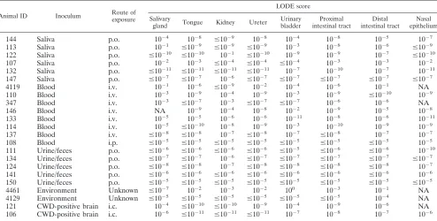
FIG. 2. Representative LODE scores for deer inoculated with saliva orally (deer 144) (A), blood intravenously (deer 137) (B), urine and feces
orally (deer 111) (C), and brain intracranially (deer 106) (D). Peripheral excretory tissue sPMCA scores of individual deer were conservatively
translated into LODE scores based on the lowest equivalent obex dilution (from the source deer) yielding that particular sPMCA score.
on November 7, 2019 by guest
http://jvi.asm.org/
Individual intrarun sPMCA scores show a high level of
agreement.
To determine the level of agreement between
scores assigned for each of two sPMCA replicate experiments,
we used a commercially available calculator to determine the
categorical Cohen’s kappa value. The sum of results available
from blood and tissue sPMCA experiments was analyzed, and
a summary of the categories and scores achieved in duplicate
runs on sonicators “A” and “B” are presented in Table 3.
Because the progression of scores for each experiment is a
linear one (i.e., from 0 to 3), the kappa value was weighted
linearly. The observed kappa value for the sum of sPMCA
experiments was 0.80 (standard error [SE], 0.031; 95%
confi-dence interval [CI], 0.74 to 0.87), which is considered
substan-tial to near-perfect agreement according to guidelines
pro-posed by Landis and Koch (27).
Peripheral versus central nervous system (brain) prion
am-plification in individual animals.
Serial dilutions of 1% brain
(obex) homogenates, ranging from 10
0to 10
⫺14(i.e., final
dilutions of 10
⫺2to 10
⫺16), from each study deer were
evalu-ated in the standardized three-round PMCA assay, with each
dilution assigned a score as described above for tissues.
Indi-vidual peripheral/excretory tissues were then correlated with
the lowest obex dilution receiving that particular score and
assigned a log obex dilution equivalent (LODE) score (Fig. 2
and Table 4). Tissues that failed to amplify PrP
CWDin three
rounds of sPMCA were assigned a LODE score less than or
equal to the first obex dilution that failed to amplify CWD
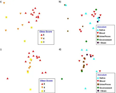
prions by sPMCA. The apparent peripheral level and
distribu-tion were dependent primarily on the central nervous system
burden (Fig. 3a and c and 4), while the inoculum source and
route of inoculation did not significantly correlate with the
peripheral burden (Fig. 3b and d and 4).
Peripheral PrP
CWDintensity and distribution correlate with
central nervous system burden.
The distribution and intensity
of PrP
CWDwere significantly related to the obex score for both
presentations of our data (
F
2,19⫽
6.2 and
P
⫽
0.0001 for the
raw sPMCA score;
F
2,19⫽
2.3 and
P
⫽
0.042 for the relative
PrP
CWDburden [determined by PERMANOVA]) (Fig. 3a and
c). Individuals with an obex score of 6 differed from individuals
with scores of 3 or 4 (
P
value of
⬍
0.001 in both cases for the
scoring measure;
P
⫽
0.05 and 0.08, respectively, for the
rela-tive PrP
CWDburden [determined by a
post hoc
test]), but the
distribution and intensity of PrP
CWDamong the latter two
groups did not differ from one another (
P
⫽
0.44 for raw
sPMCA score;
P
⫽
0.17 for relative PrP
CWDburden
[deter-mined by a
post hoc
test]). What is also apparent in these analysis
is that as the obex score increased, there was generally less
indi-vidual variation in peripheral PrP
CWDaccumulation (as
repre-sented by a more tight clustering of individuals with an obex score
of 6 than of individuals with an obex score of 3 or 4) (Fig. 3a and
c). The peripheral distribution and intensity of PrP
CWD,
con-versely, were not significantly related to the source of the
inocu-lum (
F
4,17⫽
1.67 and
P
⫽
0.072 for the raw sPMCA score;
F
4,17⫽
1.42 and
P
⫽
0.185 for the relative PrP
CWDburden
[determined by PERMANOVA]) (Fig. 3b and d).
DISCUSSION
[image:6.585.45.539.82.332.2]Among the transmissible spongiform encephalopathies
(TSEs), chronic wasting disease is unique in its high level of
TABLE 4. Summary of individual tissue LODE scores for CWD-infected deer
aAnimal ID Inoculum Route of
exposure
LODE score Salivary
gland Tongue Kidney Ureter
Urinary bladder
Proximal intestinal tract
Distal intestinal tract
Nasal epithelium
144
Saliva
p.o.
10
⫺410
⫺8ⱕ
10
⫺910
⫺810
⫺410
⫺810
⫺510
⫺7113
Saliva
p.o.
10
⫺1ⱕ
10
⫺9ⱕ
10
⫺9ⱕ
10
⫺910
⫺310
⫺810
⫺6ⱕ
10
⫺9122
Saliva
p.o.
ⱕ
10
⫺10ⱕ
10
⫺1010
⫺1ⱕ
10
⫺1010
⫺910
⫺910
⫺7ⱕ
10
⫺10107
Saliva
p.o.
10
⫺210
⫺3ⱕ
10
⫺4ⱕ
10
⫺4ⱕ
10
⫺410
⫺310
⫺310
⫺2132
Saliva
p.o.
ⱕ
10
⫺11ⱕ
10
⫺11ⱕ
10
⫺11ⱕ
10
⫺1110
⫺710
⫺1010
⫺710
⫺11147
Saliva
p.o.
ⱕ
10
⫺7ⱕ
10
⫺710
⫺6ⱕ
10
⫺7ⱕ
10
⫺7ⱕ
10
⫺7ⱕ
10
⫺7ⱕ
10
⫺74119
Blood
i.v.
10
⫺110
⫺6ⱕ
10
⫺910
⫺210
⫺410
⫺610
⫺1NA
110
Blood
i.v.
10
⫺310
⫺910
⫺410
⫺910
⫺310
⫺9ⱕ
10
⫺1010
⫺9347
Blood
i.v.
10
⫺3ⱕ
10
⫺710
⫺3ⱕ
10
⫺7ⱕ
10
⫺710
⫺610
⫺6NA
146
Blood
i.v.
NA
10
⫺910
⫺410
⫺810
⫺210
⫺910
⫺510
⫺8133
Blood
i.v.
10
⫺510
⫺510
⫺610
⫺610
⫺1110
⫺810
⫺610
⫺11114
Blood
i.v.
10
⫺5ⱕ
10
⫺1010
⫺810
⫺910
⫺310
⫺1010
⫺910
⫺9137
Blood
i.v.
ⱕ
10
⫺8ⱕ
10
⫺810
⫺7ⱕ
10
⫺810
⫺7ⱕ
10
⫺810
⫺710
⫺7108
Blood
i.p.
ⱕ
10
⫺5ⱕ
10
⫺5ⱕ
10
⫺5ⱕ
10
⫺5ⱕ
10
⫺5ⱕ
10
⫺5ⱕ
10
⫺510
⫺5111
Urine/feces
p.o.
ⱕ
10
⫺6ⱕ
10
⫺6ⱕ
10
⫺6ⱕ
10
⫺6ⱕ
10
⫺5ⱕ
10
⫺6ⱕ
10
⫺610
⫺10134
Urine/feces
p.o.
ⱕ
10
⫺7ⱕ
10
⫺710
⫺6ⱕ
10
⫺7ⱕ
10
⫺7ⱕ
10
⫺7ⱕ
10
⫺7ⱕ
10
⫺7124
Urine/feces
p.o.
ⱕ
10
⫺8ⱕ
10
⫺810
⫺7ⱕ
10
⫺8ⱕ
10
⫺8ⱕ
10
⫺8ⱕ
10
⫺810
⫺7141
Urine/feces
p.o.
ⱕ
10
⫺6ⱕ
10
⫺6ⱕ
10
⫺6ⱕ
10
⫺6ⱕ
10
⫺6ⱕ
10
⫺6ⱕ
10
⫺610
⫺6150
Urine/feces
p.o.
ⱕ
10
⫺5ⱕ
10
⫺5ⱕ
10
⫺5ⱕ
10
⫺5ⱕ
10
⫺5ⱕ
10
⫺5ⱕ
10
⫺5ⱕ
10
⫺54461
Environment
Unknown
ⱕ
10
⫺710
⫺210
⫺310
⫺210
010
⫺310
⫺1NA
4129
Environment
Unknown
ⱕ
10
⫺5ⱕ
10
⫺5ⱕ
10
⫺5ⱕ
10
⫺5ⱕ
10
⫺5ⱕ
10
⫺510
⫺4NA
121
CWD-positive brain
i.c.
10
⫺4ⱕ
10
⫺10ⱕ
10
⫺1010
⫺910
⫺410
⫺910
⫺6NA
106
CWD-positive brain
i.c.
10
⫺6ⱕ
10
⫺11ⱕ
10
⫺11ⱕ
10
⫺1110
⫺710
⫺810
⫺710
⫺6a
Serial PMCA scores were initially determined for obex dilutions from individual animals. Tissues from the respective animals were then conservatively assigned a
score based on the equivalent log obex dilution equivalent (LODE). Tissues that failed to amplify PrPCWD
in three rounds received a LODE score equal to or less than
the first obex dilution of the source animal scoring a “0.” These findings suggest that peripheral levels vary widely compared to PrPCWD
levels in the central nervous system but on occasion may approach levels seen for the obex of infected animals.
on November 7, 2019 by guest
http://jvi.asm.org/
transmissibility and, thus, prevalence in free-ranging
popula-tions. Here we report the presence of substantial
prion-con-verting activity in excretory tissues proximate to the saliva,
urine, and feces shed by CWD-infected deer. While these
results do not speak to infectivity, our previous research has
shown that conventional test-negative, sPMCA-positive tissues
and bodily fluids may indeed harbor infectivity (17, 18). The
use of sPMCA as a surrogate for a bioassay to detect PrP
CWDin excretory tissues may help to explain the efficient horizontal
transmission of CWD but also raises questions regarding the
source and mechanism of prion shedding from glandular and
mucosal tissues.
Historical cross-sectional studies of cervid herds where
CWD is endemic have largely failed to demonstrate, by
con-ventional immunohistochemistry or Western blotting,
pro-tease-resistant prions in organs involved in the production and
excretion of saliva, urine, or feces (2, 14, 49). These assays rely
on proteolytic, heat, and/or formic acid treatments, practices
which may preclude the identification of recently described
protease-sensitive forms of infectious prion proteins, denoted
sPrP
RES(9, 22, 39, 40, 52). Anecdotally, however,
protease-resistant PrP has been identified in ectopic lymphoid
aggre-gates in the kidneys of CWD-exposed deer by IHC, although
no conclusions could be drawn regarding any relationship of
this phenomenon with prionuria (19). Protease-resistant PrP
has also been demonstrated in the lingual epithelium of
ham-sters inoculated with the hyper (HY) strain of transmissible
mink encephalopathy (10) and in the salivary glands of sheep
naturally exposed to scrapie (55). Alternatively, bioassays have
demonstrated infectious prions in the salivary glands of
scrapie-infected goats (16), the tongue and nasal epithelium of
bovine spongiform encephalopathy (BSE)-infected cattle (3),
and the urine of animals with concurrent nephritis (18, 42). As
sPMCA was also shown to be capable of amplifying both
pro-tease-resistant and -sensitive forms of PrP
Sc(36), the absence
of protease-resistant forms in these tissues would not preclude
positive amplification, since other evidence exists for
protease-sensitive infectious prions (9, 22, 36). Taken together, the
above-described observations raise the following interesting
questions regarding the mechanisms involved in prionsialia
and prionuria. (i) From where do the infectious prions in
bodily fluids arise? (ii) Are the infectious prions in excreta
present as traditional, protease-resistant forms at very low
lev-els or as a more elusive protease-sensitive species? (iii) Are
infectious prions transmitted in cell-free or cell-associated
forms?
[image:7.585.101.486.69.377.2]Excreta—urine, saliva, and feces—are made up of
compo-nents from the organs and tissues responsible for their
produc-tion, including aqueous, cellular, and proteinaceous
constitu-ents. We hypothesized that these organs may be involved in
FIG. 3. NMDS ordination plots showing the relationship between peripheral PrP
CWDdistribution and intensity based on the raw sPMCA score
(a and b) and LODE score/relative PrP
CWDburden (c and d). The obex score (a and c) significantly correlated with peripheral accumulation (
P
⫽
0.0001 and 0.042), demonstrating that as central nervous system levels increased, peripheral PrP
CWDshowed greater distribution and intensity and
less variability. While the correlation between the inoculum source and peripheral accumulation did not correlate significantly (b and d), a loose
association was observed (
P
⫽
0.072 and 0.185).
on November 7, 2019 by guest
http://jvi.asm.org/
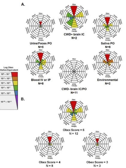
FIG. 4. Radar plot summary of tissue sPMCA and LODE results. Animals were grouped according to the source of inoculum (blood and saliva,
etc.) (A) and obex PMCA score (B). Central concentric rings represent a score of “1,” with scores progressing radially outward up to a score of
“6.” For each radar plot, colored wedges indicate the mean tissue sPMCA score for that particular group, while wedge colors indicate the mean
LODE score. Patterns of distribution varied with both the source of inoculum and obex burden, with the most widely distributed patterns identified
for animals inoculated with blood either i.v. (IV) or i.p. (IP) and animals with an obex score of “6.” IC, intracranially; PO, orally.
on November 7, 2019 by guest
http://jvi.asm.org/
prion production and/or excretion, and we found a range of
CWD prion amplifications from such tissues. Variation in
CWD prion-amplifying activities in tissues significantly
corre-lated with the apparent PrP
CWDburden within the obex of the
individual animal, while a trending relationship was observed
between peripheral distribution and intensity and the source of
inoculum and route of exposure (e.g., whole blood i.v. and
urine and feces p.o., etc.). Perhaps with more animals per
group or longer incubation times, this overall trend between
inoculum and peripheral distribution may have become
statis-tically significant.
These two findings point to an increased risk of bodily fluid
infectivity with disease progression and, potentially, source of
exposure. The tissue distribution variation with the route of
inoculation has been described previously for both viral and
bacterial infections (6, 29, 37, 45), so this finding is perhaps not
surprising. Likewise, in the case of neurotropic viruses, virus
levels in peripheral tissue often positively correlate with the
central nervous system burden (8). Evaluation of the prion
distribution in tissues of naturally infected cervids may reveal
patterns corresponding with those described in this study, i.e.,
widespread peripheral accumulation of prions that vary
ac-cording to the central nervous system burden and, potentially,
the exposure source.
The detection of amplifiable prions in peripheral excretory
tissues call into question (i) whether peripheral prion
amplifi-cation occurs
in situ
or, alternatively, whether excretory tissues
merely serve a more passive role in prion excretion and (ii) the
kinetics of prion excretion, i.e., how the levels of prion
excre-tion and infectivity in bodily fluids change over the course of
the disease. It is generally accepted that TSE pathogenesis
follows a pattern of centripetal spread from the periphery to
the central nervous system (either solely via the peripheral
nervous system or after amplification in the lymphoreticular
system), followed by central amplification and centrifugal
spread back to the periphery (4, 5, 11, 15, 53, 54). We found
that some peripheral tissue levels rivaled those in the obex of
the same animal, perhaps suggesting concurrent early
dissem-ination to the central nervous system and peripheral organs. A
further understanding of the kinetics of centripetal and
cen-trifugal prion dissemination could emanate from a sequential
sPMCA evaluation of the peripheral prion distribution in
ex-posed animals.
In summary, the present study demonstrates for the first
time prion-amplifying activity in organs and tissues associated
with prion shedding. The ultimate source and mechanism of
release into bodily fluids remain unknown. The elevated and
consistent activity found in salivary gland and urinary bladder
may suggest an active role in prion excretion. These findings
minimally warrant additional, more detailed, and longitudinal
studies of the nature and kinetics of excreted prions.
ACKNOWLEDGMENTS
We sincerely thank all of those who have made important
contribu-tions to the manuscript, including David Osborne and Sally Dahmes,
without whom the primary experiments with cervids could not have
been performed, and Michael Miller, who provided initial samples of
CWD
⫹brain and bodily fluids. Candace Mathiason was integral in
assisting with the design and implementation of blinded sPMCA of
tissues. Other notable contributors who provided assistance with assay
development and interpretation include Timothy Kurt and Amy Nalls.
For long-term care and sample collection from source deer and
trans-genic mice, we sincerely thank Sheila Hays, Kelly Anderson, and
Jea-nette Hayes-Klug. Without each of their contributions and unique
skills, this work could not have been completed.
REFERENCES
1.Angers, R. C., et al.2009. Chronic wasting disease prions in elk antler velvet.
Emerg. Infect. Dis.15:696–703.
2.Balachandran, A., et al.2010. Experimental oral transmission of chronic wasting disease to red deer (Cervus elaphus elaphus): early detection and
late stage distribution of protease-resistant prion protein. Can. Vet. J.51:
169–178.
3.Balkema-Buschmann, A., et al.2011. BSE infectivity in the absence of detectable PrP(Sc) accumulation in the tongue and nasal mucosa of
termi-nally diseased cattle. J. Gen. Virol.92:467–476.
4.Bartz, J. C., C. Dejoia, T. Tucker, A. E. Kincaid, and R. A. Bessen.2005. Extraneural prion neuroinvasion without lymphoreticular system infection.
J. Virol.79:11858–11863.
5.Beekes, M., and P. A. McBride.2007. The spread of prions through the body in naturally acquired transmissible spongiform encephalopathies. FEBS J.
274:588–605.
6.Bravata, D. M., et al.2007. Inhalational, gastrointestinal, and cutaneous anthrax in children. A systematic review of cases: 1900 to 2005. Arch.
Pedi-atr. Adolesc. Med.161:896–905.
7.Browning, S. R., et al.2004. Transmission of prions from mule deer and elk with chronic wasting disease to transgenic mice expressing cervid PrP. J.
Vi-rol.78:13345–13350.
7a.Castilla, J., et al.2005. Detection of prions in blood. Nat. Med.11:982–985. 8.Charlton, K. M., G. A. Casey, and J. B. Campbell.1983. Experimental rabies in skunks: mechanisms of infection of the salivary glands. Can. J. Comp.
Med.47:363–369.
9.Colby, D. W., et al.2010. Protease-sensitive synthetic prions. PLoS Pathog.
6:e1000736.
10.DeJoia, C., B. Moreaux, K. O’Connell, and R. A. Bessen.2006. Prion
infec-tion of oral and nasal mucosa. J. Virol.80:4546–4556.
11.Dormont, D.2002. Prion diseases: pathogenesis and public health concerns.
FEBS Lett.529:17–21.
12.Eklund, C. M., R. C. Kennedy, and W. J. Hadlow.1967. Pathogenesis of
scrapie virus infection in the mouse. J. Infect. Dis.117:15–22.
13.Foster, J. D., D. W. Parnham, N. Hunter, and M. Bruce.2001. Distribution of the prion protein in sheep terminally affected with BSE following
exper-imental oral transmission. J. Gen. Virol.82:2319–2326.
14.Fox, K. A., J. E. Jewell, E. S. Williams, and M. W. Miller.2006. Patterns of PrPCWD accumulation during the course of chronic wasting disease infec-tion in orally inoculated mule deer (Odocoileus hemionus). J. Gen. Virol.
87:3451–3461.
15.Glatzel, M., and A. Aguzzi.2000. Peripheral pathogenesis of prion diseases.
Microbes Infect.2:613–619.
16.Hadlow, W. J., et al.1974. Course of experimental scrapie virus infection in
the goat. J. Infect. Dis.129:559–567.
17.Haley, N., C. Mathiason, M. D. Zabel, G. C. Telling, and E. Hoover.2009. Detection of sub-clinical CWD infection in conventional test-negative deer
long after oral exposure to urine and feces from CWD⫹deer. PLoS One
4:e7990.
18.Haley, N. J., D. M. Seelig, M. D. Zabel, G. C. Telling, and E. A. Hoover.2009. Detection of CWD prions in urine and saliva of deer by transgenic mouse
bioassay. PLoS One4:e4848.
19.Hamir, A. N., R. A. Kunkle, J. M. Miller, and S. M. Hall.2006. Abnormal prion protein in ectopic lymphoid tissue in a kidney of an asymptomatic white-tailed deer experimentally inoculated with the agent of chronic wasting
disease. Vet. Pathol.43:367–369.
20.International Society for Infectious Diseases.24 January 2010, posting date. Chronic wasting disease, cervid—USA (02) (Virginia). Archive number 20100124.0261. International Society for Infectious Diseases, Brookline, MA. http://www.promedmail.org.
21.International Society for Infectious Diseases.3 March 2010, posting date. Chronic wasting disease, cervid—USA (03) (Missouri). First report. Archive number 20100303.0697. International Society for Infectious Diseases, Brookline, MA. www.promedmail.org.
22.Jansen, C., et al. 2010. The first case of protease-sensitive prionopathy (PSPr) in the Netherlands: a patient with an unusual GSS-like clinical
phe-notype. J. Neurol. Neurosurg. Psychiatry81:1052–1055.
23.Keane, D. P., et al.2008. Chronic wasting disease in a Wisconsin white-tailed
deer farm. J. Vet. Diagn. Invest.20:698–703.
24.Kitamoto, T., S. Mohri, and J. Tateishi.1989. Organ distribution of protein-ase-resistant prion protein in humans and mice with Creutzfeldt-Jakob
dis-ease. J. Gen. Virol.70(Pt. 12):3371–3379.
25.Kurt, T. D., et al.2007. Efficient in vitro amplification of chronic wasting
disease PrPRES. J. Virol.81:9605–9608.
26.Kurt, T. D., G. C. Telling, M. D. Zabel, and E. A. Hoover.2009. Trans-species
on November 7, 2019 by guest
http://jvi.asm.org/
amplification of PrP(CWD) and correlation with rigid loop 170N. Virology
387:235–243.
27.Landis, J. R., and G. G. Koch.1977. The measurement of observer
agree-ment for categorical data. Biometrics33:159–174.
28.Ligios, C., et al.2007. Intraepithelial and interstitial deposition of
patholog-ical prion protein in kidneys of scrapie-affected sheep. PLoS One2:e859.
28a.Lowry, R. 2011. VassarStats: website for statistical computation. http: //faculty.vassar.edu/lowry/VassarStats.html.
29.Maisner, A., J. Neufeld, and H. Weingartl.2009. Organ- and endotheliotro-pism of Nipah virus infections in vivo and in vitro. Thromb. Haemost.
102:1014–1023.
30.Mathiason, C. K., et al.2010. B cells and platelets harbor prion infectivity in
the blood of deer infected with chronic wasting disease. J. Virol.84:5097–
5107.
31.Mathiason, C. K., et al. 2009. Infectious prions in pre-clinical deer and transmission of chronic wasting disease solely by environmental exposure.
PLoS One4:e5916.
32.Mathiason, C. K., et al.2006. Infectious prions in the saliva and blood of
deer with chronic wasting disease. Science314:133–136.
33.Miller, M. W., E. S. Williams, N. T. Hobbs, and L. L. Wolfe.2004. Environ-mental sources of prion transmission in mule deer. Emerg. Infect. Dis.
10:1003–1006.
34.Mulcahy, E. R., J. C. Bartz, A. E. Kincaid, and R. A. Bessen.2004. Prion
infection of skeletal muscle cells and papillae in the tongue. J. Virol.78:
6792–6798.
34a.Murayama, Y., et al.2007. Urinary excretion and blood level of prions in
scrapie-infected hamsters. J. Gen. Virol.88:2890–2898.
35.Notari, S., et al.2010. Multiorgan detection and characterization of pro-tease-resistant prion protein in a case of variant CJD examined in the United
States. PLoS One5:e8765.
36.Pastrana, M. A., et al.2006. Isolation and characterization of a proteinase
K-sensitive PrPSc fraction. Biochemistry45:15710–15717.
37.Prentice, M. B., and L. Rahalison.2007. Plague. Lancet369:1196–1207. 38.Saborio, G. P., B. Permanne, and C. Soto.2001. Sensitive detection of
pathological prion protein by cyclic amplification of protein misfolding.
Na-ture411:810–813.
39.Safar, J., et al.1998. Eight prion strains have PrP(Sc) molecules with
dif-ferent conformations. Nat. Med.4:1157–1165.
40.Safar, J. G., et al.2005. Diagnosis of human prion disease. Proc. Natl. Acad.
Sci. U. S. A.102:3501–3506.
41.Sakaguchi, S., et al.1993. Kinetics of infectivity are dissociated from PrP
accumulation in salivary glands of Creutzfeldt-Jakob disease
agent-inocu-lated mice. J. Gen. Virol.74(Pt. 10):2117–2123.
42.Seeger, H., et al.2005. Coincident scrapie infection and nephritis lead to
urinary prion excretion. Science310:324–326.
43.Seelig, D. M., G. L. Mason, G. C. Telling, and E. A. Hoover.2010. Patho-genesis of chronic wasting disease in cervidized transgenic mice. Am. J.
Pathol.176:2785–2797.
44.Sigurdson, C. J.2008. A prion disease of cervids: chronic wasting disease.
Vet. Res.39:41.
45.Singh, G. K., N. P. Singh, and S. K. Garg.1987. Studies on pathogenesis of
fowl pox: virological study. Acta Virol.31:417–423.
46.Siso, S., et al.2006. Prion protein in kidneys of scrapie-infected sheep. Vet.
Rec.159:327–328.
47.Siso, S., et al.2008. Occurrence and cellular localization of PrPd in kidneys
of scrapie-affected sheep in the absence of inflammation. J. Pathol.215:126–
134.
48.Soto, C., G. P. Saborio, and L. Anderes.2002. Cyclic amplification of protein misfolding: application to prion-related disorders and beyond. Trends
Neu-rosci.25:390–394.
49.Spraker, T. R., et al.1997. Spongiform encephalopathy in free-ranging mule deer (Odocoileus hemionus), white-tailed deer (Odocoileus virginianus) and Rocky Mountain elk (Cervus elaphus nelsoni) in northcentral Colorado. J.
Wildl. Dis.33:1–6.
50.Tamguney, G., et al.2009. Asymptomatic deer excrete infectious prions in
faeces. Nature461:529–532.
51.Tattum, M. H., S. Jones, S. Pal, J. Collinge, and G. S. Jackson.2010. Discrimination between prion-infected and normal blood samples by protein
misfolding cyclic amplification. Transfusion50:996–1002.
52.Thackray, A. M., L. Hopkins, and R. Bujdoso.2007. Proteinase K-sensitive disease-associated ovine prion protein revealed by conformation-dependent
immunoassay. Biochem. J.401:475–483.
53.Unterberger, U., T. Voigtlander, and H. Budka.2005. Pathogenesis of prion
diseases. Acta Neuropathol.109:32–48.
54.van Keulen, L. J., A. Bossers, and F. van Zijderveld.2008. TSE pathogenesis
in cattle and sheep. Vet. Res.39:24.
55.Vascellari, M., et al.2007. PrPSc in salivary glands of scrapie-affected sheep.
J. Virol.81:4872–4876.
56.Williams, E. S., and S. Young.1980. Chronic wasting disease of captive mule
deer: a spongiform encephalopathy. J. Wildl. Dis.16:89–98.
57.Williams, E. S., and S. Young.1982. Spongiform encephalopathy of Rocky
Mountain elk. J. Wildl. Dis.18:465–471.
on November 7, 2019 by guest
http://jvi.asm.org/