0022-538X/09/$12.00 doi:10.1128/JVI.00888-09
Copyright © 2009, American Society for Microbiology. All Rights Reserved.
Control of Herpes Simplex Virus Replication Is Mediated through an
Interferon Regulatory Factor 3-Dependent Pathway
䌤
Vineet D. Menachery
1,3and David A. Leib
1,2*
Departments of Ophthalmology and Visual Sciences,1Molecular Microbiology,2and Program in Immunology,3
Washington University School of Medicine, St. Louis, Missouri, 63110
Received 2 May 2009/Accepted 8 September 2009
The type I interferon (IFN) cascade is critical in controlling viral replication and pathogenesis. Recognition pathways triggered by viral infection rapidly induce the type I IFN cascade, often in an IFN regulatory factor 3 (IRF-3)-dependent fashion. This dependence predicts that loss of IRF-3 would render early recognition pathways inoperative and thereby impact virus replication, but this has not been observed previously with herpes simplex virus type 1 (HSV-1) in vitro. In this study, HSV-1-infected IRF-3ⴚ/ⴚbone marrow-derived dendritic cells (BMDCs) and macrophages supported increased HSV replication compared to control cells. In addition, IRF-3-deficient BMDCs exhibited delayed type I IFN synthesis compared to control cells. However, while IFN pretreatment of IRF-3ⴚ/ⴚBMDCs resulted in reduced virus titers, a far greater reduction was seen after IFN treatment of wild-type cells. This suggests that even in the presence of exogenously supplied IFN, IRF-3ⴚ/ⴚBMDCs are inherently defective in the control of HSV-1 replication. Together, these results dem-onstrate a critical role for IRF-3-mediated pathways in controlling HSV-1 replication in cells of the murine immune system.
Herpes simplex virus type 1 (HSV-1) is a ubiquitous human pathogen with high seroprevalence in adults (51). HSV-1 is associated with numerous human diseases ranging from the common cold sore in immunocompetent individuals to her-petic encephalitis in neonatal and immunocompromised hosts. A member of theAlphaherpesvirusfamily, HSV-1 exhibits two distinct phases of infection (49). Acute infection typically oc-curs at peripheral epithelial sites and is characterized by lytic infection and spread. In contrast, the virus shifts from lytic to latent infection in sensory neurons, which is characterized by limited gene expression and the persistence of viral genomes in a transcriptionally active state. Following certain stimuli, peri-odic reactivation of latency occurs and may result in shedding of infectious virus at the initial site of acute infection. Reacti-vation may also be associated with immunopathological dis-eases, most notably ocular herpetic stromal keratitis.
A role for interferons (IFNs) in controlling viral replication is well established. In recent years, viral research has focused on cellular recognition of pathogen-associated molecular pat-terns and subsequent IFN induction, leading to the discovery of Toll-like receptors (TLRs) and retinoic acid inducible gene 1 (RIG-I)-like sensing molecules (18). Such molecules respond to several virally derived pathogen-associated molecular pat-terns. These include MDA-5 and TLR-3, which recognize dou-ble-stranded RNA (13, 24); DAI and TLR-9, which recognize double-stranded DNA (14, 45); TLR-7, which recognizes sin-gle-stranded RNA (9); and RIG-I, which recognizes triphos-phate and double-stranded RNA (16, 31, 53). Subsequent work
identified the adaptor molecules necessary for antiviral path-way signaling, including MyD88, TRIF, and IPS-1 (19, 46, 52). Not surprisingly, numerous gene products from viruses such as hepatitis C virus, West Nile virus (WNV), influenza virus, and vaccinia virus have been identified to antagonize these path-ways and serve to promote viral replication and virulence by degradation, interference, or sequestration of early recognition components (3, 11).
These newly identified recognition pathways utilize IRF-3, IRF-7, and NF-B to induce IFN transcription through cog-nate binding sites on the IFN-promoter (38). During initial induction of IFN, IRF-3, and NF-B, which are constitutively expressed, become activated and translocate to the nucleus, where they bind the IFN-promoter to form the IFN enhance-some (54). The initial IFN-produced acts upon the IFN-␣ receptor in both an autocrine and paracrine manner to upregu-late IFN-stimuupregu-lated genes (ISGs), most notably IRF-7 (38). In concert with IRF-3, IRF-7 amplifies and facilitates expression of the full type I IFN cascade. In the absence of IRF-3, IFN- production is reduced but the IFN-␣ levels remain normal, suggesting that IRF-7 activity can compensate for the loss of IRF-3 (15). In contrast, IRF-7 deficiency results in significant reduction in serum IFN levels with a corresponding increase in susceptibility to virus infection. IRF-7 was therefore dubbed “the master regulator” of type I IFN-dependent immune re-sponses (15). IRF-7⫺/⫺mice challenged with HSV-1 showed
increased mortality compared to control and IRF-3⫺/⫺mice,
but no increases in virus titers were observed in IRF-3- or IRF-7-deficient cells in vitro (15). A possible explanation for this lack of phenotype in vitro is that HSV-1 may control IRF-3 activation so thoroughly that this pathway is neutralized during infection. UV-inactivated HSV-1 induces IRF-3 dimerization and activation, leading to IFN induction, suggesting that very early events in infection are responsible for triggering this cascade in the absence of viral gene expression (6, 23). ICP0,
* Corresponding author. Present address: Department of Microbi-ology and ImmunMicrobi-ology, Dartmouth-Hitchcock Medical Center, 630E Borwell Building, HB 7556, 1 Medical Center Dr., Lebanon, NH 03756. Phone: (603) 650-8616. Fax: (603) 650-6223. E-mail: david.a [email protected].
䌤Published ahead of print on 16 September 2009.
12399
on November 8, 2019 by guest
http://jvi.asm.org/
significant increase in viral replication in vitro (21, 35, 37, 55). In these studies, we examined the impact of IRF-3-mediated pathways on HSV-1 replication using cells from IRF3-deficient (IRF-3⫺/⫺) mice. The absence of IRF-3 was predicted to
pre-clude the function of early recognition pathways and thereby impact HSV-1 replication. No changes in HSV-1 replication in IRF-3-deficient mouse embryonic fibroblasts (MEFs) had been observed previously, but we reasoned that, relative to MEFs, cells of the immune system might induce more vigorous IRF-3-dependent antiviral responses, manifesting with a sig-nificant impact upon viral replication. Using IRF-3-deficient bone marrow-derived dendritic cells (BMDCs) and macro-phages (BMM) we have demonstrated that IRF-3 mediated pathways are critical for control of HSV-1 replication. More-over, control of HSV-1 replication is dependent on the type I IFN cascade in these cell types induced via IRF-3-mediated pathways.
MATERIALS AND METHODS
Cells and viruses.Viral stocks were grown and titers were determined on Vero cells (34). HSV-1 WT strain KOS was the background strain for the present study (41).
BMDCs were generated from 6 to 8-week-old C57/BL6 (Charles River Lab-oratories, Wilmington, MA) or 129S6 (Taconic, Germantown, NY) mice (25, 56). Briefly, bone marrow was flushed from femurs of mice and cells were cultured as described below. For the generation of BMDCs, bone marrow was cultured in RPMI with 10% fetal calf serum, Glutamax, sodium pyruvate, non-essential amino acids, 250 U of penicillin/ml, 250 U of streptomycin/ml, and 2% granulocyte-macrophage colony-stimulating factor for 6 to 8 days at 37°C. BMDCs were then collected, counted, and divided into aliquots for infection at several multiplicities of infection (MOIs) by the addition of virus in a minimal volume of medium for 30 min at 37°C. Cells were then spun at low speed, and inocula were removed, washed, resuspended, and plated in 35-mm wells for the
duration of the experiment. BMDCs were also generated from IRF-3⫺/⫺(15),
IRF-7⫺/⫺(15), STAT-1⫺/⫺(29) (Taconic, Germantown, NY), and IFN-␣//␥
receptor⫺/⫺(AG129) mice (47).
BMM were cultured as described previously (56). Briefly, bone marrow was cultured in Dulbecco modified Eagle medium supplemented with 10% fetal calf serum, 5% heat-inactivated horse serum, 20% L-929 conditioned medium, 250 U of penicillin/ml, 250 U of streptomycin/ml for 7 days on non-tissue-culture-treated plates. At day 7, cells were washed with a 0.02% EDTA solution, col-lected, and counted. The cells were plated in 35-mm wells and rested for 3 days. The BMM were infected at MOIs of 0.01 and 1 by the addition of virus in a minimal volume of medium for 30 min at 37°C, removal of inoculum, and followed by the addition of complete medium.
IFN-ELISA.BMDCs were mock treated or infected at an MOI of 5 with HSV-1 and cultured in 1 ml of medium. Cultured supernatants were harvested at 3, 6, 9, and 12 h postinfection and spun at low speeds to remove cells.
Superna-tants were stored at⫺20°C before assay of IFN-in the medium using 50l of
harvested medium in a mouse IFN- enzyme-linked immunosorbent assay
(ELISA) as described in the kit protocol (PBL Biomedical Laboratories, Pisca-taway, NJ).
Statistics.All statistical calculations were determined by using the Studentt
test and are relative to control cells unless otherwise stated.
RESULTS
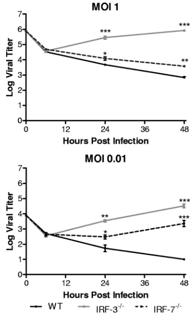
Control of HSV-1 replication in BMDCs is IRF-3 dependent in vitro. A previous study using IRF-3- and IRF-7-deficient MEFs demonstrated that the absence of either signaling mol-ecule did not significantly alter HSV-1 replication (15). Work performed in this laboratory is in agreement with these previ-ous observations (data not shown). BMDCs were chosen for infection in the present study due to their function as immune sentinels, their strong responses to IFN, and their critical role in controlling HSV-1 infection in vivo (17, 43, 44). IRF-3⫺/⫺
BMDCs yielded at least 10 times more HSV-1 than control cells at both 24 and 48 h postinfection at each MOI tested (Fig. 1). In contrast, HSV-1-infected IRF-7⫺/⫺ BMDCs did not
yield any increased virus titers compared to WT control BM-DCs. These results suggested that pathways for control of HSV-1 replication in BMDCs are dependent on IRF-3 but independent of IRF-7.
BMM require IRF-3 for control of HSV-1 replication. Pri-mary BMM were infected in order to further assess the role of IRF-3-mediated pathways in immune cells (2, 5). These adher-ent BMM were also tested to exclude the possibility that the replication pattern of HSV differed between MEFs and BMDCs because of differences in adherence to plastic substrates in culture. The results, however, demonstrated that the pattern of viral replication in the IRF-3⫺/⫺BMM resembled that seen
in BMDCs, with increased viral yields compared to control cells (Fig. 2). Increases of 10- to 100-fold in viral yields were demonstrated at both 24 and 48 h postinfection at MOIs of 0.01, 1, and 5 (data not shown). Interestingly, IRF-7⫺/⫺BMM
also supported increased viral replication. In contrast to BM-DCs, IRF-7-deficient BMM permitted a 10- to 100-fold in-crease in viral replication compared to controls at 24 and 48 h postinfection. This increase in virus titers was greater in mag-nitude at the lower MOI, but the impact of IRF-7 loss on HSV replication in BMM was less than the impact of loss of IRF-3. These data suggest a role for both IRF-3 and IRF-7 in control of HSV-1 replication in BMM. BMDCs, however, have no requirement for IRF-7 in controlling HSV-1 replication, dem-onstrating a difference in the innate immune response between macrophages and dendritic cells. Overall, in both cell types, IRF-3-mediated pathways are required to control HSV-1 rep-lication in vitro.
BMDCs lacking IFN receptors permit increased viral rep-lication in a STAT-1-dependent manner.Having identified a
on November 8, 2019 by guest
http://jvi.asm.org/
role for IRF-3-mediated pathways in controlling HSV-1 repli-cation in BMDCs and BMM, the focus was shifted to differ-entiating between two non-mutually exclusive mechanisms by which IRF-3 could be controlling HSV-1 replication. First, it is possible that IRF-3⫺/⫺BMDCs have delayed or reduced type
I IFN responses, disrupting the type I IFN cascade, and result-ing in increased viral replication. Second, it is possible that other IRF-3-dependent processes or gene products are directly controlling HSV-1 replication. To address these possibilities, BMDCs lacking both type I and type II IFN receptors were infected with HSV-1 (Fig. 3). These cells lack IFN binding and signaling but contain IRF-3 and thereby maintain elements of the early recognition pathway via IRF-3-dependent gene ex-pression. The IFN receptor-deficient BMDCs permitted in-creased viral growth in a similar fashion to IRF-3⫺/⫺BMDCs
and suggested that the type I IFN cascade was responsible for controlling HSV-1 replication (Fig. 3). Similar increases in viral replication were also seen in STAT-1⫺/⫺BMDCs.
To-gether, these data confirm that viral replication is significantly limited in these cell types through IFN-driven STAT-1 signal-ing. Although these data do not completely exclude other IRF-3-dependent processes, the results strongly suggest that the increased viral yields in IRF-3⫺/⫺BMDCs and BMM are due
to a delayed or defective type I IFN cascade.
IRF-3-deficient BMDCs have a defect in IFN-induction compared to WT control cells.IFN-plays a critical role in inducing an antiviral state and controlling viral infection (18). A deficit or a delay in IFN-induction would likely allow increased viral replication, as seen in IRF-3⫺/⫺BMDCs. In order to
exam-ine this question, IFN- protein levels were determined by ELISA in control and IRF-3⫺/⫺ BMDCs after infection with
HSV-1 (Fig. 4). BMDCs were infected at an MOI of 5 to ensure uniform infection and minimize the contribution of bystander IFN. Even at this high MOI, IRF-3⫺/⫺BMDCs yielded a
statis-tically significant increase in HSV-1 titer at 12 and 24 h postin-fection. Examining IFN-protein, IRF-3⫺/⫺BMDCs exhibited
decreased and delayed IFN-production relative to WT control BMDCs. WT BMDCs produced detectable levels of IFN-as early as 6 h postinfection and continued to escalate at 9 and 12 h postinfection. In contrast, IRF-3-deficient BMDCs only produced measurable levels at 12 h postinfection, suggesting a defect in the initiation of IFN- production. The IRF-3⫺/⫺ BMDCs were,
however, capable of producing IFN-late in the experimental infection, thereby potentially allowing control of viral replication at these later times.
[image:3.585.72.258.67.368.2]IFN␣R-blocking antibody augments viral growth in WT control and IRF-3-deficient BMDCs.To demonstrate that pro-duction of type I IFN was a primary defect, WT and IRF-3⫺/⫺
FIG. 1. In vitro replication in bone marrow derived dendritic cells. Primary BMDCs were infected with WT HSV-1 at an MOI of 1 or 0.01. At indicated times postinfection, cells and supernatants were harvested and virus titers assayed on Vero cells. The results shown are the mean titers of three independent experiments.*,P⬍0.05;**,
P⬍0.01.
FIG. 2. In vitro replication in bone marrow derived macrophages. Primary BMM were infected with WT HSV-1 at an MOI of 1 or 0.01. At the indicated times postinfection, cells and supernatants were har-vested, and virus titers were assayed on Vero cells. The results shown are mean titers of four independent experiments.*,P⬍0.05;**,P⬍
0.01;***,P⬍0.001.
on November 8, 2019 by guest
http://jvi.asm.org/
[image:3.585.325.511.68.374.2]BMDCs were infected and then treated with antibodies that block the IFN␣R or with control IgG1 antibody (Fig. 5 and data not shown). We postulated that if the restriction of HSV-1 replication in this system was dependent on type I IFN induc-tion, then WT and IRF-3-deficient BMDCs should yield sim-ilar virus titers in the presence of the blocking antibody. Con-trol IgG1 had no impact on viral replication in either cell type (data not shown). In contrast, the addition of IFN␣R blocking antibodies allowed both WT and IRF-3⫺/⫺BMDCs to produce
higher yields of HSV-1 such that viral growth curves for these two disparate cell types were similar under these conditions (Fig. 5). It was also notable that untreated IRF-3⫺/⫺BMDC
cultures yielded titers similar to those of antibody-treated BMDCs at 24 h postinfection. In contrast, by 48 h postinfec-tion, antibody-treated IRF-3⫺/⫺BMDCs yielded 10-fold more
virus than untreated cultures. Together, these data demon-strate that the type I IFN cascade is responsible for controlling HSV-1 replication in WT BMDCs and that, at late time points, IRF-3-deficient BMDCs can exert partial type I IFN-depen-dent control of HSV-1 replication.
IFN induction from WT BMDCs fails to restore control of HSV-1 replication to IRF-3-deficient BMDCs in vitro. The preceding data suggested that IFN induction was defective in IRF-3-deficient BMDCs but that these cells were still capable
of controlling viral infection once the type I IFN cascade had been initiated. The question arose, therefore, if the initial IFN induction and synthesis were restored, could IRF-3⫺/⫺
BMDCs limit HSV-1 replication to levels seen in WT control cells? We therefore investigated whether bystander IFN, pro-duced by WT cells, could restore control of viral replication to IRF-3⫺/⫺BMDCs by mixing them in culture at a 1:1 ratio. The
mixed cell population was then infected with HSV-1 at an MOI of 0.01 and viral replication measured (Fig. 6). Viral growth kinetics under these conditions were intermediate between those observed in WT (low viral growth) and IRF-3⫺/⫺(high
viral growth) BMDCs. At 48 h postinfection, the mixed BMDC population gave a 10-fold increase in viral yield over WT cells alone and a 10-fold decrease in viral yield over IRF-3⫺/⫺
BMDCs alone. The results show that IRF-3⫺/⫺BMDCs are
incapable of controlling viral replication even in the presence of bystander IFN induced by viral infection of WT cells. An-other possibility, although less likely, is that the presence of IRF-3⫺/⫺BMDCs resulted in a reduced total type I IFN
con-centration, thereby permitting increased replication in WT BMDCs. In either case, HSV-1 replication in IRF-3-deficient
FIG. 3. In vitro replication in BMDCs lacking IFN signaling. Pri-mary BMDCs derived from WT and IFN-␣//␥ receptor-deficient (AG129) or STAT1-deficient mice were infected with WT HSV-1 at an MOI of 1 or 0.01. At indicated times postinfection, cells and superna-tants were harvested, and virus titers were assayed on Vero cells. The results shown are the mean titers of three independent experiments.*,
[image:4.585.329.509.69.387.2]P⬍0.05;**,P⬍0.01;***,P⬍0.001.
FIG. 4. IFN- secretion by infected BMDCs. Primary BMDCs were infected with WT HSV-1 at an MOI of 5. At indicated times postinfection, cells and supernatants were harvested. Cells were re-moved by low-speed centrifugation, and supernatants were assayed for IFN-by ELISA. The results in the upper panel are mean totals from three independent experiments. Cells and supernatants were also as-sayed for virus titers at 6, 12, and 24 h postinfection, and virus titers were assayed on Vero cells. The results in the lower panel shown are the mean titers of three independent experiments.*,P⬍0.05.
on November 8, 2019 by guest
http://jvi.asm.org/
BMDCs was not limited in the context of bystander cell-pro-duced IFN.
IRF-3-deficient BMDCs primed with IFN partially restore control of HSV-1 replication.The results from the cell mixing experiments suggested that IRF-3⫺/⫺BMDCs were unable to
respond fully to IFN production by WT cells. However, the ability to generate a delayed IFN-response coupled with the IFN-dependent decrease in virus titers at late time points sug-gested that IRF-3⫺/⫺BMDCs were capable of inducing the
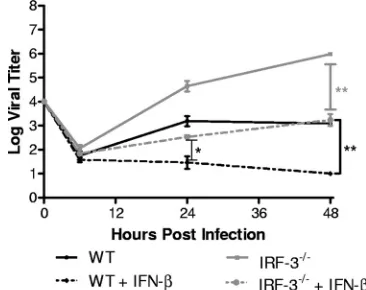
type I IFN cascade, but with low efficiency. One possible model is that cells lacking IRF-3 are inherently slowed in their re-sponse to IFN and need additional time to properly prime in order to fully control HSV-1 replication in vitro. To test this, WT and IRF-3⫺/⫺ BMDCs were pretreated overnight with
IFN-and challenged with HSV-1, and viral yields were mea-sured (Fig. 7). IFN pretreatment of IRF-3⫺/⫺BMDCs
signif-icantly decreased HSV-1 replication compared to untreated IRF-3⫺/⫺cells with a⬎100-fold decrease in virus titers at 48 h
postinfection. Titers observed were comparable to those in untreated WT control BMDCs. However, pretreatment of WT control cells resulted in further decreases in viral replication, to levels at, or below, the level of detection. These results together suggest that IRF-3-deficient BMDCs were capable of strongly responding to IFN, but the overall immune response in controlling HSV-1 replication was still defective compared to WT control cells.
DISCUSSION
Despite mice or cells lacking IFN receptors being signifi-cantly more susceptible to viral infection (22, 32), loss of IRF-3 and IRF-7 had surprisingly little impact on HSV-1 replication in vitro (15). Several groups have suggested the lack of a growth phenotype in IRF3⫺/⫺ cells may be due to HSV-1
maintaining strict control over IRF-3-dependent pathways through various viral genes, including ICP0, ICP27, ICP34.5, and vhs, thereby neutralizing the impact of IRF-3-mediated pathways (6, 10, 23, 26–28, 30, 35, 42). In the present study, we have demonstrated that HSV-1 replication was controlled in an IRF-3-dependent manner in two types of immune cells. This control was dependent on type I IFN and STAT-1 signal-ing with a primary defect in IFN production in IRF-3⫺/⫺cells.
Even in the presence of exogenously supplied IFN, however, HSV-1 replication was only partially controlled in IRF-3⫺/⫺
BMDCs. Overall, the data presented here provide evidence that IRF-3-mediated pathways have a significant impact on HSV-1 replication in certain cell types.
Previous studies examining HSV-1 and IRF-3⫺/⫺ used
[image:5.585.327.510.70.215.2]highly permissive MEFs, whereas in the present study dendritic cells and macrophages were chosen. Given the roles of den-dritic cells and macrophages as sentinels of the immune system capable of controlling viral infection in vivo, it is likely that these cells induced a more vigorous immune response and were thereby less permissive to infection than MEFs (2, 5, 17, 43, 44). In the case of HSV, a virus with multiple mechanisms to subvert IFN responses, loss of IRF-3 can only manifest with
FIG. 5. IFN␣R blockade in BMDCs. Primary BMDCs were in-fected with WT HSV-1 at an MOI of 0.01. After infection, BMDCs were plated in media containing 5g of IFN␣R blocking antibody (MAR1-5A3)/ml for the duration of the experiment. At the indicated times postinfection, cells and supernatants were harvested, and virus titers were assayed on Vero cells. The results shown are the mean titers of three independent experiments.*,P⬍, 0.05;**,P⬍0.01.
FIG. 6. In vitro replication after mixing BMDC populations. Pri-mary WT and IRF-3⫺/⫺BMDCs were mixed at a 1:1 ratio and infected
at an MOI of 0.01. At the indicated times postinfection, cells and supernatants were harvested, and virus titers were assayed on Vero cells. The results shown are the mean titers of three independent experiments.*,P⬍0.05.
FIG. 7. In vitro replication after IFN-pretreatment of BMDCs. Primary WT and IRF-3⫺/⫺BMDCs were pretreated with 100 U of
mouse IFN-/ml for 16 h. Cells were then infected with WT HSV-1 at an MOI of 0.01. At the indicated times postinfection, cells and super-natants were harvested, and virus titers were assayed on Vero cells. The results shown are the mean titers of three independent experi-ments.*,P⬍0.05;**,P⬍0.01.
on November 8, 2019 by guest
http://jvi.asm.org/
[image:5.585.71.254.527.667.2]lication may be more accurately measured in more restrictive immune cell types. This hypothesis is especially relevant to HSV-1, which compared to WNV and mouse norovirus, has more genes for IFN regulation, produces less double-stranded RNA, and exhibits less sensitivity to type I IFN.
Not surprisingly, in the absence of IRF-3, BMDCs and BMM were unable to efficiently control HSV-1 replication (Fig. 1 and 2). Although the IFN receptors are intact in these cells, the early recognition signaling likely cannot proceed ef-ficiently without IRF-3, leading to a delay in the type I IFN cascade. Later, once secondary rounds of infection have begun, alternate recognition pathways, most likely mediated through IRF-7, can lead to the induction of type I IFN. This recognition by a secondary pathway is supported by the observed late production of IFN- (Fig. 4) and the concomitant decreased viral replication in IRF-3⫺/⫺BMDCs at late time points (Fig.
5). Although IRF-3 is constitutively expressed in both BMDCs and BMM, basal expression of IRF-7 varies according to cell type (33). Plasmacytoid dendritic cells (pDCs) constitutively express IRF-7, while IRF-7 expression is reduced in conven-tional BMDCs compared to IRF-3. BMM exhibited basal ex-pression of both IRF-3 and IRF-7, potentially explaining increased replication in IRF-7⫺/⫺BMM but not in IRF-7⫺/⫺
BMDCs (8). These studies observed a parallel trend in WNV replication in IRF-7⫺/⫺BMM and BMDCs, as seen here with
HSV-1. However, in both cell types, the presence of IRF-7 cannot compensate for the loss of IRF-3-mediated pathways. These results suggest that IRF-3-mediated pathways provide the major pathway of control of HSV-1 replication.
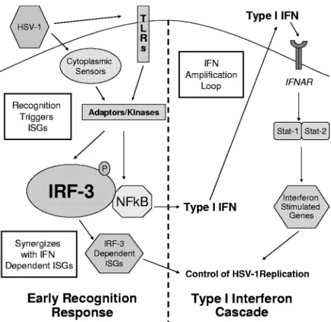
Together, our results demonstrate that the early recognition response through IRF-3-mediated pathways controls HSV-1 replication in BMDCs and leads to the following model (Fig. 8). Upon virus entry, an as-yet-undetermined sensor recog-nizes HSV-1 and triggers a signaling cascade that activates IRF-3. IRF-3 activation leads to production of IRF-3-depen-dent gene products, type I IFN, and an ensuing type I IFN cascade, resulting in control of the HSV-1 infection. In the absence of the type I IFN cascade, achieved by knockout (Fig. 3) or receptor blockade (Fig. 5), BMDCs are unable to control viral replication. Similarly, ablating IRF-3 and the early recog-nition response results in increased viral replication due to delayed and reduced IFN- production (Fig. 1, 2, and 4). Exogenous IFN provided by bystander cells (Fig. 6) or pre-treatment (Fig. 7) partially restores control of HSV-1 replica-tion in IRF-3⫺/⫺BMDCs, and yet these cells remain defective
in their control of HSV-1 replication compared to treated WT cells.
Several non-mutually exclusive possibilities exist to explain this persistent defect in the ability of IRF-3⫺/⫺ BMDCs to
control HSV-1 replication (Fig. 8). One possibility is a defec-tive autocrine and paracrine IFN amplification response. While WT BMDCs quickly respond to IFN through STAT-1 and IRF-3 signaling pathways, IRF-3⫺/⫺ BMDCs can only
respond through STAT-1-dependent, but IRF-3-independent pathways. The absence of IRF-3 thereby severely decreases or ablates the expression of several gene products, including IFN-, IFN-␣4, and IFN-␣5 (1), resulting in less robust IFN signaling. A second possibility is that IRF-3-dependent ISGs synergize with type I IFN receptor-dependent ISGs and con-trol HSV-1 replication but fail to be produced robustly in IRF-3⫺/⫺BMDCs. A third possibility is that virus recognition
may be required to augment the ongoing immune response. IFN primed IRF-3⫺/⫺BMDCs may produce IFN effectors, but
a lack of viral recognition signaling results in a delayed effector response from ISGs. IRF-3⫺/⫺BMDCs may therefore require
HSV-1 recognition signaling through a secondary pathway be-fore fully committing to a complete IFN effector response, and this delay could result in the observed increased viral replica-tion compared to WT controls.
Together, these data demonstrate that immune cells lacking IRF-3 are inherently defective in the control of HSV infection. These data, however, conflict with previously published in vivo data following intravenous (i.v.) infection (15). A possible
ex-FIG. 8. Model for continued defect in IRF-3-deficient BMDCs. Postattachment, HSV-1 infection is recognized through an unknown sensor mechanism that leads to activation of IRF-3. The early recog-nition pathway mediates the production of type I IFN- and IRF-3-dependent ISGs, leading to the control of HSV-1 replication via the type I IFN cascade. However, pretreatment with IFN does not restore HSV-1 replication in IRF-3⫺/⫺BMDCs to WT levels. The continued
defect is potentially due to three, nonexclusive mechanisms outlined in white squares: defective IFN amplification, defective antiviral trigger signaling, and IRF-3-dependent gene synergy with the antiviral re-sponse. One or more of these mechanisms leads to continued defect in the control of HSV-1 replication in IRF-3⫺/⫺BMDCs compared to
WT BMDCs after IFN treatment.
on November 8, 2019 by guest
http://jvi.asm.org/
[image:6.585.304.542.66.297.2]planation is that after i.v. infection, IFN was being produced by pDCs. pDCs, typically found in the lymph nodes away from the site of infection, are a major producer of type I IFN, and they rely on TLR-9 and IRF-7 pathways to induce IFN in response to HSV-1 (4, 20, 40). After i.v. infection, therefore, pDC pro-duction of type I IFN likely overcomes the IFN deficit and thereby is able to control HSV-1 replication in the absence of IRF-3. Previous in vitro studies in MEFs suggested a role for HSV-1 gene components in interfering with and neutralizing the activity of IRF-3 (10, 23, 26–28, 30, 36, 42, 48). In the cell types used in the present study, heightened immune responses likely reduced the efficacy of one or more viral immunoregu-latory components or presented too great of a challenge for the viral activities to counter. Therefore, the efficacy of HSV-1 genes in antagonism of IRF-3 likely depends on the overall capacity of the infected cell to mount an immune response to the incoming virus. Ongoing experiments in our laboratory seek to determine the precise molecules responsible for HSV-1 recognition. As mentioned previously, several candidates in the early recognition pathways have been implicated (21, 35, 37, 55), and cells lacking these components are currently being tested for their ability to control viral replication in BMDCs. Furthermore, in vivo studies in IRF-3 deficient animals are currently under way in order to examine HSV-1 replication and pathogenesis in peripheral and neuronal tissues.
ACKNOWLEDGMENTS
This study was supported by NIH grants to D.A.L. (EY 09083) and the Department of Ophthalmology and Visual Sciences (P30EY02687) from the National Eye Institute. V.D.M. was also supported by an NIH Research Training Grant in the Visual Sciences (T32 EY013360-09). Support from Research to Prevent Blindness to the Department and a Senior Scientific Investigator Award to D.A.L. are gratefully acknowl-edged.
IRF-3⫺/⫺, IRF-7⫺/⫺mice were generously provided by T.
Tanagu-chi, and AG129 mice were generously provided by R. Zinkernagel. We especially thank Marco Colonna and Steve McCartney for providing granulocyte-macrophage colony-stimulating factor and for helpful in-sight into BMDC cultures. We also thank M. Diamond for helpful feedback and provision of mice and members of the Leib lab for helpful discussions.
REFERENCES
1.Andersen, J., S. VanScoy, T. F. Cheng, D. Gomez, and N. C. Reich.2008. IRF-3-dependent and augmented target genes during viral infection. Genes
Immun.9:168–175.
2.Bauer, D., S. Mrzyk, N. van Rooijen, K. P. Steuhl, and A. Heiligenhaus.
2000. Macrophage-depletion influences the course of murine HSV-1
kera-titis. Curr. Eye Res.20:45–53.
3.Bowie, A. G., and L. Unterholzner.2008. Viral evasion and subversion of
pattern-recognition receptor signalling. Nat. Rev. Immunol.8:911–922.
4.Cella, M., D. Jarrossay, F. Facchetti, O. Alebardi, H. Nakajima, A. Lanza-vecchia, and M. Colonna.1999. Plasmacytoid monocytes migrate to inflamed lymph nodes and produce large amounts of type I interferon. Nat. Med.
5:919–923.
5.Cheng, H., T. M. Tumpey, H. F. Staats, N. van Rooijen, J. E. Oakes, and R. N. Lausch.2000. Role of macrophages in restricting herpes simplex virus
type 1 growth after ocular infection. Investig. Ophthalmol. Vis. Sci.41:1402–
1409.
6.Collins, S. E., R. S. Noyce, and K. L. Mossman.2004. Innate cellular re-sponse to virus particle entry requires IRF3 but not virus replication. J. Virol.
78:1706–1717.
7.Daffis, S., M. A. Samuel, B. C. Keller, M. Gale, Jr., and M. S. Diamond.2007. Cell-specific IRF-3 responses protect against West Nile virus infection by
interferon-dependent and -independent mechanisms. PLoS Pathog.3:e106.
8.Daffis, S., M. A. Samuel, M. S. Suthar, B. C. Keller, M. Gale, Jr., and M. S. Diamond.2008. Interferon regulatory factor IRF-7 induces the antiviral alpha interferon response and protects against lethal West Nile virus
infec-tion. J. Virol.82:8465–8475.
9.Diebold, S. S., T. Kaisho, H. Hemmi, S. Akira, and C. Reis e Sousa.2004. Innate antiviral responses by means of TLR7-mediated recognition of
single-stranded RNA. Science303:1529.
10.Eidson, K. M., W. E. Hobbs, B. J. Manning, P. Carlson, and N. A. DeLuca.
2002. Expression of herpes simplex virus ICP0 inhibits the induction of
interferon-stimulated genes by viral infection. J. Virol.76:2180–2191.
11.Foy, E., K. Li, R. Sumpter, Jr., Y. M. Loo, C. L. Johnson, C. Wang, P. M. Fish, M. Yoneyama, T. Fujita, S. M. Lemon, and M. Gale, Jr.2005. Control of antiviral defenses through hepatitis C virus disruption of retinoic
acid-inducible gene-I signaling. Proc. Natl. Acad. Sci. USA102:2986–2991.
12.Fredericksen, B. L., M. Smith, M. G. Katze, P. Y. Shi, and M. Gale, Jr.2004. The host response to West Nile Virus infection limits viral spread through
the activation of the interferon regulatory factor 3 pathway. J. Virol.78:
7737–7747.
13.Gitlin, L., W. Barchet, S. Gilfillan, M. Cella, B. Beutler, R. A. Flavell, M. S. Diamond, and M. Colonna.2006. Essential role of mda-5 in type I IFN responses to polyriboinosinic:polyribocytidylic acid and
encephalomyocardi-tis picornavirus. Proc. Natl. Acad. Sci. USA103:8459–8464.
14.Hemmi, H., O. Takeuchi, T. Kawai, T. Kaisho, S. Sato, H. Sanjo, M. Ma-tsumoto, K. Hoshino, H. Wagner, K. Takeda, and S. Akira.2000. A Toll-like
receptor recognizes bacterial DNA. Nature408:740–745.
15.Honda, K., H. Yanai, H. Negishi, M. Asagiri, M. Sato, T. Mizutani, N. Shimada, Y. Ohba, A. Takaoka, N. Yoshida, and T. Taniguchi.2005. IRF-7 is the master regulator of type-I interferon-dependent immune responses.
Nature434:772.
16.Hornung, V., J. Ellegast, S. Kim, K. Brzozka, A. Jung, H. Kato, H. Poeck, S. Akira, K. K. Conzelmann, M. Schlee, S. Endres, and G. Hartmann.2006.
5⬘-Triphosphate RNA is the ligand for RIG-I. Science314:994–997.
17.Kassim, S. H., N. K. Rajasagi, X. Zhao, R. Chervenak, and S. R. Jennings.
2006. In vivo ablation of CD11c-positive dendritic cells increases suscepti-bility to herpes simplex virus type 1 infection and diminishes NK and T-cell
responses. J. Virol.80:3985–3993.
18.Katze, M. G., Y. He, and M. Gale, Jr.2002. Viruses and interferon: a fight
for supremacy. Nat. Rev. Immunol.2:675–687.
19.Kawai, T., K. Takahashi, S. Sato, C. Coban, H. Kumar, H. Kato, K. J. Ishii, O. Takeuchi, and S. Akira.2005. IPS-1, an adaptor triggering RIG-I- and
Mda5-mediated type I interferon induction. Nat. Immunol.6:981–988.
20.Krug, A.2004. Herpes simplex virus type 1 activates murine natural
inter-feron-producing cells through Toll-like receptor 9. Blood103:1433.
21.Krug, A., G. D. Luker, W. Barchet, D. A. Leib, S. Akira, and M. Colonna.
2004. Herpes simplex virus type 1 activates murine natural
interferon-pro-ducing cells through Toll-like receptor 9. Blood103:1433–1437.
22.Leib, D. A., T. E. Harrison, K. M. Laslo, M. A. Machalek, N. J. Moorman, and H. W. Virgin.1999. Interferons regulate the phenotype of wild-type and
mutant herpes simplex viruses in vivo. J. Exp. Med.189:663–672.
23.Lin, R., R. S. Noyce, S. E. Collins, R. D. Everett, and K. L. Mossman.2004. The herpes simplex virus ICP0 RING finger domain inhibits IRF3- and
IRF7-mediated activation of interferon-stimulated genes. J. Virol.78:1675–
1684.
24.Matsumoto, M., S. Kikkawa, M. Kohase, K. Miyake, and T. Seya.2002. Establishment of a monoclonal antibody against human Toll-like receptor 3 that blocks double-stranded RNA-mediated signaling. Biochem. Biophys.
Res. Commun.293:1364–1369.
25.McCartney, S. A., L. B. Thackray, L. Gitlin, S. Gilfillan, H. W. Virgin, and M. Colonna.2008. MDA-5 recognition of a murine norovirus. PLoS Pathog.
4:e1000108.
26.Melchjorsen, J., J. Siren, I. Julkunen, S. R. Paludan, and S. Matikainen.
2006. Induction of cytokine expression by herpes simplex virus in human monocyte-derived macrophages and dendritic cells is dependent on virus
replication and is counteracted by ICP27 targeting NF-B and IRF-3. J. Gen.
Virol.87:1099–1108.
27.Melroe, G. T., N. A. DeLuca, and D. M. Knipe.2004. Herpes simplex virus 1 has multiple mechanisms for blocking virus-induced interferon production.
J. Virol.78:8411–8420.
28.Melroe, G. T., L. Silva, P. A. Schaffer, and D. M. Knipe.2007. Recruitment of activated IRF-3 and CBP/p300 to herpes simplex virus ICP0 nuclear foci:
potential role in blocking IFN-beta induction. Virology360:305–321.
29.Meraz, M. A., J. M. White, K. C. Sheehan, E. A. Bach, S. J. Rodig, A. S. Dighe, D. H. Kaplan, J. K. Riley, A. C. Greenlund, D. Campbell, K. Carver-Moore, R. N. DuBois, R. Clark, M. Aguet, and R. D. Schreiber.1996. Targeted disruption of the Stat1 gene in mice reveals unexpected physiologic
specificity in the JAK-STAT signaling pathway. Cell84:431–442.
30.Mossman, K. L., H. A. Saffran, and J. R. Smiley.2000. Herpes simplex virus
ICP0 mutants are hypersensitive to interferon. J. Virol.74:2052–2056.
31.Myong, S., S. Cui, P. V. Cornish, A. Kirchhofer, M. U. Gack, J. U. Jung, K. P. Hopfner, and T. Ha.2009. Cytosolic viral sensor RIG-I is a 5⬘
-triphosphate-dependent translocase on double-stranded RNA. Science323:1070–1074.
32.Pasieka, T. J., B. Lu, S. D. Crosby, K. M. Wylie, L. A. Morrison, D. E. Alexander, V. D. Menachery, and D. A. Leib.2008. Herpes simplex virus virion host shutoff attenuates establishment of the antiviral state. J. Virol.
82:5527–5535.
33.Prakash, A., E. Smith, C. K. Lee, and D. E. Levy.2005. Tissue-specific
on November 8, 2019 by guest
http://jvi.asm.org/
USA103:17343–17348.
38.Sato, M.2000. Distinct and essential roles of transcription factors IRF-3 and IRF-7 in response to viruses for IFN-alpha/beta gene induction. Immunity
13:539.
39.Sheehan, K. C., K. S. Lai, G. P. Dunn, A. T. Bruce, M. S. Diamond, J. D. Heutel, C. Dungo-Arthur, J. A. Carrero, J. M. White, P. J. Hertzog, and R. D. Schreiber.2006. Blocking monoclonal antibodies specific for mouse IFN-alpha/beta receptor subunit 1 (IFNAR-1) from mice immunized by in vivo
hydrodynamic transfection. J. Interferon Cytokine Res.26:804–819.
40.Siegal, F. P., N. Kadowaki, M. Shodell, P. A. Fitzgerald-Bocarsly, K. Shah, S. Ho, S. Antonenko, and Y. J. Liu.1999. The nature of the principal type 1
interferon-producing cells in human blood. Science284:1835–1837.
41.Smith, K. O.1964. Relationship between the envelope and the infectivity of
herpes simplex virus. Proc. Soc. Exp. Biol. Med.115:814–816.
42.Sobol, P. T., and K. L. Mossman.2006. ICP0 prevents RNase L-independent
rRNA cleavage in herpes simplex virus type 1-infected cells. J. Virol.80:
218–225.
43.Sprecher, E., and Y. Becker.1987. Herpes simplex virus type 1 pathogenicity in footpad and ear skin of mice depends on Langerhans cell density, mouse
genetics, and virus strain. J. Virol.61:2515–2522.
44.Sprecher, E., and Y. Becker.1986. Skin Langerhans cells play an essential
role in the defense against HSV-1 infection. Arch. Virol.91:341–349.
45.Takaoka, A., Z. Wang, M. K. Choi, H. Yanai, H. Negishi, T. Ban, Y. Lu, M. Miyagishi, T. Kodama, K. Honda, Y. Ohba, and T. Taniguchi.2007. DAI
Nahmias, S. M. Berman, and L. E. Markowitz.2006. Trends in herpes simplex virus type 1 and type 2 seroprevalence in the United States. JAMA
296:964–973.
52.Yamamoto, M., S. Sato, K. Mori, K. Hoshino, O. Takeuchi, K. Takeda, and S. Akira.2002. Cutting edge: a novel Toll/IL-1 receptor domain-containing adapter that preferentially activates the IFN-beta promoter in the Toll-like
receptor signaling. J. Immunol.169:6668–6672.
53.Yoneyama, M.2004. The RNA helicase RIG-I has an essential function in double-stranded RNA-induced innate antiviral responses. Nature Immunol.
5:730.
54.Yoneyama, M., W. Suhara, Y. Fukuhara, M. Fukuda, E. Nishida, and T. Fujita.1998. Direct triggering of the type I interferon system by virus infec-tion: activation of a transcription factor complex containing IRF-3 and CBP/
p300. EMBO J.17:1087–1095.
55.Zhang, S.-Y., E. Jouanguy, S. Ugolini, A. Smahi, G. Elain, P. Romero, D. Segal, V. Sancho-Shimizu, L. Lorenzo, A. Puel, C. Picard, A. Chapgier, S. Plancoulaine, M. Titeux, C. Cognet, H. von Bernuth, C.-L. Ku, A. Casrouge, X.-X. Zhang, L. Barreiro, J. Leonard, C. Hamilton, P. Lebon, B. Heron, L. Vallee, L. Quintana-Murci, A. Hovnanian, F. Rozenberg, E. Vivier, F. Gei-ssmann, M. Tardieu, L. Abel, and J.-L. Casanova.2007. TLR3 deficiency in
patients with herpes simplex encephalitis. Science317:1522–1527.
56.Zhang, X., R. Goncalves, and D. M. Mosser.2008. The isolation and
char-acterization of murine macrophages. Curr. Protoc. Immunol.14:14.1.