Replication-Defective Alleles
Robert A. Fridell, Lourdes Valera, Dike Qiu,* Melissa J. Kirk, Chunfu Wang, Min Gao Department of Virology, Bristol-Myers Squibb Research and Development, Wallingford, Connecticut, USA
Hepatitis C virus NS5A has three structural domains, is required for RNA replication and virion assembly, and exists in hypo-and hyperphosphorylated forms. Accumulated data suggest that phosphorylation is involved in modulating NS5A functions. We performed a mutational analysis of highly conserved serine residues in the linker region between domains I and II of genotype 2a JFH1 NS5A. As with genotype 1b Con1 NS5A, we found that specific serine residues were important for efficient hyperphosphor-ylation of JFH1 NS5A. However, in contrast with Con1 replicons, we observed a positive correlation between hyperphosphoryla-tion and JFH1 replicon replicahyperphosphoryla-tion. We previously demonstratedtrans-complementation of a hyperphosphorylation-deficient, replication-defective JFH1 replicon. Our results suggested that the defective NS5A encoded by this replicon, while lacking one NS5A function, was capable of performing a separate replication function. In this report, we examined an additional set of repli-cation-defective NS5A mutations intrans-complementation assays. While some behaved similarly to the S232I replicon, others displayed a uniquetrans-complementation phenotype, suggesting that NS5Atrans-complementation can occur by two distinct modes. Moreover, we were able, for the first time, to demonstrate intragenic complementation of replication-defective NS5A alleles. Our results identified three complementation groups: group A, comprising mutations within NS5A domain I; group B, comprising mutations affecting serine residues important for hyperphosphorylation and a subset of the domain I mutations; and group C, comprising a single mutation within the C-terminal region of domain II. We postulate that these complementation groups define three distinct and genetically separable functions of NS5A in RNA replication.
H
epatitis C virus (HCV) is an enveloped positive-sensesingle-stranded RNA virus of the genus Hepacivirusin the family
Flaviviridae. The⬃9.6-kb HCV genome encodes an⬃ 3,000-ami-no-acid (aa) polyprotein that is cleaved by viral and cellular pro-teases into 10 mature proteins: core, E1, E2, P7, NS2, NS3, NS4A,
NS4B, NS5A, and NS5B (reviewed in references1and2). Core is
the viral capsid protein, while E1 and E2 are highly glycosylated envelope proteins important for viral entry. P7, a small transmem-brane ion channel protein, and NS2, a memtransmem-brane-associated pro-tease, are required for assembly and release of infectious virions. The remaining nonstructural (NS) proteins are essential compo-nents of the HCV RNA replication complex. NS5B is an RNA-dependent RNA polymerase that catalyzes synthesis of positive-and negative-strpositive-and RNA. NS3 positive-and its cofactor NS4A function as a serine protease responsible for releasing mature NS proteins from the polyprotein precursor. NS3 also possesses RNA helicase and NTPase activities essential for RNA replication. NS4B is an integral membrane protein likely involved in formation of intra-cellular membranous compartments where viral replication oc-curs. The final viral component of the replication complex, NS5A, is a multifunctional protein whose precise roles in RNA replica-tion and other aspects of the HCV life cycle are still being eluci-dated.
Interactions between NS5A and a variety of host cell proteins, as well as other HCV NS proteins, have been described (reviewed
in reference3). While the biological relevance of many of these
interactions remains unclear, others play important roles in HCV
replication (3). NS5A consists of an N-terminal amphipathic
al-pha helix (amino acids 5 to 25) that targets NS5A to intracellular membranes and three structural domains (I, II, and III) separated
by regions of low-complexity sequence (LCS I and LCS II) (4–7).
Domain I (aa 28 to 213) contains a zinc-binding motif, is relatively well conserved among HCV isolates, and is essential for RNA
rep-lication. NS5A domain I has been crystallized in two distinct,
non-overlapping homodimer configurations (8,9). A groove formed
between the monomers in the Tellinghuisen et al. (9) dimer is a
potential RNA binding pocket, and several studies have confirmed
the capacity of NS5A to bind RNA (10–13). The RNA binding
groove is not present in the NS5A dimer reported by Love et al.
(8), suggesting the possibility that different dimer conformations
of NS5A could possess distinct biological properties. Domains II (aa 250 to 342) and III (aa 356 to 447) are less well conserved than
domain I and are natively unfolded (14,15). Domain II is
impor-tant for RNA replication and may also contribute to the RNA
binding properties of the protein (10,11). Domain III is largely
dispensable for RNA replication, but it is essential for virion
as-sembly (16,17).
When NS5A is expressed in tissue culture cells in the context of a contiguous NS3-NS5A polyprotein, at least two alternatively phosphorylated forms of the protein, which can be distinguished
by electrophoretic mobility, are produced (18,19).
Hypophos-phorylated NS5A (p56) is phosHypophos-phorylated at one or more residues in the central and C-terminal part of the protein, while hyper-phosphorylated NS5A (p58) is believed to also be hyper-phosphorylated at one or more highly conserved serine residues within the sur-face-exposed LCS I (aa 214 to 249) connecting domains I and II
Received12 October 2012Accepted5 December 2012
Published ahead of print12 December 2012
Address correspondence to Robert A. Fridell, Robert.fridell@bms.com.
* Present address: Dike Qiu, Discovery Biology, Bristol-Myers Squibb Research and Development, Hopewell, New Jersey, USA.
Copyright © 2013, American Society for Microbiology. All Rights Reserved.
doi:10.1128/JVI.02861-12
on November 7, 2019 by guest
http://jvi.asm.org/
(20,21). Differential phosphorylation potentially regulates several functions of NS5A in RNA replication and virion assembly
(re-viewed in reference22).
Like many positive-sense RNA viruses, HCV RNA replication occurs within virus-induced intracellular membrane alterations that provide a structural site for assembly and function of the replication machinery and may also function to shield the virus
from host cell antiviral responses (23,24). As a consequence of the
membrane compartmentalization and sequestration of viral rep-lication complexes, diffusion of virus-encoded reprep-lication pro-teins between replication complexes encoded by different viral
genomes may be limited, leading to a preference forcis-dominant
replication functions. For HCV,cis-acting RNA replication
func-tions have been reported for NS3, NS4A, and NS5B, whiletrans
-acting functions have been reported for NS5A and NS4B (25–28).
We previously usedtrans-complementation assays with HCV
replicons encoding NS5A proteins with different sensitivities to the HCV replication complex inhibitor declatasvir (DCV) (BMS-790052) to demonstrate two distinct functions of NS5A in RNA
replication, one that was efficiently rescued bytrans
-complemen-tation and another that displayed a strongcis-dominance. In this
study, we examined the correlation between NS5A hyperphos-phorylation and replication in the JFH1 subgenomic replicon sys-tem, while also expanding our previous results by examining
trans-complementation and intragenic complementation of a range of NS5A replication-defective mutants. Our results identify three distinct functions of NS5A required for HCV RNA replica-tion.
MATERIALS AND METHODS
Cells, inhibitors, and recombinant DNA clones.Human hepatoma (Huh-7.5) and baby hamster kidney (BHK1) cells were maintained as previously described (29). A plasmid encoding a bicistronic replicon with the Con1 HCV 5=untranslated region (5=UTR) and internal ribosomal entry site (IRES) sequences, aRenillaluciferase (Rluc) reporter gene, and an encephalomyocarditis virus (EMCV) IRES followed by the NS3 through 3=UTR region from the JFH1 strain has been described (26). NS5A amino acid substitutions were introduced into the replicon plasmid by replacing a BspE1-BspE1 fragment with PCR-generated fragments and were confirmed by sequence analysis. A Con1 replicon with a Rluc re-porter has been described (30). Declatasvir (BMS-790052) has been de-scribed (31).
Replicon assays.Transient replication assays were performed as pre-viously described (26,29). Briefly, replicon transcripts, generated with a Ribomax T7 express system (Promega Corp., Madison, WI) from XbaI-linearized plasmids, were transfected into Huh7.5 cells by using DMRIE-C (liposome formulation of the cationic lipid DMRIE [1,2-dimyristyloxypropyl-3-dimethyl-hydroxy ethyl ammonium bromide] and cholesterol) reagent (Invitrogen, Corp., Carlsbad, CA). Ten micro-grams of RNA was used for single-replicon transfections, while 20g (10 g of each replicon) was used for cotransfections. After 24 h, transfected cells were transferred to 96-well tissue culture plates (⬃10,000 cells/well in 200l) and treated with serial dilutions of inhibitors (1l/well) in di-methyl sulfoxide (DMSO). After an additional 72 h, plates were harvested forRenillaluciferase assays as described previously (29). Replication ca-pacities were measured as replication windows (uninhibited signal/back-ground signal) as described previously (26,29). Background signals were derived from wells treated withⱖ1M DCV, and uninhibited signals were from DMSO-treated wells. Dose-response curves and inhibitor half-maximal effective concentrations (EC50s) were generated with IDBS XLfit
(dose response one-site model 205) as described previously (32). Transient protein expression and immunoblotting assays.HCV nonstructural proteins were expressed from replicon plasmids in BHK-21
cells by using a modified vaccinia virus-T7 (MVA-T7) expression system or following transfection of replicon RNA into Huh7.5 cells as previously described (26,33). Western blot analysis of NS5A and NS3 proteins was performed as previously described (26). NS5A (7B5) and NS3 (2E1) monoclonal antibodies (BioFront Technologies, Tallahassee, FL) were used at 1:1,000 dilutions. A rabbit anti-mouse horseradish peroxidase-conjugated secondary antibody (Abcam, Cambridge, MA) was used at a 1:10,000 dilution.
RESULTS
Several serine residues in the LCS I linker region connecting NS5A domains I and II are highly conserved among NS5A isolates, in-cluding between the genotype 1b Con1 isolate and the genotype 2a
JFH1 isolate (Fig. 1A). In Con1 subgenomic replicons,
serine-to-alanine replacements of a subset of serine residues in this region have been shown to decrease NS5A hyperphosphorylation and
enhance replication (34). To assess the importance of the
con-served serine residues in the JFH1 strain, individual serine-to-alanine substitutions were introduced into a subgenomic JFH1
replicon containing aRenillaluciferase (Rluc) reporter gene. The
mutant replicons were assessed for replication in transient repli-cation assays, and NS5A expression was examined by Western blot analysis following vaccinia virus-T7 polymerase-mediated (VV-T7) expression of the replicon-encoded proteins. Serine-to-ala-nine substitutions at positions 225, 229, 232, and 235 markedly reduced replication levels while also decreasing NS5A
hyperphos-phorylation (Fig. 1B). Very similar NS5A expression patterns were
observed following transfection of replicon RNA directly into
Huh7.5 cells (Fig. 1B, bottom blot), confirming that the effect of
the mutations on hyperphosphorylation was not an artifact of the VV-T7 expression system. The most severe replication defects were seen with replicons bearing S229A and S235A amino acid substitutions, which were indistinguishable from a negative-con-trol GND replicon with a glutamic acid-to-asparagine substitu-tion at the catalytic site of NS5B. In agreement with previous
re-sults (26), a JFH1 replicon with an S232I amino acid substitution
was also severely impaired for both hyperphosphorylation and replication. In contrast, alanine replacement of serine residues at positions 222, 228, 230, and 238 did not appreciably impair
repli-cation capacity or NS5A hyperphosphorylation (Fig. 1B). These
results suggest that phosphorylation of serine residues at positions 225, 229, 232, and 235 might be important for replication of the JFH1 replicon. To further assess this possibility, glutamic acid residues were introduced as phosphoserine mimics at these posi-tions. Replicons with S225E, S232E or S235E amino acid
substi-tutions replicated at⬎80% of the wild-type (WT) level (Fig. 1C),
consistent with replication being facilitated by phosphorylation of these residues. In contrast, a replicon with an S229E amino acid substitution displayed the same replication-null phenotype as a replicon with an S229A substitution, indicating that introduction of negative charge at this position was not sufficient to promote efficient replication, and distinguishing this serine residue from serine residues at positions 225, 232, and 235. Similar levels of NS5A were expressed from each of these replicons, suggesting that the associated replication phenotypes were not related to
differ-ences in protein stability (Fig. 1C). Introducing glutamic acids at
positions 229, 232, and 235 yielded NS5A polypeptides that mi-grated slightly slower on SDS-polyacrylamide gels than
polypep-tides with alanine residues at these positions (Fig. 1C). Similar
shifts in gel migration upon replacing serine with glutamic acid
have been observed with other proteins (35,36). For a control,
on November 7, 2019 by guest
http://jvi.asm.org/
hyperphosphorylation and replication phenotypes of Con1 repli-cons with isoleucine or alanine replacements for S232 were
exam-ined. As expected (34,37), in the context of the Con1 replicon,
these amino acid substitutions resulted in increased replication and decreased hyperphosphorylation relative to the wild-type
rep-licon (Fig. 1D).
A replication-defective JFH1 replicon with an S232I
substitu-tion can be rescued (trans-complemented) by coexpression of a
helper replicon encoding a functional NS5A (26,27). To
deter-mine whether replication defects associated with S229A, S229E,
and S235A amino acid substitutions could also betrans
-comple-mented, Rluc replicons harboring these mutations were trans-fected into Huh7.5 cells together with a helper replicon that car-ried a neomycin-resistant marker (neo) but did not possess a luciferase reporter (JFH1-neo), thus permitting replication of the defective replicons to be monitored by luciferase enzyme activity.
The control S232I replicon was efficientlytrans-complemented,
with a replication capacity of⬃14% (replication window of 271)
(Fig. 2A) of the WT JFH1 control replicon. Replicons harboring S229A, S229E, and S235A amino acid substitutions were similarly
trans-complemented, with mean replication windows of 157, 182, and 185, respectively. In contrast, and in agreement with previous
results (27,34), a replicon with an active site mutation in NS5B
(GND) was nottrans-complemented (Fig. 2A).
We previously showed that replication of a trans
-comple-mented JFH1-S232I replicon was sensitive to the NS5A inhibitor
DCV even when the helper replicon was DCV resistant (26). To
determine whether the S229A, S229E, and S235A replicons
be-haved similarly, DCV EC50s were calculated from luciferase
sig-nals derived fromtrans-complementation assays performed with
DCV-sensitive (WT-neo) and DCV-resistant (F28S-neo) helper
replicons (Fig. 2B). A replicon variant with a F28S amino acid
substitution in NS5A was chosen for these experiments because it
is highly resistant to DCV (mean EC50of 566 compared to 0.056
nM for the WT replicon [Fig. 2B]) and because it replicates as well
as the WT JFH1 replicon in transient replication assays (26). Very
similar levels oftrans-complementation were obtained with the
DCV-sensitive and DCV-resistant helper replicons in the absence
of DCV (data not shown). As shown inFig. 2B, the defective
rep-licons (reprep-licons with S232I, S229A, S229E, and S235A) were
sen-sitive to DCV (EC50ⱕ0.05 nM) regardless of whether the helper
replicon was DCV sensitive or DCV resistant, indicating that the rescued replicon did not adopt the inhibitor sensitivity phenotype
of the helper replicon. In agreement with previous results (26), we
also observed that the DCV sensitivity of sensitive (WT) and re-sistant (F28S) Rluc replicons did not substantially change when they were coexpressed with a neo replicon of the opposite sensi-tivity, indicating that neither the DCV-sensitive phenotype nor
the DCV-resistant phenotype was trans-dominant (Fig. 2B).
Taken together, these results suggest that the
hyperphosphoryla-FIG 1Highly conserved serine residues in the LCS I region of NS5A are important for JFH1 replication and NS5A hyperphosphorylation. (A) Align-ment of Con1 and JFH1 NS5A amino acid residues 222 to 238 is shown, and the conserved serine residues are boxed. (B) JFH1 replicons withRenilla lucif-erase (Rluc) reporter genes and with the indicated amino acid substitutions were transiently expressed in Huh7.5 cells in the presence (1M) and absence (DMSO) of DCV as described in Materials and Methods. Replication capaci-ties were determined from luciferase activicapaci-ties approximately 96 h after trans-fection and are plotted as replication windows (relative light units [RLU] from DMSO-treated cells/RLU from DCV-treated cells) relative (rel.) to the value for the parental control (WT), which was set at 100. Values are means plus standard deviations (error bars) from at least two independent experiments performed in duplicate. A replicon with a mutation affecting the catalytic site of NS5B (GND) was used as a negative replication control. Western blots showing NS5A proteins transiently expressed from the indicated replicons by using a vaccinia virus expression system (VV-T7) or following transfection of replicon RNA into Huh-7 cells (RNA trx) are shown below the graph (see Materials and Methods for details). The positions of hyperphosphorylated (hyper-P) and hypophosphorylated (hypo-P) NS5A phosphoforms are indi-cated to the right of the top blot. Not all of the mutants were included on the bottom blot. (C) Replicons with the indicated amino acid substitutions were transiently expressed in Huh7.5 cells, and replication windows were measured
as described above for panel B. The Western blot below the graph shows NS5A proteins expressed from the replicons by using the VV-T7 expression system. (D) The replication capacities of the indicated Con1 replicons were plotted as replication windows following transient expression in Huh7.5 cells as de-scribed above for panel B. A Western blot showing NS5A proteins expressed from the replicons by using the VV-T7 expression system is shown below the blot. WT refers to a replicon without adaptive mutations, and GDD* refers to a replicon in which the GDD motif at the NS5B catalytic site was changed to AAG.
on November 7, 2019 by guest
http://jvi.asm.org/
[image:3.585.62.265.66.520.2]tion-deficient NS5A proteins encoded by the replication-defective replicons participated in replication of the defective Rluc repli-cons and were inhibited by DCV.
The trans-complementation experiments thus far described used defective replicons with mutations altering serine residues
implicated in NS5A hyperphosphorylation (Fig. 1) (34). To
deter-mine whether replicons with NS5A mutations outside this region
could also betrans-complemented, a set of mutations associated
with severe replication defects was identified by alanine
replace-ment mutagenesis (Fig. 3A). Alanine scanning of the region
im-mediately downstream of the NS5A N-terminal amphipathic al-pha helix identified three amino acid substitutions (P29A, L31A, and P32A) that decreased replication to levels similar to those of the GND negative-control replicon. Cysteine-to-alanine substitu-tions at NS5A residues involved in zinc binding (C39A and C80A) also yielded replication-defective replicons, as did valine-to-ala-nine and glycine-to-alavaline-to-ala-nine substitutions at NS5A residues 121 (V121A) and 337 (G337A). Analogous amino acid substitutions in the Con1 replicon have also been shown to be associated with
replication defects (7,10,38). Finally, a replicon with linked P32A
and S232I amino acid substitutions was also severely impaired for
replication (Fig. 3A). The replication defects of a subset of these
mutant replicons (P32A, C39A, V121A, and G337A) were con-firmed by examining luciferase activities at various time points after transfection of Rluc replicon RNAs into Huh7.5 cells. While the luciferase values generated from the JFH1-WT parental repli-con increased over 144 h, the signals from the mutant replirepli-cons steadily decreased in a pattern that was very similar to that of the
negative-control GND replicon (Fig. 3B). Mature NS3 and NS5A
proteins were detected by Western blot analysis when the
replica-FIG 2Replication of defective replicons bearing mutations affecting con-served serine residues in the central region of NS5A aretrans-complemented by replication-competent replicons. (A) Rluc replicons with the indicated amino acid substitutions were transiently expressed in Huh7.5 cells in the presence and absence of a helper replicon that did not have a luciferase re-porter (JFH1-neo). Replication windows were determined from DCV-treated and untreated cells as described in the legend toFig. 1B.trans -complementa-tion was measured as an increase in the replica-complementa-tion window upon coexpression of the helper replicon. The replication window of the parental Rluc replicon (WT) is shown for comparison (mean replication window of 1,891). A repli-con with an inactivating NS5B mutation (GND) was used as a negative repli-control (mean replication window of 1.5). (B) DCV EC50s, calculated from luciferase assays performed 96 h after coexpression of the indicated replication-defective Rluc replicons with DCV-sensitive (JFH-neo WT) or DCV-resistant (JFH-neo F28S) helper replicons, are plotted on a log10 scale. DCV EC50s from transient expression assays with the JFH1 parental (WT) and DCV-resistant (F28S) replicons in the absence (black bars) and presence of helper replicons are shown for comparison. Values are means⫾standard deviations (error bars)
from two or more independent experiments each performed in duplicate. FIG 3Identification of point mutations associated with severe RNA replica-tion phenotypes. (A) Replicareplica-tion capacities of JFH1 replicons with the indi-cated amino acid substitutions were assessed in transient replication assays as described in the legend toFig. 1Band are plotted relative to the parental control (WT). (B) Replicons with the indicated amino acid substitutions were transiently expressed in Huh7.5 cells, and luciferase assays were performed at 4, 30, 96, and 144 h after transfection. Relative light units (RLU) are plotted for each replicon at the indicated time points. Values are the means plus standard deviations (error bars) from two experiments each performed in duplicate. A replicon with a mutation affecting the GDD catalytic motif in NS5B (GND) served a negative control. (C) HCV polyproteins were transiently expressed from the indicated replicons by using a VV-T7 expression system, and NS3 and NS5A proteins were detected by Western analysis of parallel blots. Two inde-pendent blots are shown to illustrate the potential effect of C39A, C80A, and V121A amino acid substitutions on NS5A hyperphosphorylation.␣-NS3, anti-NS3 antibody.
on November 7, 2019 by guest
http://jvi.asm.org/
[image:4.585.62.265.63.289.2] [image:4.585.300.540.68.445.2]tion-defective replicons were expressed in a vaccinia virus-T7
polymerase transient expression system (Fig. 3C), indicating that
the mutations did not severely affect polyprotein processing. As expected, less hyperphosphorylated NS5A was observed from the replicon with the linked P32A plus S232I mutations. In addition, replicons with C39A, C80A, and V121A amino acid substitutions also appeared to express less hyperphosphorylated NS5A than the
WT control (Fig. 3C).
Having identified a set of replication-defective replicons, we
next asked whether any of these mutant replicons could betrans
-complemented. In fact, several of the mutant replicons were res-cued by coexpression with a helper replicon; however, the levels of replication were variable and, in most cases, were much lower
than that observed with the S232I replicon (Fig. 4A). A replicon
with a G337A substitution was trans-complemented most
effi-ciently (mean replication window of 120, or⬃6.4% if the WT
JFH1 replicon). In contrast, the P29A, L31A, P32A, V121A, and
P32A plus S232I replicons weretrans-complemented less
effi-ciently, with mean replication windows ranging from 7 to 42 (0.4 to 2.2% of the WT JFH1 replicon). The C39A and C80A replicons weretrans-complemented very poorly, if at all, with mean
repli-cation windows of 4 and 2, respectively (Fig. 4A). Except for the
C39A and C80A variants, replication windows generated from luciferase signals obtained when the NS5A mutant replicons were coexpressed with helper replicons were sufficiently large to
calcu-late DCV EC50s. DCV EC50 data fromtrans-complementation
assays with DCV-sensitive (WT-neo) and DCV-resistant
(F28S-neo) helper replicons are shown inFig. 4B. Representative
dose-response curves from experiments with two of the mutant
repli-cons (L31A and V121A) are also included inFig. 4C. The results
from these experiments were distinct from those observed with the S232I replicon. The P29A, P31A, P32A, V121A, and P32A plus S232I replicons were DCV sensitive when rescued with a DCV-sensitive helper replicon (WT-neo), but they fully adopted the resistant phenotype when the helper replicons were DCV resistant
(F28S-neo) (Fig. 4BandC), suggesting that the helper-encoded
NS5A was predominantly involved in replication of the defective Rluc replicon. The G337A replicon was also sensitive to DCV
when it wastrans-complemented with a DCV-sensitive replicon,
but an intermediate DCV sensitivity was observed when the
G337A replicon was rescued with the DCV-resistant replicon (Fig.
4B). Dose-response curves generated fromtrans
-complementa-tion assays with the G337A Rluc replicon are shown inFig. 5Aand
B. While a normal-shaped sigmoidal curve was obtained when the
helper was DCV sensitive (WT-neo [Fig. 5A]), a biphasic curve
with two apparent inflection points was observed when the helper
replicon was DCV resistant (F28S-neo [Fig. 5B]). These results
suggested the possibility that distinct sensitive and DCV-resistant replicon populations were involved in the replication of the G337A replicon when the helper replicon was DCV resistant. To explore this possibility, we compared the G337A replicon dose-response curves with those obtained when JFH1-WT and JFH1-F28S Rluc replicons were transiently expressed separately and together in Huh7.5 cells. As expected, normal sigmoidal curves with single inflection points were obtained from the
indi-vidually expressed replicons (Fig. 5C and D). However, when
equal amounts of the WT and F28S replicon RNAs were cotrans-fected into Huh7.5 cells, a biphasic curve with two inflection
points was obtained (Fig. 5E). This is the expected result if two
distinct, and similarly replicating, Rluc replicon populations were
FIG 4Replicons with different replication-inactivating mutations in NS5A display distincttrans-complementation phenotypes. Rluc replicons with the indicated amino acid substitutions were transiently expressed in the presence and absence of a helper replicon that did not have a luciferase reporter (JFH1-neo). Replication windows were determined from luciferase activities as de-scribed in the legend toFig. 1B. The replication window observed from a parental Rluc replicon (WT) is shown for comparison. A replicon with an inactivating NS5B mutation (GND) was used as a negative control. (B) DCV EC50s, calculated from luciferase assays performed 96 h after coexpression of the indicated Rluc replicon with a sensitive (JFH-neo WT) or DCV-resistant (JFH-neo F28S) helper replicon, are plotted on a log10 scale. For comparison, DCV EC50s obtained from transient expression assays with the JFH1 parental (WT) and DCV-resistant (F28S) replicons in the absence of a helper replicon are also plotted (black bars). EC50s could not be calculated for the C39A and C80A replicons because of small replication windows. Values are means⫾standard deviations (error bars) from two or more independent experiments with each experiment performed twice. ND, not determined. (C) Representative DCV dose-response curves derived fromtrans -complementa-tion assays with defective V121A and L31A Rluc replicons and DCV-sensitive (WT-neo) and DCV-resistant (F28S-neo) helper replicons are shown. The dose-response curves were plotted relative to luciferase activity in the absence of inhibitor (100% control). Data points are means⫾standard deviations. Solid lines show the DCV concentration at 50% of control (EC50).
on November 7, 2019 by guest
http://jvi.asm.org/
[image:5.585.305.533.67.509.2]present, one that was DCV sensitive (first inflection point) and another that was DCV resistant (second inflection point).
Super-imposing the dose-response curves from the individual WT (Fig.
5C) and F28S replicons (Fig. 5D) onto the curve generated from
the cotransfection (WT⫹F28S [Fig. 5E]) confirmed that the two
inflection points corresponded to the DCV EC50s expected for
sensitive and resistant replicon populations (Fig. 5F). The
similar-ity between the WT⫹F28S curve (Fig. 5E) and the curve
gener-ated when the G337A Rluc replicon wastrans-complemented with
the DCV-resistant replicon (F28S-neo) (Fig. 5B) suggests that
dis-tinct DCV-sensitive and DCV-resistant replicon populations were also present in the latter case, implying that NS5A proteins en-coded by both the helper replicon (DCV resistant, F28S-neo) and the defective replicon (DCV sensitive, G337A-Rluc) were in-volved in replication of the defective Rluc replicon.
These results prompted us to reexamine the dose-response
curves from trans-complementation assays with the replicons
bearing mutations to serine residues in NS5A LCS I (Fig. 2).
Rep-FIG 5Dose-response curves fromtrans-complementation assays reveal DCV-sensitive and DCV-resistant replicon populations. Replicons were transiently expressed in Huh7.5 cells, and DCV dose-response graphs were generated from luciferase assays performed⬃96 h posttransfection. The dose-response curves were plotted relative to luciferase activity in the absence of inhibitor (100% control). (A and B) Dose-response graphs derived fromtrans-complementation assays with the G337A Rluc replicon and DCV-sensitive (WT-neo) and DCV-resistant (F28S-neo) helper replicons, respectively. The data in panel B appear to be biphasic with two inflection points (arrows) and could not be fitted to a curve unless points were excluded (not shown). (C and D) Results generated from individual transfections of WT and DCV-resistant (F28S) Rluc replicons. (E) Data obtained following cotransfection of equal amounts of WT and F28S Rluc replicon RNA. (F) Overlay of the data shown in panels C to E. (G and H) Dose-response graphs derived fromtrans-complementation assays with the S232I replicon and the indicated helper replicons. Double inflection points in the dose-response curves plotted in panels B, E, and H are indicated by arrows. Dashed lines indicate the projected DCV concentrations at the inflection point. Solid lines mark the DCV concentration at 50% of control (EC50).
on November 7, 2019 by guest
http://jvi.asm.org/
[image:6.585.135.454.58.516.2]resentative examples of dose-response curves derived fromtrans
-complementation of the defective S232I replicon are shown inFig.
5GandH. Similar curves were also obtained fromtrans
-comple-mentation of the S229A, S229E, and S235A replicons (data not shown). A plateau observed in the lower portion of the curve (higher DCV concentrations) generated from the S232I replicon
that was rescued with the DCV-resistant replicon (F28S-neo) (Fig.
5H, arrow) suggests that a mixture of sensitive and
DCV-resistant replicon populations was also present in this case, al-though the percentage of resistant replicons was lower than in the case of the G337A replicon, and the observed plateau did not
noticeably alter the DCV EC50calculated from the luciferase signal
(Fig. 2Band5). Overall, these results indicated that NS5Atrans -complementation can occur by two distinct, but not mutually exclusive, modes, a relatively efficient mode with the rescued rep-licon retaining its original DCV sensitivity and a relatively ineffi-cient mode with the rescued replicon adopting the DCV sensitiv-ity of the helper replicon.
The results from thetrans-complementation assays hinted at
the possibility that different NS5A mutations might be affecting
different NS5A functions, thus suggesting that intragenictrans
-complementation of NS5A alleles might also be possible. To test this possibility, pairs of replication-defective Rluc replicons were cotransfected into Huh7.5 cells, and replication was assessed from the resulting luciferase activity. The P32A, S232I, and G337A alleles were chosen as representatives for testing in combination
with the panel of NS5A mutations. As shown inFig. 6, intragenic
complementation of NS5A alleles was observed, with each of the test replicons displaying a distinct complementation pattern. The increased luciferase signals observed upon complementation were
sensitive to DCV (Fig. 6) and to an HCV protease inhibitor (data
not shown), confirming that they resulted from replicon replica-tion. However, since both defective replicons carried Rluc report-ers, it was not possible to determine the relative contribution to replication of the individual replicons. The P32A allele was suc-cessfully complemented by replicons with mutations affecting the central serine residues of NS5A (229A/E, S232I, and S235A) and with the G337A mutant replicon, but it was not complemented by replicons with mutations affecting other amino acid residues within the N-terminal region of NS5A (P29A, L31A, C39A, C80A,
V121A, or P32A plus S232I) (Fig. 6A). In addition to being
com-plemented by the P32A allele, the S232I allele was comcom-plemented by the P29A, L31A, and G337A alleles, but not by the C39A, C80A,
and V121A alleles (Fig. 6B). Finally, the G337A allele was
comple-mented by all of the mutant replicons, including the replicon with
two amino acid substitutions (P32A plus S232I) (Fig. 6C). On the
basis of their abilities to complement the P32A, S232I, and G337A replicons, we classified the replication-defective NS5A alleles into
three complementation groups (groups A, B, and C [Table 1]),
consistent with NS5A performing at least three distinct RNA
rep-lication functions (see the model inFig. 7).
DISCUSSION
The function of NS5A hyperphosphorylation in HCV replication remains ambiguous. In one model, NS5A phosphorylation mod-ulates HCV replication, with hypophosphorylated NS5A playing a role in RNA replication and hyperphosphorylation inducing a
transition to virion assembly (reviewed in reference22). Much of
the evidence supporting this model comes from studies per-formed with the genotype 1b Con1 replicon. In the present study,
we examined the relationship between NS5A hyperphosphoryla-tion and replicahyperphosphoryla-tion of a genotype 2a JFH1 replicon by performing a mutational analysis of conserved serine residues in the LCS I between NS5A domains I and II. On the whole, our results suggest a positive correlation between NS5A hyperphosphorylation and JFH1 replicon replication: serine-to-alanine amino acid substitu-tions that reduced NS5A hyperphosphorylation impaired replica-tion (S225A, S229A, S232A, and S235A), while substitureplica-tions that did not appreciably reduce hyperphosphorylation did not impair replication (S222A, S228A, S230A, and S238A). Moreover, JFH1 replicons with glutamic acid residues as phosphoserine mimics at positions 225, 232, and 235 replicated at close to WT levels, sug-gesting that phosphorylation of one or more of these residues could be important for efficient replication. In contrast, S229 was unique in that it could not be functionally replaced by either alanine or glutamic acid. For each of the serine-to-alanine sub-stitutions that we examined, the effects on NS5A hyperphos-phorylation were very similar to those observed by analogous
substitutions in the Con1 replicon (34), suggesting that
hyper-phosphorylation in these strains occurs via similar pathways. However, while we observed a positive correlation between
hyper-phosphorylation and replication in the JFH1 replicon (Fig. 1B),
FIG 6Intragenic complementation of replication-null NS5A alleles. Combi-nations of replication-null Rluc replicons with the indicated amino acid sub-stitutions in NS5A were transiently expressed in Huh7.5 cells. Replication windows were derived from luciferase readings taken from DCV-treated and untreated cells 96 h after transfection as described in the legend toFig. 1B.
on November 7, 2019 by guest
http://jvi.asm.org/
[image:7.585.318.523.64.409.2]the opposite effect has been observed with the Con1 replicon (34) (Fig. 1D). One possible explanation for this difference was
pro-vided by Neddermann et al. (39) who proposed that a critical ratio
of hyper- and hypophosphorylated NS5A might be required for efficient replication. According to this model, the difference be-tween the Con1 and JFH1 strains may reflect different optimal ratios of the NS5A phosphoforms. In any event, available data from both strains point toward a critical role of the conserved serine residues in LCS I in modulating both NS5A hyperphos-phorylation and replicon replication.
trans-Complementation assays with HCV replicons have been
used to identifycis- andtrans-acting functions of HCV NS
pro-teins required for RNA replication. To date, NS3, NS4A, and
NS5B have displayedcis-acting requirements, while at least some
RNA replication defects of NS5A and NS4B can be provided in
trans(25–27,40). In this study, we identified two distinct modes
by which replicons with NS5A defects can betrans-complemented
by coexpression with a functional helper replicon. The first mode was characterized by the defective replicon completely adopting the NS5A inhibitor sensitivity phenotype of the helper replicon (Fig. 4). This was the dominant mode oftrans-complementation observed with replicons harboring mutations in the N-terminal region of NS5A (P29A, L31A, P32A, and V121A). We propose that
this type oftrans-complementation involves the exchange of the
NS5A encoded by the helper replicon into the replication complex
of the defective replicon. This “direct”trans-complementation
occurred relatively inefficiently, with maximum replication
ca-pacities⬃2% that of the WT JFH1 replicon, consistent with
find-ings from previous studies indicating that very little exchange of NS5A occurs between replication complexes encoded by different
replicons (26,40). Jones et al. (27) reported similarly lowtrans
-complementation efficiencies of certain NS4B mutants. They
sug-gested that NS4Btrans-complementation might occur only when
helper and defective RNA genomes are incorporated into the same replication foci, a potentially rare event. Our results suggest that
trans-complementation of some NS5A mutations might be simi-larly limited.
In the second mode oftrans-complementation, the defective
replicon did not completely adopt the inhibitor sensitivity pheno-type of the helper replicon but instead maintained at least a par-tially sensitive phenotype, even when the helper replicon was highly resistant to the inhibitor, thus implying that the sensitive NS5A encoded by the defective replicon participated in an RNA replication function that was blocked by the NS5A inhibitor. This
type oftrans-complementation was observed with defective
rep-licons that had mutations affecting key serine residues required for efficient hyperphosphorylation (S229A/E, S232I, and S235A) and also with a replicon with a G337A amino acid substitution in
the C-terminal region of NS5A domain II (Fig. 2and4). We
pro-pose that this mode oftrans-complementation occurs when the
defective NS5A is capable of performing a function as a compo-nent of the replication complex but lacks another function that is provided by the helper replicon and that may occur outside the
replication complex. This type oftrans-complementation might
be viewed as “indirect”trans-complementation, since it does not
require the helper-encoded NS5A to be exchanged into the repli-cation complex of the defective replicon. The indirect mode of
trans-complementation can be relatively efficient, as exemplified by the replication capacities achieved with replicons with muta-tions affecting hyperphosphorylation-associated serine residues
(⬃8 to 14% that of the WT control replicon [Fig. 2A]). In cases
where indirecttrans-complementation occurred, distinct
sensi-tive and resistant replicon populations were observed (Fig. 5),
implying that both the helper-encoded and defective-replicon-encoded NS5A proteins were involved in replication of the defec-tive replicon. These results could be explained if “direct” and
“indirect” modes oftrans-complementation were operating
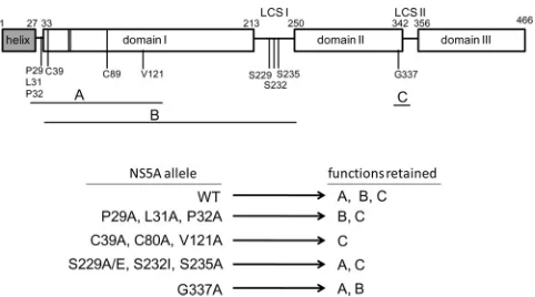
[image:8.585.40.287.87.260.2]si-FIG 7Model of NS5A complementation groups and associated functions. A diagram of the domain organization of the 466-amino-acid JFH1 NS5A pro-tein is shown, based on analogy to the proposed organizational domain struc-ture of the Con1 NS5A protein (7). The positions of three structural domains (domains I, II, and III) and interconnecting low-complexity linker sequences (LCS I and LCS II) are indicated. A membrane-associated N-terminal amphi-pathic␣helix (helix) is connected to domain I by a proposed hinge (aa 28 to 32). Zinc-coordinating residues in domain I (C39, C57, C59, and C80) are shown as vertical lines. The approximate positions of residues shown by mu-tational analysis to be critical for RNA replication are indicated. The horizontal lines beneath the diagram indicate the complementation groups (A, B, and C) to which the mutations affecting these residues were assigned (Table 1). We propose that each complementation group is associated with a distinct NS5A function (also labeled A, B, and C to match the complementation groups) required for RNA replication. The schematic below the NS5A diagram illus-trates the proposed functions retained by the indicated NS5A alleles. Note that C39A, C80A, and V121A belong to complementation groups A and B and therefore are deficient for functions A and B while retaining function C.
TABLE 1Intragenic complementation of replication-defective NS5A allelesa
JFH1-Rluc replicon
Complementation of the following replication-defective NS5A alleleb:
Complementation group(s)c
P32A S232I G337A
P29A ⫺ ⫹ ⫹ A
L31A ⫺ ⫹ ⫹ A
P32A ⫺ ⫹⫹ ⫹ A
C39A ⫺ ⫺ ⫹ A, B
C80A ⫺ ⫺ ⫹ A, B
V121A ⫺ ⫺ ⫹⫹ A, B
S229A ⫹ ⫺ ⫹⫹ B
S229E ⫹⫹ ⫺ ⫹⫹ B
S232I ⫹⫹ ⫺ ⫹⫹ B
S235A ⫹⫹ ⫺ ⫹⫹ B
G337A ⫹ ⫹⫹ ⫺ C
P32A⫹S232I ⫺ ⫺ ⫹ A, B
aPairs of replication-defective replicons were assessed for complementation (Fig. 6). b⫺
, replication window of⬍2;⫹, replication window of 10 to 100;⫹⫹, replication window of⬎100.
c
Group A, unable to complement P32A; group B, unable to complement S232I; group C, unable to complement G337A.
on November 7, 2019 by guest
http://jvi.asm.org/
[image:8.585.301.542.424.558.2]multaneously, such that the inhibitor-sensitive replication
re-sulted from indirecttrans-complementation, and the
inhibitor-resistant replication resulted from directtrans-complementation.
Replicons with C39A and C80A NS5A amino acid
substitu-tions weretrans-complemented very poorly, if at all, even
com-pared to the other replicons that were inefficientlytrans
-comple-mented (Fig. 4A). C39 and C80 are well-characterized residues
that, together with C57 and C59, coordinate a zinc atom that is probably required to maintain the structural integrity of NS5A
domain I (7,9). The inability to successfullytrans-complement
replicons with these mutations could be explained if the misfolded proteins interact nonproductively with other replication complex components and occlude the integration of helper replicon-encoded NS5A into the defective replication complex. Alternatively, since the C39A and C80A amino acid substitutions are predicted to block
NS5A dimerization (41), it is also possible that directtrans
-comple-mentation requires the formation of heterodimers between NS5A molecules expressed from the helper and defective replicons.
In addition totrans-complementation, we were able to
dem-onstrate intragenic complementation of replication-defective NS5A alleles. Our results identified three NS5A complementation
groups (groups A, B, and C [Table 1]), each of which is potentially
associated with a distinct RNA replication function (Fig. 7).
Com-plementation group A, defined by the inability to complement a replicon with a P32A allele, includes mutations affecting residues in the N-terminal region of NS5A domain I (P29A, L31A, P32A, C39A, C80A, and V121A). Residues 29, 31, and 32 are highly
con-served among sequences in the European HCV database (42) and
are located in a proposed hinge region linking the
membrane-anchoring N-terminal amphipathic␣-helix to the zinc-binding
motif of domain I. Residues C39 and C80 are important for zinc
binding, RNA binding, and NS5A dimerization (7,41), and V121,
located in-sheet 5 downstream of the zinc-binding motif, is
required for an interaction with the cellular protein FKBP8 (38).
Among the domain I alleles, P29A, L31A, and P32A were capable of complementing the S232I allele, while C39A, C80A, and V121A were not. These results indicate that the C39A, C80A, and V121A alleles also belong to complementation group B, which was de-fined by the inability to complement the S232I allele and which
also includes the S229A, S229E, and S235A alleles (Table 1).
Rep-licons with C39A, C80A, and V121A amino acid substitutions appeared to express reduced levels of hyperphosphorylated NS5A
compared to the WT replicon control (Fig. 3C), potentially
estab-lishing a phenotypic link between these alleles and the LCS I serine alleles. In fact, accumulating evidence suggests a functional asso-ciation between LCS I and NS5A domain I. For example, Hwang et
al. (13) defined a minimal NS5A RNA binding domain (amino
acids 33 to 249) that requires Zn2⫹and includes LCS I, and Lim et
al. (41) recently showed that the LCS I linker region, in addition to
domain I, is important for efficient NS5A dimerization. These RNA binding and dimerization studies were carried out with
non-phosphorylated NS5A (13,41), implying that NS5A
hyperphos-phorylation is not required for these activities. These findings are consistent with a model in which a hypophosphorylated NS5A dimer or multimer binds RNA and is directly involved in RNA replication, possibly as an integral component of the replication complex. We postulate that it is this function that is retained by the S232I allele but is lacking in complementation group A do-main I alleles. A separate pool of NS5A, possibly made available via hyperphosphorylation, could perform a non-replication-complex
function, for example by interacting with cellular components to establish a favorable environment for RNA replication. This sec-ond function would be retained by the P29A, L31A, and P32A alleles but would be lacking in complementation group B alleles. Recent studies demonstrating an NS5A domain I-mediated
inter-action with phosphatidylinositol 4-kinase III␣ (PI4KIII␣),
required for maintaining the integrity of the membranous
repli-cation compartment (43–45), identify a function that may be
af-fected by mutations belonging to complementation group B. Complementation group C was defined by the inability to complement the G337A NS5A allele. All of the other NS5A mu-tants that we examined, including a double mutant with P32A and S232I amino acid substitutions, were capable of complementing the G337A allele, suggesting that this amino acid substitution is associated with a third NS5A function required for replication (Fig. 7). G337 is located near the C terminus of NS5A domain II within the second of two cyclophilin A (CypA) binding sites
iden-tified within JFH1 NS5A (46). It will be interesting to determine
whether the G337A amino acid substitution affects CypA binding and whether complementation group C is functionally defined by
the interaction between NS5A and CypA. Lim et al. (41) recently
showed that NS5A proteins with C39A and C80A amino acid sub-stitutions retained the ability to bind CypA, suggesting that these mutations do not affect the overall structural integrity of NS5A domain II. These results are consistent with our finding that these alleles can complement the G337A-associated replication defect.
Our results suggest that NS5A performs at least three functions required for RNA replication. These functions are associated with mutations in domains I and II which comprise complementation groups A, B, and C. NS5A also plays a critical role in virion assem-bly, and mutations affecting this process map to the C-terminal
region of NS5A domain III (16,17,47,48). At least some of the
NS5A mutations that affect virion assembly do not affect RNA replication, indicating that the assembly function can be
geneti-cally separated from RNA replication functions (16,17,47). These
findings suggest the likelihood that the assembly mutations define a fourth NS5A complementation group. On the whole, these re-sults support the hypothesis that NS5A is a multifunctional, mod-ular protein with several key roles in the HCV life cycle.
ACKNOWLEDGMENTS
We thank Nick Meanwell and Mark Cockett for leadership and for criti-cally reading the manuscript and our colleagues Donald O’Boyle, Jin-hua Sun, Peter Nower, and Burt Rose for kindly sharing ideas and reagents.
REFERENCES
1.Poenisch M, Bartenschlager R.2010. New insights into structure and replication of the hepatitis C virus and clinical implications. Semin. Liver Dis.30:333–347.
2.Suzuki T, Ishii K, Aizaki H, Wakita T.2007. Hepatitis C viral life cycle. Adv. Drug Deliv. Rev.59:1200 –1212.
3.Cordek DG, Bechtel JT, Maynard AT, Kazmierski WM, Cameron CE.
2011. Targeting the NS5A protein of HCV: an emerging option. Drugs Future36:691–711.
4.Brass V, Bieck E, Montserret R, Wölk B, Hellings JA, Blum HE, Penin F, Moradpour D.2002. An amino-terminal amphipathic␣-helix medi-ates membrane association of the hepatitis C virus nonstructural protein 5A. J. Biol. Chem.277:8130 – 8139.
5.Penin F, Brass V, Appel N, Ramboarina S, Montserret R, Ficheux D, Blum HE, Bartenschlager R, Moradpour D.2004. Structure and func-tion of the membrane anchor domain of hepatitis C virus nonstructural protein 5A. J. Biol. Chem.279:40835– 40843.
6.Shi ST, Lee K-J, Aizaki H, Hwang SB, Lai MMC.2003. Hepatitis C virus
on November 7, 2019 by guest
http://jvi.asm.org/
RNA replication occurs on a detergent-resistant membrane that cofrac-tionates with caveolin-2. J. Virol.77:4160 – 4168.
7.Tellinghuisen TL, Marcotrigiano J, Gorbalenya AE, Rice CM.2004. The NS5A protein of hepatitis C virus is a zinc metalloprotein. J. Biol. Chem.
279:48576 – 48587.
8.Love RA, Brodsky O, Hickey MJ, Wells PA, Cronin CN.2009. Crystal structure of a novel dimeric form of NS5A domain I protein from hepatitis C virus. J. Virol.83:4395– 4403.
9.Tellinghuisen TL, Marcotrigiano J, Rice CM. 2005. Structure of the zinc-binding domain of an essential component of the hepatitis C virus replicase. Nature435:374 –379.
10. Tellinghuisen TL, Foss KL, Treadaway JC, Rice CM.2008. Identification of residues required for RNA replication in domains II and III of the hepatitis C virus NS5A protein. J. Virol.82:1073–1083.
11. Foster TL, Belyaeva T, Stonehouse NJ, Pearson AR, Harris M.2010. All three domains of the hepatitis C virus nonstructural NS5A protein con-tribute to RNA binding. J. Virol.84:9267–9277.
12. Huang L, Hwang J, Sharma SD, Hargittai MRS, Chen Y, Arnold JJ, Raney KD, Cameron CE.2005. Hepatitis C virus nonstructural protein 5A (NS5A) is an RNA-binding protein. J. Biol. Chem.280:36417–36428. 13. Hwang J, Huang L, Cordek DG, Vaughan R, Reynolds SL, Kihara G, Raney KD, Kao CC, Cameron CE.2010. Hepatitis C virus nonstructural protein 5A: biochemical characterization of a novel structural class of RNA-binding proteins. J. Virol.84:12480 –12491.
14. Hanoulle X, Verdegem D, Badillo A, Wieruszeski J-M, Penin F, Lippens G.2009. Domain 3 of non-structural protein 5A from hepatitis C virus is natively unfolded. Biochem. Biophys. Res. Commun.381:634 – 638. 15. Liang Y, Ye H, Kang CB, Yoon HS.2007. Domain 2 of nonstructural
protein 5A (NS5A) of hepatitis C virus is natively unfolded. Biochemistry (Mosc.)46:11550 –11558.
16. Appel N, Zayas M, Miller S, Krijnse-Locker J, Schaller T, Friebe P, Kallis S, Engel U, Bartenschlager R.2008. Essential role of domain III of nonstructural protein 5A for hepatitis C virus infectious particle assembly. PLoS Pathog.4:e1000035. doi:10.1371/journal.ppat.1000035.
17. Tellinghuisen TL, Foss KL, Treadaway J.2008. Regulation of hepatitis C virion production via phosphorylation of the NS5A protein. PLoS Pathog.
4:e1000032. doi:10.1371/journal.ppat.1000032.
18. Koch OJ, Bartenschlager R.1999. Modulation of hepatitis C virus NS5A hyperphosphorylation by nonstructural proteins NS3, NS4A, and NS4B. J. Virol.73:7138 –7146.
19. Neddermann P, Clementi A, De Francesco R.1999. Hyperphosphory-lation of the hepatitis C virus NS5A protein requires an active NS3 pro-tease, NS4A, NS4B, and NS5A encoded on the same polyprotein. J. Virol.
73:9984 –9991.
20. Kaneko T, Tanji Y, Satoh S, Hijikata M, Asabe S, Kimura K, Shimotohno K.1994. Production of two phosphoproteins from the NS5A region of the hepatitis C viral genome. Biochem. Biophys. Res. Commun.205:320 –326. 21. Tanji Y, Kaneko T, Satoh S, Shimotohno K.1995. Phosphorylation of
hepatitis C virus-encoded nonstructural protein NS5A. J. Virol.69:3980 – 3986.
22. Huang Y, Staschke K, De Francesco R, Tan S-L.2007. Phosphorylation of hepatitis C virus NS5A nonstructural protein: a new paradigm for phos-phorylation-dependent viral RNA replication? Virology364:1–9. 23. Egger D, Wolk B, Gosert R, Bianchi L, Blum HE, Moradpour D, Bienz
K.2002. Expression of hepatitis C virus proteins induces distinct mem-brane alterations including a candidate viral replication complex. J. Virol.
76:5974 –5984.
24. Mackenzie J.2005. Wrapping things up about virus RNA replication. Traffic6:967–977.
25. Appel N, Herian U, Bartenschlager R.2005. Efficient rescue of hepatitis C virus RNA replication by trans-complementation with nonstructural protein 5A. J. Virol.79:896 –909.
26. Fridell RA, Qiu D, Valera L, Wang C, Rose RE, Gao M.2011. Distinct functions of NS5A in hepatitis C virus RNA replication uncovered by studies with the NS5A inhibitor BMS-790052. J. Virol.85:7312–7320. 27. Jones DM, Patel AH, Targett-Adams P, McLauchlan J.2009. The
hep-atitis C virus NS4B protein can trans-complement viral RNA replication and modulates production of infectious virus. J. Virol.83:2163–2177. 28. Tong X, Malcolm BA.2006. Trans-complementation of HCV replication
by non-structural protein 5A. Virus Res.115:122–130.
29. Fridell RA, Qiu D, Wang C, Valera L, Gao M.2010. Resistance analysis of the hepatitis C virus NS5A inhibitor BMS-790052 in an in vitro replicon system. Antimicrob. Agents Chemother.54:3641–3650.
30. Lemm JA, Liu M, Rose RE, Fridell R, O’Boyle DR, II, Colonno R, Gao M.2005. Replication-competent chimeric hepatitis C virus subgenomic replicons. Intervirology48:183–191.
31. Gao M, Nettles RE, Belema M, Snyder LB, Nguyen VN, Fridell RA, Serrano-Wu MH, Langley DR, Sun J-H, O’Boyle DR, II, Lemm JA, Wang C, Knipe JO, Chien C, Colonno RJ, Grasela DM, Meanwell NA, Hamann LG.2010. Chemical genetics strategy identifies an HCV NS5A inhibitor with a potent clinical effect. Nature465:96 –100.
32. O’Boyle DR, II, Nower PT, Lemm JA, Valera L, Sun J-H, Rigat K, Colonno R, Gao M.2005. Development of a cell-based high-throughput specificity screen using a hepatitis C virus-bovine viral diarrhea virus dual replicon assay. Antimicrob. Agents Chemother.49:1346 –1353. 33. Qiu D, Lemm JA, O’Boyle DR, Sun J-H, Nower PT, Nguyen V, Hamann
LG, Snyder LB, Deon DH, Ruediger E, Meanwell NA, Belema M, Gao M, Fridell RA.2011. The effects of NS5A inhibitors on NS5A phosphorylation, polyprotein processing and localization. J. Gen. Virol.92:2502–2511. 34. Appel N, Pietschmann T, Bartenschlager R.2005. Mutational analysis of
hepatitis C virus nonstructural protein 5A: potential role of differential phosphorylation in RNA replication and identification of a genetically flexible domain. J. Virol.79:3187–3194.
35. Curmi PA, Maucuer A, Asselin S, Lecourtois M, Chaffotte A, Schmitter JM, Sobel A.1994. Molecular characterization of human stathmin ex-pressed in Escherichia coli: site-directed mutagenesis of two phosphory-latable serines (Ser-25 and Ser-63). Biochem. J.300(Part 2):331–338. 36. Sakamoto K, Huang B-W, Iwasaki K, Hailemariam K, Ninomiya-Tsuji J,
Tsuji Y.2010. Regulation of genotoxic stress response by homeodomain-interacting protein kinase 2 through phosphorylation of cyclic AMP response element-binding protein at serine 271. Mol. Biol. Cell21:2966 –2974. 37. Blight KJ, Kolykhalov AA, Rice CM.2000. Efficient initiation of HCV
RNA replication in cell culture. Science290:1972–1974.
38. Okamoto T, Omori H, Kaname Y, Abe T, Nishimura Y, Suzuki T,
Miyamura T, Yoshimori T, Moriishi K, Matsuura Y.2008. A single-amino-acid mutation in hepatitis C virus NS5A disrupting FKBP8 inter-action impairs viral replication. J. Virol.82:3480 –3489.
39. Neddermann P, Quintavalle M, Di Pietro C, Clementi A, Cerretani M, Altamura S, Bartholomew L, De Francesco R.2004. Reduction of hepatitis C virus NS5A hyperphosphorylation by selective inhibition of cellular kinases activates viral RNA replication in cell culture. J. Virol.78:13306 –13314. 40. Evans MJ, Rice CM, Goff SP.2004. Genetic interactions between
hepa-titis C virus replicons. J. Virol.78:12085–12089.
41. Lim PJ, Chatterji U, Cordek D, Sharma SD, Garcia-Rivera JA, Cameron CE, Lin K, Targett-Adams P, Gallay PA.2012. Correlation between NS5A dimerization and hepatitis C virus replication. J. Biol. Chem.287:30861– 30873.
42. Combet C, Garnier N, Charavay C, Grando D, Crisan D, Lopez J, Dehne-Garcia A, Geourjon C, Bettler E, Hulo C, Mercier PL, Barten-schlager R, Diepolder H, Moradpour D, Pawlotsky J-M, Rice CM, Trepo C, Penin F, Deleage G.2007. euHCVdb: the European hepatitis C virus database. Nucleic Acids Res.35:D363–D366.
43. Lim Y-S, Hwang SB.2011. Hepatitis C virus NS5A protein interacts with phosphatidylinositol 4-kinase type III␣and regulates viral propagation. J. Biol. Chem.286:11290 –11298.
44. Tai AW, Salloum S.2011. The role of the phosphatidylinositol 4-kinase PI4KA in hepatitis C virus-induced host membrane rearrangement. PLoS One6:e26300. doi:10.1371/journal.pone.0026300.
45. Reiss S, Rebhan I, Backes P, Romero-Brey I, Erfle H, Matula P, Kaderali L, Poenisch M, Blankenburg H, Hiet MS, Longerich T, Diehl S, Ramirez F, Balla T, Rohr K, Kaul A, Bühler S, Pepperkok R, Lengauer T, Albrecht M, Eils R, Schirmacher P, Lohmann V, Bartenschlager R.
2011. Recruitment and activation of a lipid kinase by hepatitis C virus NS5A is essential for integrity of the membranous replication compart-ment. Cell Host Microbe9:32– 45.
46. Grisé H, Frausto S, Logan T, Tang H. 2012. A conserved tandem cyclophilin-binding site in hepatitis C virus nonstructural protein 5A reg-ulates Alisporivir susceptibility. J. Virol.86:4811– 4822.
47. Hughes M, Griffin S, Harris M.2009. Domain III of NS5A contributes to both RNA replication and assembly of hepatitis C virus particles. J. Gen. Virol.90:1329 –1334.
48. Masaki T, Suzuki R, Murakami K, Aizaki H, Ishii K, Murayama A, Date T, Matsuura Y, Miyamura T, Wakita T, Suzuki T.2008. Interaction of hepatitis C virus nonstructural protein 5A with core protein is critical for the production of infectious virus particles. J. Virol.82:7964 –7976.