0022-538X/08/$08.00⫹0 doi:10.1128/JVI.01022-08
Copyright © 2008, American Society for Microbiology. All Rights Reserved.
Intrahepatic Expression of Genes Affiliated with Innate and Adaptive
Immune Responses Immediately after Invasion and during Acute
Infection with Woodchuck Hepadnavirus
䌤
Clifford S. Guy,
1Patricia M. Mulrooney-Cousins,
1Norma D. Churchill,
1and Tomasz I. Michalak
1,2*
Molecular Virology and Hepatology Research Group, Division of BioMedical Science,1and Discipline of Laboratory Medicine,2
Faculty of Medicine, Health Science Centre, Memorial University, St. John’s, Newfoundland, Canada
Received 15 May 2008/Accepted 23 June 2008
The importance of effective immune responses in recovery from acute hepadnaviral hepatitis has been demonstrated. However, there is no conclusive delineation of virological and immunological events occurring in the liver immediately after hepadnavirus invasion and during the preacute phase of infection. These very early events might be of primary importance in determining the recovery or progression to chronic hepatitis and the intrinsic hepadnaviral propensity to persist. In this study, applying the woodchuck model of acute hepatitis B, the hepatic kinetics of hepadnavirus replication and activation of genes encoding cytokines, cytotoxicity effectors, and immune cell markers were quantified in sequential liver biopsies collected from 1 h postinoculation onward by sensitive real-time cDNA amplification assays. The results revealed that hepadna-virus replication is established in the liver as early as 1 hour after infection. In 3 to 6 h, significantly augmented intrahepatic transcription of gamma interferon and interleukin-12 were evident, suggesting activation of antigen-presenting cells. In 48 to 72 h, NK and NKT cells were activated and virus replication was transiently but significantly reduced, implying that this early innate response is at least partially successful in limiting virus propagation. Nonetheless, T cells were activated 4 to 5 weeks later when hepatitis became histologically evident. Collectively, our data demonstrate that virus replication is initiated and the innate response activated in the liver soon after exposure to a liver-pathogenic dose of hepadnavirus. Nevertheless, this response is unable to prompt a timely adaptive T-cell response, in contrast to infections caused by other viral pathogens.
Hepatitis B virus (HBV) is the prototypic member of the Hepadnaviridaefamily of small, enveloped, primarily hepato-tropic DNA viruses which cause acute and chronic hepatitis and hepatocellular carcinoma (reviewed in reference 9). Hep-adnaviruses display highly restricted host specificity. Thus, HBV infection is limited to humans and higher primates, while woodchuck hepatitis virus (WHV), despite sharing significant structural, genomic, and antigenic similarities with HBV, in-fects the eastern North American woodchuck (Marmota monax) (38). Notwithstanding their limited host ranges, HBV and WHV induce similar courses of hepatitis, which are pre-ceded by a long incubation period ranging from 6 weeks to 6 months for hepatitis B and from 4 to 10 weeks for WHV hepatitis (9, 34). In contrast with the appearance of viremia and adaptive T-cell immunity a few days after invasion with most viral pathogens, a significant increase in HBV replication is not observed until 3 to 5 weeks postinfection (p.i.) in chim-panzees, while activation of HBV-specific adaptive immunity occurs several weeks later (4, 17, 59).
Despite the delay in activation of an HBV-specific T-cell response, a robust, multispecific T-cell reactivity seems to be essential for both induction of acute hepatitis (AH) and the clinical recovery (50, 51), although it is unable to clear the virus entirely (43, 55, 56). Experimental evidence acquired from the
chimpanzee and HBV-transgenic mouse systems, as well as the woodchuck model of hepatitis B, has clearly indicated the importance of virus-specific CD4⫹and CD8⫹T lymphocytes and antiviral cytokines, such as alpha interferon, beta inter-feron, gamma interferon (IFN-␥), and tumor necrosis factor alpha (TNF-␣), in the downregulation of virus replication (21, 35, 36, 48), while bystander recruitment of virus-nonspecific T cells and other immune cell subsets greatly contributes to liver inflammation and hepatocyte destruction (30, 58). In addition, the role of antiviral cytokines, particularly IFN-␥, has been ascribed to the ability to inhibit replication of hepadnavirus in a noncytopathic manner (15, 17). In this regard, elevations in the intrahepatic expression of IFN-␥appeared sufficient in chimpanzees to reduce HBV viremia before the peak onset of hepatitis (17). Also, an increased intrahepatic expression of IFN-␥was consistently detected for many years following re-covery from WHV-induced AH, implying its role in control of residual (occult) WHV replication continuing after resolution of acute disease (21).
Natural killer (NK) and NK T (NKT) cells are components of the innate immune system. They are enriched among the resident lymphomononuclear cells in the liver and are capable of producing IFN-␥and other cytokines within minutes follow-ing viral invasion (reviewed in reference 8). The results from studies with HBV transgenic mice suggest that activation of NKT cells via CD1d-restricted␣-galactosylceramide can down-regulate virus replication (22, 23), while nonclassical NKT cells may recognize HBV antigens expressed in the liver (1). It has been suggested that NK- or NKT-derived IFN-␥may be the principal mediator of HBV downregulation in acutely infected * Corresponding author. Mailing address: Molecular Virology and
Hepatology Research Group, Faculty of Medicine, Health Sciences Centre, Memorial University, St. John’s, NL, Canada A1B 3V6. Phone: (709) 777 7301. Fax: (709) 777 8279. E-mail: timich@mun.ca.
䌤Published ahead of print on 2 July 2008.
8579
on November 8, 2019 by guest
http://jvi.asm.org/
chimpanzees (17), while indirect evidence from the woodchuck model suggests that activated, cytotoxic NK or NKT cells may contribute to hepatocyte killing and recovery from WHV in-fection (20).
Nonetheless, despite findings suggesting that hepadnavi-ruses may directly activate intrahepatic immune cells capable of producing cytokines promoting antiviral defense and favor-ing a T-helper-cell type 1 response (17, 59), comprehensive data from a natural model of hepadnaviral infection are lack-ing. Furthermore, characterization of the liver immune re-sponse in the first hours and days following hepadnavirus in-vasion has not yet been accomplished. The aim of the current study was to recognize, using the woodchuck model of hepatitis B, the nature and the kinetics of intrahepatic immune re-sponses occurring soon after exposure to WHV and during the preacute and acute phases of hepadnaviral infection by quan-tifying hepatic transcriptional activities of genes encoding crit-ical proinflammatory and antiviral cytokines and markers spec-ifying individual immune cell subsets.
MATERIALS AND METHODS
Study design.Prior to inoculation with WHV, liver biopsies (designated LBx 1) (Fig. 1) were obtained by laparotomy from 29 young-adult, healthy, WHV-naive woodchucks and their fragments were cryopreserved for subsequent DNA and RNA isolations and fixed in formalin for histological examination. All woodchucks were intravenously infected with the same WHV/tm3 inoculum
carrying wild-type WHV at a dose of 1.1⫻1010DNase-protected virus genome
equivalents (vge) (GenBank accession number AY334075) (41). The animals were randomly subdivided into groups, each comprised of at least two animals (Table 1). Beginning at 1 h post-WHV injection, liver biopsies (designated LBx 2) were collected according to the scheme shown in Fig. 1. Subsequent liver samples were obtained 6 weeks thereafter (designated LBx 3) and at the end of follow-up (designated LBx 4) (Fig. 1). Serum samples were collected weekly until 10 weeks p.i. and then monthly for up to 6 to 36 months postinoculation. Animals were housed in the Woodchuck Hepatitis Research Facility at Memorial Uni-versity, St. John’s, Newfoundland, Canada. All animal experimental protocols were approved by the Institutional Committee on Animals Bioethics and Care.
Serological assays and assessment of liver inflammation.Detection of serum WHV surface antigen (WHsAg) and antibodies to WHV core antigen (anti-WHc) was done using specific immunoassays, as previously described (10, 41). Histological examination of paraffin-embedded liver tissue fragments was per-formed after routine processing and staining, as described elsewhere (21, 42).
The morphological assessment of hepatocellular and extrahepatocellular in-tralobular and periportal lesions and overall degree of hepatitis was graded on a numerical scale from 0 to 3, applying criteria previously described (21, 39, 40).
Nucleic acid isolations.Total DNA was extracted from 250-l serum samples or approximately 50 mg of liver tissue using proteinase K digestion and the phenol-chloroform-isoamyl alcohol method as previously described (42). Total RNA was isolated from liver biopsy samples using Trizol reagent (Invitrogen Life Technologies, Burlington, Ontario, Canada), as per the manufacturer’s instruc-tion. RNA was treated with DNase to remove potentially contaminating DNA using an RNase-free DNase digestion kit (Sigma Chemical Company, Oakville, Ontario, Canada) prior to reverse transcription to cDNA, as previously described (18).
Detection of WHV DNA and RNA by real-time PCR.Quantitative assessment of the WHV DNA load was done by real-time PCR analysis using DNA derived
from the equivalent of 25l of serum or 50 ng of liver biopsy sample DNA and
the Roche LightCycler instrument (Roche Diagnostics, Laval, Quebec, Canada).
WHV DNA amplicons were generated using the forward primer 5⬘-ATGCAC
CCATTCTCTCGAC and the reverse primer 5⬘-CTGAGCAGCTTGGTTAG
AGT, yielding a 221-bp fragment which was detected by Sybr green I incorpo-ration. Copy numbers of WHV DNA were calculated by extrapolation from a standard curve which was generated using 10-fold serial dilutions of plasmid
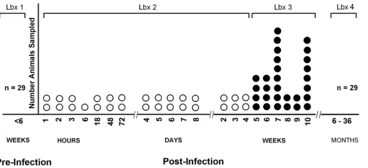
FIG. 1. Schematic representation of the timeline of acquisition of liver tissue samples from a cohort of WHV-infected woodchucks investigated in the current study. Liver biopsies (LBx 1) were obtained from each healthy animal prior to inoculation with WHV (n⫽29). The animals were then blindly randomized and each injected with 1.1⫻1010vge of WHV. The second liver biopsy (LBx 2) was taken beginning at 1 h p.i. as shown
[image:2.585.114.473.66.230.2]in the scheme. The third liver biopsy (LBx 3) was collected 6 weeks after Lbx 2. The animals (n⫽29) were followed for 6 to 36 months prior to autopsy (Lbx 4).
TABLE 1. Experimental groups according to the time elapsing between inoculation with WHV and acquisition of liver
biopsy sample
Time period post-WHV inoculation
No. of liver biopsies
Liver biopsy sequential no.
0 29 1
1–3 h 5 2
3–6 h 2 2
18–48 h 3 2
48–72 h 3 2
4–8 days 10 2
2 wk 2 2
3 wk 2 2
4 wk 2 2
5 wk 4 3
6 wk 4 3
7 wk 9 3
8 wk 2 3
9 wk 2 3
10 wk 8 3
⬎6 mo 29 4
on November 8, 2019 by guest
http://jvi.asm.org/
containing known copy numbers of complete, recombinant WHV DNA. WHV RNA was similarly detected using 50 ng liver total RNA. The specificity of real-time PCR products was always confirmed by nucleic acid hybridization (NAH), i.e., Southern blot hybridization analysis, using complete recombinant WHV DNA as a probe and autoradiography, as previously described (10, 41).
The sensitivity of the real-time PCR assay was⬃200 vge/g DNA,⬃200
cop-ies/g RNA, or⬃50 vge/ml serum.
Detection of WHV cccDNA.WHV covalently closed circular DNA (cccDNA), representing virus genome replicative intermediate, was detected in liver tissue
samples by PCR amplification with a sensitivity of 102
vge/g total DNA, as
previously described in detail (26, 41). Briefly, 4g of DNA was digested with
mung bean nuclease prior to PCR amplification with oligonucleotide primers spanning the nicked region of the partially double-stranded WHV DNA genome. The specificity of PCR amplicons was routinely confirmed by NAH, as previously described (10, 42).
Identification of woodchuck cellular gene sequences.To facilitate analysis of the spectrum and the dynamics of intrahepatic immune response in WHV in-fection, a number of woodchuck gene sequences encoding markers specifying different immune cell subtypes, immune cell effector molecules, and cytokines were determined, applying a strategy previously reported (18). In general, wood-chuck gene sequences were identified by reverse transcription-PCR (RT-PCR) using degenerate oligonucleotide primers whose sequences were deduced through interspecies comparison of the sequences available in GenBank. The resulting amplicons were cloned into the PCRII TOPO TA cloning system (Invitrogen) and the excised fragments sequenced in both directions. Based on the woodchuck sequences determined, the pairs of gene-specific primers were designed. Table 2 presents the list of woodchuck genes for which hepatic expres-sion was quantified in the current study and the primer pairs used for their quantitative detection.
Analysis of woodchuck gene expression by real-time PCR.Real-time RT-PCR assays were developed for each cellular effector molecule and the cytokine gene analyzed, using the Roche LightCycler instrument with Sybr green I detection and PCR primer pairs specified in Table 2. Changes in gene expression levels were determined by comparison to the baseline level of each gene’s transcription detected in the liver biopsy sample obtained prior to infection with WHV for each individual animal after normalization to expression of the housekeeping
gene-actin. Following measurement of a given gene’s expression in a particular
[image:3.585.43.285.107.357.2]liver sample from an individual animal, the mean expression level was deter-mined for all liver biopsy samples within the experimental groups showed in Table 1.
Inactivation of WHV inoculum.To ascertain that the observed changes in the intrahepatic gene expression levels were truly related to infection with WHV but not to injection with serum components present in the WHV inoculum, a control experiment was performed. Thus, the WHV/tm3 inoculum was inactivated by
treatment with 50g/ml psoralen (Sigma) combined with exposure to 365 nm
UV light for 90 min at 4°C. Animals were injected intravenously with 0.5 ml of
psoralen-inactivated inoculum containing the equivalent of 1.1⫻1010
vge or with 0.5 ml of similarly treated healthy woodchuck serum prior to euthanasia at 3 days postinjection.
Statistical analysis.A two-tailed, unpaired Studentttest with 95% confidence
interval was applied to compare the means of sample groups investigated, andP
values of⬍0.05 were considered statistically significant.
Accession numbers of woodchuck gene sequences identified.The accession numbers for the woodchuck gene sequences established in the course of this study have been submitted to GenBank under the following accession numbers: for the CD4 gene, EF621765; for the CD8 gene, EF621766; for the CD40 ligand (CD40L), EF621170; for CD1d, EF621767; for NKp46, EF621768; and for in-terleukin-8 (IL-8), EF-126348.
RESULTS
Serologic and hepatic profiles of WHV infection.Inoculation of woodchucks with a WHV dose of 1.1⫻1010vge resulted in
transiently serum WHsAg-positive, self-limiting acute infec-tion in all 29 animals. The mean time of WHsAg appearance was 18.5 days, while its average duration in the circulation was 35.8 days. Anti-WHc remained detectable until the end of the observation period (Fig. 2A). The pattern of serum WHsAg positivity implied, based on the known profiles of serological markers of WHV infection (41, 42), that a self-limited episode of AH had developed in all woodchucks investigated. This was confirmed by histological examination of serial liver biopsy samples collected.
Considering morphological alterations encountered in liver tissue, their characteristics and the timing of their occurrence closely followed those previously reported for experimental acute WHV infection (38, 42). In brief, the appearance of scattered lymphocytes and neutrophils in sinusoids and peri-portal areas, accompanied by mild proliferations of sinusoidal lining endothelium and bile ducts, was the first manifestation, which occurred at 4 to 5 weeks p.i. In the next few days, scattered lobular infiltrations, consisting mainly of lympho-cytes, degenerative or necrotic changes of single hepatolympho-cytes, progressing hyperplasia of Kupffer cells, and limited portal infiltrations, occurred. Subsequently, more-severe lobular in-filtrations and hepatocyte degenerative and necrotic changes and the appearance of acidophilic bodies were observed. At the peak of AH, usually occurring between weeks 6 and 8 p.i., both lobular and periportal inflammatory changes were evi-dent, the portal infiltrations became most intense and ex-panded occasionally through limiting plates, and multifocal necroses of parenchyma with varying numbers of acidophilic bodies were found. However, the severity of AH varied, and while the changes in some woodchucks were intense, reaching histological degree of hepatitis 2.5, in others they were mild and hepatitis did not exceed grade 1.5. Following resolution of AH, intermittent minimal lobular and portal inflammatory al-terations and periods of normal or nearly normal liver mor-phology were observed, as previously reported (21, 41, 42).
In previous studies, WHV DNA in the circulation and the TABLE 2. Primer sequences used in this study for real-time
RT-PCR quantifications of expression of woodchuck genes encoding immune cell markers, cytokines,
and cytotoxicity effector molecules
Gene Primer sequencea
Expected size of amplicon (bp) GenBank accession no.
CD3 F-CTGGGACTCTGCCTCTTATC 536 AF232727
R-GCTGCCCTTTCCGGATGGGCTC
CD4b F-GGAGAATAAGAAGATAGAGG 560 EF621765
R-TCAAGAGTCACAGTCAGG
CD8b F-AACGAGGGCTACTATTTCTGCTC 309 EF621766
R-GTTTCCGGTGGTGACAGATGA
CD40 ligandb F-AGCATGTGTGCTACAGT 214 EF621770
R-CCGCCCTGAGTAAGATT
CD1db F-TCCTAGATTAGGGAAGTCAGAAC 222 EF621767
R-GCTCGGAGATACCACG
NKp46b F-TTGCCACCTAGTGACAG 200 EF621768
R-CACCAGGAGCATCACC
CD95 ligandb F-CCATTTAACAGGTAAGCCC 250 AF152368
R-TCATCATCTTGCCCTCC
Perforinb F-GCATCAACAATGACTGGCGGG 302 AF298158
R-TGAAGTGGGTGCCGTAGTTGTGG
IL-4 F-TTTGCTGTCCCCAAGAAC 200 AF333965
R-CCTGGATTCACTCACGG
IL-8b F-GGTAACCTGCCTACTTTC 207 EF216348
R-GTTCAGGCAAAGCTCT
IL12p40 F-tTGGATTGGCACCCTGACAC 198 X97019
R-GCATCTGGCTCAGAACTTCAC
IFN-␥ F-AGGAGCATGGACACCATCA 215 AF232728
R-cCGACCCCGAATCGAAG
TNF-␣ F-TGAGCACTGAAAGTATGATCC 283 AF333967
R-TGCTACAACATGGGCTACAG
2⬘,5⬘-OAS F-TCAGGCAAAGGCACTACCC 150 AF082498
R-aCTTCTCTTTCGGACATGCT
a
F, forward primer; R, reverse primer.
b
Woodchuck gene sequence identified in the current study.
on November 8, 2019 by guest
http://jvi.asm.org/
virus replication status in the liver were never assessed during the first few hours or days after exposure to the virus. In the current study, WHV DNA was quantified in serum and liver tissue acquired from 1 h p.i. onward. Not surprisingly, WHV DNA was detectable in serum at the first time point of exam-ination, i.e., 1 h p.i. (Fig. 2A). This reflected the carryover of WHV from the inoculum containing a massive amount of virus, i.e., 1.1⫻1010DNase-protected vge. Subsequently, the
serum level of WHV DNA progressively decreased until 48 to 72 h p.i. and then slowly increased until a sudden expansion at 3 to 4 weeks p.i. (Fig. 2A), culminating in peak detection at week 7 p.i. (a mean level of 3.2 ⫻ 1010vge/ml). The mean
WHV DNA level exhibited minor fluctuations during the peak acute phase of WHV infection, occurring between weeks 6 and 9 p.i., prior to a sudden protraction by more than 7 logs after week 9 p.i. (Fig. 2A). However, in agreement with our previous findings (10, 21, 42), WHV DNA remained consistently detect-able in serum at a mean level of 175 vge/ml during follow-up lasting up to 3 years p.i.
WHV DNA and WHV mRNA transcripts were also
detect-able in hepatic tissue beginning at 1 h after inoculation with virus. The WHV DNA load transiently but significantly (P⫽ 0.004) and uniformly declined between 48 to 72 h p.i. in all animals, reaching a level approximately 10-fold lower than that detected between 1 and 6 h p.i. (Fig. 2A). Paralleling WHV DNA detection in serum, the intrahepatic level of WHV DNA exhibited an exponential increase at 3 to 4 weeks p.i. (Fig. 2A) prior to reaching the mean peak detection of 4⫻109vge/g
total DNA at 7 weeks p.i. Despite a greater than 4-log reduc-tion in the hepatic WHV DNA load beginning at week 9 p.i., WHV DNA was consistently detectable in the liver for up to 3 years p.i. at a mean level of 2.3⫻104vge/g total liver DNA.
It remained unclear whether the WHV DNA detected in the liver in the first few hours postexposure reflected the virus originating from the inoculum, which was passing through or was trapped within hepatic tissue, or the virus actively repli-cating in hepatocytes. However, the finding of a significant lowering in the hepatic load of WHV DNA at 48 to 72 h p.i., which was replenished 24 h later, strongly suggested that the virus was already actively replicating, at least from 96 h p.i. forward.
To directly determine the status of WHV replication in the liver, real-time RT-PCR detecting WHV mRNA was applied. The results showed that low quantities of WHV transcripts (2.6⫻102copies/g total RNA) were identifiable as early as
1 h p.i. (Fig. 2A). In contrast with hepatic WHV DNA, the WHV RNA level progressively increased in the liver and achieved the maximum at a mean level of 7.2⫻108copies per
g total RNA between weeks 8 and 9 p.i. (Fig. 2A). A possi-bility of contamination of cDNA preparations with viral DNA was excluded by DNase treatment of RNA samples prior to the RT reaction and by PCR amplification of both transcribed and nontranscribed mRNA preparations. As shown in Fig. 2B, only RNA samples which were reverse transcribed demonstrated WHV cDNA signals, while those similarly treated in the ab-sence of reverse transcriptase remained negative even when the amplification products were analyzed by NAH. This con-firmed strict specificity of the WHV RNA detections. Taken together, the results demonstrated that infection with a mas-sive, liver-pathogenic dose of WHV results in almost immedi-ate establishment, although at a low level, of hepadnavirus replication in the liver, as evidenced by detection of viral tran-scripts as early as 1 h p.i.
In addition, we analyzed expression of WHV cccDNA by a PCR/NAH assay specifically identifying this replicating DNA intermediate. The results revealed that the WHV cccDNA signal was detectable in the liver from 18 h p.i. onward (data not shown).
WHV invasion promptly activates APC in the liver. Since injection with WHV resulted in the initiation of virus replica-tion as early as 1 h p.i., it was of interest to determine whether the virus may at this early stage activate antigen-presenting cells (APC), which should be the first cell type recognizing viral intrusion. In this regard, the liver expression of IL-12, a key cytokine produced by APC involved in innate immune re-sponses (29), IL-8, a strong chemoattractant mediating chemo-taxis of phagocytic cells (24), CD1d, a key molecule facilitating antigen presentation by APC to NKT cells (1, 25), and CD40L, involved in activation of APC via CD40-CD40L, was quantified FIG. 2. Results on serological and molecular analyses of WHV
infection markers after inoculation with WHV at 1.1⫻1010vge per
animal. (A) Circulating WHsAg and antibodies to WHV core antigen (anti-WHc) were detected by specific enzyme-linked immunosorbent assay in serial serum samples collected as described in Materials and Methods. The mean times of the appearance and duration of elevated levels of serum sorbitol dehydrogenase, a biochemical indicator of hepatic injury in woodchucks, were extrapolated from another group of 40 animals intravenously infected with the same liver-pathogenic dose of WHV (N. D. Churchill and T. I. Michalak, unpublished data). WHV DNA was quantified by real-time PCR in DNA extracted from serial serum or liver biopsy samples. WHV RNA levels were deter-mined by real-time RT-PCR using cDNA transcribed from total RNA isolated from liver biopsies. WHV DNA or WHV RNA copy numbers are presented as the mean expression levels determined for each group of animals as defined in Table 1. (B) Identification of WHV mRNA in liver biopsy samples collected between 1 and 3 h post-infection with WHV. Total RNA was extracted from liver tissue samples obtained at 1 h (n⫽2), 2 h (n⫽2), and 3 h (n⫽2) after infection. RNA samples were treated with DNase prior to transcription (RT⫹) or not (RT⫺) to cDNA, as described in Materials and Methods. WHV cDNA equiv-alents of 50 ng of total RNA were quantified by real-time PCR (not shown), and the amplicons derived were separated by agarose gel electrophoresis and analyzed by Southern blot hybridization using32
P-labeled complete, recombinant WHV DNA as a probe.
on November 8, 2019 by guest
http://jvi.asm.org/
by using real-time RT-PCR assays specifically developed for the purpose of this study.
Figure 3 shows that IL-12 achieved a maximum hepatic expression (⬃20-fold increase over the preinfection level;P⫽ 0.02) between 3 and 6 h p.i. (Fig. 3A, left panel). Interestingly, in contrast with this early increase, the remaining course of WHV infection, including the period of histologically evident AH with peaking histologically evident liver injury occurring between weeks 6 and 8 p.i. (Fig. 3A, right panel), was without any noticeable increase in expression of this cytokine.
The transcription level of IL-8 tended to rise (a 12.5-fold increase) within 1 to 3 h p.i. above that detected in the period prior to infection; however, this increase did not reach a sta-tistically significant value (P⫽0.1) (Fig. 3A, left panel). The maximum expression of the cytokine (a 35-fold increase over the preinfection level;P⫽0.05) occurred at week 7 p.i., during the phase of acute liver inflammation (Fig. 3A, right panel).
Since a strong induction of IL-12 expression detected at 3 to 6 h p.i. could be directly associated with activation of APC (29), the intrahepatic expression of CD1d, which facilitates antigen presentation to NKT cells, was also evaluated. The results showed that the level of CD1d mRNA reached a peak (a⬃3-fold increase over the preinfection level;P⫽0.03) by 48 to 72 h p.i. (Fig. 3B, left panel). Then, the level subsided until WHV replication was drastically augmented at 3 to 4 weeks p.i.
(Fig. 3B, right panel). Thus, the upregulated transcription of CD1d at 48 to 72 h was associated with a significant one-log decrease in the hepatic WHV DNA load and a decline of WHV DNA in circulation. Furthermore, the concomitant in-crease (P⫽ 0.02) in CD40L expression (Fig. 3B, left panel) suggested activation of APC via CD40-CD40L interaction.
Activation of hepatic NK and NKT cells follows infection with WHV.WHV infection induced two distinct phases of very early immune activation in the liver, one between 3 and 6 h and a second at 48 to 72 h p.i. Since experimental evidence from other viral infections indicates the ability of NK and NKT cells to respond within minutes to hours to virus by secretion of IFN-␥or by acquisition of cytotoxic function (8), expression of the gene encoding the NK receptor NKp46 was investigated. Furthermore, since IL-12 is recognized as a key cytokine which influences IFN-␥ secretion by NK cells and also increases NK-mediated cytotoxicity, which is primarily facilitated by the perforin pathway (44), perforin mRNA levels were also quan-tified. The results showed that the earliest increase in tran-scription of IL-12, occurring between 3 to 6 h p.i. (Fig. 3A, left panel), coincided with significantly augmented transcription of IFN-␥ at the same time (3.3-fold;P ⫽ 0.018) (Fig. 4B, left panel). However, apparent increases in the expression of NKp46 (a 2.5-fold induction) and perforin (a 3.4-fold induc-FIG. 3. WHV infection upregulates intrahepatic genes indicative of activation of APC. Profiles of expression of IL-12 and IL-8 (A) and CD1d and CD40L (B) are shown. Gene mRNA levels were quantified by real-time RT-PCR in liver biopsy samples. Data shown represent the mean expression levels for each group of liver biopsy samples analyzed at the time points indicated and are presented relative to the maximum level detected for each gene analyzed. Mean levels of intrahepatic WHV DNA were determined as described in Materials and Methods and in the legend to Fig. 2A. WHV DNA profiles are shown for the time periods between 0 and 2 weeks p.i. (left panel) and between 2 and 10 weeks p.i. (right panel) as a reference. Abbreviations for times post-inoculation with WHV: hr, hours; d, days; wk, weeks.
on November 8, 2019 by guest
http://jvi.asm.org/
[image:5.585.43.541.73.364.2]tion) at 3 to 6 h p.i. (Fig. 4A and B, left panels, respectively) did not reach statistically meaningful levels (for both,P⫽0.13).
As was shown in Fig. 3B, the early expression of CD1d peaked at 48 to 72 h p.i. This finding raised the possibility that APC may at this stage display an increased capacity to present antigens to CD1d-restricted NKT cells (25). The activation of NKT cells may result in upregulated expressions of IFN-␥and IL-4 (2). As the data indicated, enhanced CD1d expression was indeed associated with an increase in IFN-␥transcription at 48 to 72 h p.i. (fourfold; P⫽ 0.032). Conversely, a twofold in-crease in IL-4 transcription (Fig. 4B, left panel) did not achieve the level of statistical significance (P⫽0.065).
After week 2 p.i., IFN-␥and perforin mRNA levels reached
their maximum, not surprisingly, at week 7 p.i., during the peak of histologically evident liver injury, which is characterized by the most extensive liver lymphomononuclear infiltrations (Fig. 5A) (34, 38). Intriguingly, intrahepatic transcription of IL-4 and that of the NK cell marker, NKp46, reached a maximum at week 3 p.i., i.e., before the exponential viral expansion in the liver (Fig. 4B, right panel).
[image:6.585.45.540.68.512.2]Activation of innate immune effector mechanisms in re-sponse to hepadnaviral infection was further supported by an augmented expression of the antiviral 2⬘,5⬘-oligoadenylate syn-thetase (OAS) (Fig. 4C). Thus, detection of WHV DNA and WHV RNA, as early as 1 to 3 h p.i., was associated with some increase in the OAS mRNA level (1.7-fold), as determined by FIG. 4. Expression profiles of hepatic genes indicative of activation of NK and NKT cell subsets in woodchucks inoculated with WHV. Liver biopsy samples were analyzed by real-time RT-PCR for expression of cytokines and activation molecules associated with NK cells (A) or NKT cells (B) or OAS (C). Data shown represent the mean expression levels for each group of animals at the time points indicated and are presented relative to the maximum level detected for each gene analyzed. Mean detection levels for intrahepatic WHV DNA were determined and are presented as described in the legend to Fig. 3.
on November 8, 2019 by guest
http://jvi.asm.org/
real-time RT-PCR, but this was not significant (P⫽0.14) (Fig. 4C, left panel). However, at 48 to 72 h p.i., a significant in-crease (P⫽0.05) in OAS (Fig. 4C, left panel) correlated with a significant (P⫽0.03) elevation in expression of IFN-␥(Fig. 4B, left panel), a known inducer of OAS (45, 60). This oc-curred at the time when the WHV load significantly decreased in the liver. Taken together, the coincidence of these events supports the notion that the transient upregulation of hepatic IFN-␥occurring in the first 72 h following hepadnaviral expo-sure was biologically relevant and exerted a strong antiviral effect.
Intrahepatic CD4ⴙ and CD8ⴙ T cells are quiescent for weeks after WHV infection.The prominent reduction in hep-adnavirus replication and resolution of AH appear reliant upon a strong and multispecific antiviral T-cell response, which is characterized by secretion of IFN-␥and TNF-␣(15, 16). To assess whether the observed initial elevations in intrahepatic expression of IFN-␥(Fig. 4B, left panel) could be due to the presence of activated T cells, hepatic transcription of genes encoding CD4, CD8, and CD3 T-cell markers was quantified. It was found that CD4 and CD8 mRNA levels tended to increase (by 20 to 25%) in the first 3 h post-WHV exposure, but these increases did not reach a statistically significant dif-ference (P⫽0.18 and 0.12, respectively) over the preinfection levels. The CD4 and CD8 mRNA levels remained consistently low (10 to 15% of the preinfection levels; P ⫽ 0.3 and 0.2, respectively) until week 4 p.i. (Fig. 5A, left panel). However, not surprisingly, very prominent increases in the mRNA levels of CD4 (8.3-fold;P⫽0.017) and CD8 (6.7-fold;P⫽0.03) were detected during the peak of histologically evident liver
inflam-mation, which also correlated with detection of the maximal level of IFN-␥mRNA (a 31.3-fold increase;P⫽0.003). On the other hand, the accompanied elevation (a 17-fold increase) in TNF-␣ mRNA did not achieve a statistically different level (P⫽0.07) (Fig. 5A, right panel).
In contrast to CD4 and CD8, the level of CD3 mRNA showed a distinctive peak (a 2.9-fold increase;P⫽0.03) at 48 to 72 h p.i. (Fig. 5A, left panel). Subsequently, the CD3 ex-pression level became again augmented beginning at week 3 p.i., preceding the rise in CD4 and CD8 transcription by approximately 1 week. Then, the expression profiles of CD3, CD4, and CD8 paralleled each other both during and after the acute phase of hepatitis (Fig. 5A, right panel). These results suggested that upregulated intrahepatic expression of IFN-␥ (Fig. 4B, left panel) detected at 48 to 72 h p.i. reflected acti-vation of cells of the innate immune system rather than con-ventional T-cell subsets; however, a contribution of the latter cannot be completely excluded. In the same time period, a tendency to elevate transcription of TNF-␣(a 2.2-fold increase over the preinfection level) was also observed, but this increase did not achieve a statistically meaningful difference (P⫽0.08) (Fig. 5A, left panel). The augmented coincident expression of CD3, CD1d (Fig. 3B, left panel), and IFN-␥ (Fig. 4B, left panel) at 48 to 72 h p.i. implied that NKT cells also became activated soon after exposure to pathogenic hepadnavirus.
In addition, the enhanced expression of CD3, preceding the rise in CD4 and CD8 expression during the acute phase of WHV hepatitis (Fig. 5A, right panel) and coinciding with the augmented transcription of NKp46, may suggest that the acti-vation of both NKp46-positive NK cells and CD3-positive NKT FIG. 5. Expression of T-cell-affiliated genes in sequential liver samples collected from animals infected with a liver-pathogenic dose of WHV. Profiles of expression of CD3, CD4, and CD8 T-cell markers and TNF-␣(A) and cytotoxic effector molecules CD95L mRNA and perforin mRNA (B) are shown. Data shown represent mean expression levels and are presented relative to the maximum level detected for each gene analyzed. Mean levels of intrahepatic WHV DNA were determined and are shown for reference (see the legend to Fig. 3).
on November 8, 2019 by guest
http://jvi.asm.org/
cells was taking place before major infiltration of the liver with CD4⫹ and CD8⫹ T lymphocytes (Fig. 5A, right panel). In addition, it appears that CD3-positive NKT cells at this rela-tively late stage of infection, i.e., week 3 p.i., were responding by producing IL-4 (Fig. 4B, right panel).
Upregulated transcription of CD95L and perforin corre-lates with early immune response to WHV.The intrahepatic expression of perforin was significantly augmented (1.9-fold; P⫽0.05) at 48 to 72 h p.i. (Fig. 5B, left panel). To ascertain whether an increase in the local cytotoxic activity in the liver might be responsible for the transient decrease in hepatic WHV DNA, expression of CD95L, an effector molecule capa-ble of inducing death of hepatocytes, which are naturally en-dowed with CD95 (49), was quantified by real-time RT-PCR. As shown in Fig. 5B (left panel), CD95L was significantly (P⫽ 0.03) upregulated (2.6-fold) at 48 to 72 h p.i., suggesting that indeed augmented hepatic cytotoxicity, possibly mediated by both perforin and CD95L, might contribute to the transient depletion of WHV DNA seen at 48 to 72 h p.i. In the time period after week 2 p.i., CD95L expression in the liver became noticeably enhanced, reaching a maximum (a 4.8-fold increase; P⫽0.01) at week 4 p.i. (Fig. 5B, right panel). Surprisingly, this CD95L mRNA peak preceded both the expansion of virus in hepatic tissue and the rise in CD4 and CD8 mRNA levels coinciding with the appearance of histologically evident AH (Fig. 5A, right panel). On the other hand, this peak of CD95L expression occurred together with the upregulated transcrip-tion of CD3, suggesting a possible contributranscrip-tion of NKT cells to the increased intrahepatic detection of CD95L. However, we have previously reported that hepatocytes also constitutively express CD95L (18). Thus, it cannot be completely excluded that an increase in the intrahepatic CD95L mRNA level was due at least in part to the elevated transcription of CD95L in hepatocytes. It is potentially possible, since IFN-␥upregulates CD95L transcription in hepatocytes and intrahepatic expres-sion of this cytokine was augmented (P⫽0.032) between 3 and 6 h p.i. (Fig. 4B, left panel).
Liver immune activation is reproducibly induced soon after invasion with WHV.To further ascertain that WHV induced activation of an intrahepatic immune response as early as 72 h p.i., two additional healthy woodchucks were intravenously injected with 1.1⫻1010vge of the WHV/tm3 inoculum and
analyzed at 3 days p.i. In agreement with the first set of data, significant elevations in CD3 (P⬍0.0001), NKp46 (P⫽0.03), and CD1d (P⬍0.0001) expression levels were detected 72 h after injection with virus, suggesting again an accumulation and/or local activation of NK and NKT cells (Fig. 6A). How-ever, transcription of CD4 and CD8 was also meaningfully upregulated (by 47% and 21%;P⫽0.001 and 0.003, respec-tively) compared with the hepatic expression of the genes prior to WHV infection (Fig. 6A). Despite these increases, we did not notice any morphological evidence of lymphomononuclear infiltrations, suggesting that this augmented expression could be due to the activation of the cells already residing within the liver.
Quantification of the cytokine expression at 72 h p.i. also confirmed the data obtained from the first experiment demon-strating significant elevation of IFN-␥and a trend toward aug-mented expression of IL-4, possibly reflecting activation of NKT cells (Fig. 6B). In this supplementary experiment, both
IFN-␥(P ⫽ 0.004) and IL-4 (P ⬍0.0001) transcription was significantly augmented over the preinfection levels. In addi-tion, a meaningful (P⬍0.0001) increase in TNF-␣may reflect activation of intrahepatic macrophages, as was previously sug-gested in regard to a transient elevation in IL-12 mRNA (Fig. 3A, left panel) at 3 to 6 h p.i., which had subsided by 72 h p.i. (Fig. 3A, left panel, and 6B). Similarly, as shown in Fig. 4C (left panel), detection of OAS mRNA was significantly (P⫽0.0017) elevated at 72 h (Fig. 6B), confirming that the invading WHV is recognized by effector immune cells. Finally, expression of both the cytotoxic effector molecules CD95L (P⬍0.0001) and perforin (P⫽0.047) was significantly elevated at 72 h p.i. (Fig. 6C), raising again the possibility that antiviral cytotoxic mech-anisms also are activated in the liver at this very early stage of hepadnavirus infection.
Infectious WHV is required to activate early hepatic im-mune response. To exclude the possibility that exposure to components of serum carrying the virus but not to WHV itself might be responsible for activating the intrahepatic immune response, additional control experiments were performed us-ing an inactivated WHV inoculum or healthy woodchuck se-rum. For this purpose, a sample of woodchuck serum serving as WHV/tm3 inoculum and containing 1.1⫻ 1010WHV vge
and serum from a healthy woodchuck were treated with pso-ralen under UV light. As shown in Fig. 6D, inactivated WHV inoculum or similarly treated control normal woodchuck se-rum was unable to upregulate expression of the hepatic genes, which have been previously found to be significantly aug-mented at 72 h p.i. Specifically, there were no increases in IFN-␥or CD3 mRNA levels and no induction of OAS (Fig. 6D). These results confirmed that productive WHV infection but not exposure to serum components or circulating viral antigens was responsible for activation of intrahepatic immune responses as found in our study.
DISCUSSION
A unique feature of hepadnaviral hepatitis is a prolonged incubation period where no apparent clinical symptoms or biochemical manifestations of liver injury are evident. Previous studies, using both the woodchuck model of hepatitis B (11, 34, 35) and HBV-infected chimpanzees (47, 62), have suggested that viral replication in the liver remains largely undetectable until 3 to 4 weeks p.i., at which time exponential virus expan-sion leads to infection of all or almost all hepatocytes. These studies have also demonstrated that antiviral immunity, medi-ated predominantly by virus-specific CD8⫹ cytotoxic T lym-phocytes via both noncytolytic and cytotoxic mechanisms, is finally responsible for downregulation of hepadnaviral replica-tion and clinical recovery from hepatitis. Furthermore, manip-ulations of the antiviral immune response in HBV-transgenic mice have suggested an involvement of innate immune cell subsets in inhibition of viral replication (22, 23). Previous ob-servations with HBV-infected chimpanzees (62) and WHV-infected woodchucks (20) have also suggested an involvement of the innate immune system in controlling hepadnavirus in-fection. However, these studies commenced evaluation of virus replication and intrahepatic immune responses not earlier than 1 week postinfection (62).
Since the half-life of HBV in serum is estimated to be as
on November 8, 2019 by guest
http://jvi.asm.org/
FIG. 6. Challenge with WHV but not with inactivated virus or normal woodchuck serum upregulates genes indicative of activation of hepatic immune response. For panels A to C, two adult healthy woodchucks were injected with WHV 72 h prior to euthanasia. Intrahepatic expression of genes encoding immune cell subsets (A), antiviral and proinflammatory cytokines and OAS (B), or cytotoxic effector molecules (C) were quantified by real-time RT-PCR. Data represent the mean gene expression levels determined for both woodchucks, with each cDNA sample analyzed for expression of a particular gene in triplicate. In control experiments (D), woodchucks were inoculated with 0.5 ml healthy woodchuck serum or with 0.5 ml of serum used as WHV inoculum containing 1⫻1010vge which were treated with psoralen and UV as described in Materials
and Methods. Animals were sacrificed 72 h p.i., and hepatic transcription levels of the genes indicated were quantified by real-time RT-PCR. Gene expression levels shown in panels A to D are presented relative to those determined in liver biopsy samples obtained from the respective animals prior to injection with the infectious inoculum, inactivated inoculum, or inactivated normal woodchuck serum. Differences marked with an asterisk were significant atPvalues indicated in Results.
on November 8, 2019 by guest
http://jvi.asm.org/
short as 4 h (46), although it might even be shorter (13), it could be expected that infection of hepatocytes may occur promptly after exposure to virus, at least in situations when the host is exposed to liver-pathogenic doses of virions (41). Fur-thermore, it is acknowledged that cells of the innate immune system are activated within minutes to hours following invasion with viral pathogens (8). Taken together, we hypothesized that inoculation with WHV doses known to induce serologically and histologically evident AH, i.e., exceeding 103to 104virions
(41), should result in activation of the hepatic innate immunity, although one pertinent study using three chimpanzees infected with HBV, in which expression of the innate immune response-affiliated genes was assessed by cDNA microarray analysis (61), suggested otherwise. To investigate these issues, a large cohort of WHV-naive woodchucks was infected with a well-characterized WHV inoculum containing 1.1⫻1010
DNase-protected vge per dose. This large cohort of animals permitted reliable determination of viral kinetics and the status of intra-hepatic immune activation starting as early as 1 h p.i.
Our quantitative analysis, applying a real-time PCR assay, demonstrated WHV DNA in serum (⬃4⫻ 106 vge/ml) and
liver tissue (⬃6⫻105vge/g total DNA) at 1 h after injection
with virus. However, these high levels of the WHV genomes detected almost immediately after inoculation almost certainly originated to a large degree, if not entirely, from carryover of the inoculum injected. On the other hand, quantification of WHV RNA provided a direct insight into the status of virus replication in hepatic tissue. At 1 h p.i., WHV transcripts were found at levels approximating 200 to 400 copies perg total liver RNA. This indicated that the virus was able to enter hepatocytes, repair its partially double-stranded DNA, and transcribe DNA to mRNA within 1 h after injection. In this regard, our study is the first where the evidence of such early replication of hepadnavirus in vivo has been documented. In our work, viral mRNA was detected by sensitive real-time RT-PCR (sensitivity of⬍200 vge/g RNA). In one pertinent but not fully compatible study, in which HBV RNA was eval-uated by an RNase protection assay with HBV-infected chim-panzees, HBV pregenomic RNA transcripts were detected in the liver beginning at 3 to 4 weeks p.i., with subsequent expo-nential expansion between weeks 4 and 6 p.i. (61). The differ-ence between ours and the study mentioned is almost certainly due to the greater sensitivity of the PCR-based WHV mRNA detection. Our finding of WHV replication in the liver as early as 1 h p.i. is not unique and appears to be compatible with replication kinetics delineated for some other viruses. For ex-ample, evidence of de novo synthesis of measles virus RNA in HeLa cells was found as early as 2 h p.i. when analyzed by real-time RT-PCR; however, the earlier time points postinocu-lation were not examined in this study (52). Also, transcription of the nucleopolyhedrovirus in its host was detected at 1 h p.i. by RT-PCR (14).
Our attempt to detect WHV cccDNA in the liver in the first few hours after inoculation was not successful. This replicative intermediate of the WHV genome, which constitutes an obli-gate prerequisite for the generation of hepadnaviral mRNA transcripts (32), was for the first time identified at 18 h p.i. The discrepancy between the time of the earliest detection of WHV mRNA and WHV cccDNA was not surprising and was most certainly related to a greater sensitivity of the WHV mRNA
RT-PCR assay than the PCR assay available for WHV cccDNA detection (at least 5- to 10-fold) and to naturally occurring lower copy numbers of WHV cccDNA than WHV mRNA in infected cells. In one related study, in which early kinetics of duck HBV replication were investigated using in vitro-infected primary duck hepatocytes examined by classical Southern blot hybridization methods, virus cccDNA and sin-gle-stranded DNA, both indicative of active replication, were detected at 48 h p.i. (53).
While the hepatic load of WHV mRNA transcripts progres-sively increased starting from 1 h p.i., there was only a slight parallel increase in the WHV DNA level until 3 weeks p.i. compared with that detected at 48 to 72 h p.i. From week 3 to 6 weeks p.i., a strong coordinated expansion of levels of WHV DNA and mRNA was apparent, suggesting exponential viral replication. This result is in agreement with previous findings from HBV infection showing that exponential viral replication includes proportional increases in expression of both hepad-naviral genomes and replicative intermediates (47, 62). Subse-quently, a parallel increase in viral RNA transcripts and DNA, although of a lower magnitude, continued until 8 to 9 weeks p.i. However, there was a noticeable transient, but not statis-tically meaningful, decrease in the detection of both nucleic acid forms in the liver and WHV DNA in serum around week 6 p.i. From week 10 p.i. forward, a progressive decline in hepatic loads of WHV RNA and DNA occurred, lasting until 30 weeks p.i. Nonetheless, traces of WHV DNA and RNA remained detectable in hepatic tissue until the end of the observation period, which was as long as 3 years p.i. in some animals. This finding was consistent with the results of previous studies where the life-long persistence of low-level replication of infectious WHV after seemingly complete serological and biochemical resolution of AH was documented (10, 21, 42; reviewed in reference 37).
The microenvironment of the liver displays unique immu-nological properties which have been ascribed to hepatic APC, including liver sinusoidal endothelial cells and Kupffer cells, and to the disproportionate occurrence of NK and NKT cells (reviewed in reference 54). Furthermore, recruitment of NK and NKT cells into the liver from the splenic compartment has been observed following viral infection (57). Our results sug-gested that WHV infection resulted in apparent sequential activation of APC and NK or NKT cells within the liver, lead-ing to a temporary decrease in the viral load. Specifically, a significant (P⬍0.004) one-log decrease in hepatic WHV DNA content was detected in all animals whose livers were sampled between 48 to 72 h after infection. The hepatic WHV DNA level returned to that seen prior to this decrease approximately 24 h later, implying that the rebound was due to active WHV replication. This temporary reduction of WHV DNA was as-sociated with significantly augmented hepatic transcription of antiviral mediators, such as IFN-␥and OAS, cellular markers CD1d and CD3, and cytolytic effector molecules CD95L and perforin. This strongly argues that an activation of intrahepatic innate immunity caused this transient but significant decline in the viral load, although without apparent modification in the level of virus transcription. These results are the first of this kind, and they do not conform with those reported for exper-imental HBV infection in chimpanzees, which suggested that innate immunity is not activated by hepadnaviral infection
on November 8, 2019 by guest
http://jvi.asm.org/
(61). However, the aforementioned study began examination of the gene expression in serial liver biopsies from 1 week p.i. and applied less-sensitive microarray analysis, while evaluation of the host response in our study was commenced within 1 h after WHV inoculation and applied 5- to 10-fold more-sensi-tive real-time RT-PCR assays.
Experimental evidence accumulated from infections with other viral pathogens clearly indicates that NK and NKT cells have the ability to respond to virus by production of IFN-␥or by acquisition of a cytotoxic function within minutes or hours after infection (8). In this regard, recognition of viral antigens by the activating NK receptor NKp46 has been implicated as a key stimulus during influenza virus infection (31). Our data demonstrated that as early as 3 to 6 h after exposure to WHV, there was a significant increase in intrahepatic expression of IFN-␥. This was accompanied by trends, although without reaching statistically significant difference, toward elevated ex-pression of NKp46 (a 2.5-fold induction) and perforin (a 3.4-fold induction), a key effector molecule mediating NK cell cytotoxicity. This may suggest that NK cells are activated al-most immediately following WHV infection, although the cur-rent data are not conclusive in this regard. These events oc-curred in parallel with a significant upregulated expression of IL-12, implying that the initial production of IFN-␥may have augmented the activation of APC.
Based on detection of WHV transcription shortly after ex-posure and the fact that hepadnaviral envelope proteins may activate NKT cells (1), it is reasonable to suggest that early activation of NK cells could lead to enhanced presentation of WHV antigens to CD1d-restricted NKT cells, culminating in elevations of intrahepatic IFN-␥and IL-4 at 48 to 72 h p.i. In support of this possibility, CD3 expression was also found to exhibit a distinct peak (a 2.9-fold increase comparing to the preinfection level) at 48 to 72 h p.i. (see Fig. 5A, left panel). Since CD1d-restricted NKT cells express a T-cell receptor (TCR) comprised of TCR␣/chains in combination with the CD3 receptor complex (25), significant elevations in CD3 but not CD4 or CD8 T-cell markers could be interpreted in sup-port of the idea that WHV infection also activated intrahepatic NKT cells, resulting in the increased expression of IFN-␥. It has been shown that activation of CD1d-restricted NKT cells inhibits viral replication in HBV-transgenic mice via noncyto-pathic mechanisms mediated by IFN-␥(22). Overall, the re-sults obtained suggest that WHV infection shortly after inva-sion may first activate NK cells and subsequently NKT cells, with the latter possibly contributing to a transient decrease in viral DNA. The activation of the intrahepatic innate immunity was transient, waning by 72 h p.i. Therefore, it might not be surprising that this innate immune response was undetected in the liver in previous studies which commenced evaluation of expression of the genes affiliated with this response at 1 week after inoculation of chimpanzees with HBV (61).
Infection with lymphocytic choriomeningitis virus has been shown to induce NKT-cell activation, resulting in IFN-␥and IL-4 expression, which is immediately followed by a decrease in their levels and a subsequent increase several weeks later (19, 28). Interestingly, peak expression of the NK marker, NKp46, occurred in our study at 3 weeks p.i., coinciding with significant elevations in IL-4 expression, in the absence of upregulated transcription of IFN-␥. Although we cannot determine the
cellular site of augmented expression of IL-4, the histological analysis showed a lack of lymphomononuclear inflammatory infiltrations in the liver at that time. Thus, NKT cells may represent the predominant cell type responsible for the in-creased hepatic expression of IL-4 in our study. Since specific antibodies for detection of woodchuck NKT cells or IL-4 are currently lacking, the explanation of this possibility will require further investigation when such reagents become available. Furthermore, WHV infection skewing toward the T-helper-cell type 2 response, characterized by IL-4 and the absence of IFN-␥expression, is another enticing possibility.
Despite activation of innate immune cell subsets in the liver during days following WHV infection, coinciding with signifi-cant reductions in viral load, this initial antiviral response waned and failed to promptly induce a CD4⫹and CD8⫹T-cell response until 5 to 6 weeks later. These findings are in contrast with those encountered during infections with other viral pathogens, which tend to induce timely sequential activation of innate and adaptive immune cell subsets, leading normally to the self-resolution of acute infection. The prolonged period of antigen-specific immunological ignorance to hepadnaviral in-fection, as depicted in our study by a lack of hepatic expression of CD4 and CD8 T-cell marker genes and an absence of liver inflammation following activation of the innate immune re-sponse or in the studies by others as a lack of hepadnavirus-specific T-cell responses (4, 35), may be partially explained by the tolerance-inducing effect of the liver. The capacity of the liver to induce tolerance to oral or allogeneic antigens is now well recognized (3, 5, 27). It is thought that this occurs via several mechanisms, including suboptimal T-cell priming and induction of T-cell anergy (reviewed in references 6 and 12). In our study, the expression of CD4 and CD8 was transiently elevated immediately following infection. In ad-dition, inflammatory mediators, including IFN-␥, have been shown to influence the expression of adhesion molecules on endothelial cells, which have been implicated in mediating T-cell trapping in the liver (33). Furthermore, transient ac-tivation of antigen-specific T cells has been observed in TCR-transgenic mouse models, wherein cognate antigen presentation was restricted to hepatocytes, leading to dys-functional priming of naive T cells (5, 7). Thus, initial trap-ping of CD4 and CD8 T cells, followed by suboptimal prim-ing or deletion, may potentially facilitate hepadnaviral subversion of the adaptive immune response.
In summary, our findings provide new insights into the fea-tures of the immune responses associated with hepadnaviral infection. The data obtained indicate that infection induced by a liver-pathogenic dose of hepadnavirus rapidly initiates viral replication in hepatic tissue and activates the local immune system. The infection appears to almost immediately stimulate intrahepatic innate immune cells, including APC, NK, and NKT cells, which coincides with a transient but profound sup-pression of the hepatic virus load. However, in contrast with other viral infections, this very early immune activity does not precipitate a swift adaptive T-cell immune response. The rea-son behind this is unknown, but it could be due to as yet unidentified viral factors or a consequence of the liver’s ability to induce immune tolerance.
on November 8, 2019 by guest
http://jvi.asm.org/
ACKNOWLEDGMENTS
We thank Colleen L. Trelegan for her expert technical assistance, Luke Grenning for assistance during woodchuck laparotomies, and Judy Foote for histological processing of liver biopsies. We also thank Julia Pohling for her contribution to identification of woodchuck IL-8 gene sequence.
This research was supported by grant MOP-14818 from the Cana-dian Institutes of Health Research (to T.I.M.). C.S.G. was supported in part by a Canadian Liver Foundation doctoral fellowship. T.I.M. is the Canada Research Chair (Tier 1) in Viral Hepatitis/Immunology, sup-ported by the Canadian Institutes of Health Research and the Canada Foundation for Innovation.
REFERENCES
1.Baron, J. L., L. Gardiner, S. Nishimura, K. Shinkai, R. Locksley, and D. Ganem.2002. Activation of a nonclassical NKT cell subset in a transgenic
mouse model of hepatitis B virus infection. Immunity16:583–594.
2.Bendelac, A., P. B. Savage, and L. Teyton.2007. The biology of NKT cells.
Annu. Rev. Immunol.25:297–336.
3.Berg, M., G. Wingender, D. Djandji, S. Hegenbarth, F. Momburg, G. Ham-merling, A. Limmer, and P. Knolle.2006. Cross-presentation of antigens from apoptotic tumor cells by liver sinusoidal endothelial cells leads to
tumor-specific CD8⫹T cell tolerance. Eur. J. Immunol.36:2960–2970.
4.Bertoletti, A., and C. Ferrari.2003. Kinetics of the immune response during
HBV and HCV infection. Hepatology38:4–13.
5.Bertolino, P., D. G. Bowen, G. W. McCaughan, and B. Fazekas de St Groth.
2001. Antigen-specific primary activation of CD8⫹T cells within the liver.
J. Immunol.166:5430–5438.
6.Bertolino, P., G. W. McCaughan, and D. G. Bowen.2002. Role of primary intrahepatic T-cell activation in the ‘liver tolerance effect.’ Immunol. Cell
Biol.80:84–92.
7.Bertolino, P., M. C. Trescol-Biemont, and C. Rabourdin-Combe. 1998.
Hepatocytes induce functional activation of naive CD8⫹T lymphocytes but
fail to promote survival. Eur. J. Immunol.28:221–236.
8.Biron, C. A., K. B. Nguyen, G. C. Pien, L. P. Cousens, and T. P. Salazar-Mather.1999. Natural killer cells in antiviral defense: function and
regula-tion by innate cytokines. Annu. Rev. Immunol.17:189–220.
9.Chisari, F. V., and C. Ferrari.1995. Hepatitis B virus immunopathogenesis.
Annu. Rev. Immunol.13:29–60.
10.Coffin, C. S., T. N. Pham, P. M. Mulrooney, N. D. Churchill, and T. I. Michalak.2004. Persistence of isolated antibodies to woodchuck hepatitis
virus core antigen is indicative of occult infection. Hepatology40:1053–1061.
11.Cote, P. J., B. E. Korba, R. H. Miller, J. R. Jacob, B. H. Baldwin, W. E. Hornbuckle, R. H. Purcell, B. C. Tennant, and J. L. Gerin.2000. Effects of age and viral determinants on chronicity as an outcome of experimental
woodchuck hepatitis virus infection. Hepatology31:190–200.
12.Crispe, I. N.2003. Hepatic T cells and liver tolerance. Nat. Rev. Immunol.
3:51–62.
13.Dandri, M., J. M. Murray, M. Luetgehetmann, T. Volz, A. Lohse, and J. Petersen.2008. Virion half-life in chronic hepatitis B infection is strongly
correlated with viral load. J. Hepatol.48(Suppl.):S30–S31.
14.Duffy, S. P., E. M. Becker, B. H. Whittome, C. J. Lucarotti, and D. B. Levin.
2007. In vivo replication kinetics and transcription patterns of the nucle-opolyhedrovirus (NeabNPV) of the balsam fir sawfly, Neodiprion abietis.
J. Gen. Virol.88:1945–1951.
15.Guidotti, L. G., K. Ando, M. V. Hobbs, T. Ishikawa, L. Runkel, R. D. Schreiber, and F. V. Chisari.1994. Cytotoxic T lymphocytes inhibit hepatitis B virus gene expression by a noncytolytic mechanism in transgenic mice.
Proc. Natl. Acad. Sci. USA91:3764–3768.
16.Guidotti, L. G., and F. V. Chisari.2001. Noncytolytic control of viral infec-tions by the innate and adaptive immune response. Annu. Rev. Immunol.
19:65–91.
17.Guidotti, L. G., R. Rochford, J. Chung, M. Shapiro, R. Purcell, and F. V. Chisari.1999. Viral clearance without destruction of infected cells during
acute HBV infection. Science284:825–829.
18.Guy, C. S., J. Wang, and T. I. Michalak.2006. Hepatocytes as cytotoxic effector cells can induce cell death by CD95 ligand-mediated pathway.
Hepa-tology43:1231–1240.
19.Hobbs, J. A., S. Cho, T. J. Roberts, V. Sriram, J. Zhang, M. Xu, and R. R. Brutkiewicz.2001. Selective loss of natural killer T cells by apoptosis
follow-ing infection with lymphocytic choriomenfollow-ingitis virus. J. Virol.75:10746–
10754.
20.Hodgson, P. D., M. D. Grant, and T. I. Michalak.1999. Perforin and Fas/Fas ligand-mediated cytotoxicity in acute and chronic woodchuck viral hepatitis.
Clin. Exp. Immunol.118:63–70.
21.Hodgson, P. D., and T. I. Michalak.2001. Augmented hepatic interferon gamma expression and T-cell influx characterize acute hepatitis progressing to recovery and residual lifelong virus persistence in experimental adult
woodchuck hepatitis virus infection. Hepatology34:1049–1059.
22.Kakimi, K., L. G. Guidotti, Y. Koezuka, and F. V. Chisari.2000. Natural
killer T cell activation inhibits hepatitis B virus replication in vivo. J. Exp.
Med.192:921–930.
23.Kakimi, K., T. E. Lane, F. V. Chisari, and L. G. Guidotti.2001. Cutting edge: inhibition of hepatitis B virus replication by activated NK T cells does not
require inflammatory cell recruitment to the liver. J. Immunol.167:6701–
6705.
24.Kobayashi, Y.2008. The role of chemokines in neutrophil biology. Front.
Biosci.13:2400–2407.
25.Kronenberg, M., and L. Gapin.2002. The unconventional lifestyle of NKT
cells. Nat. Rev. Immunol.2:557–568.
26.Lew, Y. Y., and T. I. Michalak.2001. In vitro and in vivo infectivity and pathogenicity of the lymphoid cell-derived woodchuck hepatitis virus. J.
Vi-rol.75:1770–1782.
27.Limmer, A., J. Ohl, G. Wingender, M. Berg, F. Jungerkes, B. Schumak, D. Djandji, K. Scholz, A. Klevenz, S. Hegenbarth, F. Momburg, G. J. Hammer-ling, B. Arnold, and P. A. Knolle.2005. Cross-presentation of oral antigens by liver sinusoidal endothelial cells leads to CD8 T cell tolerance. Eur.
J. Immunol.35:2970–2981.
28.Lin, Y., T. J. Roberts, C. R. Wang, S. Cho, and R. R. Brutkiewicz.2005. Long-term loss of canonical NKT cells following an acute virus infection.
Eur. J. Immunol.35:879–889.
29.Ma, X., and G. Trinchieri.2001. Regulation of interleukin-12 production in
antigen-presenting cells. Adv. Immunol.79:55–92.
30.Maini, M. K., C. Boni, C. K. Lee, J. R. Larrubia, S. Reignat, G. S. Ogg, A. S. King, J. Herberg, R. Gilson, A. Alisa, R. Williams, D. Vergani, N. V. Naou-mov, C. Ferrari, and A. Bertoletti.2000. The role of virus-specific CD8(⫹) cells in liver damage and viral control during persistent hepatitis B virus
infection. J. Exp. Med.191:1269–1280.
31.Mandelboim, O., N. Lieberman, M. Lev, L. Paul, T. I. Arnon, Y. Bushkin, D. M. Davis, J. L. Strominger, J. W. Yewdell, and A. Porgador.2001. Recognition of haemagglutinins on virus-infected cells by NKp46 activates
lysis by human NK cells. Nature409:1055–1060.
32.Mason, W. S., M. S. Halpern, J. M. England, G. Seal, J. Egan, L. Coates, C. Aldrich, and J. Summers.1983. Experimental transmission of duck hepatitis
B virus. Virology131:375–384.
33.Mehal, W. Z., F. Azzaroli, and I. N. Crispe.2001. Antigen presentation by liver cells controls intrahepatic T cell trapping, whereas bone marrow-de-rived cells preferentially promote intrahepatic T cell apoptosis. J. Immunol.
167:667–673.
34.Menne, S., and P. J. Cote.2007. The woodchuck as an animal model for pathogenesis and therapy of chronic hepatitis B virus infection. World J.
Gastroenterol.13:104–124.
35.Menne, S., J. Maschke, M. Lu, H. Grosse-Wilde, and M. Roggendorf.1998. T-cell response to woodchuck hepatitis virus (WHV) antigens during acute self-limited WHV infection and convalescence and after viral challenge.
J. Virol.72:6083–6091.
36.Menne, S., C. A. Roneker, M. Roggendorf, J. L. Gerin, P. J. Cote, and B. C. Tennant.2002. Deficiencies in the acute-phase cell-mediated immune re-sponse to viral antigens are associated with development of chronic
wood-chuck hepatitis virus infection following neonatal inoculation. J. Virol.76:
1769–1780.
37.Michalak, T. I.2007. Characteristics and consequences of experimental occult hepatitis B virus infection in the woodchuck model of hepatitis B.
Curr. Topics Virol.6:1–13.
38.Michalak, T. I.1998. The woodchuck animal model of hepatitis B. Viral
Hepat. Rev.4:139–165.
39.Michalak, T. I., and B. Lin.1994. Molecular species of hepadnavirus core and envelope polypeptides in hepatocyte plasma membrane of woodchucks
with acute and chronic viral hepatitis. Hepatology20:275–286.
40.Michalak, T. I., B. Lin, N. D. Churchill, P. Dzwonkowski, and J. R. Desousa.
1990. Hepadna virus nucleocapsid and surface antigens and the antigen-specific antibodies associated with hepatocyte plasma membranes in
exper-imental woodchuck acute hepatitis. Lab. Investig.62:680–689.
41.Michalak, T. I., P. M. Mulrooney, and C. S. Coffin.2004. Low doses of hepadnavirus induce infection of the lymphatic system that does not engage
the liver. J. Virol.78:1730–1738.
42.Michalak, T. I., I. U. Pardoe, C. S. Coffin, N. D. Churchill, D. S. Freake, P. Smith, and C. L. Trelegan.1999. Occult lifelong persistence of infectious hepadnavirus and residual liver inflammation in woodchucks convalescent
from acute viral hepatitis. Hepatology29:928–938.
43.Michalak, T. I., C. Pasquinelli, S. Guilhot, and F. V. Chisari.1994. Hepatitis B virus persistence after recovery from acute viral hepatitis. J. Clin. Investig.
93:230–239.
44.Moretta, A., E. Marcenaro, S. Parolini, G. Ferlazzo, and L. Moretta.2008. NK cells at the interface between innate and adaptive immunity. Cell Death
Differ.15:226–233.
45.Mullan, P. B., A. M. Hosey, N. E. Buckley, J. E. Quinn, R. D. Kennedy, P. G. Johnston, and D. P. Harkin.2005. The 2⬘,5⬘-oligoadenylate synthetase/RNaseL pathway is a novel effector of BRCA1- and interferon-gamma-mediated
apoptosis. Oncogene24:5492–5501.
46.Murray, J. M., R. H. Purcell, and S. F. Wieland.2006. The half-life of
hepatitis B virions. Hepatology44:1117–1121.
on November 8, 2019 by guest
http://jvi.asm.org/
47.Murray, J. M., S. F. Wieland, R. H. Purcell, and F. V. Chisari.2005. Dynamics of hepatitis B virus clearance in chimpanzees. Proc. Natl. Acad.
Sci. USA102:17780–17785.
48.Nakamura, I., J. T. Nupp, M. Cowlen, W. C. Hall, B. C. Tennant, J. L. Casey, J. L. Gerin, and P. J. Cote.2001. Pathogenesis of experimental neonatal woodchuck hepatitis virus infection: chronicity as an outcome of infection is associated with a diminished acute hepatitis that is temporally deficient for the expression of interferon gamma and tumor necrosis factor-alpha
mes-senger RNAs. Hepatology33:439–447.
49.Ogasawara, J., R. Watanabe-Fukunaga, M. Adachi, A. Matsuzawa, T. Ka-sugai, Y. Kitamura, N. Itoh, T. Suda, and S. Nagata.1993. Lethal effect of
the anti-Fas antibody in mice. Nature364:806–809.
50.Penna, A., M. Artini, A. Cavalli, M. Levrero, A. Bertoletti, M. Pilli, F. V. Chisari, B. Rehermann, G. Del Prete, F. Fiaccadori, and C. Ferrari.1996. Long-lasting memory T cell responses following self-limited acute hepatitis
B. J. Clin. Investig.98:1185–1194.
51.Penna, A., G. Del Prete, A. Cavalli, A. Bertoletti, M. M. D’Elios, R. Sor-rentino, M. D’Amato, C. Boni, M. Pilli, F. Fiaccadori, and C. Ferrari.1997. Predominant T-helper 1 cytokine profile of hepatitis B virus
nucleocapsid-specific T cells in acute self-limited hepatitis B. Hepatology25:1022–1027.
52.Plumet, S., W. P. Duprex, and D. Gerlier.2005. Dynamics of viral RNA
synthesis during measles virus infection. J. Virol.79:6900–6908.
53.Qiao, M., C. A. Scougall, A. Duszynski, and C. J. Burrell.1999. Kinetics of early events in duck hepatitis B virus replication in primary duck
hepato-cytes. J. Gen. Virol.80:2127–2135.
54.Racanelli, V., and B. Rehermann.2006. The liver as an immunological organ.
Hepatology43:S54–S62.
55.Rehermann, B., C. Ferrari, C. Pasquinelli, and F. V. Chisari.1996. The hepatitis B virus persists for decades after patients’ recovery from acute viral
hepatitis despite active maintenance of a cytotoxic T-lymphocyte response.
Nat. Med.2:1104–1108.
56.Rehermann, B., P. Fowler, J. Sidney, J. Person, A. Redeker, M. Brown, B. Moss, A. Sette, and F. V. Chisari.1995. The cytotoxic T lymphocyte response to multiple hepatitis B virus polymerase epitopes during and after acute viral
hepatitis. J. Exp. Med.181:1047–1058.
57.Salazar-Mather, T. P., and K. L. Hokeness.2003. Calling in the troops: regulation of inflammatory cell trafficking through innate
cytokine/chemo-kine networks. Viral Immunol.16:291–306.
58.Sitia, G., M. Isogawa, K. Kakimi, S. F. Wieland, F. V. Chisari, and L. G. Guidotti.2002. Depletion of neutrophils blocks the recruitment of antigen-nonspecific cells into the liver without affecting the antiviral activity of hepatitis B virus-specific cytotoxic T lymphocytes. Proc. Natl. Acad. Sci. USA
99:13717–13722.
59.Thimme, R., S. Wieland, C. Steiger, J. Ghrayeb, K. A. Reimann, R. H. Purcell, and F. V. Chisari.2003. CD8(⫹) T cells mediate viral clearance and
disease pathogenesis during acute hepatitis B virus infection. J. Virol.77:
68–76.
60.Wang, J., S. A. Gujar, L. Cova, and T. I. Michalak.2007. Bicistronic wood-chuck hepatitis virus core and gamma interferon DNA vaccine can protect from hepatitis but does not elicit sterilizing antiviral immunity. J. Virol.
81:903–916.
61.Wieland, S., R. Thimme, R. H. Purcell, and F. V. Chisari.2004. Genomic analysis of the host response to hepatitis B virus infection. Proc. Natl. Acad.
Sci. USA101:6669–6674.
62.Wieland, S. F., H. C. Spangenberg, R. Thimme, R. H. Purcell, and F. V. Chisari.2004. Expansion and contraction of the hepatitis B virus
transcrip-tional template in infected chimpanzees. Proc. Natl. Acad. Sci. USA101:
2129–2134.