http://eprints.whiterose.ac.uk/113468/
Version: Published Version
Article:
Doheny-Adams, Timothy, Redeker, Kelly Robert orcid.org/0000-0002-1903-2286, Kittipol,
Varanya et al. (2 more authors) (2017) Development of an efficient glucosinolate extraction
method. Plant Methods. pp. 1-14. ISSN 1746-4811
https://doi.org/10.1186/s13007-017-0164-8
[email protected] https://eprints.whiterose.ac.uk/ Reuse
This article is distributed under the terms of the Creative Commons Attribution (CC BY) licence. This licence allows you to distribute, remix, tweak, and build upon the work, even commercially, as long as you credit the authors for the original work. More information and the full terms of the licence here:
https://creativecommons.org/licenses/
Takedown
If you consider content in White Rose Research Online to be in breach of UK law, please notify us by
METHODOLOGY
Development of an eicient
glucosinolate extraction method
T. Doheny‑Adams
*, K. Redeker, V. Kittipol, I. Bancroft and S. E. Hartley
Abstract
Background: Glucosinolates, anionic sulfur rich secondary metabolites, have been extensively studied because of their occurrence in the agriculturally important brassicaceae and their impact on human and animal health. There is also increasing interest in the biofumigant properties of toxic glucosinolate hydrolysis products as a method to con‑ trol agricultural pests. Evaluating biofumigation potential requires rapid and accurate quantiication of glucosinolates, but current commonly used methods of extraction prior to analysis involve a number of time consuming and hazard‑ ous steps; this study aimed to develop an improved method for glucosinolate extraction.
Results: Three methods previously used to extract glucosinolates from brassicaceae tissues, namely extraction in cold methanol, extraction in boiling methanol, and extraction in boiling water were compared across tissue type (root, stem leaf ) and four brassicaceae species (B. juncea, S. alba, R. sativus, and E. sativa). Cold methanol extraction was shown to perform as well or better than all other tested methods for extraction of glucosinolates with the exception of glucoraphasatin in R. sativus shoots. It was also demonstrated that lyophilisation methods, routinely used during extraction to allow tissue disruption, can reduce inal glucosinolate concentrations and that extracting from frozen wet tissue samples in cold 80% methanol is more efective.
Conclusions: We present a simpliied method for extracting glucosinolates from plant tissues which does not require the use of a freeze drier or boiling methanol, and is therefore less hazardous, and more time and cost efective. The presented method has been shown to have comparable or improved glucosinolate extraction eiciency relative to the commonly used ISO method for major glucosinolates in the Brassicaceae species studied: sinigrin and glucona‑ sturtiin in B. juncea; sinalbin, glucotropaeolin, and gluconasturtiin in S. alba; glucoraphenin and glucoraphasatin in R. sativus; and glucosatavin, glucoerucin and glucoraphanin in E. sativa.
© The Author(s) 2017. This article is distributed under the terms of the Creative Commons Attribution 4.0 International License (http://creativecommons.org/licenses/by/4.0/), which permits unrestricted use, distribution, and reproduction in any medium, provided you give appropriate credit to the original author(s) and the source, provide a link to the Creative Commons license, and indicate if changes were made. The Creative Commons Public Domain Dedication waiver (http://creativecommons.org/ publicdomain/zero/1.0/) applies to the data made available in this article, unless otherwise stated.
Background
Glucosinolates, B-thioglucoside N-hydroxysulfate deriva-tives, are secondary metabolites found in brassicaceae and related families [1]. Over 120 glucosinolates, which difer in variable aglycone side chains derived from an alpha-amino acid, have been identiied and classiied into aliphatic, aromatic and indole glucosinolates [2, 3]. Due to their prevalence in cultivated vegetables, spices, oils and animal feed, glucosinolates and their hydrolysis products have been much studied in the context of their efects on human and animal nutrition [4, 5]. Glucosi-nolates and their breakdown products have also been a
focus of studies in dietary prevention of disorders linked to oxidative stress such as cancer and gastric ulcers [2, 6, 7] and more recently, potential undesirable dietary efects such as genotoxicity of glucosinolate breakdown prod-ucts in broccoli [8] and Pak Choi [9]. he breakdown of glucosinolates has also been studied because of their potential use as agricultural pesticides in a technique known as biofumigation. In biofumigation a glucosi-nolate-rich crop is mulched into the ield, releasing toxic secondary glucosinolate by-products, in order to reduce the incidence of pests, weeds and diseases in the follow-ing arable and horticultural crops [10–13].
Evaluating biofumigation potential requires rapid and accurate quantiication of glucosinolates, but current commonly used methods of extraction prior to analy-sis involve a number of time consuming and potentially
Open Access
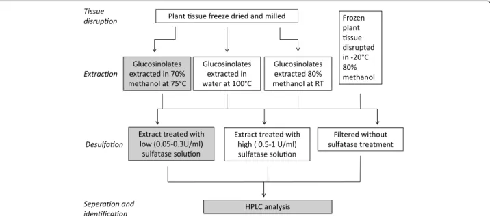
hazardous steps. hese steps are (1) lyophilisation, or freeze drying, and tissue disruption, (2) extraction in water or methanol, (3) puriication of extract, typically by desulfation on DEAE Sephadex, and (4) separation and analysis of (desulfo)glucosinolates. hese steps are out-lined in Fig. 1 and discussed in more depth below. his study aimed to improve glucosinolate extraction methods by inding alternatives to commonly used steps which are unnecessary or likely to introduce variability.
Myrosinase, an enzyme found in brassicaceae and compartmentalised in cells in close proximity to glu-cosinolates, is responsible for hydrolysing glucosinolates upon plant tissue disruption. Accurate analysis of glu-cosinolates therefore requires inactivation of myrosi-nase prior to tissue disruption. his is achieved by irst freezing then freeze drying the tissue which allows dis-ruption by milling or grinding to occur in the absence of water (Fig. 1). Lyophilisation, or freeze drying, is used to remove water from glucosinolate-containing tissues while preventing myrosinase mediated glucosinolate hydrolysis through thermal inhibition. Publications on freeze drying plant tissue have focussed primarily on the production of heat or its implications in generat-ing oxygen sensitive foodstufs (e.g. space, military or extreme-sport foodstufs and instant cofee) [14]. To our knowledge, no study has yet examined the eiciency of freeze drying in maintaining glucosinolate concentra-tions. Freeze drying functions on the principle of sub-limation: pressure is reduced below the triple point of water (6.12 mbar, 0.01 °C) at which point sublimation of
ice from the sample occurs. he cooling efect of subli-mation should be high enough to ensure the sample remains below 0 °C for the initial stage of freeze drying, thus minimizing enzyme-driven glucosinolate hydrolysis. Rapid sample loading and rapid initial pressure drop are also required to avoid sample defrosting before pressure is reduced below 6.12 mbar. Leaves have a high surface area to volume ratio and may defrost quickly, activating myrosinase and reducing inal glucosinolate concentra-tion. Despite the importance of the freeze drying process in glucosinolate extraction, many authors do not report details which are likely to afect inal concentrations of glucosinolates (e.g. how samples are transported, temper-ature of the room, whether a heating/cold plate is used and time taken for the pressure to drop).
he most commonly used methods for extraction of glucosinolates from plant material are based on the ISO 9167-1 method [15; highlighted in grey in Fig. 1], which was designed for extraction of glucosinolates from B.
napus seed and has been adapted to suit the needs of
researchers examining glucosinolate proiles of other plant species and tissue types. Although freeze drying is not explicitly detailed in the ISO 9167-1 method, it is an implicit requirement in order to avoid myrosinase medi-ated glucosinolate hydrolysis during disruption of leaf, stem or root tissues. Once the plant tissue is prepared, the ISO 9167-1 extraction is carried out at 75 °C in 70% methanol for 10 min. Heating the sample is thought to be an essential step to denature myrosinase, thus preventing enzymatic hydrolysis of glucosinolates [16]. Samples are
Plant ssue freeze dried and milled Frozen plant ssue disrupted in -20°C 80% methanol Glucosinolates
extracted in 70% methanol at 75°C
Glucosinolates extracted in water at 100°C
Glucosinolates extracted 80% methanol at RT
Extract treated with low (0.05-0.3U/ml)
sulfatase soluon
Extract treated with high ( 0.5-1 U/ml) sulfatase soluon
Filtered without sulfatase treatment
HPLC analysis
Tissue disrupon
Extracon
Desulfaon
Seperaon and idenficaon
subsequently desulfated by ion exchange chromatogra-phy on a DEAE Sephadex column to remove impurities. Desulfoglucosinolates are then separated and identiied using HPLC with a reverse phase C18 column and a UV or MS detector. Hazards associated with boiling metha-nol [17] and the time required for extractions using this method have led researchers to seek alternatives. Replac-ing heated methanol with boilReplac-ing water is reported to have comparable [18, 19], and in some cases better [20], extraction eiciencies. Although most glucosinolates are thermostable for the typical 10–30 min heating period, indole glucosinolates such as 4-hydroxy-glucobrassicin and 4-methoxyglucobrassicin have been reported to degrade quickly at temperatures below 100 °C [21]. In addition, prior to 2002 the major glucosinolate in leaves of E. sativa, 4-mercaptobutyl glucosinolate, was missed because it self-dimerises via formation of disulphide link-ages during extraction [22]. A major challenge therefore to ensuring consistent and repeatable GSL analysis is to create extraction conditions in which myrosinase is inac-tive, and glucosinolates do not self-react or degrade. A single study, conducted exclusively on radish roots, has demonstrated that cold extraction in 80% methanol does not cause appreciable reduction in glucosinolate concen-trations compared to more conventional heated extrac-tion methods [23]. However, myrosinase activity can vary dramatically [24] and whether this method is suitable for extraction of glucosinolates from other glucosinolate containing plants has not previously been assessed.
A desulfation step is often carried out post extraction to purify desulfoglucosinolates and improve accuracy and identiication from HPLC. However, the desulfation reac-tion of glucosinolates can be afected by feedback inhibi-tion of the enzyme which causes incomplete desulfainhibi-tion of glucosinolates [25]. In addition, rhamnopyranosyloxy-benzyl glucosinolates extracted from M. oleifera have been shown to be completely converted and degraded by the desulfation puriication step [26]. Due to these draw-backs, and the additional time and potential error extra steps can introduce, some authors have skipped the puri-ication and desulfation steps entirely [19, 26, 27] (Fig. 1).
We have tested each stage of glucosinolate analysis from the roots, stems and leaves of B. juncea, S. alba, R. sativus, E. sativa and B. napus and suggest a number of adjustments/improvements which can be made to reduce the costs, time and variability associated with glucosi-nolate analysis. Speciically, this study aims to address the following questions:
1) How do lyophilisation conditions afect glucosinolate concentrations?
2) Is lyophilisation a necessary step for glucosinolate extraction from green tissues?
3) Do extractions in hot methanol, cold methanol and boiling water yield comparable glucosinolate concen-trations across a range of brassicaceae species and tissue types?
4) How do desulfation time and enzyme concentration afect inal glucosinolate concentrations?
5) Is desulfation a necessary step for glucosinolate extraction from green tissue?
Methods
Plant material
B. napus used in the freeze drying tests were grown in 1 L pots illed with Terra-green in a controlled temperature glasshouse (regulated from 17.6 to 27.7 °C). At 3–4 weeks post germination, leaves were removed and halved down the limits of the midrib, excluding the midrib from the inal sample. Leaf halves were immediately frozen in liquid nitrogen and stored at −80 °C for a maximum of
1 week.
B. juncea (cv. ISCI99), R. sativus (cv. Bento), S. alba (cv. Ida Gold) and E. sativa (cv. Nemat) plants were grown by Barworth agriculture ltd. in a sandy loam soil domi-nated ields (coordinates: 53.000371, −0.290404) from
31/07/2014 to 25/09/2014. Total stem and total leaves were cut from lowering plants and immediately frozen in liquid nitrogen; root samples were gently washed and dried before freezing in liquid nitrogen. Samples were stored at −80 °C for a maximum of 2 months.
Freeze drying
Samples wrapped loosely in aluminium foil were trans-ported on dry ice and loaded into one of two freeze driers (Table 1). Maximum loading time was 30 s.
Tissue disruption
(i) Freeze dried plant tissue was homogenised to a roughly ground powder (approximately 0.1 cm particle size) using a grinder (Lloytron, E5601BK) Homogenised ground samples were milled at a fre-quency of 20 Hz for 10 min (Retch, MM400) with 2 steel ball bearings to a ine powder (particle diameter <0.1 mm). Samples were then sealed and stored at 20 °C for up to 9 months.
(ii) Frozen fresh B. napus leaf halves (experiment 2, Table 2) were placed in 2 ml eppendorf vials and stored at −20 °C. 1.755 ml of 80% methanol pre-cooled at −20 °C, 25 µl of 5 mM sinigrin and 2 small ball bearings were added. Samples were milled for 10 min at frequency 20 Hz (TissueLyser II, Qiagen). Final concentrations of methanol were estimated by incorporating average leaf moisture content of fresh
moisture content accounted for <1% of inal liquid volume.
where cMeOHf is inal methanol concentration (%), cMeOHi is initial methanol concentration (90%), VMeOHi is initial methanol volume (1.755 ml), mav is the average moisture content per dry weight (in this case 0.22 ml/g), mdl dry mass of leaf sample (g).
Glucosinolate extraction
Extractions were carried out in one of three ways (Fig. 1). In each case 50 µl of a 5 mM gluctropaeolin (for B. juncea samples) or 20 mM sinigrin (for all other samples) inter-nal standard was added.
Hot methanol extraction (based on the ISO 9167‑1 method)
0.1 g of plant material was preheated at 75 °C for 3 min in a 20 ml falcon tube. 4.95 ml of 70:30 methanol:water, pre-heated to 75 °C and the internal standard was added. he
(1)
CMeOHf = cMeOHi×VMeOHi mav×mdl+VMeOHi
sample was incubated at 75 °C for 10 min, and manually shaken every 2 min. he sample was then centrifuged at 4000 rpm (Jouan, model:B 3.11) for 10 min. Supernatent was stored at −20 °C or desulfated directly.
Cold methanol extraction (Ishida et al. [23])
5 ml of 80:20 methanol:water at 20 °C was added to 0.1 g plant tissue and the internal standard was added. he sample was shaken and left to stand for 30 min at room temperature. he sample was then mixed at 70 rpm with a platform rocker for a further 30 min (Bibby, STR6) before centrifugation at 4000 rpm (Jouan, model:B 3.11) for 10 min. Supernatent was then iltered through a 0.22 µm syringe ilter (Millex GP) for direct injection on HPLC, or uniltered if applied to Sephadex column in a puriication step.
Boiling water extraction (adapted from Herzallah and Holley [19])
25 ml of boiling water was added to 0.1 g of freeze dried and milled plant tissue in a 150 ml erlenmeyer lask and the internal standard was added. Sample was heated at 100 °C and stirred with a magnetic stirrer hot plate for
Table 1 Freeze drier characteristics
Freeze drier Room temp (°C) Cooling plate Time to 5 mbar (s) Lowest pressure (mbar)
Freezer temperature (°C)
Model
A 22 Yes 90 0.12 −45 Lyotrap, LTE scientiic ltd.
1 chamber
B 28 No 65 0.16 −53 Thermo, Heto Powerdry
LL3000 4–6 chambers
Table 2 Summary of methods used
Experiment Fig Species Tissue Freeze
drying/tis-sue disruption
Extraction Desulfation HPLC
1—Efect of freeze drier on GSL con‑ centration
2 B. napus Leaves FD‑A or FD‑B/mill Cold methanol 0.3 U/ml for 24 h ISO 9167‑1 method
2—Comparison of GSL extraction from freeze dried tissue with extraction from wet tissue
3 B. napus Leaves FD‑A or −20 °C methanol
Cold methanol 0.3 U/ml for 24 H ISO 9167‑1 method
3—Comparison of extraction methods
6, 7 R. sativus B. juncea S. alba E. sativa
Leaves, stems, roots FD‑A Hot methanol, Cold methanol, Boiling water
0.3 U/ml for 24 H ISO 9167‑1 method
4—Comparison of desulfation/purii‑ cation methods
8, 9 R. sativus B. juncea S. alba E. sativa
Leaves, stems, roots FD‑A Cold methanol 0.3 U/ml for 12, 24, 48 h, and 5 U/ml for 16 h or iltration
ISO 9167‑1 method for desulfoGSL, Herzallah and Holly
10 min. Sample was heated for a further 4 h at 70 °C before centrifugation at 4000 rpm (Jouan, model:B 3.11) for 10 min. Sample was topped up to 20 ml with deion-ised water.
Puriication and determination of activity of sulfatase Sulfatase from Helix pomatia type H-1 (Sigma, S9626) was puriied according to Wathalet et al. [25]. 25 mg of sulfatase was added to 1 ml 40% ethanol and centrifuged at 8000 rmp for 1 min (eppendorf centrifuge, 54,151). he supernatant was transferred to a fresh 2 ml eppendorf tube, 1 ml of pure ethanol was added to precipitate the sulfatase before being centrifuged at 8 krmp for 1 min. he supernatant was discarded and the sulfatase pellet air dried and redissolved in 2 ml of water.
Activity of sulfatase was determined based on the ISO 9167-1 method. 1 ml of bufered 0.15 mM sinigrin solu-tion (3 ml of 5 mM sinigrin, adjusted to 100 ml with a solution containing 40 ml 0.2% ethylene diamine, 73 ml 0.2% acetic acid; adjusted to pH 5.8) in a quartz cuvette was placed in a UV spectrometer set to 229 nm. At t = 0, 25 µl of diluted and undiluted puriied sulfatase
was added to the cuvette and measurements taken over the course of 4 h. he tangent to t = 0 was plotted and
its gradient (ΔA/Δt) measured. Activity was calculated using Eq. (2):
where ΔA/Δt is the gradient at t = 0 and Ae is the
difer-ence between absorbance at equilibrium and absorbance at t = 0.
he activity for Sulfatase from Helix pomatia type H-1 (Sigma, S9626) given by the supplier is determined by desulfation of p-nitrocatechol sulfate and is an order of magnitude higher than the activity measured for desulfa-tion of sinigrin using this method.
Desulfation of glucosinolates
As per the ISO 9067-1 method, columns were prepared with 0.5 ml Sephadex slurry (2 g DEAE Sephadex beads in 30 ml 2 M acetic acid.) and activated with 2 ml imizad-ole formate (6 M). Columns were washed twice with 1 ml water. he column was washed twice with 1 ml 20 mM sodium acetate (pH 4.0) and 75 µl of puriied sulfatase was added (5 or 0.3 U/ml). Columns were incubated at room temperature for either 12, 24 or 48 h before elution of desulfoglucosinolates with two 1 ml volumes of water. For the reduction of disulphide linkages, from dimer-ized desulfoglucosatavin in E. sativa extracts 3 g TCEP (Tris(2-carboxyethyl)phosphine hydrochloride powder Sigma, C4706) was added to 1 ml of desulfated extract. Desulfoglucosinolates were stored at −20 °C before high
(2)
Activity(U/ml)= �
A×5.7 �tAe
performance liquid chromatography analysis (Additional ile 1).
For the high sulfatase treatment, between 0.5 and 1 ml of sample was added due to insuicient sample volume remaining.
HPLC
A Waters 600E system controller attached to a Waters 717 autosampler, Waters 996 photodiode array detector and SphereClone 5µ ODS(2) column (Phenomonex) were used for separation and detection of desulfo and intact glucosinolates.
HPLC analysis of desulfoglucosinolates—adapted from ISO 9167‑1
A reverse phase C18 column (Phenomonex, Sphere-Clone 5µ ODS(2), 150 mm × 4.6 mm) was equilibrated
for 30 min with a mobile phase which consisted of 100% diH2O. Flow rate was set to 1 ml/min and samples sepa-rated according to programme for desulfoglucosinolates detailed in Table 3. Mobile phase solutions were degassed for 30 min in a sonicator (Decon, Sussex England).
Solution A: 100% diH2O
Solution B: 70:30, diH2O:acetonitrile
Desulfoglucosinolates were quantiied using 229 nm wavelength within the UV spectrum. he HPLC PDA detector allowed a full spectrum analysis from 180 to 800 nm, allowing comparative UV–visible spectra analy-sis, which aided in identifying unknown glucosinolates. hrough standard injections and HPLC–MS identiica-tion we were able to conirm the id’s of these reported glucosinolates. Desulfated puriied standards: sinigrin (sigma aldrich), glucotropaeolin, glucoraphenin, gluc-oraphanin, glucerucin, glucobrassicin, gluconasturtiin, sinalbin, progoitrin and glucoiberin (phytoplan).
Mass spectrometry
Major glucosinolates for which no commercial standard is available were identiied using an MS detector (Bruker maXis UHR-TOF) with the following settings:
Table 3 Mobile phase conditions for separation of desul-foglucosinolates
Time % Solution A % Solution B Transition
0 100 0
30 0 100 Linear gradient
35 0 100
40 100 0 Linear gradient
Source: Standard electrospray (low split 1/10 from LC)
Nebulizer: 2.0 bar Dry gas: 6.0 L/min Dry gas heater: 25 °C Capillary voltage: 3500 V Ion polarity: positive Spectra rate: 1 Hz
HPLC analysis of intact glucosinolates—adapted from Herzallah and Holly [19]
A C18 column (Phenomonex, SphereClone 5μ ODS(2)) was equilibrated for 3 h with a mobile phase which con-sisted of 80 mL (0.02 M) TBA (tetrabutylammonium bromide) and 20 mL ACN (acetonitrile) with detection at 229 nm. he low rate was set at 1.0 ml/min and sepa-rated according to programme for desulfoglucosinolates detailed in Table 3.
Solution A: 100% TBA (0.02 M)
Solution B: 70:30, TBA (0.02 M):acetonitrile
Glucosinolates were quantiied using the chromato-gram from 229 nm and standard curves were constructed using pure sinigrin (sigma aldrich), glucotropaeolin, glu-coraphenin, glucoraphanin, glucerucin, glucobrassicin, gluconasturtiin, sinalbin, progoitrin and glucoiberin (phytoplan).
In the case of glucoraphasatin in R. sativus leaves and glucotropaeolin in B. juncea minor alterations were made to avoid peaks co-eluting. he mobile phase programme for R. sativus leaves was 100% A for 5 min, followed by a 35 min linear gradient to 66% B followed by a 5 min lin-ear gradient to 100% B followed by a 5 min linlin-ear gradi-ent to 100% A. For B. juncea leaves, an isocratic 85:15, TBA (0.02 M):acetonitrile mobile phase for 70 min was used.
Determination of myrosinase activity
Activity of pure myrosinase was tested in water and 80% methanol solutions containing 0.25 mM sinigrin and 0.1 mM ascorbic acid, a myrosinase cofactor [30]. Myrosinase was added at t = 0 and absorbance of sini-grin at 229 nm was measured over the course of an hour. Activity was measured at room temperature (25 °C).
Determination of glucosinolate thermostability
A 50 µl of 10 mM sinigrin, 10 mM glucotropaeolin, 10 mM glucobrassicin solution was added to 0.95 ml water or 70% methanol preheated to 100 or 75 °C respec-tively and sealed in 1.5 ml eppendorf tubes. Samples were maintained at either 100 or 75 °C for 5, 10, 30 and 60 min and intact glucosinolate concentrations analysed with
HPLC following the adapted Herzallah and Holly method [19].
Calculation of glucosinolate content
Glucosinolate content, expressed in µmol/g were calcu-lated according to the ISO 9067-1 method (Eq. 3):
where Ag is the peak area corresponding to desulfoglu-cosinolate; As is the peak area corresponding to internal standard; n is the quantity, in micromoles, of the inter-nal standard; m is the mass of the test portion; Kg is the response factor of the desulfoglucosinolate relative to the internal standard; w is the moisture and volatile matter content, expressed as a percentage by mass of the test sample.
Statistical analysis
Paired two tailed t test analysis were carried out on total B. napus glucosinolate content per leaf half in experi-ments 1 and 2 with Microsoft excel (Table 2). For deter-mination of signiicance of efect of method on inal glucosinolate content estimates in experiments 3 and 4 (Table 2), repeat measure ANOVA analyses were car-ried out for each glucosinolate with R statistical software package (version 3.3.1).
Results and discussion
Lyophilisation
Modiications to the ISO9167-1 method (speciically created for the extraction and analysis of glucosinolates from oil rape seed samples) are required for analysis of plant green tissues (leaves, stems and roots). A number of prior-to-analysis steps, such as sampling in the ield, cleaning (if required), freezing, crushing, storage or/and shipping and reduction of sample amount have been dis-cussed by Wathelet et al. [28] and are not revisited here. hese preliminary steps are followed by lyophilisation, or freeze drying, to remove water from glucosinolate con-taining tissues while preventing myrosinase mediated glucosinolate hydrolysis through thermal inhibition. his process allows subsequent tissue disruption without risk-ing glucosinolate degradation.
We tested reproducibility of glucosinolate concentra-tions extracted after lyophilisation in separate freeze dri-ers (Table 4). Fresh B. napus leaves were halved, loosely wrapped in foil, lash frozen in liquid nitrogen and transported in dry ice to be dried in separate freeze dri-ers (Table 4). Total glucosinolate concentrations were signiicantly higher in samples dried in freeze drier A than freeze drier B (Fig. 2a). In addition, samples dried
(3) Glucosinolate content = Ag
As × n
m×Kg×
100
in freeze drier B developed a darker hue and deformed more than samples in dried in freeze drier A (Fig. 2b). Plant tissue samples have been shown to deform dur-ing the freeze drydur-ing process when temperatures exceed the glass transition state and melting point of water [29]. It is likely that samples placed into freeze drier B may have defrosted before the pressure had reduced below the 6.12 mbar required for sublimation due to higher temperatures and the lack of cooling plate. As a result, enzyme mediated hydrolysis of glucosinolates may have occurred at the initial stage. Additionally, as sublima-tion slows over time due to the remaining water vapour passing through a dry layer of increasing thickness and because water is increasingly more tissue bound, the sample temperature may have increased to above 0 °C in freeze drier B, causing defrosting.
hese results underline the need for a more substan-tive study to assess optimal conditions for freeze drying plant tissues for glucosinolate analysis. It is clear that dif-ferences in freeze drying can introduce signiicant vari-ability in retained glucosinolate concentrations (Fig. 2a).
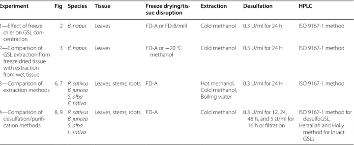
A cold methanol extraction method may be suicient to (1) inactivate myrosinase and (2) eiciently extract glucosinolates, precluding the need for the lyophilisa-tion step altogether. We tested this by comparing glucosi-nolates extracted from one half of a B. napus leaf in 80% methanol without freeze drying against glucosinolates
extracted from the other half, irst dried in freeze drier A and then extracted using the cold methanol extraction method.
No signiicant diference in inal glucosinolate con-centration was found between the two methods (Fig. 3). Freeze drying is an energy intensive and costly process requiring long drying times under continuous vacuum and the signiicant efect of freeze drier parameters on inal glucosinolate concentrations (Fig. 2a) highlights a potential source of variation between studies. If long term storage of plant tissue samples is not required, skip-ping the freeze drying step and extracting glucosinolates directly into cold methanol (−20 °C) is cheaper, quicker
and less hazardous.
Extraction
Some authors have highlighted that glucosinolates, spe-ciically indole glucosinolates, are heat sensitive and are signiicantly degraded in temperatures ≥75 °C in <10 min
[21]. his has serious implications for accuracy and relia-bility of the ISO 9167-1 extraction method, which recom-mends extractions occur in boiling 70% methanol (75 °C) for 10 min, as well as the less commonly used boiling water extraction (100 °C). In order to irst test whether thermal degradation of glucosinolates was likely to occur with these methods we measured the glucosinolate con-centrations of pure sinigrin (aliphatic), glucotropaeolin (aromatic) and glucobrassicin (indole) in boiling water (Fig. 4) and boiling 70% methanol (data not shown). Sini-grin and glucotropaeolin did not signiicantly decrease over 60 min suggesting that extraction in boiling water or methanol is unlikely to afect the concentrations of these glucosinolates. However, glucobrassicin was thermally degraded at 100 °C and data from extractions carried out at these temperatures or above (such as with microwave based methods) may underestimate the concentration of
Table 4 Freeze drier characteristics
Freeze drier
Room temp (°C)
Cooling plate
Time to 5 mbar (s)
Lowest pressure (mbar)
Freezer tem-perature (°C)
A 22 Yes 90 0.12 −45
B 28 No 65 0.16 −53
0 2 4 6 8 10 12 14
A B
Mean total glucosinolate content (µmol/g)
A
B
a
b
glucobrassicin and other indole glucosinolates. Boiling an extract in water for 10 min degrades glucobrassicin by an estimated 7%.
Activity of pure myrosinase was tested at 25 °C in water and 80% methanol solutions containing 0.25 mM sinigrin and 0.1 mM ascorbic acid, a myrosinase cofactor [30]. Absorbance of sinigrin at 229 nm, at room temperature (25 °C), was measured over the course of an hour after myrosinase addition. Myrosinase was inactive in 80% methanol (Fig. 5) suggesting that heating methanol at 75 °C for 10 min in order to inactivate myrosinase may be an unnecessary step for extracting glucosinolates from plant tissue.
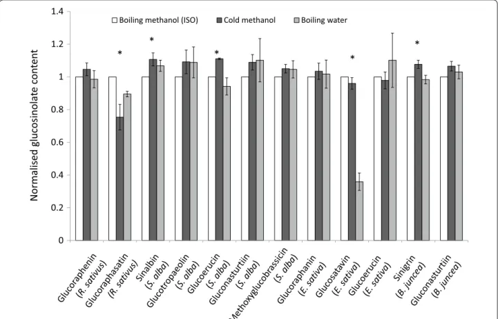
Glucosinolates from B. juncea, S. alba, R. sativus and E. sativa leaves, stems and roots were extracted (1) in boiling water for 10 min followed by a 4 h incubation at 70 °C, (2) in 70% methanol at 75 °C, or (3) in 80% meth-anol at room temperature (~20 °C) for 30 min standing followed by 30 min shaking at 70 rpm. All extracts were centrifuged and desulfated with sulfatase according to the ISO 9167-1 method. Major glucosinolates from these species can be found in Table 5.
Figure 6 compares glucosinolate concentrations
obtained using the cold methanol method and boiling water method normalised against the ISO 9167-1 boil-ing methanol method. For most glucosinolates, across most tissue types and species, the three extraction methods yield similar results. We found that extraction with cold methanol produced a signiicantly higher esti-mated concentration of sinalbin in S. alba and sinigrin in B. juncea than the hot methanol extraction (Fig. 6). Surprisingly, given the sensitivity of glucobrassicin
0 20 40 60 80 100
0 20 40 60 80 100
To
ta
l glucosinolat
e
content
fr
om fr
ee
ze
dried
ex
tr
acon (µmol/
g)
Total glucosinolate content from wet extracon (µmol/g)
Fig. 3 There is no diference in inal glucosinolate concentrations between freeze drying or direct extraction in −20 °C methanol. B. napus leaves were cut in half and frozen. One half was freeze dried prior to glucosinolate extraction, the other half was extracted directly into −20 °C methanol (n = 12; paired t test, p= 0.15; R2= 0.96). The
dashed line represents equivalence of x and y
0 0.2 0.4 0.6 0.8 1 1.2
5 10 30 60
Normalised glucosinolate concentr
aon
s
Time (min)
Sinigrin Glucotropaeolin Glucobrassicin
* * *
to thermal degradation (Fig. 4), extraction in boiling water did not signiicantly reduce the concentration of the indole glucosinolate: methoxyglucobrassicin rela-tive to the other two methods. However, glucosatavin was extracted with lower eiciency from leaves of E. sativa using the boiling water method (Fig. 6). It seems unlikely that this glucosinolate is less thermostable than other glucosinolates and was therefore degraded by the extraction method since reduced extraction ei-ciencies are not observed for stem and root samples. here are no published explanations or hypotheses that might help to explain the observed lower extraction
eiciencies for glucosatavin using the boiling water method. Glucoraphasatin extraction using cold metha-nol appears to be signiicantly less efective than the standard ISO method (Fig. 6), however this was driven by poor extraction eiciencies from R. sativus stems (Fig. 7). Ishida et al. reported a signiicant 5% increase in glucoraphasatin concentrations extracted from R. sati-vus roots using the cold methanol method [23]. In this study, extraction eiciencies of glucoraphenin in R. sati-vus roots with a cold methanol method were compara-ble to extraction eiciencies using the boiling methanol method (Fig. 7).
0 0.2 0.4 0.6 0.8 1 1.2 1.4
0 10 20 30 40 50 60 70
Absorbance (@229nm)
Time (min) Water
80% Methanol
Fig. 5 Spectrophotometric analysis of sinigrin hydrolysis kinetics in water and 80% methanol (n= 3) by puriied myrosinase (0.05 mg/ml) at room temperature (25 °C)
Table 5 Glucosinolates examined in this study
L, S and R correspond to leaf, stem and root respectively. Letters in underline represent major glucosinolates of those tissues (>10 µmol/g dry weight)
Common name Chemical name Structure Species, tissue type
Sinigrin 2‑Propenyl Aliphatic B. juncea L, S, R
Glucoraphenin 4‑Methylsulinyl‑3‑butenyl Aliphatic R. sativus L, S, R Glucoraphanin 4‑Methylsulinylbutyl Aliphatic E. sativa L, S, R
Glucosatavin Mercaptobutyl Aliphatic E. sativa L, S, R
Glucoraphasatin or hydroxyglucoerucin 4‑Methylthio‑3‑butenyl Aliphatic R. sativus L, S, R
Glucoerucin Methylthiobutyl Aliphatic E. sativa S, R
S. alba, R
Sinalbin 4‑Hydroxybenzyl Aromatic S. alba L, S, R
Glucotropaeolin Benzyl Aromatic S. alba L, S, R
Gluconasturtiin Phenylethyl Aromatic B. juncea R
No glucosinolates were detected in a subset of sam-ples extracted in cold water indicating the presence of active myrosinase leading to their degradation (data not shown). However, the cold methanol extraction did not signiicantly afect the concentration of the internal standard relative to the boiling methanol method (data not shown), providing additional evidence that myrosi-nase is inactivated in 80% methanol without heating (Fig. 5).
hese data demonstrate that 80% cold methanol can be used instead of boiling methanol to extract glucosi-nolates across a broad spectrum of brassicaceae species and tissue types. With the exception of glucoraphasatin in R. sativus shoots, replacing hot 70% methanol with cold 80% methanol did not signiicantly reduce glucosi-nolate concentrations, yet marginally increased recov-ery of sinalbin in S. alba and sinigrin in B. juncea. It is advised, due to reduction in steps and hazard as well as improved or comparable glucosinolate recovery, that
Normalised glucosinolat
ec
on
te
nt
*
*
*
*
0 0.2 0.4 0.6 0.8 1 1.2 1.4
Boiling methanol (ISO) Cold methanol Boiling water
*
Fig. 6 Extraction of glucosinolates (≥1 µmol/g) in plant tissues across the three extraction methods. Glucosinolate concentrations from the cold methanol and boiling water extraction methods are normalised to the glucosinolate concentrations obtained from the ISO9167‑1 (75 °C methanol) method (n = 4–12). Error bars represent standard error. Asterisks represent a signiicant efect of extraction method on glucosinolate concentration (repeat measure ANOVA, p < 0.05)
0 0.2 0.4 0.6 0.8 1 1.2
Root Stem
Boiling methanol Cold methanol
Normalised glucoraphasan conten
t
a cold methanol extraction is used instead of a boiling methanol extraction for most glucosinolate containing green tissues.
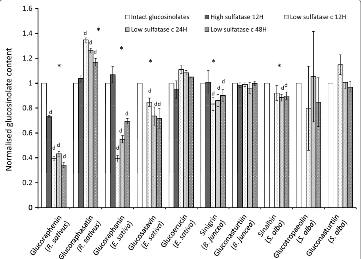
Puriication
Puriication of extract according to the ISO 9167-1 method is carried out by introducing 1 ml of extract to a column containing 0.5 ml of Sephadex solution. he column is rinsed with a 20 mM acetate bufer at pH 4.0 to avoid possible reduction of indole glucosinolates recovery [28]. 75 µl of sulfatase solution with an activity above 0.05 U/ml is applied and left to act overnight. We tested the extrac-tion eiciency of the ISO 9167-1 puriicaextrac-tion step at the described pH 4.0, at 20 °C for 12, 24 and 48 h. Complete desulfation of glucosinolates in rapeseed extract required a minimum of 11 h in operating conditions of 30 °C and pH 5.8 [25] so it was expected that an overnight 12 h desulfa-tion period may be insuicient for complete desulfadesulfa-tion of samples at room temperature. Figure 8 shows absorbance values for representative desulfoglucosinolate solutions from B. juncea, S. alba, R. sativus and E. sativa extracts treated with sulfatase solution for 12, 24 or 48 h. In most cases, 12 and 24 h incubation periods were insuicient for complete desulfation of glucosinolates. Glucoraphenin decreased in all R. sativus leaf samples tested, from 24 to 48 h, while recovery of the internal standard increased, suggesting that speciically this desulfoglucosinolate is degraded during the puriication process (Fig. 8).
Not all glucosinolates are desulfated on the column at the same rate [31], meaning that incomplete desulfation of extractions is likely to yield imprecise results: overes-timating or underesoveres-timating the inal concentration of glucosinolates which are desulfated quicker or slower respectively than the internal standard. In addition, rela-tive and total concentrations of glucosinolates and deg-radation or rearrangement of glucosinolates during this process can also afect inal concentrations [26, 31]. Use of higher sulfatase concentrations than outlined in the ISO method has been suggested for glucosinolate analy-sis in B. napus and B. oleracea [25, 31]. Figure 9 compares relative glucosinolate concentrations from B. juncea, S. alba, R. sativus and E. sativa puriied with a low activ-ity sulfatase solution (0.3 U/ml) for 12, 24 and 48 h, a high activity sulfatase solution (5 U/ml) and intact glu-cosinolates. All concentrations have been normalised to the intact glucosinolate values. Desulfated glucosi-nolates concentrations obtained with high concentra-tion sulfatase compared well with intact glucosinolates (Fig. 9). However, both high sulfatase as well as low sul-fatase treatments yielded lower glucoraphenin content estimates. Coupled with the reduction of the recovery of desulfoglucoraphenin from 24 to 48 h (Fig. 8), these data suggest that glucoraphenin is degraded or transformed during the desulfation process.
Shorter desulfation times and lower sulfatase con-centrations resulted in underestimation of the
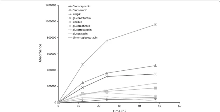
0 200000 400000 600000 800000 1000000 1200000
0 10 20 30 40 50 60
Absorbance
Time (h)
Glucoraphanin Glucoerucin sinigrin gluconasturin sinalbin glucoraphenin glucotropaeolin glucosatavin dimeric glucosatavin
concentrations of glucoraphenin from R. sativus, gluc-oraphanin and glucosatavin from E. sativa, sinigrin from B. juncea, and sinalbin from S. alba and an overestima-tion of the concentraoverestima-tions of glucoraphasatin in R. sati-vus roots (Fig. 9). he overnight (12–24 h) incubation with 0.3 U/ml sulfatase solution yields inaccurate results for most major glucosinolates examined in this study. he ISO9167-1 method suggests that a diluted puri-ied sulfatase solution with an activity exceeding 0.05 U/ ml should be used, which is shown to be insuicient for glucosinolate analysis from plant samples and conditions examined in this study (Fig. 9). Instead, if a desulfation step is carried out, use of a higher concentration of puri-ied sulfatase (in this case, 5 U/ml) is advised.
In all E. sativa leaf samples tested, recovery of mono-meric desulfo-glucosatavin decreased and recovery of dimeric desulfo-glucosatavin increased between 24 and
48 h. Bennet et al. [22] previously hypothesised that dimeric glucosatavin is unlikely to be found in vivo and is probably an artefact of the extraction process. We can conirm that glucosatavin forms dimers as a result of the desulfation step of the extraction and that without carry-ing this step out and instead quantifycarry-ing intact glucosi-nolates, no dimeric glucosatavin was detected in these samples.
Given that glucoraphenin concentration estimates are lower from methods employing a desulfation step, and that this step is also responsible for the dimerization of glucosatavin, analysis of intact glucosinolates is prefer-able in most instances. It is out of the scope of this study to compare or improve separation and detection meth-ods but it should be noted that major glucosinolates in this study were accurately measured by a HPLC–UV method adapted from Herzallah and Holley [19]. For
1.4
1.6
Intact glucosinolates High sulfatase 12H Low sulfatase c 12H
Low sulfatase c 24H Low sulfatase c 48H d
*
1.2
d
d
ontent
*
*
*
*
*
1
d
dd
d d
d
dd
inolat
ec
o
0.6
0.8
d
d d
ed glucos
0.4
d d
d
d
Normalis
0
0.2
0
examination of low abundance glucosinolates, and to avoid any potential inaccuracies due to contamination it is advised that an alternative HPLC method such as those suggested in Lee et al. or Forster et al. be used instead [26, 32].
Suggested method for glucosinolate extraction
Tissue disruption
Depending on whether freeze drying is required:
1a Freeze samples loosely wrapped in foil in liquid nitro-gen and store at −80 °C. Transport samples to freeze drier in dry ice. Rapidly load samples onto a cool plate in freeze drier and ensure the pressure drops to below 5 mbar in under 2 min. Mill samples once dried and store in airtight containers in the dark.
or
1b Freeze 50 mg samples in liquid nitrogen in 2 ml eppendorf tubes and store at −80 °C (for larger sam-ples use larger tubes). Add a volume of 80% metha-nol precooled to −20 °C ensuring that inal methanol concentration remains above 78% according to Eq. (1) in materials and methods. Add an appropriate volume of internal standard sinigrin or glucotropaeolin (e.g. 100 µM inal concentration). Disrupt tissue by add-ing 2 small ball bearadd-ings and agitatadd-ing with a tissue lyser (e.g. tissuelyserII, Qiagen) for 10 min at 20 rev/s. Alternatively use a plastic pestle to thoroughly grind the sample taking care that to keep the media below 0 °C. Continue directly to 2b.
Extraction
2a For freeze dried tissue (1a). To 0.1 g tissue, add 5 ml of 80% methanol and 50 µL of 20 mM sinigrin solu-tion. hen
2b Shake sample once and leave to stand for 30 min. Shake sample for a further 30 min (70 rev/s). Centri-fuge at 4000 rpm and transfer supernatant to a fresh tube.
Desulfation
If desulfation is required, a high concentration sulfatase solution should be prepared by dissolving 15–25 mg sul-fatase in 1 ml 40% ethanol and centrifuge at 8000 rmp for 1 min. Transfer supernatant to a fresh 2 ml eppendorf tube and add 1 ml of pure ethanol to precipitate the sul-fatase and centrifuge at 8000 rpm for 1 min. Discard the supernatant and air dry the pellet before re-dissolving in 2 ml of water. Proceed with desulfation according to ISO9167-1 method.
Conclusions
In this study we compared diferent methods for extract-ing and purifyextract-ing glucosinolates from B. napus, B. junea, S. alba, E. sativa and R. sativus green tissues to highlight unnecessary or hazardous steps. We have presented a simpliied method for extracting glucosinolates from plant tissues which does not require the use of a freeze drier or boiling methanol, and is therefore less hazardous, and more time and cost efective. he presented method has been shown to have comparable or improved glu-cosinolate extraction eiciency relative to the commonly used ISO method for major glucosinolates in the Bras-sicaceae species studied: sinigrin and gluconasturtiin in B. juncea; sinalbin, glucotropaeolin, and gluconasturtiin in S. alba; glucoraphenin and glucoraphasatin (roots but not shoots) in R. sativus; and glucosatavin, glucoerucin and glucoraphanin in E. sativa.
Authors’ contributions
TDA, KR, and SEH organized the project. VK carried out sample prepara‑ tion and glucosinolate extractions on B. napus leaves. TDA performed all other experiments, analyzed the data, and wrote the paper; SEH, IB and KR reviewed and edited the manuscript. All authors read and approved the inal manuscript.
Acknowledgements
We are grateful to Catherine Lilley, (University of Leeds), Peter Urwin (Uni‑ versity of Leeds), Andy Barker (Barworth Agriculture ltd.) and Helen Barker (Barworth Agriculture ltd.) for collection of leaf, stem and root samples used in this study. We would also like to thank Thomas Hartley (University of York) for statistical advice. We would also like to express our gratitude to the BBSRC for funding this study. Finally, we would like to thank the reviewers for their time and efort in reviewing this manuscript.
Competing interests
The authors declare that they have no competing interests.
Availability of data and materials
Material used in this study is stored at the University of York and is available on request. Datasets analysed in this study are available from the corresponding author on request.
Funding
This work was supported by UK Biotechnology and Biology Sciences Research Council (BB/L002124/1) and (BB/K020463/1).
Received: 18 August 2016 Accepted: 11 March 2017
References
1. Clarke DB. Glucosinolates, structures and analysis in food. Anal Methods. 2010;2(4):310–25.
Additional ile
• We accept pre-submission inquiries
• Our selector tool helps you to find the most relevant journal
• We provide round the clock customer support
• Convenient online submission
• Thorough peer review
• Inclusion in PubMed and all major indexing services
• Maximum visibility for your research
Submit your manuscript at www.biomedcentral.com/submit
Submit your next manuscript to BioMed Central
and we will help you at every step:
2. Fahey JW, Zalcmann AT, Talalay P. The chemical diversity and distribution of glucosinolates and isothiocyanates among plants. Phytochemistry. 2001;56(1):1–5.
3. Mithen RF, Dekker M, Verkerk R, Rabot S, Johnson IT. The nutritional signiicance, biosynthesis and bioavailability of glucosinolates in human foods. J Sci Food Agric. 2000;80(7):967–84.
4. VanEtten CH, Daxenbichler ME, Wolf IA. Natural glucosinolates (thioglu‑ cosides) in foods and feeds. J Agric Food Chem. 1969;17(3):483–91. 5. Cartea ME, Velasco P. Glucosinolates in Brassica foods: bioavail‑
ability in food and signiicance for human health. Phytochem Rev. 2008;7(2):213–29.
6. Talalay P, Fahey JW. Phytochemicals from cruciferous plants pro‑ tect against cancer by modulating carcinogen metabolism. J Nutr. 2001;131(11):3027S–33S.
7. Shapiro TA, Fahey JW, Wade KL, Stephenson KK, Talalay P. Chemo‑ protective glucosinolates and isothiocyanates of broccoli sprouts metabolism and excretion in humans. Cancer Epidemiol Biomark Prev. 2001;10(5):501–8.
8. Latté KP, Appel KE, Lampen A. Health beneits and possible risks of broc‑ coli—an overview. Food Chem Toxicol. 2011;49(12):3287–309. 9. Wiesner M, Schreiner M, Glatt H. High mutagenic activity of juice from
pak choi (Brassica rapa ssp. chinensis) sprouts due to its content of 1‑methoxy‑3‑indolylmethyl glucosinolate, and its enhancement by elicitation with methyl jasmonate. Food Chem Toxicol. 2014;31(67):10–6. 10. Ngala BM, Haydock PP, Woods S, Back MA. Biofumigation with Brassica
juncea, Raphanus sativus and Eruca sativa for the management of ield populations of the potato cyst nematode Globodera pallida. Pest Manag Sci. 2015;71(5):759–69.
11. Lord JS, Lazzeri L, Atkinson HJ, Urwin PE. Biofumigation for control of pale potato cyst nematodes: activity of Brassica leaf extracts and green manures on Globodera pallida in vitro and in soil. J Agric Food Chem. 2011;59(14):7882–90.
12. Mattner SW, Porter IJ, Gounder RK, Shanks AL, Wren DJ, Allen D. Factors that impact on the ability of biofumigants to suppress fungal pathogens and weeds of strawberry. Crop Prot. 2008;27(8):1165–73.
13. Bellostas N, Kachlicki P, Sørensen JC, Sørensen H. Glucosinolate proiling of seeds and sprouts of B. oleracea varieties used for food. Sci Hortic. 2007;114(4):234–42.
14. Ratti C. Hot air and freeze‑drying of high‑value foods: a review. J Food Eng. 2001;49(4):311–9.
15. ISO 9167‑1, 1992 NA 057‑05‑05 AA—Joint committee of DIN and DGF for the analysis of fats, oils and products thereof, related and primary products. (2012): rapeseed—determination of glucosinolate content— part 1: method using high‑performance liquid chromatography (ISO 9167–1:1992/DAM 1:2012), German version EN ISO 9167‑1:1995/prA1: 2012.).
16. Ares AM, Nozal MJ, Bernal JL, Bernal J. Optimized extraction, separation and quantiication of twelve intact glucosinolates in broccoli leaves. Food Chem. 2014;1(152):66–74.
17. Church AS, Witting MD. Laboratory testing in ethanol, methanol, ethylene glycol, and isopropanol toxicities. J Emerg Med. 1997;15(5):687–92.
18. Rangkadilok N, Nicolas ME, Bennett RN, Premier RR, Eagling DR, Taylor PW. Determination of sinigrin and glucoraphanin in Brassica species using a simple extraction method combined with ion‑pair HPLC analysis. Sci Hortic. 2002;96(1):27–41.
19. Herzallah S, Holley R. Determination of sinigrin, sinalbin, allyl‑and benzyl isothiocyanates by RP‑HPLC in mustard powder extracts. LWT Food Sci Technol. 2012;47(2):293–9.
20. Stoin DF, Dogaru RD. Researches regarding the isolation, puriication and analysis of sinigrin glucosinolate from Brassica nigra and Armoracia rusticana. Bull USAMV‑CN. 2007;63:77–82.
21. Oerlemans K, Barrett DM, Suades CB, Verkerk R, Dekker M. Thermal degra‑ dation of glucosinolates in red cabbage. Food Chem. 2006;95(1):19–29. 22. Bennett RN, Mellon FA, Botting NP, Eagles J, Rosa EA, Williamson G. Iden‑
tiication of the major glucosinolate (4‑mercaptobutyl glucosinolate) in leaves of Eruca sativa L. (salad rocket). Phytochemistry. 2002;61(1):25–30. 23. Ishida M, Kakizaki T, Ohara T, Morimitsu Y. Development of a simple and rapid extraction method of glucosinolates from radish roots. Breed Sci. 2011;61(2):208–11.
24. Piekarska A, Kusznierewicz B, Meller M, Dziedziul K, Namieśnik J, Bartoszek A. Myrosinase activity in diferent plant samples; optimisation of meas‑ urement conditions for spectrophotometric and pH‑stat methods. Ind Crops Prod. 2013;31(50):58–67.
25. Wathelet JP, Mabon N, Marlier M. Determination of glucosinolates in rapeseed improvement of the oicial HPLC ISO method (precision and speed). In: Proceedings of the 10th international rapeseed congress. Gosford: The Regional Institute Ltd; 1999 p. 185.
26. Förster N, Ulrichs C, Schreiner M, Müller CT, Mewis I. Development of a reliable extraction and quantiication method for glucosinolates in Moringa oleifera. Food Chem. 2015;1(166):456–64.
27. Troyer JK, Stephenson KK, Fahey JW. Analysis of glucosinolates from broc‑ coli and other cruciferous vegetables by hydrophilic interaction liquid chromatography. J Chromatogr A. 2001;919(2):299–304.
28. Wathelet JP, Iori R, Leoni O, Rollin P, Quinsac A, Palmieri S. Guidelines for glucosinolate analysis in green tissues used for biofumigation. Agroindus‑ tria. 2004;3(3):257–66.
29. Karathanos VT, Anglea SA, Karel M. Structural collapse of plant materials during freeze‑drying. J Therm Anal Calorim. 1996;47(5):1451–61. 30. Burmeister WP, Cottaz S, Rollin P, Vasella A, Henrissat B. High resolution
X‑ray crystallography shows that ascorbate is a cofactor for myrosinase and substitutes for the function of the catalytic base. J Biol Chem. 2000;275(50):39385–93.
31. Hennig K, Verkerk R, Bonnema G, Dekker M. Pitfalls in the desulpha‑ tion of glucosinolates in a high‑throughput assay. Food Chem. 2012;134(4):2355–61.