0022-538X/84/030741-07$02.00/0
Copyright © 1984, AmericanSociety forMicrobiology
Mapping of the
Structural Gene for the Herpes Simplex Virus Type
2
Counterpart of
Herpes
Simplex Virus Type 1 Glycoprotein C and
Identification of a Type 2 Mutant Which Does Not Express This
Glycoprotein
KATHLEEN M. ZEZULAKAND PATRICIA G. SPEAR*
Department of Microbiology, The University of Chicago, Chicago, Illinois 60637 Received 31 August1983/Accepted 7 November 1983
ThegeneencodingglycoproteinF(gF)ofherpes simplex virustype 2(HSV-2) was mapped to the region
oftheviralgenomefrom0.62 to 0.64 mapunits. This region is colinear with, and partiallyhomologous
to,
the region ofthe HSV-1 genome previously shown to encode gC. Mapping of the gF gene was done by
insertion of HSV-2DNAfragmentsinto thethymidine kinase gene of anHSV-1 virus and screening of the resultantrecombinant viruses for the expression ofgF. In this way, DNA sequences necessary for the
expression of gF in infected cells were also delimited. Because several plaque morphology mutants
(syncytialmutants) of HSV-1 have previously been shown to be gC-, a syncytial mutant of HSV-2
(Gp)
was testedfortheexpressionof gF. It wasfound to be gF-,indicatingthat gF isnot essentialforreplicationofHSV-2 in cell culture, justasgCis notessentialfor replication of HSV-1. This result also suggests that the gF- and gC- phenotypes are related in the same, as yet undefined, way to the expression of a syncytial marker. A proposal to change the name of HSV-2 gF to gC (gC-2) is discussed.
Herpes simplex virus types 1 and2
(HSV-1
andHSV-2)
specify severalglycoproteins
that are expressed on bothviral and infected cell surfaces. The HSV-1
glycoproteins
have beendesignated gB, gC, gD, and gE (31). ForHSV-1gB, gD, andgE, relatedcounterparts of similar
electropho-reticmobility have been identified in HSV-2 (6, 8, 20,21,23-25,29)andhave beenassignedthe same
alphabetic
designa-tions. Until recently HSV-1 gC was thought to be typespecific. Evidence has recently emerged, however, indicat-ingthatgF(1,2) istheHSV-2counterpartofHSV-1
gC.
We have reported mapping results (22) thatsuggested
that the geneforHSV-2
gF couldbecolinearwith the geneforHSV-1 gC. Zweigetal. (37)described amonoclonal antibody to
gFthat cross-reactswithgC,and we havedemonstratedthat a rabbit antiserum
prepared against
HSV-1 membranepro-teins contains anti-gC antibodies that cross-react with
gF
(36).
Here we presentmorerefined
mapping
results that show that thegenefor HSV-2 gF is located within a2.9-kilobase (kb) region ofthe HSV-2 genome (0.62 to 0.64 mapunits)
thatis colinear withthegeneforgC (0.63to0.64map units [9,
14])
in HSV-1.This mappingwasperformed
by insertion ofappropriate HSV-2 DNA fragments into thethymidine
kinase (TK)gene ofthegC- strain HSV-1(MP)and demon-strationthatinsertion ofparticular fragments correlatedwithexpression
ofgFbytherecombinant viruses.Mapping
of the geneforgF in thiswayalso definedboundaries ofsequences necessary fortheexpression
ofgF in infected cells.Although the functions of HSV-1
gC
and HSV-2gFarenot known, spontaneous HSV-1 gC- mutants have emergedincell culture(4, 11,12),
suggesting
thatgC
isnotnecessary for viral replication in such cell systems. These spontaneous HSV-1 gC- mutants were originally recognized because of their syncytium-forming (syn) phenotype. Although it has been shown that the absence of gC-1 expression is notdirectly responsible for the synphenotype (13, 14),
sponta-* Correspondingauthor.
neousemergenceofthe mutantphenotypes(gC- andsyn) is oftencoincident,
suggesting
some as yetundefined relation-ship between thetwo.Additional analogies between HSV-1 gCand HSV-2gFareprovidedhere bytheidentification ofanHSV-2gF- mutantin whichthegF- phenotypeisfound inassociation withasyncytial phenotype.Therefore, neither gCnorgF is essentialforviralreplicationincellculture,and spontaneous emergence of both the gF- and gC- mutant
phenotypes can be coincident with the expression ofasyn
marker.
MATERIALS AND METHODS
Cells and viruses.HEp-2(human
epidermoid carcinoma-2)
cellsandAfricangreenmonkeykidney (Vero) cells obtained from the American Type Culture Collection were used inthesestudies.Thecellsweregrown asmonolayer cultures in Dulbecco modified Eagle minimal essential medium
supple-mentedwith 10% fetal bovineserum. Viruses usedwerethe
gC- strain HSV-1(MP) (12) and HSV-2 strains 333 (7; obtained from F.Rapp, Hershey MedicalCenter, Hershey, Pa.) and
Gp
(4). Therecombinant virusesproduced inthesestudies aredescribed below.
Plasmids.pRB103[26;BamHIfragmentQ from HSV-1[F]
DNA cloned into pBR322] was provided by B. Roizman. Plasmids constructed forthegeneration of thymidine
arabi-noside-resistant
(AraTr)
insertion mutant viruses werede-rived from pRB103 (Fig. 1). For pKZ669 and pKZ672, the
4.9-kb
Sacd
fragment of HSV-2 DNA, as indicated, wasligated in different orientations into the single SacI site of
pRB103, interrupting the coding sequence of the HSV-1 TK gene (17, 32). For pKZ801 and pKZ802 the 2.9-kb
SalI
fragment of HSV-2 DNA was inserted in different orienta-tions into the singleBglII
site of pRB103 by blunt endligation after an end-filling reaction with the Klenow
frag-mentofEscherichiacoli DNApolymeraseI.
BglII
siteswereregenerated by this ligation, except for one
BglII
site in pKZ801, whichwas lost (Fig. 1). Theseinsertions interruptthe TKgene between tne site for theinitiationof
transcrip-741
on November 10, 2019 by guest
http://jvi.asm.org/
Recombinant Plasmids
HSV-2 DNA
Fragments
pKZ672
B
pKZ669
Ba Sc So
I I
a-0
So Bg SoScBgBo
I I9 I I
la
scsa
Bgsa
sa
scBg Ba
11 I I II I I
0.608 0.621 0.630 0.642
map
units
i
|\t\\\\\\\\\\\\\\\\\\! | ScSa
So BgSoSc
J
o_I
I I x19
b\\\\\\
4.9
kb
pKZ801
pKZ802
pKZ701
pKZ705
4- 0O
Ba Sc
Bg Bg
(Bg) Ba
II II
Ba Sc Bg Bg Bg Bo
I
III
VIIIIIIl
///~//1
I
Ba
-4-4- /
Sc Sa
So BgBa
II I I I
So Bg So
2.9 kb
ScSo So
Bg
I.1k
3.1
k
b
4--v /0
Ba
ScSa
BgBa
BgSaSc
pKZ750
-IIIII-A
1.8 kb
FIG. 1. Recombinant plasmids derived from pRB103 [BamHI-Q fragment of HSV-1(F) DNAinserted intopBR322] and HSV-2 DNA fragments used in their construction. HSV-2(333)DNAfragmentswereinserted into siteswithintheHSV-1 TK gene located in theBamHI-Q fragment of pRB103. HSV-1 BamHI-Q sequencesareindicatedby open boxes, and HSV-2DNAsequencesareindicated bystripedboxes, thedirection of the stripe denoting theirorientation. pBR322 sequences (not drawntoscale)areindicatedby lines.Sequences homologousto uninterruptedTKmRNAialongwiththedirection oftranscription (17, 32),areindicatedby the brokenarrows.The locations of the HSV-2 DNAfragments within the HSV-2 genome areindicated on the right. Map unitswerecalculatedbyusing the values of 0.580 and 0.630 for the left and right boundaries, respectively, of theHSV-2BglII-N DNAfragment. Abbreviations: Bg,
BgIII;
Sa, Sall;Ba,BamHI; Sc,Sacl.tion and the codingsequence (17, 32). pKZ701 andpKZ705 arepresumably identicalplasmids constructed by ligation of
the SacI-BgII 3.1-kb HSV-2 DNA fragment between the SaclandBgIII sites of pRB103, resulting in the loss of500
base pairs of the TKgene. pKZ750, containing the 1.8-kb BglII-SacIfragment of HSV-2 DNA, was also constructed
by ligation ofthisfragmentbetween theBglIIandSacI sites ofpRB103.
Preparation of viral DNA. HSV-1(MP) viral DNA was
isolated frominfectedVerocellsbyaprocedure (3) adapted from that described by Walboomers and ter
Schegget
(33)with additional modifications. Briefly, infected cells were
washedwith
phosphate-buffered
saline, suspended in0.01 MTris-hydrochloride
buffer(pH 7.6)containing
0.01 M EDTAand 1% Nonidet
P-40,
heldonice for10min,andcentrifuged
atlowspeedtopelletthenuclei.Tothe
cytoplasmic fraction,
sodiumdodecylsulfatewasaddedto0.6%,
and pronasewasaddedto 1mg/ml, followedby incubationat
37°C
for4hand
centrifugation toequilibrium
on NaIdensity gradients
con-taining ethidium bromide. The
single
viral DNA band wasvisualizedbyexposure to UVlight and, after recovery, was
dialyzed overnight againstTEbuffer(0.01M
Tris-hydrochlo-ridebuffer[pH7.6],
0.001MEDTA), extractedwithisoamyl alcohol, anddialyzedexhaustively
againstTEbuffer. ViralDNA from AraTr recombinant viruses produced in these studies was prepared as above, except that after pronase treatment, the DNA wassuccessivelyextracted withphenol, phenol-chloroform-isoamyl alcohol (50:48:2), and chloro-form-isoamyl alcohol (24:1)andthen ethanol precipitated.
--- -tk probe _ r- insert probe-
-- 23-
9.4-6.6- g- a -m a
44- _ _
-00
2.3-
2.0-3 MP - 1 2 3 4 2 3
N z 801-
802-le o:
m- IL
-MPn 1 2 3 4 2 3
0 0
N i 801-
802-Y O~
QZ Qk
FIG. 2. Southern blot analysis of genomic DNAs from AraTr viruses that were digested with BamHI and hybridized to 32P-labeled pRB103DNA(TK probe)or to the32P-labeled2.9-kbHSV-2 SallDNAfragment(0.621to0.641mapunits)purifiedfrom agarose gels (insert probe). The TK probe labeled pBR322 sequences, as well as BamHI Qfragment sequences fromHSV-1 DNA, whereas the insert probe labeled only the HSV-2DNAsequencesindicated. TheAraTr viral isolateswerefrom transfections with eitherpKZ801 orpKZ802DNA and MP DNA.BamHI-digestedDNAsfrom
HSV-1(MP),pKZ801, and pRB103arealsoshown. Themolecularweight markersontheleft, shown inkb, are fromanHindIIIdigest of A DNA.
t
on November 10, 2019 by guest
http://jvi.asm.org/
[image:2.612.108.508.98.349.2] [image:2.612.318.554.433.615.2]Cloning procedures, purification of plasmid DNA, and
purification of restricted DNA fragments from agarose. The methods used for the construction and screening of cloned DNA fragments and for the isolation of plasmid DNA by centrifugation to equilibrium in cesium chloride gradients were asdescribed by Post et al. (26). In addition, ligation of restricted DNA fragments with noncomplementary, protrud-ing 5' ends by blunt-end ligation after an end-fillprotrud-ing reaction with the Klenow fragment of E. coli polymerase I was as
described by Wartell and Reznikoff (34). Digestion of DNA withrestriction enzymeswas asspecifiedby the
manufactur-er (New England Biolabs). Purification of restricted DNA fragments from agarose was byelectrophoresisofthe DNA through SeaPlaque (FMC Corp.,Rockland, Maine) agarose,
meltingofthe gel slices at 66°C, addition ofNaCIto 0.1 M,
phenol extraction, and dialysis against TE buffer.
Southern transfer of DNA and hybridization. Viral or
plasmid DNA (or both) was digested with appropriate
re-striction enzymes, electrophoresed on 0.75% agarose gels, andtransferred tonitrocellulose by theprocedure of South-ern(30).Hybridizationsweredoneat65°C in6xSSC(0.9 M
NaCl, 0.09 M sodiumcitrate), 5x Denhardt solution (0.1%
Ficoll, 0.1% polyvinylpyrrolidone, 0.1%bovine serum
albu-min), 0.5% sodium dodecyl sulfate, 50 ,ug of calf thymus DNA per ml,30% formamide,and thedenatured
32P-labeled
DNAprobe. Afterhybridization for12 to 24 h,nitrocellulose
filters were washed asdescribed by Maniatis et al. (15). Nick
translation kits obtained from New England Nuclear Corp.
(Boston,Mass.) were used toproduce the32P-labeled DNA
probes.
Construction of AraTr recombinant viruses. Vero cells
werecotransfectedwith mixtures ofHSV-1(MP)viral DNA and the appropriate BamHI-digested plasmid DNA
(pKZ669, pKZ672, pKZ801, pKZ802, pKZ701, pKZ705, or
pKZ750). Calcium phosphate precipitates of DNA were
prepared which contained 0.25 or 0.4 ,ug of HSV-1(MP)
r ___--- tk probe--e
23
-
9.4-6.6- * 4.4- 40
insert probe
---Oak
2.3-
2.0-MP 4 5 6 10 9 11 MP 4 5 6 10 9 11
Nz 705- 701- 705- c 705- 701-
705-a.~~~~~~~~~~~~~~~Q
FIG. 3. Southern blot analysis of genomic DNAs from AraTr viruses that were digested with BamHI and hybridized to 32p-labeledpRB103 DNA (TKprobe)ortothe32P-labeled 2.9-kb HSV-2 SalI DNA fragment(0.621to0.641mapunits)purifiedfromagarose
gels (insert probe). The AraTr viral isolates were obtained from
transfectionswith either pKZ701 or pKZ705 DNA andMPDNA. BamHI-digestedDNAsfromHSV-1(MP),pKZ701,andpRB103are
also shown.The molecularweightmarkersontheleft,shown inkb,
arefromanHindlIl digestofADNA.
tk probe---- insert probe -T
23-9.4-
-
6.6-4.4- _3 MD
2.3-2
.0-0 nMP 1 2 3 4 5
-____
Nhem
0-~
50N.
o r MP 1 2 3 4 5
u 0o N -;
t an
750-Y a
fl 0
FIG. 4. Southern blot analysis ofgenomic DNAs from AraTr viruses that were digested with BamHI and hybridized to
32p-labeledpRB103DNA(TKprobe)or tothe32P-labeled2.9-kbHSV-2 SallDNAfragment (0.621 to0.641mapunits)purified from agarose gels (insert probe). The AraTr viral isolates were obtained from transfections with pKZ750 DNA and MP DNA. BamHI-digested DNAsfrom HSV-1(MP), pKZ750, andpRB103arealso shown. The molecular weight markers on the left, shown in kb, are from an
HindlIl digests of X DNA.
DNA, 20,ug of salmon sperm
DNA,
and0.05, 0.15,
or0.45 ,ugofplasmid
DNA per 0.5ml,
and cellswere treated withthis DNA
by
theprocedure
of Grahamand Van der Eb(10)
as modified by
Wigler
et al.(35). Progeny
virus from thetransfected cells were
plated
onVerocells underagarosein thepresenceofAraT(Raylo
Chemical, Edmonton, Alberta,
Canada)at 100,ug/ml (18)
toselectforAraTrrecombinants. AraTr isolateswereplaque
purified
twice in thepresence of AraT, exceptfor isolates 750-1through
750-4,
which werenot plaque
purified.
Antibodies and
immunoprecipitations.
Hybridoma
cell lines secreting monoclonal antibodies directedagainst
HSV-1 orHSV-2
glycoproteins
were isolatedby
M. Para in ourlaboratory. Themonoclonal antibodiesused in these studies (in the form of ascites
fluid)
werepreviously
described(19,
22) andareasfollows: III188 directedagainst
gF-2
(anti-gF-2), 1173 directedagainst
gC-1
(anti-gC-1),
and 111114directedagainst
gD-1 andgD-2
(anti-gD-1/2).
Monoclonalantibody
151.2,specific
forgC-1,
was agift
fromJ. Glorioso andM.Levine
(University
ofMichigan,
AnnArbor).
Rabbitantise-rum R#71, prepared against HSV-2 gF, was previously
described(36).
Immunoprecipitations were performed as previously
de-scribed (36) with extracts from infected HEp-2 cells that were labeled with
[35S]methionine
from 4 to 24 h after infection, and the precipitated products were analyzed on8.5% sodium dodecyl sulfate-polyacrylamide gels cross-linked withN,N'-diallyltartardiamide (11).
RESULTS
Construction of recombinant viruses. To more precisely
define the location of the gene for gF-2 in the HSV-2 genome, recombinant viruses were constructed that
con-tained defined regionsof HSV-2(333) DNA inserted into the
. . an _
on November 10, 2019 by guest
http://jvi.asm.org/
[image:3.612.317.556.75.264.2] [image:3.612.61.301.455.641.2]+ + + -+ -
200- 30-
94-68 _ ,
43-333 1 2 3 4 2 3
801-
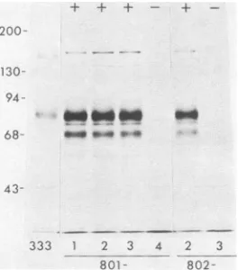
802-FIG. 5. Polypeptidesprecipitated by anti-gF-2 (111188)antibody from extracts ofHEp-2 cells infected with HSV-2(333) or AraTr viruses.TheAraTrviruseswere from transfections with pKZ801 or pKZ802DNAand MP DNA.Infected cells werelabeled with25,uCi of[35S]methioninepermlfrom4to 24hafter infection. The+and
-symbols showthe resultsofSouthern blot analyses performed with theDNAfromeachoftheAraTrviruses and indicate thepresence orabsence, respectively, of HSV-2DNA insertions withinthe TK genesof theAraTrviruses.
TK gene of the gC- strain HSV-1(MP), and these viruses werethen analyzed for the expression of gF. Figure 1 shows therecombinant plasmids produced forthese constructions and the map locations of the HSV-2 DNA fragments that were inserted into these plasmids. HSV-2 DNA fragments chosen for insertion were a 4.9-kb SacI fragment (located
between 0.608 to 0.642 map units in the genome) that
hybridized to the HSV-1(F) Sall R DNA fragment, within whichthe gene for gC-1is located (9, 14), and subregions of thisfragment.
Toproducetherecombinantviruses, plasmid DNAswere
digested with BamHI and, togetherwithHSV-1(MP) DNA, were used to transfect Vero cells. Viral progeny were
selected forlossofTKexpressionin the presence ofAraT, and the DNAs for these viral isolates were analyzed to
determine whether they had insertions in their TK genes.
Fig. 2, 3, and 4 show Southern blot analyses ofgenomic
DNAsfrom AraTr isolates digestedwith BamHI and hybrid-ized to probes for the TK gene or for the HSV-2 DNA sequences inserted intothe TK gene.
Figure2 showsthat three of fourAraTr isolates obtained
fromtransfectionswithpKZ801(801-1,801-2,and801-3)and one AraTr isolate obtained from transfection with pKZ802 (802-2) contained insertions. These insertions were
charac-terized by loss of the 3.6-kb BamHI-Q DNA fragment (within whichTK sequences arelocated; 17, 32) present in the parental MP strain and the appearance ofa new6.5-kb DNAfragmentthathybridizedbothtotheTKprobeand the insert probe and comigrated with the 6.5-kb fragment in pKZ801. The 7.0-kb HSV-1 DNAfragment,which contains the gene for gC-1 and therefore hybridizes to the insert
probe, comigratedwith this new6.5-kbfragment onthisgel, partially obscuring the hybridization results with the insert
probe.
In similarfashion, Fig. 3 shows that four AraTr isolates
(705-4, 705-5, 705-6, and 701-10) from transfections with either pKZ701 or pKZ705 contain insertions, and Fig. 4 shows that three AraTr isolates (750-3, 750-4, and 750-5) contain insertions after transfections with pKZ750. Similar
results were also obtained in analyses of genomic DNAs
from AraTr viral isolates obtained after transfections with pKZ672 and pKZ669 (datanot shown).
The AraTr isolates shown in Fig. 2, 3, and 4 that donot haveinsertions of HSV-2 DNAsequences into theTK gene arepresumed to be spontaneoustk- mutants.
Analysisof AraTr recombinantvirusesforexpressionofgF. To determine whether the AraTr recombinant viruses expressed gF, immunoprecipitation experiments were per-formed in which [35S]methionine-labeled infected cell ex-tractswerereacted withanti-gF-2 (III188) antibody, anti-gF-2antiserum R#71, orboth.
[image:4.612.115.247.74.224.2]Recombinant viruses that contained the HSV-2 4.9-kb Sacl fragment (0.608 to 0.642 map units) insertedin either orientation into their TK genes expressed gF (data not shown).Similarly, recombinant viruses that contained inser-tionsof the HSV-2 2.9-kbSall DNA fragment (0.621to0.641 mapunits), inserted in either orientation into their TK genes, also expressed gF (Fig. 5). As expected, viruses without insertions (801-4 and 802-3) did not express gF.
Figure 6 shows immunoprecipitations with AraTr recom-binantvirusesthat containedinsertions of the HSV-23.1-kb SacI-BglII DNA fragment (0.608 to 0.630 map units)orthe 1.8-kbBglII-SacIDNAfragment (0.630to0.642mapunits). Insertion of these DNA fragments did not result in the expression of gF, demonstrating that DNA sequences neces-saryfor gF expressionare notwholly contained within either region. Figure 7 summarizes the results obtained from analysisof the recombinant viruses. These results map the geneforgFto a region of the HSV-2genome (0.62 to0.64 mapunits)that is colinear with thegeneforgC (0.63to0.64 map units; 9, 14) in the HSV-1 genome.
A
+ +-I-
-B
--+ + -I
200-
130- 94-
68-
43-4 5 6 9 10 11 un MP 1 2 3 4 5
[image:4.612.318.553.413.581.2]~~~ ~ ~
...
705- 701- 705-
750-FIG. 6. (A) Immunoprecipitates obtained withanti-gF-2 (R#71) antiserum fromextracts ofuninfected HEp-2 cells (un)orHEp-2 cells infected with MPor AraTrviruses. The AraTrviruseswere obtained from transfectionswithpKZ701orpKZ705DNAandMP DNA.Infected cellswerelabeledwith 25p.Ciof[35S]methionineper ml from 4 to 24 h after infection. Uninfected cells were labeled during thissametimeperiod. (B)Immunoprecipitatesobtained with
a combination of both anti-gF-2 (111188) antibody and anti-gF-2 (R#71) antiserum fromextractsofHEp-2 cells infected with AraTr viruses obtained from transfections with pKZ750 DNA and MP DNA.Infectedcellswerelabeled with30,uCiof[35S]methionineper mlfrom4to24 hafterinfection. The + and- symbolspresentin both A and B show theresults of Southern blotanalysesperformed with the DNA from each of the AraTr viruses and indicate the presence orabsence, respectively,ofHSV-2DNAinsertions within theTKgenesofthe AraTrviruses.
on November 10, 2019 by guest
http://jvi.asm.org/
a
b
I pF
0.0
L
ZI~
I I I~~~~~~~~~III
-~~~~~~~~~~~
ba
lac'
c aI I I I . I
0.1
0.2
0.3
04
0.5
0.6
0.7
tk
gFexpression when inserted into HSV- I
tk
geneOrientation
Same
+
Opposite
0.81
gC
Sa
/& /
Sc
+
ND 3.1 kb
Sc ND
--/
/
0.9
1.0
4.9kb
SaBs
Bg
2.9
kbSa
Bg
1.8kbBg
FIG. 7. Location of thegeneforgFontheHSV-2genomeand thegenesforgC(9, 14)and TK (17, 32)ontheHSV-1genomeanda
sum-mary of the results obtained from screening AraTr viruses that contained insertions of HSV-2 DNA sequences in their TK genes for
expression ofgF. HSV-2 DNAfragmentswereinserted intothe HSV-1 TKgenein thesameorientationastheyoccurin the HSV-2genome, orin theopposite orientation. The+ symbol denotes expression of gF, and the-symboldenotes absence of expression of gF. ND indicates
notdone. The vertical dashed line marks the boundary between the L and Scomponentsof the HSVgenomeand thewhite boxesrepresent
thereiteratedsequencesof the L and Scomponents(27). Abbreviations: Sa, Sall; Sc, SacI; Bs, BstEII; Bg,BgII;Pv,P'uI.
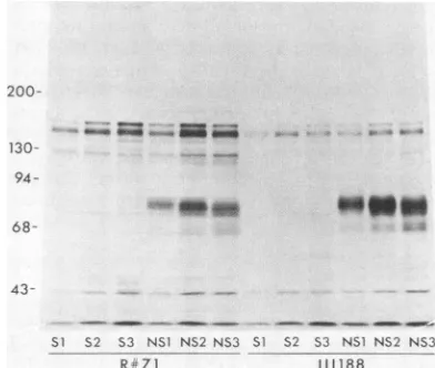
Functional correlation between gC-1 and gF-2by the identi-ficationof gF- mutants of HSV-2. A syn mutant ofHSV-2 wasexaminedtodetermine whether this mutant,inanalogy with some HSV-1 syn mutants, was gF-.
HSV-2(Gp)
is a spontaneoussyncytial variant ofHSV-2(G) isolated by Cas-saietal.(4). These authors showed thatGp
virionscontained aglycoprotein of the appropriate electrophoretic mobilityto be gF-2, and we found that gF was precipitated fromGp-infectedcellextractsby anti-gF-2(III188)antibody (datanot shown). However, examination of
Gp
plaque morphologyonboth HEp-2 and Vero cells demonstrated two different phenotypes in the viral population: syncytial plaques and smaller plaques in which no fusion was evident, although progeny from these plaques provedto have limited fusion-inducing capacity. Progeny from three syncytial plaques (S1, S2, and S3) and three of the otherplaques (NS1, NS2, and NS3)were isolated from Vero cell monolayers and plaque
purified three times, and then viral stocks of these isolates
wereprepared in HEp-2 cells. Immunoprecipitation experi-ments performed with these six viral isolates showed that NS1,NS2, and NS3 expressed gF, whereasSi, S2, and S3 did not(Fig. 8). Analysis of the DNAs of these six isolates with the restriction enzyme BglII resulted in fragment pat-terns similar to that ofHSV-2(G) and dissimilar to that of HSV-1 (datanotshown), confirming that all six isolateswere
HSV-2strains.Also,noneof the sixisolates expressed gC-1 as assayed by immunoprecipitations with anti-gC-1 (151.2
and II73) antibodies, but all six isolates did express gD as
assayed by immunoprecipitations with anti-gD-1/2 (111114) antibody (datanotshown). These results indicatethatgF-2, like gC-1, is nonessential for viral growth in tissue culture andthat expression of gF- andsynphenotypes iscoincident
in certain HSV-2syn mutants.
200-
130-
94-* NI
_-68- _o"
z._
_
43-SI $2 53 NS1 N$2 N53 S1 $2 $3 NSI NS2 NS3
R - 71 111188
FIG. 8. Polypeptides precipitated by anti-gF-2 (111188) antibody
oranti-gF-2antiserumR#71fromextractsofHEp-2 cellsinfected withHSV-2(Gp)isolates S1, S2, S3, NS1, NS2orNS3.The infected
cellswerelabeled with 20,uCiof[35S]methionineperml from 4 to24 h after infection.
map
units
HSV- I
HSV-2
P S
PvSc
Sa
Sc gF
on November 10, 2019 by guest
http://jvi.asm.org/
[image:5.612.80.536.80.374.2] [image:5.612.337.533.512.678.2]DISCUSSION
The results presented here, together with other evidence
summarized above (22, 36, 37), demonstrate that gF is the HSV-2 counterpart of HSV-1 gC.
The gene for gF maps within a 2.9-kb Sall DNAfragment
(approximately 0.62 to 0.64 map units). This HSV-2 region
shares DNA homology with the HSV-1
SalI-R
DNAfrag-ment, within which the gC gene is located (9, 14), and thegC gene (approximately 0.63 to 0.64 map units) is colinearwith the gene for gF. Because gF is expressed uponinsertion of
the 2.9-kbSallDNA fragment, in either orientation, intothe HSV-1 TK gene, both coding and cis-acting regulatoryDNA sequences necessary for expression of gF in infected cells are probably located within this DNA fragment. Analysis of
viruses containing
SacI-BglII
HSV-2 DNA fragments insert-ed into the TK gene demonstratinsert-ed that sequences both tothe left and right of theBglII
site (0.630 map units) in the2.9-kbSallDNA fragment are necessary for the expression ofgF. DNA sequence determinations of the HSV-1 and HSV-2 genomes in this region (9; R. Frink, K. Draper, M. Swain, D. Galloway, and
E.
Wagner, manuscript in preparation) have shown that, based on the best sequence fit, this BgiII sitelines up with the HSV-1 genome 35 base pairs to the rightof the
EcoRI
site, which is located within the HSV-1 gCstructural gene approximately 450 base pairs downstreamof
the putative initiation codon. Also, an mRNA species that has been located in this region of the HSV-2 genome is the same size as the mRNA species encoding HSV-1 gC (ap-proximately 2.5 kb; Frink et al., in preparation), suggesting that gF may be translated from this mRNA. Comparisons of the complete nucleotide sequences of the genes for gF and gC are of particular importance and interest because gFand gC exhibit differences in size and antigenicity (1, 2, 22, 36, 37) that suggest more divergence between these HSV-1 and HSV-2 genes than is evident for other glycoprotein genes.
Comparison of the mapping data presented here with the data of Marsden et al. (16) suggests that the 63K HSV-2 glycoprotein they mapped to the region from 0.57 to 0.66 map units is gF.
Identification of an HSV-2 gF- mutant, in association with a syn phenotype, demonstrates not only that gF is analogous to gC in being nonessential for viral growth in cell culture, but also that it may share the same, as yet unde-fined, relationship to expression of a syn marker.
Based on evidence presented here and on the previously established antigenic relatedness of gC-1 and gF-2 (36, 37),a proposal was made at the Eighth International Herpesvirus Workshop at Oxford (31 July to 5 August 1983) to change the name of gF-2 to gC-2, in keeping with criteria agreed upon for the naming of HSV glycoproteins (5). It was also proposed that the glycoprotein previously designated gC-2 (28) be considered unnamed until it is further characterized, and that the name gF be retired from future use in designat-ing HSV glycoproteins.
ACKNOWLEDGMENTS
We thank B. Roizman for the gift ofa plasmid, J. Glorioso and M. Levine for the gift of a monoclonal antibody, and G. T.-Y. Leefor providing a preparation of HSV-1 (MP) viral DNA and for providing DNA from pTYL302 (pTYL302 contains HSV-2[333] DNA be-tween the BamHI sites at 0.607 and 0.690 map units), which was used in the construction of pKZ672 and pKZ669. We also thank Richard Aspell and Mary Lynn Parish for excellent technical assistance and E. K. Wagner and D. Galloway for communicating results before their publication.
This work was supported by grants from the American Cancer
Society and Public Health Service grants CA21776 and CA19264 from the National Cancer Institute (to P.G.S.). K.M.Z. was a trainee of the National Service ResearchAward 5 T32GM07197.
LITERATURE CITED
1. Balachandran, N., D. Harnish, R. A. Killington, S. Bacchetti, and W. E. Rawls.1981. Monoclonalantibodiesto two glycopro-teinsofherpes simplex virustype2. J. Virol. 39:438-446. 2. Balachandran, N., D. Harnish, W. E. Rawls, and S. Bacchetti.
1982.Glycoproteins of herpes simplex virustype2 asdefinedby monoclonalantibodies. J.Virol. 44:344-355.
3. Camacho, A.,and P.G.Spear.1978.Transformationofhamster embryo fibroblasts byaspecific fragment of the herpessimplex virusgenome. Cell15:993-1002.
4. Cassai, E.,R.Manservigi, A. Corallini, and M. Terni.1975/1976. Plaquedissociation ofherpes simplex viruses: biochemicaland biological characters of the viral variants. Intervirology 6:212-223.
5. Cohen, G., I. Halliburton, and R. Eisenberg. 1981. Glycopro-teins of herpesvirus, p. 549-554. In A. J. Nahmias, W. R. Dowdle, and R. F. Schinazi (ed.), The humanherpesviruses, an interdisciplinary perspective. Elsevier-North Holland Publish-ing Co., New York.
6. Cohen,G.H., M. Katze, C. Hydrean-Stern, and R. J.Eisenberg. 1978. Type-common CP-1 antigen of herpes simplex virus is associated with a 59,000-molecular-weight envelope glycopro-tein. J. Virol. 27:172-181.
7. Duff, R., and F. Rapp. 1971. Properties of hamster embryo fibroblasts transformed in vitro after exposure to ultraviolet-irradiated herpes simplex virus type 2. J. Virol. 8:469-477. 8. Eisenberg, R.J., M. Ponce deLeon, L. Pereira, D. Long, and
G. H. Cohen. 1982. Purification of glycoprotein gD ofherpes simplex virus types 1 and 2by use ofmonoclonal antibody. J. Virol. 41:1099-1104.
9. Frink,R.J.,R. Eisenberg, G. Cohen, and E. K. Wagner. 1983. Detailedanalysis oftheportionofthe herpes simplex virus type 1 genome encoding glycoprotein C.J. Virol. 45:634-647. 10. Graham,F.L.,and A.J.Van der Eb.1973.A newtechniquefor
the assay ofinfectivity of human adenovirus 5 DNA.Virology 52:456-467.
11. Heine, J. W., R. W. Honess, E. Cassai, and B. Roizman. 1974. Proteins specified by herpes simplex virus. XII. The virion polypeptides oftype 1 strains. J. Virol. 14:640-651.
12. Hoggan, M. D., and B. Roizman. 1959. The isolation and properties ofavariant ofherpes simplexproducing multinucle-ated giant cells in monolayer cultures in the presence of antibody. Am. J. Hyg. 70:208-219.
13. Honess, R. W., A. Buchan,I. W. Halliburton, and D. H. Wat-son. 1980. Recombination and linkage between structural and regulatory genes of herpes simplex virus type 1: study ofthe functional organization of the genome. J. Virol. 34:716-742. 14. Lee, G. T.-Y., K. L. Pogue-Geile, L. Pereira, and P. G. Spear.
1982. Expressionof herpessimplexvirusglycoprotein C froma DNAfragment inserted into the thymidine kinase gene ofthis virus. Proc. Natl. Acad. Sci. U.S.A. 79:6612-6616.
15. Maniatis, T., E. F. Fritsch, and J. Sambrook. 1982. Molecular cloning, a laboratorymanual. ColdSpring HarborLaboratory, Cold Spring Harbor, N.Y.
16. Marsden, H. S., N. D. Stow, V. G.Preston, M. C.Timbury,and N. M. Wilkie. 1978. Physical mapping ofherpes simplex virus-induced polypeptides. J. Virol. 28:624-642.
17. McKnight, S. L. 1980. The nucleotide sequence and transcript mapof the herpes simplex virusthymidine kinasegene. Nucleic AcidsRes. 8:5949-5964.
18. Mocarski, E. S., L. E. Post, andB. Roizman. 1980. Molecular engineering of the herpes simplex virus genome: insertion of a second L-Sjunction into the genome causes additional genome inversions. Cell22:243-255.
19. Noble, A. G., G. T.-Y.Lee,R.Sprague, M. L.Parish, and P. G. Spear. 1983. Anti-gD monoclonal antibodies inhibit cell fusion induced byherpessimplex virus type 1. Virology 129:218-224. 20. Norrild, B., H. Ludwig, and R. Rott. 1978. Identification ofa
on November 10, 2019 by guest
http://jvi.asm.org/
commonantigen of herpes simplex virus, bovine herpes mam-millitis virus, and B virus. J. Virol. 26:712-717.
21. Para, M. F., L. Goldstein, and P. G. Spear. 1982. Similarities anddifferences in the Fc-binding glycoprotein (gE) of herpes simplex virus types 1 and2andtentative mapping of the viral genefor this glycoprotein. J. Virol. 41:137-144.
22. Para, M. F., K. M. Zezulak, A. J. Conley, M. Weinberger, K. Snitzer, and P. G. Spear. 1983. Use of monoclonal antibodies against two 75,000-molecular-weight glycoproteins specified by herpes simplex virus type 2 in glycoprotein identification and genemapping. J.Virol. 45:1223-1227.
23. Pereira, L., D. V. Dondero, D. Gallo, V. Devlin, and J. D. Woodie. 1982.Serological analysis of herpes simplex virus types 1and 2 with monoclonalantibodies. Infect. Immun. 35:363-367. 24. Pereira, L., D. Dondero, B. Norrild, and B. Roizman. 1981. Differentialimmunologic reactivity and processing of glycopro-teins gA and gB of herpes simplex virus types 1 and 2 made in Vero and HEp-2 cells. Proc. Natl. Acad. Sci. U.S.A. 78:5202-5206.
25. Pereira, L., T. Klassen, and J. R. Baringer. 1980. Type-common andtype-specific monoclonal antibody to herpes simplex virus type 1. Infect. Immun. 29:724-732.
26. Post,L.E.,A.J. Conley,E.S. Mocarski, and B. Roizman. 1980. Cloning of reiterated and nonreiterated herpes simplex virus 1 sequencesasBamHIfragments. Proc. Natl. Acad. Sci. U.S.A. 77:4201-4205.
27. Roizman, B. 1979. The structure and isomerization of herpes simplex virus genomes. Cell 16:481-494.
28. Ruyechan, W.T., L. S. Morse, D. M. Knipe, and B. Roizman. 1979. Moleculargenetics of herpes simplex virus.II. Mapping of the majorviral glycoproteins and of the genetic loci specifying
the social behavior ofinfected cells. J. Virol. 29:677-697. 29. Showalter, S. D., M. Zweig, andB.Hampar. 1981.Monoclonal
antibodiestoherpessimplex virus type1proteins, including the immediate-early protein ICP 4. Infect. Immun. 34:684-692. 30. Southern, E. M. 1975. Detection ofspecific sequences among
DNAfragments separated by gelelectrophoresis. J. Mol. Biol. 98:503-517.
31. Spear, P. G. 1980.Herpesviruses, p.709-750.In H. A.Blough and J. M. Tiffany (ed.), Cellmembranes and viral envelopes, vol. 2. Academic Press, Inc., London.
32. Wagner, M. J., J.A.Sharp, and W. C. Summers.1981. Nucleo-tide sequence of thethymidine kinase gene of herpes simplex virus type 1. Proc. Natl. Acad. Sci. U.S.A. 78:1441-1445. 33. Walboomers, J. M. M., and J. ter Schegget.1976. A newmethod
for the isolation of herpes simplex virus type2 DNA.Virology 74:256-258.
34. Wartell, R. M., and W. S. Reznikoff. 1980. Cloning DNA restriction endonuclease fragments with protruding single-stranded ends. Gene 9:307-319.
35. Wigler, M.,A.Pellicer,S.Silverstein,R.Axel,G.Urlaub, andL. Chasin.1979.DNA-mediatedtransfer of the adenine phosphori-bosyltransferase locus into mammalian cells. Proc. Natl. Acad. Sci.U.S.A. 76:1373-1376.
36. Zezulak, K. M., and P.G. Spear. 1983. Characterization ofa herpessimplex virus type 275,000-molecular-weight glycopro-tein antigenically relatedtoherpes simplex virus type 1 glyco-protein C. J. Virol. 47:553-562.
37. Zweig, M.,S. D. Showalter, S. V. Bladen, C. J. Heilman, Jr., and B.Hampar. 1983.Herpessimplex virus type2glycoprotein gF and type 1 glycoproteingC have related antigenic determi-nants.J. Virol. 47:185-192.