A Thesis Submitted for the Degree of PhD
at the
University of St Andrews
1967
Full metadata for this item is available in
St Andrews Research Repository
at:
http://research-repository.st-andrews.ac.uk/
Please use this identifier to cite or link to this item:
http://hdl.handle.net/10023/13850
by
A . 8 GR API -P RAC A 881
A tiiosis preoented to the University St. Andrews
E^pr the Dej^ree Doctor of^ PbilOGOpby
Department of Diochemietry,
The quality of this reproduction is dependent upon the quality of the copy submitted. In the unlikely event that the author did not send a com plete manuscript and there are missing pages, these will be noted. Also, if material had to be removed,
a note will indicate the deletion.
uest
ProQuest 10170914
Published by ProQuest LLO (2017). Copyright of the Dissertation is held by the Author. All rights reserved.
This work is protected against unauthorized copying under Title 17, United States C ode Microform Edition © ProQuest LLO.
ProQuest LLO.
789 East Eisenhower Parkway P.Q. Box 1346
D H C L A R A T l O r ;
1 üereUy Ueclaro tüat the followlOti tüosie Is based on %fOrk carried out by mo* that the
theei» io my own composition* nnd that no port of it hue boon presonted previously for a higher degree.
The research woo carried out in the Department of Biochemistry in the United College of St.
I hereby certify that A. S B R A I* 11^! I - b' R A C .A S SI I\ 31 has opent nine terme engaged in research work under my direction and that he has fulfilled the
cotiditlonc; of Ordinauce I^o.lG (St. Andrews) and
that he is qualified to oubmit the accompanying
ACADfiMÏC M, *; "frvTPf *». iiîi’COKDWw»-'V
I matriculated at the University of Padova
(Italy) 1x1 October 1953, and graduated with the degree of Doc toi' of Medicine, fir at clacs with medal5 in î\ovember 1959.
From December 1969 to September 1956 I held the post of lecturer in the Department of Histology, Padova University.
In. September 196B Ï acceptod the poot of
aoeictant lecturer in the Department of Diochomiotry, St# /uidrowo University, and I a?ae appointed to a
ACKAO\a.O)JGMBfVT3
X thank Professor G. i*U Tristram for his constant intorest during the couroo of this study.
Financial support from the Scottish Home and Health Department during the last year of
thi^ reoeerch ±5 gratefully acknowledged.
I also thank Dr. J.U. Smith foj? his advice and collaboration in part of this work.
Thanks are also due to Dr. T.J. Peters for his aid in the end-group esfcimationc, to Mr. J Crawford for his skilful technical assistance in physicocuemieal experiments and to Mr. H.J. hlyth for Che photography.
Finally, I wish to thank my wife, Laura, for carrying out the amino acid analyses and,
c () i\ _T U T S
Page
IKTRODLCTXOH ... X
AlATnRlALS nnd M U T H O D S ... 9
RBSlii;rs ... 36
3)ISCU8SI0H ... 84
1 ^ î? *■*'» 1 > il f f ' i f"\ *I
of bovine septal cartilage are collagen and a protein-* polysaccharide complex, the macromolecular structure
of which has been extensively studied since Schattou and Schubert (1964) showed that aqueous extracts of this tissue contained a high-moXecular-weight component consisting of chondroitin sulphate apparently bound to a non-collagenous protein.
The proteiii-polysaccharide once extracted from the tissue, by high speed homogenisation in large volumes of water (Mnlawista and Schubert, 1958), can then be separated into a light fraction (PP-X.) and a heavy
fraction (PP-li) by centrifugation (Gerber, Franklin and Schubert, i960). Analytical ultracentrifugation and
zone electrophoresis studies have shown that below pH 10, PP-L behaves as a single entity (Schubert, 1964). On
the other hand, there is no evidence that PP-H is a
However, analysis of the *protein* moiety has shown that only 68% of its nitrogen is recoverable as amino
acids (Partridge and Davis, 1968), and on papain digestion some 16% of keratosulphato is liberated (Gregory and Roden, 1961),
Webber and BayXey (1956) estimated, by sedimentation and viscosity methods, that the molecular weight of
calcium chloride-extractecl protein^polysaccharide complex
6
was 1 X 10 . They also suggested that the complex had a random coil configuration and that it consisted of some 20 chondroitin sulphate molecules, of molecular weight
4
approximately 4 x 10 , linked by polypeptide chains in an end-to-end arrangement. Bernard! (1957 a,b), working with a similar preparation, confirmed the random coil
6 configuration and reported a molecular weight of 1,98 x 10 Mathews and Lozaityte (1968) pointed out that
light-scattering and viscosity data supported the existence of a rod-like fundamental molecular unit of molecular weight 4 X 10 . They proposed a comb-like model in
confirmed by a study of the kinetics of degradation by hyaluronidase and papain of a preparation of protein-polysaccharide (Ceeei and Bernard!, 1966). However, Partridge, Davis and Adair (1961) have suggested that PP-L, in fact, consists of smaller
s units having s molecular weight of about 7,6 x 10 , and that in each such unit a protein core, with a molecular weight of 120,000, gives attachment to some
twenty-three CSA chains, each with a molecular weight of 28,000*
It is evident, therefore, that various
preparations of chondromucoprotein exhibit widely varying molecular weights (Webber and Bayley, 1966 ; Bernard!, 1967 s,b; Mathews end Lozeityte, 1968; Partridge # , ^ * , 1961 ; Lus combe and Phelps, 1967). Mathews and Lozaityte (1958) pointed out that
chondroitin sulphate-protein molecules may form 7 aggregates of molecular weight up to 6 x 10 by
bridged by basic proteins. This hypothesis was supported by the observation that chondromucoprotein preparations purified by chromatography on
DïïAïï-cellulose have been reported to have a protein content of approximately 7.5% (Partridge, Whiting and Davie, 1965) and a molecular weight as low as 2.4 x 10^
(Partridge, 1966). Similar results were obtained
by Buddocke, Krbri and Lanka (1963), who, by purification with eetylpyridinium chloride, obtained a material of molecular weight 5.6 x 10^, but characterized by a protein content of approxijrtately 16%. The last-named authors also showed that, in the presence of Cn^ ' or Co^^, molecular aggregates of molecular weight up to several million formed.
Finally, Mashburn, Hoffman, Anderson and Meyer (1965) reported that even the mildest methods of
porcine chondromucoprotein could be separated by electrophoresis into immunologically distinct constituent8»
The collagenous nature of the fibres in mature mammalian cartilage does not seem to be in doubt,
since they have been shown to hove the same amino acid composition as collagen from other sources (Dixon and Perkins, 1966). I^ever the less, the fibres in intact
cartilage, other than articular cartilage, do not exhibit the characteristic repeating band pattern of
collagen after urnnyl acetate or phosphotungstic acid staining (Fitton Jackson, 1964),
Because protain-polysaccharide has never been visualized in intact cartilage, its relationship to the collagen fibres of that tissue is unknown. Most authors make the tacit apsumption that there is no
namely the difficulty Of extracting readily soluble protein-polyoaccharide from whole cartilage, tiio peculiar staining reaction of the collagen in that
tissue, and the chemical characteristics of PP-H.
Rinbindor and Schubert (1951), in their study on the binding of dyes and mucopolysaccUaridea by insoluble collagen in vitro, reported that the amount of chondroitin sulphate fixed by collagen varies as. a function of the pll, at which the reaction is carried out, with a maximum at pll 3.5. They interpreted these résulte by suggesting an electrostatic interaction
taking place between the sulphate groups only of the p oly00 e Chari de end the cationic groups of collagen. And because they wero unoblo to detect any binding above pH 7 they concluded there was no basis for believing that salt-like compounds could form, at
The role of oXoctrostatic forces on the
formation and etubilizatioii of the mucopolysaccharide-collagen complex has boon emphasized by Mathews (196S) in hio iiivootigatiou, by freci solution elootrophoreslG at pH 7, of -the reversible association of acid muco polysaccharides aru1 solubilized collagen. This
collagen preparation had a near-native structure with a molecular weight ranging between X and 20 million. The failure of heparin muV chondroitin su3.phate
(Mw 18*000) to form complexes with collagen at I 0,4 led the author to the conclusion that chain length os well OG number and kind of interacting charged groups per moleculo are very important features. On the other hand, since the complex formation weo destroyed by prior heating of oolubilized collagen, the author Buggoated that high molecular weight or internal etructuro of the fibrous protein are probable
Tlîio the Gis reports:
i) the olectrou mieroscopic examination of PP-L, obtained from an aqueous extract of bovine nasal cartilage, by using a now "otaining" procedure# This work ia concerned mainly with the aosesament of the length of the proteia-polyeaccliaricle
maoromolocule end of the number of chondroitin euIphate chains linked to the protein core;
ii) the electron microscopic study of the relationship of protein-polysRCCharide macromoleculoa to collagen in P P-H;
iii) the physicochemical charaeteriziition, chemical composition and amino eud-group analysis of the protoln-polysacoharicle macromoleculeo examined by
electron microscopy in PP-L;
iv) the ilia tribu tion of collagen and protoin-poly saccharide ia both anael and articular cartilage ;
v) the investigation* in vitro, of the reaction of
uxtraçtioa of chondroitin Bulphate-protein complex
Fresh bovine naeal septa were cleaned* planed into strips and extracted by the procedure of Malawista and Schubert (1968). Chondromucoprotoin was fractionated by high-Gpoed centrifugation into PP«L and PP-H by the method of Gerber et al. (1960), all manipulatione being
carried out as quickly as possible at 4^C to avoid proteolytic degradation, PP-L was precipitated from ito GOlution in O.lSRI.IiCl by addition of 2 volumes of ethanol. This material is referred to, ia the present paper, ea crude PP-L (PP-L-C),
The CSA and protein fractions of PP-L-C were
prepared by the procedure of Malawista and Schubert (1968).
PP-L-C and PP-H
In the course of the present study, it wae established that bismuth nitrate was a satisfactory electron microscope "otain" for the carbohydrate moiety of protein-polysaccharide and its derivatives. The dimensions of these carbohydrate moieties are below the
monovalent counterions by Bi , they would assume a coiled configuration in which they would be visible in the electron microscope as dark particles. The method thus permitted a study of the morphology of PP-L-C and ito derivatives v^ithin the limits diseuooed
above, and also an investigation of the relationship of protein-polysficcharide to collagen in both PP-H and whole cartilage.
Two preparations of bismuth nitrate were used. In the first, 1 g, bismuth nitrate was dissolved in 10 ml, 2M ni trie acid and this was made up to 200 ml, with distilled water, giving an 0,5% solution in 0,IM nitric acid at pll 1,2, This will bo referred to n&
"aquoouG biomuth nitrate", In the second preparation 1 g, bismuth nitrate dissolved in 10 ml, 2M nitric acid was made up to 200 ml, with acetone. This will be referred to a© "bismuth nitrate in acetone",
precipitate© which formed wore ©pun down and waaheci
oevoral times $fith %vnter or acetone# After redisperoion* the Guepensione tmre sprayed onto carbon-coated grids
which were then dried in a desiccator, Most of the grido were examined without further staining* but some of the PP-II grids were additionally stained with 2% phosphotungstic acid.
Purification of chondroitin sulphate-protein complex
For the preparation of the bismuth-purified PP-L (PP-;^-Bi)* the protoin-polysaccharide complex was
precipitated from a 2% (w/v) solution of PP-L-C in O.SM, %C1 by slow addition of 4 volumes of Di(hO«)g in acetone
(1 g. of Bi(D!0,)» was dissolved in 10 ml. of 2N,HbiOg and this woo made up to 200 ml, with acetone). The flocculent white precipitate was collected by centri-fugation and washed with acetone and with distilled
centrifuged at 78*000 for 30 minutes. The clear supernatant was exhaustively dialysed against dietilled water at 4^€ and freeze-dried. The average yield wae
80% of the starting material.
Precipitation of PP-L-C by cetyipyricliniwm
chloride on a cellulose column followed by fractional olution of the complex with a salt gradient was adopted as an alternative purification procedure. The product was catylpyridinium chloride-purified PP-L (PP-L-CPC). The method used wae that of Scott (I960)* ao modified
by Antonopouloe, Borolius* Oardell* HamnstrBm and Scott (1961)* à linear gradient of MgClg in 0.06% (w/v)
CO tylpyridinium chlox'ide was used for the elution of the column. The o lu a te was monitored for its ^^260 by a haGo-compenoating automatic reootdor* and for its p Olya ni on coûtent by the tiuH>idimetrie method of
freeze-dried. The average yield was 75% of the otortin g matori a1*
Aasay of proteolytic activity of protein-polysnccUaride p rep a ra ti on s,
The proteolytic activity of various PP-L
preparations was tested occorcling to the method described by Anson (1938)* by using as substrate bovine haemoglobin
that had previously been exhmmlively dialysed against distilled water and freeze-dried, Gitrate-phoephate buffers were used over the range pH 2 - 8 ,
Chemi ca1 do termi n a ti ona
Ash and moisture were estimated as described by Gastoe and Courts (1963).
Total nitrogen was estimated by the method of Chibnail* Rees and Williams (1943),
For hoxosamines* hydrolysis was carried out by heating the sample in a sealed tube under with 4M,HCl (2 ml, of acid/mg. of material) at lOu^^G for 8 hours. After hydrolysis, excess of acid was removed in a rotary film evaporator at 30^C*
,Cesei and PIliego (I960), Controls were run to allow for losses occurring during both hydrolysis and removal of acid.
Ogston (1964) pointed out that estimation of hexo8amine in complex polysaccharides may give low results because other substances present may be converted, during hydrolysis, into products that subsequently decrease the yield of chromogen in the reaction with acetylacetono. To check this possible source of error, samples of bovine haemoglobin, ossein gelatin and serum albumin were hydrolysed under the conditions specified for hexoaamine estimation. The recovery of known amounts of galnctosamine added to those hydrolysates was then determined. The results showed that haemoglobin did not interfere oven in 1000-fold excess. Gelatin lowered the yield if present in greater amount than 200-fold excess, but albumin, in the range 200-2000-fold excess, had the opposite effect. Mono of the proteins tested caused any significant interference in the protein concentration range of PP-L.
Bllman (1969).
Amino acid analysis
Solutions of various PP-L preparations, in 200 times their own weight of constant-boiling HCl, were heated in sealed tubes under at 110®Ç for 24 hours. Additional hydrolyses were carried out for 36 and 7U
hours with sample 1 and sample S (Table l). Bxceae of acid was removed from the hydrolysates in a rotary
film evaporator, the temperature of the water bath being maintained at 30®C*
Alkaline hydrolysis for tryptophan was carried out by the procedure of Brenner, hiederwieeer and Pataki (1966).
Amino acid analyses were carried out with a
Technicon Auto-Analyser, Since the samples contained large amounts of hexosamines, it was found necessary
to change the buffer gradient to avoid galactosamine merging with valine. This wee done by lowering the pH of the first buffer from 2.876 to 2*760 and by
Amino end-group analysis
Samples of approximately 300 mg. of dried protein-polysacoharide were dissolved in 50 ml. of O.iSM.KCl in the reaction vessel of a Radiometer pH-stat set at pH 8.4. The microsyringe was filled with 0.35M.Ma0H and was bubbled through the
solution. After the pH had been stabilized, a 5 ml. volume of a freshly prepared 5% (v/v) solution of i-fluoro-2,4-dinitrobenzene in ethanol was added. The reaction was followed on the recorder and
stopped when the rate of titration had levelled off. The viscous product was then exhaustively dialysed
against distilled water at 4®C. The dinitrophenylated material (DMF-PP-L) was collected by freeze-drying
no it was soluble even on acidification of the medium and was not precipitated by the addition of 10 volumes of acetone. Acid hydrolysis of l)MP-PP-L for 13 hours at 110®C in a aoaled tub© with a 300-fold excess of
constant-boiling HCl gave rise to marked humin formation, Attempts to separate the DMP-amino acids from the various artefacts by chromatography on a «ilicic acid column
Gucceseful and lossos occurred. Resin hydrolysis was therefore adopted. The procedure was essentially
that described by Steven (1962). Samples (200 mg.) of BMP-PP-L were mixed with 30 ml. of a thick aqueous slurry of Dowex (X 8 ; form; 200-400 mesh) in sealed tubes and heated for 30 hours at 100®C in an oven
fitted with a device for rotating the tubes about their mid-point at 10 rev./min. u-DMP-amino acids were recovered from the resin by elution with boiling water. Amino acide and the remaining DMP-derivativea wore then eluted with O.SM.MHg solution.
Samples of the ether-soluble BMP-amino acids were subjected to two-dimensional thin-layer chromatography according to Brenner _e£ (1966) on 20 cm. % 20 cm. glass plates. The plates were developed with toluene-p y r i d i i i e - 2 - c h l o r o e t h a n o l - O . solution (10;3:6;6*
after development; were sprayed with 0* 8% ninhydrin in butaii*^l-ol acidified with acetic acid. Both the resin hydrolysis products of BKP-PP*»F. were rehydrolysed with cons tan t*“hoiling HCl for 18 hours at lOG^C, in sealed tubes, and rechromatographed. This procedure was followod Because, with resin hydrolysis, breakdown of
protein to its constituent amino acids may not be complete and the presence of peptides could lead to misleading
interpretation of chromatograms.
After development, the spots of the ether-soluble l>KP-amiiio acids were removed according to the vacuum
technique of Ritter and Meyer (1963). The D W - m m i n o acids were eluted from the ’thimbles’ with chloroform-acetic acid (99si, v/v) and measured. Quantitative thin-layer chromatograms were run in quadruplicate and samples of standard Pb’P-amiiio acids were also subjected to hydrolysis and chromatography to allow correction for looses.
Analysis of the acetone simper am tant after precipitation of PF-L-Bi
Centrifugation. Acetone was removed in a rotary film evaporator; the residue was then neutralized
with iMciOH and extracted twice with butan-l-ol previously equilibrated with K-HCl. The extracto wore evaporated
to dx^yneas and then desalted on a column of Dowex 50 (S8} form; 200-400 mo»h) (Smith, .I960). The el» a te from the column was evaporated to dryness, dissolved in a small volume of 80% (v/v) acetone and subjected to thin-layer chromatography with the solvent systems described for the analysis of the water-soluble D.NP derivatives. The developed plates were sprayed with both acidified ninhydrin in biitan-l-ol and Morgan-lilsosi reagent (Waldi, 196B).
P hysicochemi ca1 me a &u rernents
The vax^ious PP-L preparations were dissolved in, and dialysed againat, 0.15M-KC1 before analyses.
A SpinCO model B analytical ultracentrifuge with
a Schlieren optical system was used for ultracentrifugation. The coristant-temperaturo control wee set at 20^0.
Partial specific volume was calculated from density measurements performed at àü®C with two 10 ml. pycnometers
Viscosity measurements were made at 20^0 with a capillary viscometer constructed as described by Schachmaa (1969),
Kasai and articular cartilage
Analytical methods
Samples of bovine nasal and articular cartilage were lyophilimod and dried to constant weight in vacuo
over concentrated sulphuric acid. The residual moisture content (which was always less than 3%) and the sulphatod ash content ware estimated by the methods of Bastoe and Courts (1963).
Acid hydrolysis was found to cause excessive
destruction of urunic acid and hexosamines, and consequently the desiccated cartilage was hydrolysed by the resin
hydrolysis procedure of Anaetcissiadis and Common (1958). Maximum yields of uronic acid were obtained after 2 hours, whereas maximum yields of hexosamines and hydroxyproline occurred after 30 hours. The eluates from the resin were analysed for hexosamines by the method of Cessi and Piliegu
deecribed by Sera fini-Cessi and Cossi (1964), Control hydroxyproline determinations on snmploe of cartilage wUicU lied been hydrolysed for 24 lion re at llO^C in
sealed tubes with redistilled B,7K hydrochloric acid were in close agreement with the results obtained after
rooin hydrolysis, ho elmstin was found in either cartilage by the procedure of Keuman and Logan (1950).
HxBtological methods
Small pieces of articular cartilage were obtained from the metatarsophalangeal joints of 18-month-old bullocks within a few minutes of death, Some of this material and thin fragments of nasal cartilage were
fixed immediately in 1% osmium tetroxide in veronal/
acetate buffer at pH 7.2, and after embedding in Araldite, sections were stained with 2% aqueous uranyl acetate or 1% aqueous phosphotungstic acid. Other pieces were stained without previous fixation by bismuth nitrate. They were immersed in O.lM nitric acid for 2 hours and
then in 0.5% bismuth nitrate in O.lM nitric acid for a further 24 hours. The material was then fixed in
Interaction of insoluble collagen and sulphated macromolecules
Preparation of collagen
Ox Achilles tendon was cut into pieces which were washed first with water and then with IM.KaCl. The
fibres were normally dissociated and extracted with IM-KaCl for 3 days at They were then washed
with successive changes of distilled water for 48 hours, and extracted three times with ethanol; chloroform (1:3, by vol.). The dry material was ground to a fir®
powder in a mill before use.
The purified collagen contained ash 0.03, moisture 12
%.
Physicochemical characterisation of heparin
The experiments were performed at 29,500 r.p.m, on samples containing approximately 1.5 x 10 /ml. heparin. Partial specific volume was calculated from measurements made in a density gradient column prepared according to the method of LinderstrjAm-Lang (Hvidt,
Johansen, Linderstr^m-Lang and VasXow, 1954; Linderstrfôm-Lang and Lanz, 1935-8; Linderstr^m-Linderstrfôm-Lang, Jacobsen and Johansen, 1938-41) as modified by Miller and McGurren Gasek (i960).
Refractive increments were measured in a differential refractometer (Polymer Consultants Ltd.) at a wavelength of 646 mp at 20®. Calculations were made on a dry weight basis.
Viscosity studies were made at 20® with a capillary viscometer constructed as described by Schachman (1957). The measurements were made over a concentration range of 0.8 - 1.9 j£ 10“ ^g./ml.
Physicochemical characterisation of chondroitin sulphate
The chondroitin sulphate used was obtained from Sigma Chemicals Ltd* (Lot 15B-0010).
Physicochemical characterisation of dextran sulphate
The dextran sulphate used was obtained from Sigma Chemicals Ltd* (Lot 15B-2320j type 500S). Molecular weight was determined in a Hewlett-Packard/Mechrolab Membrane Osmometer. The constant-temperature control was set at 20®* The membranes used were produced by Schleicher and Schuell Go* (Type B19).
Dextran sulphate was dissolved in, and dialyzed against :
1) O.IM-phosphate buffer, pH 6*5;
2) 0.IM-phosphate buffer + 0.4M#NeCl, pH 6.5.
The measurements were made over a concentration range of 4 to 18 X 10*"^g./ml.
Estimation of sulphate
The method used for sulphate estimation was that described by Gieliman and Tdlg (I960), The principle employed is the reduction of sulphate to hydrogen sulphide which is distilled over, in a hydrogen atmosphere, into
Binding of acid polysaccharides by collagen
In order to measure the degree of binding of chondroitin sulphate, heparin, PP-L-Bi and dextran sulphate by collagen, the following procedure was used.
Weighed samples of 125 mg. of dry collagen were shaken for three hours at 20® with 13.5 ml. of poly saccharide solutions in 0.OlM-phosphate-citrate buffer. The pH range examined was from 3 to 6.6. The solutions were then centrifuged. The amount of polysaccharide bound was calculated from the unbound polysaccharide in the supernatant as measured from sulphate estimations carried out before and after shaking.
The shift in pH due to reaction was also recorded.
Electron microscopy
The various residues were stained with bismuth nitrate in acetone before electron microscopic
R E S U L T S
Electron microscopy of PP-L-C and its derivatives
(a) Chondroitin sulphate (CSA)
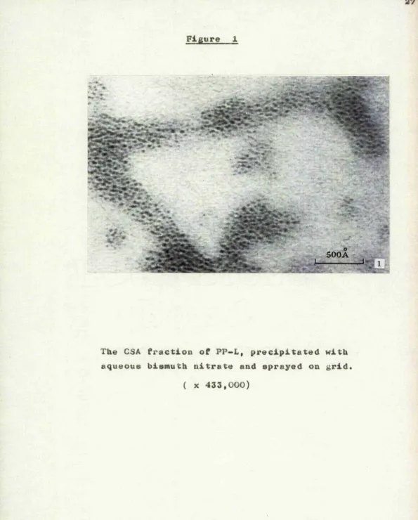
After precipitation of an 0.5% solution of CSA by aqueous bismuth nitrate, the morphology of the precipitate is as shown in figure 1, The material consists of discrete groups of dark spheroidal
particles which have an average diameter of about 0
23A, and there is no evidence of isolated individual particles between the groups..
The pH of the mixture of CSA and bismuth nitrate was 1.4, Gurd and Murray (1954) have demonstrated
that at such a pH little if any ion binding is to be expected from ionizable carboxyl groups. Consequently, it is considered probable that the bismuth ions are
entirely bound to the sulphate groups of CSA. The nitrogen content of the bismuth salt of CSA was
determined by the method of Ma and Zunzaga (1942) as 2.. 38%, whereas that of the CSA disaccharide unit
. r' If#
_ 500A*^
The CSA fraction of PP-L, precipitated with aqueous bismuth nitrate and sprayed on grid
[image:36.618.11.606.16.756.2]and 22% bismuth, and that the ratio of the number of CSA disaccharide units to the number of bismuth ions is T. 6. I t is probable therefore that many of the bismuth ions are bound to more than one sulphate group on the same ox» on different CSA chains. And this, in turn, suggests that the discrete groups of particles in Figure 1 represent networks of CSA
chains joined to one another by intermolecular cross links. This concept is in conformity with the work of Katchalsky and Zwick (1955) w h o , in their study of the effects of divalent cations on polymethacrylic acid gels showed that many of the bound cations were involved in the cross-linking of functional groups located on one or more polymethacrylic acid chains.
It is considered, therefore, that the individual
0
23 A dark particles in Figure 1 indicate those segments of the cross-linked CSA chains which have assumed a coiled configuration as a result of the neutralization of their net charge. Consequently, although Figure 1 demonstrates that bismuth ions are bound to CSA,
probably to the sulphate groups, it is considered
CSA molecules.
(fa) "Protein" fraction
When an. 0.5% solution of the "protein" fraction is precipitated fay aqueous bismuth nitrate and the suspended precipitate is sprayed onto grids, the material takes the form shown in Figure 2. The appearance is essentially similar to that of CSA in Figure 1. The spheroidal dark particles have a
0
similar average diameter of about 23 A, and are arranged in discrete groups.
Albersheim and Killias (1963), in their study of cellular staining reactions with bismuth nitrate, found that the cation was not bound to protein to
any significant extent, and in the present investigation (see below) no staining of collagen has been observed. It is probable, therefore, that the staining of the
"protein" fraction in Figure 2 is due to the binding of bismuth to the sulphate groups of the keratosulphate moiety rather than to its protein part.
It seems likely that the reaction of the bismuth ions with keratosulphate is essentially similar to
y
300Â
The "protein" fraction of PP-L, precipitated
[image:39.612.19.600.16.732.2]comparative rigidity of the protein moiety of the "protein" fraction probably reduceo the extent of intermolecular cross-linking. It is considered
therefore that the visible particles in Figure 2 represent coiled segment© of keratosulphate chains, carried by networks of cross-linked protein molecules, and consequently that the size of those particles
gives no indication of the size or form of the keratosulphate molecule.
(c) The light fraction of proteinpolysaccharide(PP-L-C) (l) The morphology of FP-L, after precipitation of an 0,5% solution by aqueous bismuth nitrate, is shown in Figure 3. It resembles the comparable preparations of CSA and of the "protein" fraction
(Figures 1,2) in that it consist© of groups of closely packed, dark, spheroidal particles# However, the
0
average size of the particles is 47 A, 50% being 0
between 45 and 50 A, so that their average volume of 55,000 A® is about ten times that of the CSA or "protein" particles#
It is considered that each group of particles
»
» V
Vf##
% »
■ ■ i>500A
[image:41.613.16.602.10.753.2]moieties, and that each individual dark particle represents a carbohydrate segment of this network in coiled configuration. Although the preparations of CSA and PP-L cannot be directly compared, a
comparison of the "protein" fraction and PP-L may be valid, since both are visualized as interweaving
protein cores, cross-linked through their carbohydrate moieties. It may be, therefore, that the greater average size of the PP-L particle compared with the "protein" particle is only an expression of the greater proportion of carbohydrate in the former material. Whatever the explanation, it is probable
that the average size of the PP-L particle has no direct relationship to the molecular weight of either PP-L or its component parts.
(2) The morphology of individual PP-L macro-molecules, which is obscured by the use of aqueous
bismuth nitrate, is made evident when precipitation is produced by bismuth nitrate in acetone; 5 ml.
of a 1% solution of PP-L in distilled water was added to 90 ml. of bismuth nitrate in acetone giving a
precipitate wae separated by centrifugation, washed repeatedly In 90% acetone and sprayed onto grids. Its appearance is shown in Figure 4. The great majority of the stained elements are dark particles arranged in single rows. The rows vary in length
0
from 1100 to 1500 A and each contains from twenty to twenty-five particles. The particles vary in
0 0
diameter in the range 15 to 47 A (average 30 A),
whereas the unstained intervals between the particles
0 0
vary in length from 17 to 60 A (average 36 A). Moreover, there is no apparent correlation between the lengths of the intervals and the size of their limiting particles.
It seems very improbable that this arrangement of particles is fortuitous. It is considered,
• • %»
•
#• #"
« X#
SOOA
. .--3
• i
having a molecular weight of 7.5 x 10^.
It is evident that precipitation of PP-L in
acetone results in a more or less complete separation of the macromoleculee. It is considered that the prevention of intermolecular cross-linking between PP-L macromolecules is a result of the greatly increased repulsion between like charges which is
to be expected in a medium of low dielectric constant. But if this factor prevents intermolecular
cross-linking it must also very largely inhibit cross-cross-linking between adjacent CSA chains along the length of the protein-polysaccharide macromolecule. This concept is strongly supported both by the almost completely extended form of the macromolecules in Figure 4, and by the coincidence of the number of particles in each macromolecule and the number of CSA chains as estimated by Partridge ^ (1961). It follows, that in this preparation, each particle very probably represents an individual carbohydrate unit, the greater part of which is CSA. The fact that the particles are not
of uniform size does not necessarily indicate that
particle does not represent a coiled CSA chain in its entirety, but only that part of the coil in which the density of bismuth is above a critical level. In other words, the size of each visible particle is a function not only of the length of
the corresponding CSA chain but of the final shape which the chain has assumed on the grid. One factor which influences this' final shape is the dehydrating effect of acetone which makes the coiled CSA chains more compact than they would be in an aqueous medium.
Electron microscopy of the heavy fraction of protein polysaccharide (PP-H)
The appearance of a suspension of PP-H, treated with aqueous bismuth nitrate is shown in Figures 5 and 6. The material consists, on the one hand, of
particulate areas (A in Figure S) in which the particles appear identical to those in Figure 5, and, on the other hand, of fibres which vary in diameter from 200 to
o
c
PP-H precipitated with aqueous bismuth nitrate and sprayed on grid. A indicates a zone of protein polysaccharide particles. B , C and D indicate
500A
Enlargement of D in Figure 5
[image:48.612.11.601.14.734.2]0
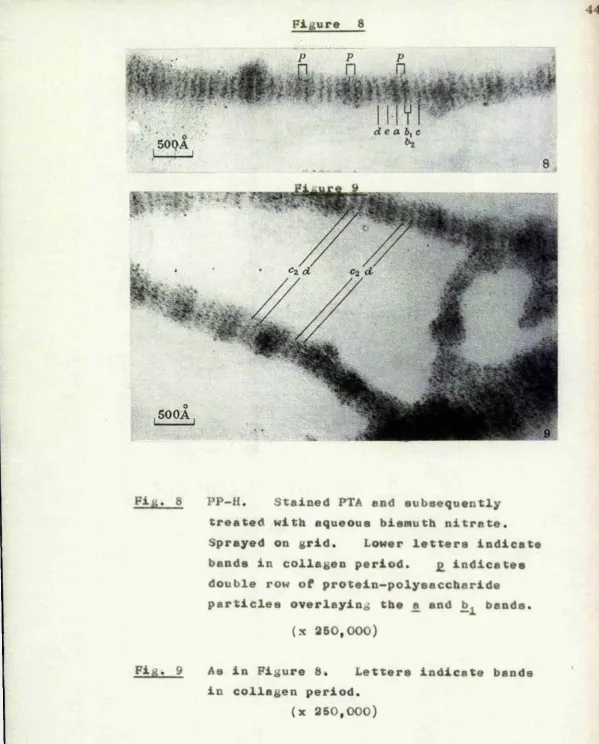
the fibres at regular intervals of about 630 A, and the collagenous nature of the fibres which
this period indicates, is confirmed by their staining pattern with phosphotimgstic acid (Figures 8 and 9).
When a suspension of purified tendon collagen was treated with aqueous bismuth nitrate, washed and
sprayed onto grids, no staining of the fibres was observed# It seems evident therefore that in PP-H protein-polysaccharide macromolecules are bound to
collagen in a rather specific manner ; This conclusion is entirely in keeping with the composition of PP-H
suggested by Schubert (1965)#
The exact arrangement of the protein-polysaccharide macromolecules on the collagen fibres of PP-H varies. In some situations (B in Figure 5) dark particles
are distributed along the whole length of the fibre, but are preferentially aggregated at intervals which conform to the native collagen period; Other fibres (C in Figure 5) exhibit short particle-free zones
0
150 to 200 A wide, between particulate zones which
0
0
interveaing light strip, comprise a zone 250 A wide in each collagen period. The latter arrangement is consistent with macromolecules of protein-poly-saccharide, in the form shown in Figure 4, being
coiled transversely round the surface of the collagen fibre at two specific sites.
It has been noted that when protein-polysaccharide in solution is precipitated by aqueous bismuth nitrate
the individual macromolecule© are joined into a network by cross-linking. But it is evident that the degree of such cross-linking must be considerably influenced by any existing attachment of the macromolecules to a rigid substrate such as collagen. It is considered therefore that the different arrangements of the
particles at B, C and D in Figure 5 are the consequence of different degrees of attachment of
protein-polysaccharide to collagen# At D the
protein-polysaccharide macromolecules are visualized as being firmly attached to collagen so that cross-linking
between the înacromolecules is minimal and they exhibit the same individual form as is shown by
particles is more closely comparable to that exhibited by protein-polysacchariae in water (Figure 5). This weaker attacEimea L may be the result of a recombina tion
of protoin-polysnccharide and collagen, separ^itod from one another in the original extraction process*
To define the sites of attachment of protein— polyoaccharidc macromoj.eculos within the native collagen period, PP-il treated with aqueous bismuth nitrate was additionally stained with phoaphotungstic acid (Figures 8 mid 9), and compared with purified tendon collagen stained with PTA alone (Figure 7)*
Using the conventional notation of Schmitt and Gross (1948) tho fibre in Figure 7 exhibits the composite a bond and the In, , b,.. e,. and d bands while the o
'WÎ!'» *3*-»» .2 • W •' f tP*/.* «V-*
bands and the c. band are iust visible* The fibre shown In Figure 3 appears to be comparable to that shown at D in Figure b, and exhibits two strips of
5 0 0 A
/ T • I, T ^
dzCL^b^ci e-i
[image:52.612.14.600.15.733.2]de a h^c
: '. 500A ■• I, :■ j
8
500A
Fi& PP—H, Stained PTA end subsequently treated with aqueous bismuth nitrate.
Sprayed on grid. Lower letters indicate bands in collagen period. £ indicates double row of protein-polysaccharide particles overlaying the £ and bands.
(x 350,000)
Fig. 9 As in Figure 8* Letters indicate bands in collagen period.
[image:53.625.9.609.13.758.2]comparable to that at C in Figure 6 ; they exhibit particulate zones covering the £ and ]) bands and particle-free zones in which the — .3 and d bands are— clearly evident.
Biizymic activity of PP-L preparations
Contamination of PP-L by proteolytic enzymes was assayed over a wide range of pH values. With PP-L-C two optima were found, at pH 2.5 and 7.. 5, with enzymic activities (expressed as m-equiv. of tyrosine liberated/ hr. /g . of PP-L) 9.0 X 10 ^ and 6,. 0 x 10 ^ respectively.
is’o proteolysis was detectable with PP-L-CPC and PP-L-Di.
Protein content and composition of PP-L-C» PP-L-CPC and PP-L-Si
To determine the protein content of the various PP-L preparations and the individual amino acid
hydrolysates are reported in Table 1, column (A), where the results are expressed as Pg. of anhydro-amino acids/10 mg. of dry and ash-free material. A corrected protein content of 14.17% was derived by extrapolation to zero hydrolysis time of the
summated amino acid contents. The same calculation procedure was applied to the analytical results of sample 1^ and a corrected protein content of 20.12% was obtained* Since the extrapolation curves of sample 1 and sample 5 had identical slopes and in both cases the percentage of zero-time recovery after 24 hours hydrolysis was 90.2, a correction coefficient of 1.108 was used for all other samples examined in the calculation of their corrected protein contents from the amino acid compositions of their 24 hour hydrolysates.
The amino acid composition values of the three PP-L-Bi hydrolysates converted into g. of anhydro-amino acids/100 g. of the protein component are
TABLB X
Amino acid analysis of FP-L-Bi (Sample S)
(A) Wt. of aiihydi’o-amino acid (jug./lOing. of PP-L-Bi, ash- and moisUire-free); (B) wfc. of anhydro-amino acid {g./lOOg. of the protein component); (C) recovery after 24hr. liydrolysis (%),
Time of hydrolysis (A) (B) Corrected
(In .)... ... ... . 24 36 72 24 36 72 values (C)
Amino acid
Hydroxyproline 0-00 0*00 000 0*00 0*00 0*00
Aspartic acid 121-69 116*50 99*79 8*58 8*22 7*04 9*86* 91*7
Threonine 74*81 71*47 55*70 5*30 5*04 3*93 6*08* 87*2
Serine 87-32 73*00 69*28 6*16 5*15 4*89 7*88t 78*3
Glutamic acid 190*19 201*38 166*57 13*42 14*21 11*75 14*21 î 94*4
Proline 116*95 107*22 88*22 8*25 7*57 6*23 946* 90*1
Glycine 77*88 78*38 57*87 5*50 5*53 4*08 5-53Î 99*3
Alanine 61*16 61*32 46*21 432 4*33 3*26 4*33î 99*8
Valine 83*87 84*26 80*91 5*92 5*95 5*71 5*95$ 99*5
Cystine (half) 0*00 0*00 0*00 0*00 0*00 0*00 0*00 (68*8)5
Methionine 0*00 0*00 0*00 0*00 0*00 000 0*00 (45*4)§
Isolencine 63*80 60*59 59*83 4*50 4*28 4*22 4*52* 99*5
Leucine 129*69 126*40 101*07 9*15 8*92 7*13 9*27t 98*7
Tyrosine 47*55 33*09 15*06 3*36 2*34 1*06 4*24* 79*3
Phenylalanine 63*45 70*85 54*17 4*48 5*00 3 82 5*00$ 89*4
Hydroxylysino 0*00 000 0*00 0 00 0*00 0*00 0*00
Lysine 57*48 57*09 43*46 4*06 4*03 3*07 445$ 97*8
Histidine 31*36 31*17 24*55 2*21 2*20 173 2*25$ 98*2
Arginine 71*96 70*38 67*33 5*08 4*97 4*75 5*18* 98*0
Tryptophan 0*00 0*00 0*00 0*00 0*00 0*00 0*00
Totals 1279*06 1243*10 1030*02 90*29 87*74 72*67 97*11
Protein content extrapolated to %ero time: 14-17%
* Extrapolation to %ero time (24hr., 36hr. and 72hr. hydrolysates), t Extrapolation to zero time (24hr. and 36hr. hydrolysates). $ Greatest valno.
alanine, valine and phenylalanine were difficult to liberate, giving maximal values after 36 hours hydrolysis# Hydrolytic destruction of all other amino acids was.apparent# Corrected concentrations of all these amino acids except four were determined by extrapolating to zero-time their concentrations in all three hydrolysates, assuming a first-order reaction over the full range of hydrolysis as
suggested by the linearity of their losses# Since a non-linear destruction of serine, leucine, lysine and histidine was found, corrected concentrations of these four amino acids were calculated by extrapolation, too, but by using the 24 hour and 36 hour hydrolysates only# For each amino acid, the percentage of zero time recovery after 24 hour hydrolysis was calculated and reported in Table 1, column (C). Again, good agreement was found with the recoveries calculated from the analytical data of sample 1.
Values are calculated as percentages of dry asli-lree samples. PP-L-G
PP-L-CPC 4
PP-L-Bi
Sample... 1 2 3 5 1 54" 1
Protein 20*12 18*18 17*48 15*96 14*17 14*14
Nitrogen — . 5*8 — 5*8 5*6 —
Total hexosamine (as — 29*3 — 30*0 30*6 —
free base)
Galactosamine : glucos- 9*6 10*6 10*5
amine ratio
Amino acid composition of the protein component
Wt. of anhydro-amino acid {g./lOOg. of protein) residues (moles/Amino acid
Hydroxjfproline 0*00 0*00 O '00 0*00 0*00 0*00
Aspartic acid 9*98 10*34 10*19 10*01 9*36 9*71 89*96
Threonine 5*46 5*45 6 * 6 8 5*56 6*08 5*55 62*46
Serine 7*11 • 7*31 6*81 7*46 7*88 7*87 98*19
Glutamic acid 14*59 15*21 15*23 15-93 14*21 14*29 119*83
Proline 9*32 7*82 9*11 9*14 9*16 9*23 102*80
Glycine 6*90 5*51 5*82 6*61 6*63 6*10 110*63
Alanine 5*00 5*37 4*64 4*99 4*33 4*36 66*37
Valine 6*93 6*88 6*06 5*74 5*95 5*90 64*89
Cystine (half) 1*22 Trace Trace 0*00 0*00 0*00
Methionine 1*54 0*00 0*00 0*00 0*00 0*00
Isolencine 3*95 4*87 4*92 4*50 4*52 4*80 44*70
Leucine 8*36 9*54 9*98 9*05 9*27 9*43 89*71
Tyrosine 4*16 4*38 4*70 4*20 4*24 3*67 26*32
Phenylalanine 4*43 4*94 6*36 5*61 5*00 4*98 36*81
Hydroxylysine 0*00 0*00 0*00 0*00 0*00 0*00
Lysine 4*02 4*94 5*43 4*81 4*15 3*82 33*76
Histidine 2*19 2*11 2*26 2*22 2*25 2*22 17*69
Arginine 7*12 5*59 6*86 6*21 5*18 4*93 35*14
Tryptophan Trace — — 0*00 0*00 0*00
TABLE 3
Bncl~gro«p analysis of PP-I,~Bi
Krr-rr-iiim-^fciiTthi’; tw\viriwiiiVT#v«iii«fnnf TrfowTr fittrrrri-nift-<-iiTiirYrnTTwWi-«-itii mi *r»« >n-< n’ffTirir. i w, g^rn#„„ndM',nr-i"n
[image:58.613.91.596.77.772.2]—10 significant amounts were found: less than 10 mole of 3H groups/mg# of PP-L.
The values for nitrogen and hexosamines contents are reported in Table 2.
Amino end-group analysis of PP-L-Bi and PP-L-C
The results of the quantitative analysis of the N-terminal amino acids of PP-L-Bi are reported in
6
Table 3 as moles/10 g. of protein-polysaccharide. Acid rehydrolysis confirmed that the DHP derivatives had all been released by the resin hydrolysis.
No chromatographic evidence was obtained, either before or after the second hydrolysis, for the
presence of 0-DNP-tyrosine.
When samples of PP-L-C were examined, a
Analysis of the acetone supernatant
Chromatographic analyses carried out on the organic material liberated from PP-L-C by acetone at low pH revealed the presence of peptides,
hexosamine and the following free amino acids:
isoleucine, phenylalanine, tyrosine, arginine, valine, alanine, glycine, glutamic acid, leucine and aspartic acidi
Sedimentation coefficients of PP-L-CPC and PP-L-Bi
The sedimentation coefficients at infinite dilution, of PP-L-CPC and PP-L-Bi, obtained by linear extrapolation of 1/S or S{1/^}o) against
c to c « O by the method of least squares, wore 12#41S and 12;34s respectively* The determination of v^q
Electron microscopy of bovine nasal and articular cartilages
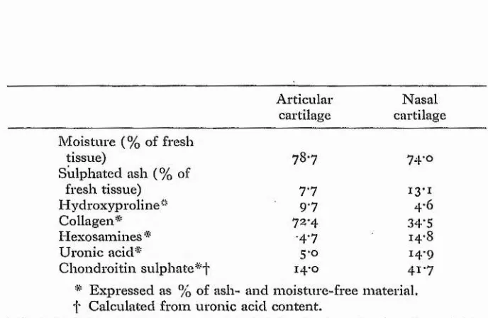
The proportions of collagen and
protein-polysaccharide vary considerably in different types of cartilage, as shown in Table 4.
In both materials the contents of uronic acid and hexosamines are approximately equal, and this indicates that nearly all the acid mucopolysaccharide of both tissues is chondroitin sulphate. The total water and sulphated ash contents are very similar in
the two tissues and it is evident, therefore, that articular cartilage contains over twice as much
collagen and about one-third the amount of chondroitin sulphate as compared with nasal cartilage. The
collagen/GSA ratio is 5.2 in articular cartilage and 0.83 in nasal cartilage.
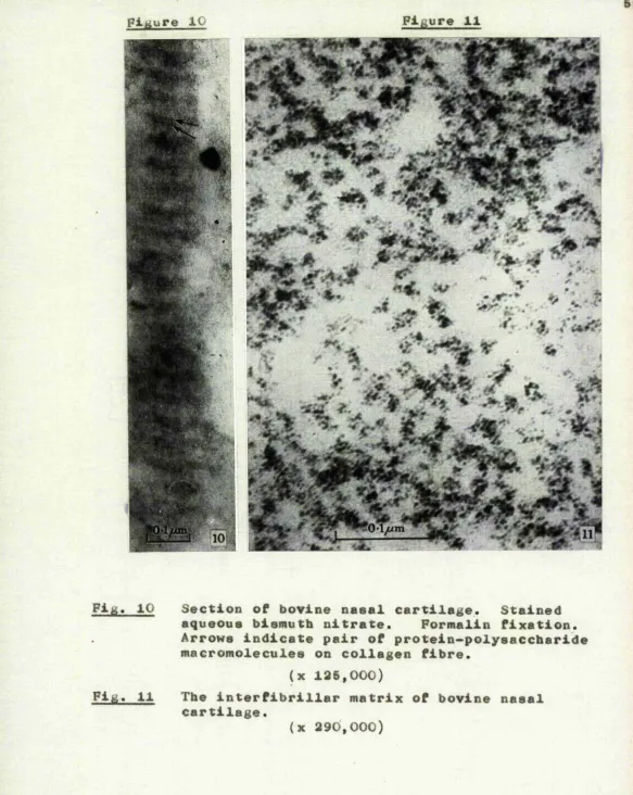
a ) Sections of intact bovine nasal cartilage
After staining with aqueous bismuth nitrate, nasal cartilage is seen to consist of isolated and
TABLE 4
Compositions of bovine articular and nasal cartilages
Articular Nasal
cartilage cartilage Moisture (% of fresh
tissue) 78*7 74*0
Sulphated ash (% of
fresh tissue) 7*7 13*i
Hydroxyproline" 9-7 4*6
Collagen* 72*4 34-5
Hexosamines * -4*7 14*8
Uronic acid* 5-0 14-9
Chondroitin sulphate 14 o 41*7
[image:62.621.155.499.273.497.2]Naturally, it is comparatively seldom that a collagen fibre is found which is exactly parallel to the plane of a section, but whenever this
orientation is closely approached, the appearance
is as shown in Figure 10. A fibre extends throughout the length of this figure, but because it is, itself, unstained, its outline is very largely defined by the presence of transversely attached rows of dark
o
particles. The rows are about 530 A apart which conforms to the period which is customarily exhibited by collagen in embedded and sectioned material (Jakus, 1956). Moreover, some of the rows are double, the
two parts being separated by a distinct clear zone. Thus although the definition of the particles in
sections is naturally below that in sprayed material, the morphology in Figure 10 is almost exactly comparable to that already noted in PP-H at D in Figure 5 and in Figure 6. On the basis of this close morphological similarity, it is considered that in intact nasal cartilage, protein-polysaccharide macromolecules, of the form indicated in Figure 4, have a transverse surface attachment to each collagen fibre in the
"
i
^ i L K • % .
^ / - .p< ^ y . ^ ^ . r Uiv;
% % %
-% 7
t %
Pig. 10 Section of bovine nasal cartilage. Stained aqueous bismuth nitrate. Formalin fixation. Arrows indicate pair of protein-polysaccharide macromolecules on collagen fibre.
(x 125,000)
Fig. 11 The interfibrillar matrix of bovine nasal cartilage.
[image:64.615.11.594.13.745.2]regions of nasal cartilage are occupied by groups of dark particles, the average dimensions of which are similar to those of the particles already noted in PP-L and PP-H* In some situations the groups are quite irregular, but in many others the particles are arranged in short single rows of four to seven# It is considered that this arrangement indicates that
the interfibrillar regions are occupied by free protein-polysaccharide macromolecules. Many of the
macro-molecules are cross-linked by bismuth and consequently the particles are aggregated into irregular groups which, though smaller, are otherwise comparable to
those in Figure 3. Other macromolecules have remained separate, as in Figure 4, probably because cross-linking with neighbouring macromolecules was inhibited by the semisolid environment in which they lie. And the small parts of these long, individual macromolecules which lie within any one section are visible as the short rows of particles already noted.
b ) Sections of intact bovine articular cartilage* Ultrastructure after osmium fixation
is shown in Figures 12 - 14. The chondrocytes exhibit the features which have been described in detail by Godman and Porter (1960), Sllberberg, Silberberg, Vogel and Wettstein (1961), Davies, Barnett, Cochrane and Palfrey (1962), Sheldon and
Kimball (1962), Revel and Hay (1963), Silberberg, Silberberg and Feir (1964), and Palfrey and Davies
(1966). In particular, the processes of the cells are characteristically blunt and smooth and are
quite distinct from the sharp spicule-like projections which are associated with contraction of the cell
from the surrounding matrix as a result of poor
fixation. The main part of the extracellular matrix (m) exhibits numerous straight fibres, between which the
interfibrillar spaces are clear. Between the cells and the main matrix, and sharply demarcated from both, is a narrow region of distinct appearance (p) which will be designated the pericellular zone.
This varies considerably in size on different aspects of the ovoid cells, being wide at the poles (Figure 12) and in the intervals between closely adjacent cells
Bovine articular cartilage. Osmium fixation, stained uranyl acetate.
[image:67.612.17.594.14.753.2]Figure 13
%
&
Bovine articular cartilage. Osmium fixation, stained uranyl acetate.
Figure 14
Bovine articular cartilage. Matrix adjacent to equator of cell. Osmium fixation, stained uranyl acetate.
[image:69.615.10.608.16.743.2]At; a higher magnification and after PTA
staining (Figures 16, 17) it is apparent that the fibres of the main matrix exhibit some degree of
preferential orientation. The fibre diameter varies
0
evenly in the range 250-900 A, but the average fibre diameter is constant throughout the region, right up to the edge of the pericellular zone. Some of the fibres are uniformly grey in appearance whereas others exhibit the banded pattern typical of native collagen. The relative numbers of fibres showing these two
appearances suggest that typical collagen staining may be restricted to those fibres which are actually
traversed by the surfaces of the section. There is no evidence of any interfibrillar material.
At this higher'magnification the pericellular zone (Figure 17) contains a mass of fine fibres with
o
diameters ranging from 100 to 250 A. The fibres follow irregular rather than straight courses and give no
Figure 16
m
Articular cartilage of rabbit. Oamium fixation, stained uranyl acetate.
[image:71.614.11.602.15.726.2]Bovine articular cartilage* Main matrix* Osmium fixation, stained PTA.
[image:72.615.11.601.13.752.2]Bovine articular cartilage. Osmium fixation, stained PTA.
[image:73.612.15.601.15.741.2]structure is discussed in a later section of this
0
paper. Between these 100- to 260-A fibres,fine and rather tenuous fibrils are often evident so that the interfibrillar spaces of the pericellular zone are generally darker than those of the main matrix.
It seems clear that the dimensions of the pericellular zone are not artefactual, for it is quite distinct from the empty pericellular region which often surrounds a poorly fixed and contracted
cell; The zone is not evident in the cartilage of small experimental animals (Figure 15) in which there tends, instead, to be a gradual increase in the average fibre diameter for some distance from the cell membrane (Silberberg et al., 1961: Revel and Hay, 1963). What is so characteristic of bovine articular cartilage is the abrupt increase of average fibre diameter from the pericellular zone to the main matrix.
Ultrastructure after bismuth s